Advertisement

Advertisement

Advertisement

Advertisement

Kidneys: Anatomy

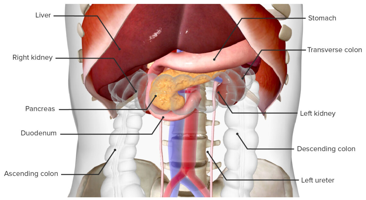
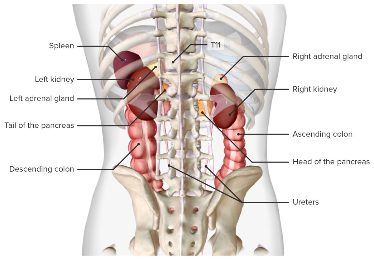
The kidneys are a pair of bean-shaped organs located retroperitoneally against the posterior wall of the abdomen on either side of the spine Spine The human spine, or vertebral column, is the most important anatomical and functional axis of the human body. It consists of 7 cervical vertebrae, 12 thoracic vertebrae, and 5 lumbar vertebrae and is limited cranially by the skull and caudally by the sacrum. Vertebral Column: Anatomy. As part of the urinary tract Urinary tract The urinary tract is located in the abdomen and pelvis and consists of the kidneys, ureters, urinary bladder, and urethra. The structures permit the excretion of urine from the body. Urine flows from the kidneys through the ureters to the urinary bladder and out through the urethra. Urinary Tract: Anatomy, the kidneys are responsible for blood filtration and excretion of water-soluble waste in the urine. The kidneys also play a major role in homeostatic processes, including electrolyte concentration, blood pressure, and acid–base regulation. Grossly, they consist of an outer cortex and inner medulla. Microscopic functional units known as nephrons filter the blood through a structure called the glomerulus, and this filtrate is then modified and concentrated as it moves through a complex tubular system. The renal arteries Arteries Arteries are tubular collections of cells that transport oxygenated blood and nutrients from the heart to the tissues of the body. The blood passes through the arteries in order of decreasing luminal diameter, starting in the largest artery (the aorta) and ending in the small arterioles. Arteries are classified into 3 types: large elastic arteries, medium muscular arteries, and small arteries and arterioles. Arteries: Histology supply the kidneys via a central opening, known as the renal hilum Hilum Lungs: Anatomy, on its medial side; large renal veins Veins Veins are tubular collections of cells, which transport deoxygenated blood and waste from the capillary beds back to the heart. Veins are classified into 3 types: small veins/venules, medium veins, and large veins. Each type contains 3 primary layers: tunica intima, tunica media, and tunica adventitia. Veins: Histology empty directly into the vena cava.

Last updated: Nov 19, 2024

Editorial responsibility: Stanley Oiseth, Lindsay Jones, Evelin Maza

Advertisement

Advertisement

Advertisement

Advertisement

Advertisement

Advertisement

Embryology

The kidney develops from embryonic mesoderm Mesoderm The middle germ layer of an embryo derived from three paired mesenchymal aggregates along the neural tube. Gastrulation and Neurulation in 3 successive forms from the nephrogenic cords Nephrogenic cords Development of the Urogenital System as they elongate in a cranial-to-caudal direction.
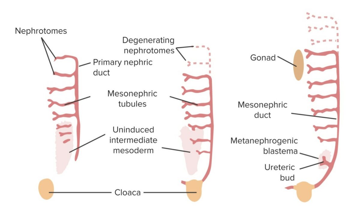
Pronephros Pronephros The primordial kidney that develops from the intermediate mesoderm in the embryos of vertebrates, and is succeeded by the mesonephros. In higher vertebrates and humans, the pronephros is a vestigial and transient structure. Development of the Urogenital System

  • Appears in week 4 as a cluster of a few cells that disintegrate shortly thereafter
  • Rudimentary and nonfunctional

Mesonephros Mesonephros One of a pair of excretory organs (mesonephroi) which grows caudally to the first pair (pronephroi) during development. Mesonephroi are the permanent kidneys in adult amphibians and fish. In higher vertebrates, proneprhoi and most of mesonephroi degenerate with the appearance of metanephroi. The remaining ducts become wolffian ducts. Development of the Urogenital System

  • Begins to develop in the thoracolumbar region around week 5 as pronephros Pronephros The primordial kidney that develops from the intermediate mesoderm in the embryos of vertebrates, and is succeeded by the mesonephros. In higher vertebrates and humans, the pronephros is a vestigial and transient structure. Development of the Urogenital System regresses
  • Regresses by week 10 
  • Consists of:
    • A longitudinal mesonephric duct (also known as the wolffian duct)
    • A series of tubules coming off the main duct and growing anteriorly toward the aorta Aorta The main trunk of the systemic arteries. Mediastinum and Great Vessels: Anatomy 
  • Begins filtering blood → filtrate travels down the mesonephric tubule → mesonephric duct → cloaca Cloaca A dilated cavity extended caudally from the hindgut. In adult birds, reptiles, amphibians, and many fishes but few mammals, cloaca is a common chamber into which the digestive, urinary and reproductive tracts discharge their contents. In most mammals, cloaca gives rise to large intestine; urinary bladder; and genitalia. Development of the Abdominal Organs allantois Allantois An extra-embryonic membranous sac derived from the yolk sac of reptiles; birds; and mammals. It lies between two other extra-embryonic membranes, the amnion and the chorion. The allantois serves to store urinary wastes and mediate exchange of gas and nutrients for the developing embryo. Development of the Abdominal Organs
  • Functions as the primitive urinary system, while the metanephros Metanephros Development of the Urogenital System develops into the permanent kidney
  • The mesonephric ducts persist and form part of the male reproductive system.
Graphical summary of the developing kidney

Graphical summary of the developing kidney:
The ureteric bud grows off of the mesonephric duct and into a collection of intermediate mesoderm cells known as the metanephric blastema. Together, this is known as the mesonephros, which develops into the kidney. The mesonephric tubules regress. In males, the mesonephric duct persists in the ejaculatory system.

Image by Lecturio.

Metanephros Metanephros Development of the Urogenital System

The permanent kidney is formed from the metanephros Metanephros Development of the Urogenital System.
  • Develops starting around the 5th week of gestation
  • Cells in the intermediate mesoderm Intermediate mesoderm Development of the Urogenital System in the pelvic region begin to differentiate into a structure called the metanephric blastema Metanephric blastema Wilms Tumor, which:
    • Ultimately becomes the cells making up the nephrons
    • Releases growth factors that stimulate the development of an outpouching off the caudal portion of the mesonephric duct called the ureteric buds
  • Ureteric buds grow toward and invade the metanephric blastema Metanephric blastema Wilms Tumor:
    • Elongating stalk of the ureteric bud develops into the ureter.
    • Within the metanephric blastema Metanephric blastema Wilms Tumor, the ureteric buds undergo a series of branchings to form the:
      • Renal pelvis Pelvis The pelvis consists of the bony pelvic girdle, the muscular and ligamentous pelvic floor, and the pelvic cavity, which contains viscera, vessels, and multiple nerves and muscles. The pelvic girdle, composed of 2 “hip” bones and the sacrum, is a ring-like bony structure of the axial skeleton that links the vertebral column with the lower extremities. Pelvis: Anatomy
      • Major calyces
      • Minor calyces
      • Collecting tubules
  • Metanephric mesodermal cap Metanephric mesodermal cap Development of the Urogenital System:
    • Mesoderm Mesoderm The middle germ layer of an embryo derived from three paired mesenchymal aggregates along the neural tube. Gastrulation and Neurulation from the metanephric blastema Metanephric blastema Wilms Tumor sitting on top of the developing collecting ducts
    • Elongates, forming the tubular system The tubular system The kidneys regulate water and solute homeostasis through the processes of filtration, reabsorption, secretion, and excretion. After the filtration of blood through the glomeruli, the tubular system takes over and is responsible for adjusting the urine composition throughout the remainder of the nephron. Tubular System of the nephrons → becomes known as the metanephric tubules
    • The metanephric tubule fuses with the collecting tubule, creating one continuous system.
  • Bowman capsule Capsule An envelope of loose gel surrounding a bacterial cell which is associated with the virulence of pathogenic bacteria. Some capsules have a well-defined border, whereas others form a slime layer that trails off into the medium. Most capsules consist of relatively simple polysaccharides but there are some bacteria whose capsules are made of polypeptides. Bacteroides: forms off the growing end of the metanephric tubule
  • Glomerular capillaries Capillaries Capillaries are the primary structures in the circulatory system that allow the exchange of gas, nutrients, and other materials between the blood and the extracellular fluid (ECF). Capillaries are the smallest of the blood vessels. Because a capillary diameter is so small, only 1 RBC may pass through at a time. Capillaries: Histology:
    • Develop off the common iliac arteries Arteries Arteries are tubular collections of cells that transport oxygenated blood and nutrients from the heart to the tissues of the body. The blood passes through the arteries in order of decreasing luminal diameter, starting in the largest artery (the aorta) and ending in the small arterioles. Arteries are classified into 3 types: large elastic arteries, medium muscular arteries, and small arteries and arterioles. Arteries: Histology
    • Become associated with Bowman capsule Capsule An envelope of loose gel surrounding a bacterial cell which is associated with the virulence of pathogenic bacteria. Some capsules have a well-defined border, whereas others form a slime layer that trails off into the medium. Most capsules consist of relatively simple polysaccharides but there are some bacteria whose capsules are made of polypeptides. Bacteroides at the end of the metanephric tubules → begin creating “urine” (Note: True waste products are removed from the fetus via the placenta Placenta A highly vascularized mammalian fetal-maternal organ and major site of transport of oxygen, nutrients, and fetal waste products. It includes a fetal portion (chorionic villi) derived from trophoblasts and a maternal portion (decidua) derived from the uterine endometrium. The placenta produces an array of steroid, protein and peptide hormones (placental hormones). Placenta, Umbilical Cord, and Amniotic Cavity.)
  • Nephrons are formed until birth.
  • Nephron maturation continues after birth.

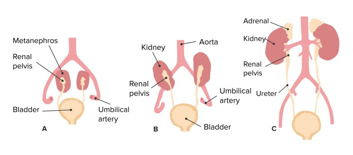
Position of the kidney and changes in vascularization

  • The kidneys are initially located in the pelvic region.
  • As the caudal portion of the body grows downward, the relative location of the kidneys “ascends” into the upper quadrants of the abdomen (failure to ascend results in a pelvic kidney).
  • As the kidneys ascend, the original blood supply degenerates.
  • New vessels (higher up) develop off the aorta Aorta The main trunk of the systemic arteries. Mediastinum and Great Vessels: Anatomy and invade the kidneys, becoming the mature renal arteries Arteries Arteries are tubular collections of cells that transport oxygenated blood and nutrients from the heart to the tissues of the body. The blood passes through the arteries in order of decreasing luminal diameter, starting in the largest artery (the aorta) and ending in the small arterioles. Arteries are classified into 3 types: large elastic arteries, medium muscular arteries, and small arteries and arterioles. Arteries: Histology.
  • If the original vessels fail to regress, they may persist as additional renal arteries Arteries Arteries are tubular collections of cells that transport oxygenated blood and nutrients from the heart to the tissues of the body. The blood passes through the arteries in order of decreasing luminal diameter, starting in the largest artery (the aorta) and ending in the small arterioles. Arteries are classified into 3 types: large elastic arteries, medium muscular arteries, and small arteries and arterioles. Arteries: Histology or veins Veins Veins are tubular collections of cells, which transport deoxygenated blood and waste from the capillary beds back to the heart. Veins are classified into 3 types: small veins/venules, medium veins, and large veins. Each type contains 3 primary layers: tunica intima, tunica media, and tunica adventitia. Veins: Histology.
Ascent of the kidneys and corresponding change in vascular supply

Ascent of the kidneys and corresponding change in vascular supply

Image by Lecturio.

Gross Anatomy

Location

  • Retroperitoneal Retroperitoneal Peritoneum: Anatomy organs
  • Located along the posterior abdominal wall Abdominal wall The outer margins of the abdomen, extending from the osteocartilaginous thoracic cage to the pelvis. Though its major part is muscular, the abdominal wall consists of at least seven layers: the skin, subcutaneous fat, deep fascia; abdominal muscles, transversalis fascia, extraperitoneal fat, and the parietal peritoneum. Surgical Anatomy of the Abdomen on either side of the vertebral column Vertebral column The human spine, or vertebral column, is the most important anatomical and functional axis of the human body. It consists of 7 cervical vertebrae, 12 thoracic vertebrae, and 5 lumbar vertebrae and is limited cranially by the skull and caudally by the sacrum. Vertebral Column: Anatomy in the paravertebral gutter
  • In front of the T12–L3 transverse processes
  • The superior pole rests against the 11th and 12th ribs Ribs A set of twelve curved bones which connect to the vertebral column posteriorly, and terminate anteriorly as costal cartilage. Together, they form a protective cage around the internal thoracic organs. Chest Wall: Anatomy.
  • The lower pole is directed laterally and anteriorly.
  • The presence of the liver Liver The liver is the largest gland in the human body. The liver is found in the superior right quadrant of the abdomen and weighs approximately 1.5 kilograms. Its main functions are detoxification, metabolism, nutrient storage (e.g., iron and vitamins), synthesis of coagulation factors, formation of bile, filtration, and storage of blood. Liver: Anatomy on the right side forces the right kidney to be slightly lower than the left.

Anatomical relations

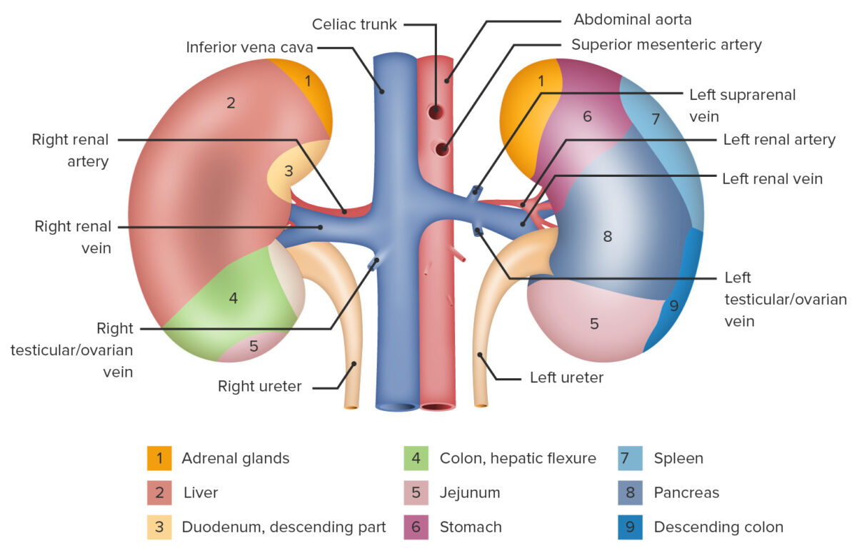
Table: Anatomic relations of the kidneys
Direction (in relation to the kidney) Right Left
Superior Right adrenal gland Left adrenal gland
Anterior
  • Upper portion: liver Liver The liver is the largest gland in the human body. The liver is found in the superior right quadrant of the abdomen and weighs approximately 1.5 kilograms. Its main functions are detoxification, metabolism, nutrient storage (e.g., iron and vitamins), synthesis of coagulation factors, formation of bile, filtration, and storage of blood. Liver: Anatomy
  • Lower portion: hepatic flexure of the colon Colon The large intestines constitute the last portion of the digestive system. The large intestine consists of the cecum, appendix, colon (with ascending, transverse, descending, and sigmoid segments), rectum, and anal canal. The primary function of the colon is to remove water and compact the stool prior to expulsion from the body via the rectum and anal canal. Colon, Cecum, and Appendix: Anatomy
  • Upper portion: stomach Stomach The stomach is a muscular sac in the upper left portion of the abdomen that plays a critical role in digestion. The stomach develops from the foregut and connects the esophagus with the duodenum. Structurally, the stomach is C-shaped and forms a greater and lesser curvature and is divided grossly into regions: the cardia, fundus, body, and pylorus. Stomach: Anatomy
  • Mid portion: pancreas Pancreas The pancreas lies mostly posterior to the stomach and extends across the posterior abdominal wall from the duodenum on the right to the spleen on the left. This organ has both exocrine and endocrine tissue. Pancreas: Anatomy
  • Lower portion: jejunum Jejunum The middle portion of the small intestine, between duodenum and ileum. It represents about 2/5 of the remaining portion of the small intestine below duodenum. Small Intestine: Anatomy
Lateral Liver Liver The liver is the largest gland in the human body. The liver is found in the superior right quadrant of the abdomen and weighs approximately 1.5 kilograms. Its main functions are detoxification, metabolism, nutrient storage (e.g., iron and vitamins), synthesis of coagulation factors, formation of bile, filtration, and storage of blood. Liver: Anatomy
  • Upper portion: Spleen Spleen The spleen is the largest lymphoid organ in the body, located in the LUQ of the abdomen, superior to the left kidney and posterior to the stomach at the level of the 9th-11th ribs just below the diaphragm. The spleen is highly vascular and acts as an important blood filter, cleansing the blood of pathogens and damaged erythrocytes. Spleen: Anatomy
  • Lower portion: Descending colon Descending colon The segment of large intestine between transverse colon and the sigmoid colon. Colon, Cecum, and Appendix: Anatomy
Posterior
  • Muscles:
    • Posterosuperior: diaphragm Diaphragm The diaphragm is a large, dome-shaped muscle that separates the thoracic cavity from the abdominal cavity. The diaphragm consists of muscle fibers and a large central tendon, which is divided into right and left parts. As the primary muscle of inspiration, the diaphragm contributes 75% of the total inspiratory muscle force. Diaphragm: Anatomy
    • Posteromedial: psoas muscle Psoas muscle A powerful flexor of the thigh at the hip joint (psoas major) and a weak flexor of the trunk and lumbar spinal column (psoas minor). Psoas is derived from the greek ‘psoa. Pyelonephritis and Perinephric Abscess
    • Directly posterior: quadratus lumborum Quadratus lumborum Posterior Abdominal Wall: Anatomy
  • Nerves:
    • Subcostal
    • Iliohypogastric
    • Ilioinguinal

Size and shape

  • Bean-shaped organs
  • Dimensions:
    • Length: 10–12 cm
    • Width: 5–7 cm
    • Depth: 2–3 cm
  • Weight: approximately 150 g

Outer layers

Surrounding the kidneys are several layers of adipose and connective tissue Connective tissue Connective tissues originate from embryonic mesenchyme and are present throughout the body except inside the brain and spinal cord. The main function of connective tissues is to provide structural support to organs. Connective tissues consist of cells and an extracellular matrix. Connective Tissue: Histology (from outside in):
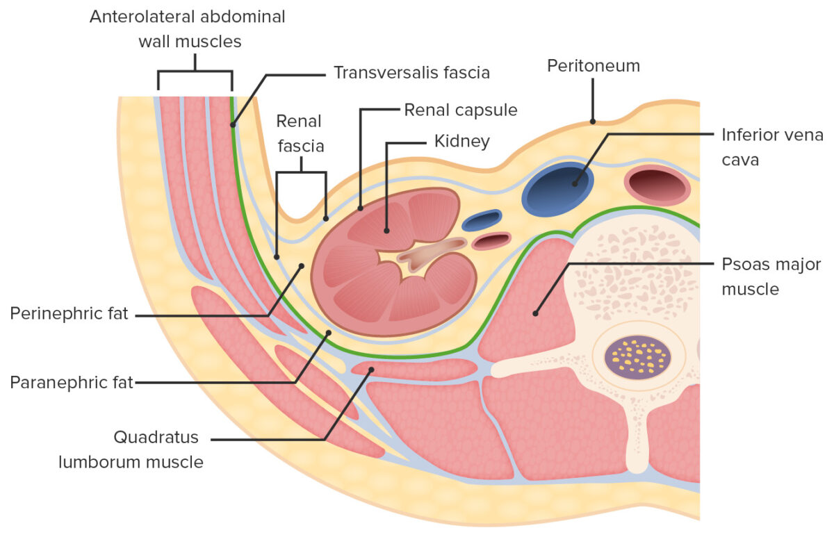
  • Paranephric fat:
    • Located posterior to the kidney, between the renal fascia Fascia Layers of connective tissue of variable thickness. The superficial fascia is found immediately below the skin; the deep fascia invests muscles, nerves, and other organs. Cellulitis and the back muscles Back muscles The back is composed of several muscles of varying sizes and functions, which are grouped into intrinsic (or primary) back muscles and extrinsic (or secondary) back muscles. The extrinsic muscles comprise the superficial and intermediate muscle groups, while the intrinsic muscles comprise the deep muscles. Muscles of the Back: Anatomy 
    • Anchors kidneys to the posterior abdominal wall Abdominal wall The outer margins of the abdomen, extending from the osteocartilaginous thoracic cage to the pelvis. Though its major part is muscular, the abdominal wall consists of at least seven layers: the skin, subcutaneous fat, deep fascia; abdominal muscles, transversalis fascia, extraperitoneal fat, and the parietal peritoneum. Surgical Anatomy of the Abdomen
    • Provides some protection and warmth
  • Renal fascia Fascia Layers of connective tissue of variable thickness. The superficial fascia is found immediately below the skin; the deep fascia invests muscles, nerves, and other organs. Cellulitis, which also encloses the adrenal gland 
  • Perinephric fat
  • Fibrous capsule Fibrous capsule Hip Joint: Anatomy
Layers of adipose and connective tissue surrounding the kidneys (transverse section)

Layers of adipose and connective tissue surrounding the kidneys (transverse section)

Image by Lecturio.

Surface features

  • Superior and inferior poles
  • Convex lateral surface
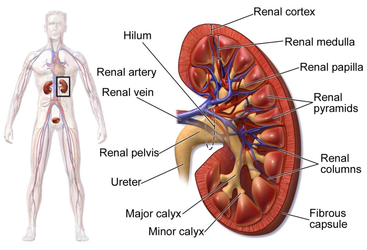
  • Concave medial surface, which creates a cavity called the renal sinus
  • The renal sinus contains:
    • Renal neurovasculature, including:
    • Renal pelvis Pelvis The pelvis consists of the bony pelvic girdle, the muscular and ligamentous pelvic floor, and the pelvic cavity, which contains viscera, vessels, and multiple nerves and muscles. The pelvic girdle, composed of 2 “hip” bones and the sacrum, is a ring-like bony structure of the axial skeleton that links the vertebral column with the lower extremities. Pelvis: Anatomy (terminal portion of the renal collecting system)
    • Adipose tissue Adipose tissue Adipose tissue is a specialized type of connective tissue that has both structural and highly complex metabolic functions, including energy storage, glucose homeostasis, and a multitude of endocrine capabilities. There are three types of adipose tissue, white adipose tissue, brown adipose tissue, and beige or “brite” adipose tissue, which is a transitional form. Adipose Tissue: Histology (continuous with the perinephric fat surrounding the kidney)
  • Renal hilum Hilum Lungs: Anatomy: the entrance to the renal sinus

Renal parenchyma

The parenchyma consists of:

  • Renal cortex:
    • The outermost part of the kidney, approximately 1 cm thick
    • Located underneath the renal capsule Renal capsule Pyelonephritis and Perinephric Abscess
    • Projections medially form the renal columns (of Bertin).
    • Microscopic structures located in the cortex:
      • Bowman capsule Capsule An envelope of loose gel surrounding a bacterial cell which is associated with the virulence of pathogenic bacteria. Some capsules have a well-defined border, whereas others form a slime layer that trails off into the medium. Most capsules consist of relatively simple polysaccharides but there are some bacteria whose capsules are made of polypeptides. Bacteroides
      • Proximal and distal tubules
      • Upper portions of the collecting ducts
  • Renal medulla:
    • Divided into units known as renal pyramids
    • 6–10 renal pyramids separated by the columns of Bertin
    • Base of pyramid toward the cortex
    • The apex:
      • Projects toward the renal sinus 
      • Is called a papilla
      • Collecting tubules drain out of the papilla into the minor calyces.
    • Microscopic structures located in the medulla/pyramids:
      • Loops of Henle 
      • Collecting ducts
  • Collecting system:
    • Collects the newly formed urine and directs it to the ureter
    • Each papilla drains into a minor calyx.
    • 2–3 minor calyces drain into a single major calyx.
    • 2–3 major calyces converge to form the renal pelvis Pelvis The pelvis consists of the bony pelvic girdle, the muscular and ligamentous pelvic floor, and the pelvic cavity, which contains viscera, vessels, and multiple nerves and muscles. The pelvic girdle, composed of 2 “hip” bones and the sacrum, is a ring-like bony structure of the axial skeleton that links the vertebral column with the lower extremities. Pelvis: Anatomy, which:
Diagram depicting renal anatomy

Diagram depicting the renal anatomy

Image: “Human Kidney Anatomy” by Blausen.com staff. License: CC BY 3.0, cropped by Lecturio.

Microscopic Anatomy

Nephrons are the functional units of the kidney; there are approximately 1.2 million nephrons in each kidney. The nephron is divided into 2 main parts: the renal corpuscle and the renal tubule, which has multiple defined segments.

Renal corpuscle

The renal corpuscle is where the blood plasma Plasma The residual portion of blood that is left after removal of blood cells by centrifugation without prior blood coagulation. Transfusion Products is filtered.

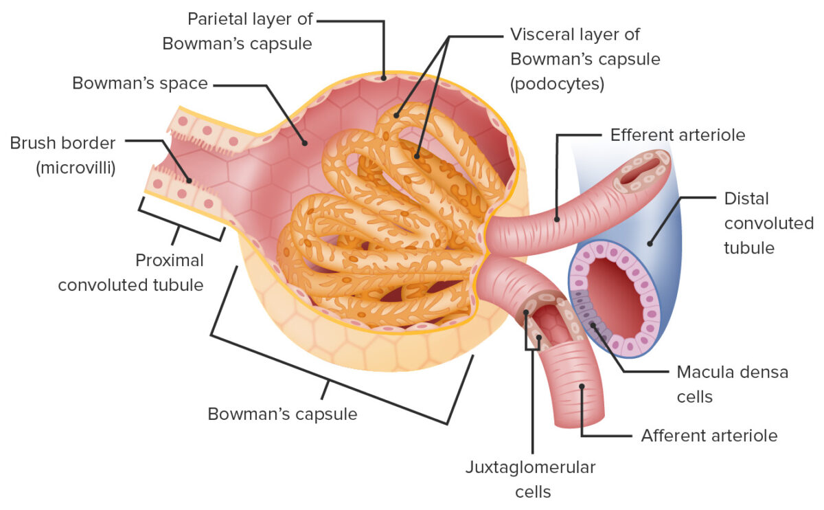
Glomerular (Bowman) capsule Capsule An envelope of loose gel surrounding a bacterial cell which is associated with the virulence of pathogenic bacteria. Some capsules have a well-defined border, whereas others form a slime layer that trails off into the medium. Most capsules consist of relatively simple polysaccharides but there are some bacteria whose capsules are made of polypeptides. Bacteroides

  • A ball-shaped structure surrounding a web of arterial capillaries Capillaries Capillaries are the primary structures in the circulatory system that allow the exchange of gas, nutrients, and other materials between the blood and the extracellular fluid (ECF). Capillaries are the smallest of the blood vessels. Because a capillary diameter is so small, only 1 RBC may pass through at a time. Capillaries: Histology
  • Has an outer parietal Parietal One of a pair of irregularly shaped quadrilateral bones situated between the frontal bone and occipital bone, which together form the sides of the cranium. Skull: Anatomy layer made up of simple squamous epithelial cells
  • Has an inner visceral layer:
    • Directly surrounds the glomerular capillaries Capillaries Capillaries are the primary structures in the circulatory system that allow the exchange of gas, nutrients, and other materials between the blood and the extracellular fluid (ECF). Capillaries are the smallest of the blood vessels. Because a capillary diameter is so small, only 1 RBC may pass through at a time. Capillaries: Histology
    • Made up of specialized epithelial cells known as podocytes 
  • Bowman space:
    • The space between the parietal Parietal One of a pair of irregularly shaped quadrilateral bones situated between the frontal bone and occipital bone, which together form the sides of the cranium. Skull: Anatomy and visceral layers of Bowman capsule Capsule An envelope of loose gel surrounding a bacterial cell which is associated with the virulence of pathogenic bacteria. Some capsules have a well-defined border, whereas others form a slime layer that trails off into the medium. Most capsules consist of relatively simple polysaccharides but there are some bacteria whose capsules are made of polypeptides. Bacteroides 
    • Collects the filtrate (urine)
    • Drains into the proximal convoluted tubule Proximal convoluted tubule The renal tubule portion that extends from the bowman capsule in the kidney cortex into the kidney medulla. The proximal tubule consists of a convoluted proximal segment in the cortex, and a distal straight segment descending into the medulla where it forms the u-shaped loop of henle. Osmotic Diuretics ( PCT PCT The renal tubule portion that extends from the bowman capsule in the kidney cortex into the kidney medulla. The proximal tubule consists of a convoluted proximal segment in the cortex, and a distal straight segment descending into the medulla where it forms the u-shaped loop of henle. Osmotic Diuretics)
Structure of the renal corpuscle

Structure of the renal corpuscle

Image by Lecturio.

Blood vessels associated with the renal corpuscle:

  • Afferent arteriole Afferent arteriole Glomerular Filtration:
    • Brings blood into the glomerulus 
    • Relatively large diameter 
  • Glomerulus (also called glomerular capillaries Capillaries Capillaries are the primary structures in the circulatory system that allow the exchange of gas, nutrients, and other materials between the blood and the extracellular fluid (ECF). Capillaries are the smallest of the blood vessels. Because a capillary diameter is so small, only 1 RBC may pass through at a time. Capillaries: Histology):
    • A web of small arterial capillaries Capillaries Capillaries are the primary structures in the circulatory system that allow the exchange of gas, nutrients, and other materials between the blood and the extracellular fluid (ECF). Capillaries are the smallest of the blood vessels. Because a capillary diameter is so small, only 1 RBC may pass through at a time. Capillaries: Histology between the afferent Afferent Neurons which conduct nerve impulses to the central nervous system. Nervous System: Histology and efferent Efferent Neurons which send impulses peripherally to activate muscles or secretory cells. Nervous System: Histology arterioles Arterioles The smallest divisions of the arteries located between the muscular arteries and the capillaries. Arteries: Histology 
    • Where filtration occurs
  • Efferent arteriole Efferent arteriole Glomerular Filtration:
  • Filtration occurs primarily because of the high hydrostatic pressure Hydrostatic pressure The pressure due to the weight of fluid. Edema in the glomerular capillaries Capillaries Capillaries are the primary structures in the circulatory system that allow the exchange of gas, nutrients, and other materials between the blood and the extracellular fluid (ECF). Capillaries are the smallest of the blood vessels. Because a capillary diameter is so small, only 1 RBC may pass through at a time. Capillaries: Histology created by the large inlet ( afferent arteriole Afferent arteriole Glomerular Filtration) and small outlet ( efferent arteriole Efferent arteriole Glomerular Filtration).

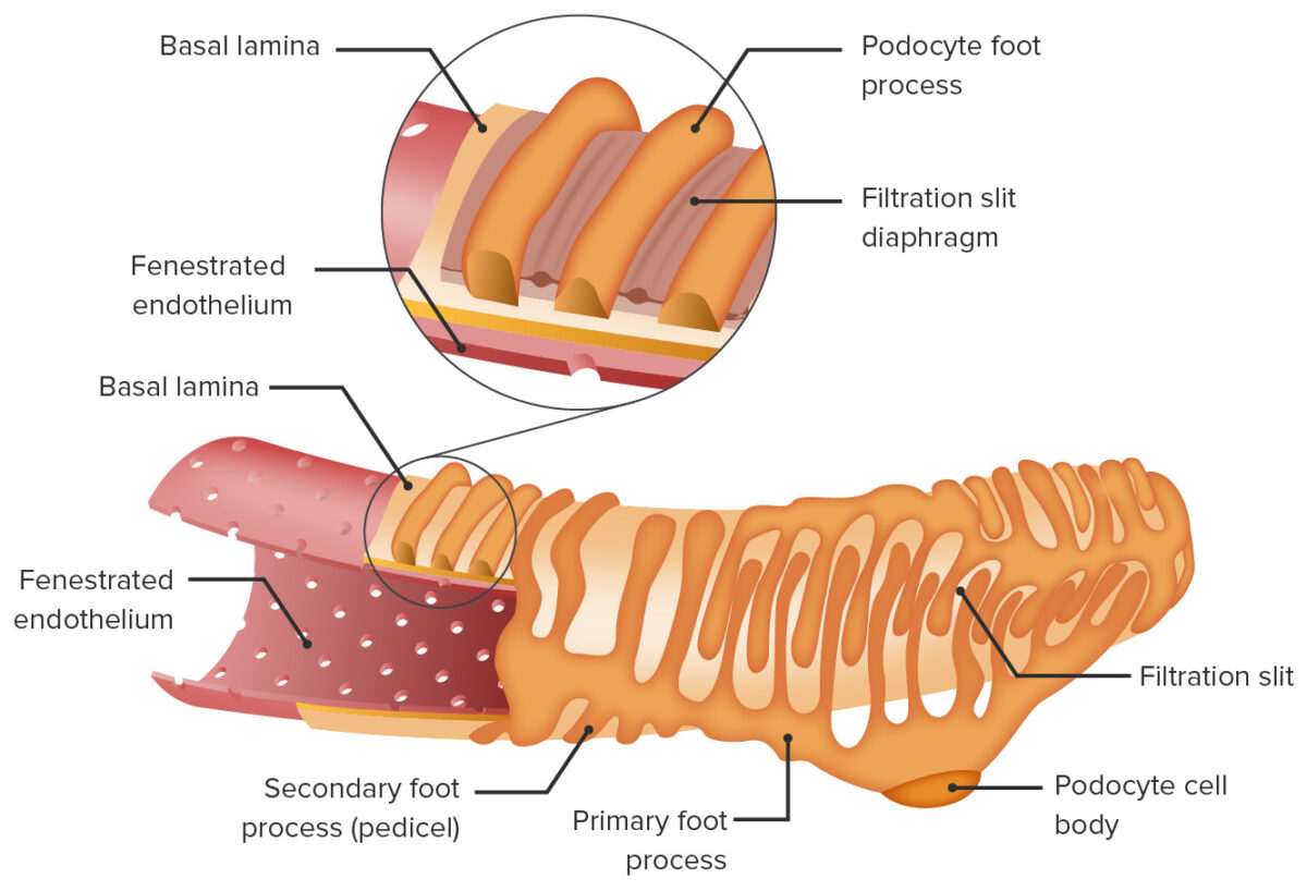
Filtration membrane:

  • Constitutes the main blood filter between the glomerular capillaries Capillaries Capillaries are the primary structures in the circulatory system that allow the exchange of gas, nutrients, and other materials between the blood and the extracellular fluid (ECF). Capillaries are the smallest of the blood vessels. Because a capillary diameter is so small, only 1 RBC may pass through at a time. Capillaries: Histology and Bowman space
  • Consists of 3 layers:
    • Fenestrated endothelium Endothelium A layer of epithelium that lines the heart, blood vessels (vascular endothelium), lymph vessels (lymphatic endothelium), and the serous cavities of the body. Arteries: Histology lining the glomerular capillaries Capillaries Capillaries are the primary structures in the circulatory system that allow the exchange of gas, nutrients, and other materials between the blood and the extracellular fluid (ECF). Capillaries are the smallest of the blood vessels. Because a capillary diameter is so small, only 1 RBC may pass through at a time. Capillaries: Histology
    • Glomerular basement membrane Basement membrane A darkly stained mat-like extracellular matrix (ecm) that separates cell layers, such as epithelium from endothelium or a layer of connective tissue. The ecm layer that supports an overlying epithelium or endothelium is called basal lamina. Basement membrane (bm) can be formed by the fusion of either two adjacent basal laminae or a basal lamina with an adjacent reticular lamina of connective tissue. Bm, composed mainly of type IV collagen; glycoprotein laminin; and proteoglycan, provides barriers as well as channels between interacting cell layers. Thin Basement Membrane Nephropathy (TBMN) (GBM): made up of a negatively charged proteoglycan gel (repels larger negatively charged molecules)
    • Podocytes ( epithelium Epithelium The epithelium is a complex of specialized cellular organizations arranged into sheets and lining cavities and covering the surfaces of the body. The cells exhibit polarity, having an apical and a basal pole. Structures important for the epithelial integrity and function involve the basement membrane, the semipermeable sheet on which the cells rest, and interdigitations, as well as cellular junctions. Surface Epithelium: Histology):
      • Form the visceral layer of Bowman capsule Capsule An envelope of loose gel surrounding a bacterial cell which is associated with the virulence of pathogenic bacteria. Some capsules have a well-defined border, whereas others form a slime layer that trails off into the medium. Most capsules consist of relatively simple polysaccharides but there are some bacteria whose capsules are made of polypeptides. Bacteroides
      • Multiple interdigitating foot processes Foot processes Nephrotic Syndrome wrap around the vessels, creating filtration slits between foot processes Foot processes Nephrotic Syndrome.
      • Slits are covered by a membrane called the slit diaphragm Diaphragm The diaphragm is a large, dome-shaped muscle that separates the thoracic cavity from the abdominal cavity. The diaphragm consists of muscle fibers and a large central tendon, which is divided into right and left parts. As the primary muscle of inspiration, the diaphragm contributes 75% of the total inspiratory muscle force. Diaphragm: Anatomy (a unique form of intercellular junction consisting of multiple proteins Proteins Linear polypeptides that are synthesized on ribosomes and may be further modified, crosslinked, cleaved, or assembled into complex proteins with several subunits. The specific sequence of amino acids determines the shape the polypeptide will take, during protein folding, and the function of the protein. Energy Homeostasis).
  • Not permeable to large molecules in blood, such as plasma Plasma The residual portion of blood that is left after removal of blood cells by centrifugation without prior blood coagulation. Transfusion Products proteins Proteins Linear polypeptides that are synthesized on ribosomes and may be further modified, crosslinked, cleaved, or assembled into complex proteins with several subunits. The specific sequence of amino acids determines the shape the polypeptide will take, during protein folding, and the function of the protein. Energy Homeostasis (e.g., albumin Albumin Serum albumin from humans. It is an essential carrier of both endogenous substances, such as fatty acids and bilirubin, and of xenobiotics in the blood. Liver Function Tests)
  • Permeable to small molecules, generally < 3 nm, including:
    • Water
    • Electrolytes Electrolytes Electrolytes are mineral salts that dissolve in water and dissociate into charged particles called ions, which can be either be positively (cations) or negatively (anions) charged. Electrolytes are distributed in the extracellular and intracellular compartments in different concentrations. Electrolytes are essential for various basic life-sustaining functions. Electrolytes (e.g., sodium Sodium A member of the alkali group of metals. It has the atomic symbol na, atomic number 11, and atomic weight 23. Hyponatremia, potassium Potassium An element in the alkali group of metals with an atomic symbol k, atomic number 19, and atomic weight 39. 10. It is the chief cation in the intracellular fluid of muscle and other cells. Potassium ion is a strong electrolyte that plays a significant role in the regulation of fluid volume and maintenance of the water-electrolyte balance. Hyperkalemia)
    • Glucose Glucose A primary source of energy for living organisms. It is naturally occurring and is found in fruits and other parts of plants in its free state. It is used therapeutically in fluid and nutrient replacement. Lactose Intolerance
    • Amino and fatty acids Acids Chemical compounds which yield hydrogen ions or protons when dissolved in water, whose hydrogen can be replaced by metals or basic radicals, or which react with bases to form salts and water (neutralization). An extension of the term includes substances dissolved in media other than water. Acid-Base Balance 
    • Vitamins
  • Can be damaged by infection and trauma
Filtration apparatus in the glomerulus

Filtration apparatus in the glomerulus

Image by Lecturio.

Renal tubule

The renal tubule is a long continuous tube that adjusts the contents of the filtrate received from the renal corpuscle. The tubules ultimately drain through the papilla into the calyces. Segments of the tubule, in order, are:
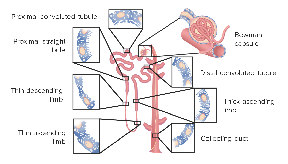
  • PCT PCT The renal tubule portion that extends from the bowman capsule in the kidney cortex into the kidney medulla. The proximal tubule consists of a convoluted proximal segment in the cortex, and a distal straight segment descending into the medulla where it forms the u-shaped loop of henle. Osmotic Diuretics:
    • Located in the cortex
    • Major site of reabsorption, especially for:
      • Electrolytes Electrolytes Electrolytes are mineral salts that dissolve in water and dissociate into charged particles called ions, which can be either be positively (cations) or negatively (anions) charged. Electrolytes are distributed in the extracellular and intracellular compartments in different concentrations. Electrolytes are essential for various basic life-sustaining functions. Electrolytes: Na+, Cl, K+, Ca CA Condylomata acuminata are a clinical manifestation of genital HPV infection. Condylomata acuminata are described as raised, pearly, flesh-colored, papular, cauliflower-like lesions seen in the anogenital region that may cause itching, pain, or bleeding. Condylomata Acuminata (Genital Warts)2+, Mg2+, HCO3, PO43– 
      • Glucose Glucose A primary source of energy for living organisms. It is naturally occurring and is found in fruits and other parts of plants in its free state. It is used therapeutically in fluid and nutrient replacement. Lactose Intolerance
      • Amino acids Amino acids Organic compounds that generally contain an amino (-NH2) and a carboxyl (-COOH) group. Twenty alpha-amino acids are the subunits which are polymerized to form proteins. Basics of Amino Acids and peptides
    • Lined with simple cuboidal epithelium Simple cuboidal epithelium Surface Epithelium: Histology with prominent microvilli known as a brush border Brush border Tubular System lining the tubule lumen (↑ the surface area for reabsorption)
    • H&E stain: dark pink owing to high amounts of mitochondria Mitochondria Semiautonomous, self-reproducing organelles that occur in the cytoplasm of all cells of most, but not all, eukaryotes. Each mitochondrion is surrounded by a double limiting membrane. The inner membrane is highly invaginated, and its projections are called cristae. Mitochondria are the sites of the reactions of oxidative phosphorylation, which result in the formation of ATP. They contain distinctive ribosomes, transfer RNAs; amino Acyl tRNA synthetases; and elongation and termination factors. Mitochondria depend upon genes within the nucleus of the cells in which they reside for many essential messenger RNAs. Mitochondria are believed to have arisen from aerobic bacteria that established a symbiotic relationship with primitive protoeukaryotes. The Cell: Organelles (↑ rates of reabsorption requires ↑ energy for active transport Active transport The movement of materials across cell membranes and epithelial layers against an electrochemical gradient, requiring the expenditure of metabolic energy. The Cell: Cell Membrane)
  • Proximal straight tubule Proximal straight tubule Tubular System ( PST PST Tubular System):
    • Major site of secretion Secretion Coagulation Studies of:
      • Organic anions Anions Negatively charged atoms, radicals or groups of atoms which travel to the anode or positive pole during electrolysis. Electrolytes (e.g., bile Bile An emulsifying agent produced in the liver and secreted into the duodenum. Its composition includes bile acids and salts; cholesterol; and electrolytes. It aids digestion of fats in the duodenum. Gallbladder and Biliary Tract: Anatomy salts, urate, drugs)
      • Organic cations Cations Positively charged atoms, radicals or groups of atoms which travel to the cathode or negative pole during electrolysis. Electrolytes (e.g., creatinine, dopamine Dopamine One of the catecholamine neurotransmitters in the brain. It is derived from tyrosine and is the precursor to norepinephrine and epinephrine. Dopamine is a major transmitter in the extrapyramidal system of the brain, and important in regulating movement. Receptors and Neurotransmitters of the CNS, drugs)
    • H&E stain: dark pink 
  • Loop of Henle Loop of Henle The U-shaped portion of the renal tubule in the kidney medulla, consisting of a descending limb and an ascending limb. It is situated between the proximal kidney tubule and the distal kidney tubule. Tubular System (located in the cortex and medulla), further divided into:
    • Thin descending and thin ascending limbs
      • Both made of simple squamous epithelium Simple squamous epithelium Surface Epithelium: Histology
      • Involved in passive transport Passive transport The passive movement of molecules exceeding the rate expected by simple diffusion. No energy is expended in the process. It is achieved by the introduction of passively diffusing molecules to an environment or path that is more favorable to the movement of those molecules. Examples of facilitated diffusion are passive transport of hydrophilic substances across a lipid membrane through hydrophilic pores that traverse the membrane, and the sliding of a DNA binding protein along a strand of DNA. The Cell: Cell Membrane of water or electrolytes Electrolytes Electrolytes are mineral salts that dissolve in water and dissociate into charged particles called ions, which can be either be positively (cations) or negatively (anions) charged. Electrolytes are distributed in the extracellular and intracellular compartments in different concentrations. Electrolytes are essential for various basic life-sustaining functions. Electrolytes, which establish the large osmotic gradient in the medulla
    • Thick ascending limb Thick ascending limb Renal Sodium and Water Regulation ( TAL TAL Renal Sodium and Water Regulation):
      • Cuboidal epithelium Epithelium The epithelium is a complex of specialized cellular organizations arranged into sheets and lining cavities and covering the surfaces of the body. The cells exhibit polarity, having an apical and a basal pole. Structures important for the epithelial integrity and function involve the basement membrane, the semipermeable sheet on which the cells rest, and interdigitations, as well as cellular junctions. Surface Epithelium: Histology
      • Heavily involved in active transport Active transport The movement of materials across cell membranes and epithelial layers against an electrochemical gradient, requiring the expenditure of metabolic energy. The Cell: Cell Membrane of electrolytes Electrolytes Electrolytes are mineral salts that dissolve in water and dissociate into charged particles called ions, which can be either be positively (cations) or negatively (anions) charged. Electrolytes are distributed in the extracellular and intracellular compartments in different concentrations. Electrolytes are essential for various basic life-sustaining functions. Electrolytes
  • Distal convoluted tubule Distal convoluted tubule The portion of renal tubule that begins from the enlarged segment of the ascending limb of the loop of henle. It reenters the kidney cortex and forms the convoluted segments of the distal tubule. Gitelman Syndrome (DCT)
    • Located in the cortex
    • Responsible for “fine tuning” the components of the urine
    • Lined with simple cuboidal epithelium Simple cuboidal epithelium Surface Epithelium: Histology without microvilli
    • H&E stain: pale pink owing to fewer mitochondria Mitochondria Semiautonomous, self-reproducing organelles that occur in the cytoplasm of all cells of most, but not all, eukaryotes. Each mitochondrion is surrounded by a double limiting membrane. The inner membrane is highly invaginated, and its projections are called cristae. Mitochondria are the sites of the reactions of oxidative phosphorylation, which result in the formation of ATP. They contain distinctive ribosomes, transfer RNAs; amino Acyl tRNA synthetases; and elongation and termination factors. Mitochondria depend upon genes within the nucleus of the cells in which they reside for many essential messenger RNAs. Mitochondria are believed to have arisen from aerobic bacteria that established a symbiotic relationship with primitive protoeukaryotes. The Cell: Organelles
    • Last part of the nephron functional unit
  • Collecting duct Collecting duct Straight tubes commencing in the radiate part of the kidney cortex where they receive the curved ends of the distal convoluted tubules. In the medulla the collecting tubules of each pyramid converge to join a central tube (duct of bellini) which opens on the summit of the papilla. Renal Cell Carcinoma (CD)
Segments of the nephron

Segments of the nephron

Image by Lecturio.

Juxtaglomerular apparatus (JGA)

A specialized group of 3 cell types in close proximity to the glomerulus. The JGA plays an important role in maintaining blood pressure and fluid homeostasis Homeostasis The processes whereby the internal environment of an organism tends to remain balanced and stable. Cell Injury and Death.
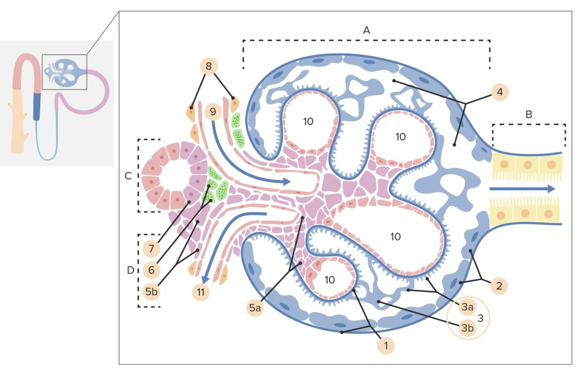
  • Juxtaglomerular (JG) cells:
    • Enlarged smooth muscle cells primarily in the afferent Afferent Neurons which conduct nerve impulses to the central nervous system. Nervous System: Histology (and some in the efferent Efferent Neurons which send impulses peripherally to activate muscles or secretory cells. Nervous System: Histology) arterioles Arterioles The smallest divisions of the arteries located between the muscular arteries and the capillaries. Arteries: Histology
    • Can dilate or constrict the arterioles Arterioles The smallest divisions of the arteries located between the muscular arteries and the capillaries. Arteries: Histology, adjusting the pressure within the glomerulus
    • Secrete renin Renin A highly specific (leu-leu) endopeptidase that generates angiotensin I from its precursor angiotensinogen, leading to a cascade of reactions which elevate blood pressure and increase sodium retention by the kidney in the renin-angiotensin system. Renal Sodium and Water Regulation in response to hypovolemia Hypovolemia Sepsis in Children and hypotension Hypotension Hypotension is defined as low blood pressure, specifically < 90/60 mm Hg, and is most commonly a physiologic response. Hypotension may be mild, serious, or life threatening, depending on the cause. Hypotension
  • Macula Macula An oval area in the retina, 3 to 5 mm in diameter, usually located temporal to the posterior pole of the eye and slightly below the level of the optic disk. It is characterized by the presence of a yellow pigment diffusely permeating the inner layers, contains the fovea centralis in its center, and provides the best phototropic visual acuity. It is devoid of retinal blood vessels, except in its periphery, and receives nourishment from the choriocapillaris of the choroid. Eye: Anatomy densa (MD) cells:
    • Specialized slender, closely spaced epithelial cells in the DCT that are adjacent to the JG cells
    • Detect sodium Sodium A member of the alkali group of metals. It has the atomic symbol na, atomic number 11, and atomic weight 23. Hyponatremia concentration of the fluid in the DCT
    • Signals JG cells to dilate or constrict arterioles Arterioles The smallest divisions of the arteries located between the muscular arteries and the capillaries. Arteries: Histology, adjusting the GFR GFR The volume of water filtered out of plasma through glomerular capillary walls into Bowman’s capsules per unit of time. It is considered to be equivalent to inulin clearance. Kidney Function Tests in order to maintain homeostasis Homeostasis The processes whereby the internal environment of an organism tends to remain balanced and stable. Cell Injury and Death
  • Mesangial cells:
    • Flat and elongated cells in the cleft between the afferent Afferent Neurons which conduct nerve impulses to the central nervous system. Nervous System: Histology and efferent Efferent Neurons which send impulses peripherally to activate muscles or secretory cells. Nervous System: Histology arterioles Arterioles The smallest divisions of the arteries located between the muscular arteries and the capillaries. Arteries: Histology
    • Connected to MD and JG cells by gap junctions Gap Junctions Connections between cells which allow passage of small molecules and electric current. Gap junctions were first described anatomically as regions of close apposition between cells with a narrow (1-2 nm) gap between cell membranes. The variety in the properties of gap junctions is reflected in the number of connexins, the family of proteins which form the junctions. The Cell: Cell Junctions
    • Role is still unclear, but potentially mediate communication Communication The exchange or transmission of ideas, attitudes, or beliefs between individuals or groups. Decision-making Capacity and Legal Competence between MD and JG cells
Structure of a renal corpuscle and the juxtaglomerular apparatus

Structure of a renal corpuscle and the juxtaglomerular apparatus:
A: Renal corpuscle
B: Proximal tubule
C: Distal convoluted tube
D: Juxtaglomerular apparatus
1: Basement membrane
2: Bowman capsule, parietal layer
3: Bowman capsule, visceral layer
3a: Podocyte pedicles
3b: Podocyte
4: Bowman space (urinary space)
5a: Mesangium—intraglomerular mesangial cells
5b: Mesangium—extraglomerular mesangial cells
6: Juxtaglomerular cells
7: Macula densa
8: Myocytes (cells of smooth muscle)
9: Afferent arteriole
10: Glomerulus capillaries
11: Efferent arteriole.

Image by Lecturio.

Types of nephrons

Nephrons are divided into cortical and juxtamedullary nephrons based on their location. 
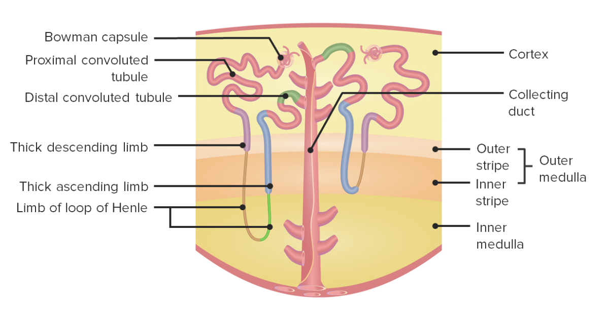
  • Cortical nephrons:
    • Located almost entirely in the cortex
    • Loops of Henle have a short course inside the medulla.
  • Juxtamedullary nephrons:
    • Located close to the corticomedullary junction
    • Loop of Henle Loop of Henle The U-shaped portion of the renal tubule in the kidney medulla, consisting of a descending limb and an ascending limb. It is situated between the proximal kidney tubule and the distal kidney tubule. Tubular System traverses deep into the medulla.
    • Responsible for maintaining high osmotic gradient within medulla
    • Allow for stronger concentration of urine
Nephron anatomy

Nephron anatomy:
The left side shows a juxtamedullary nephron, while the right side shows a cortical nephron.
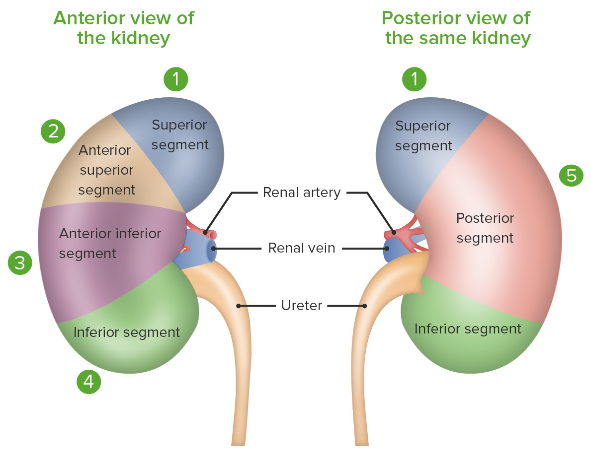
Image by Lecturio.

Neurovasculature

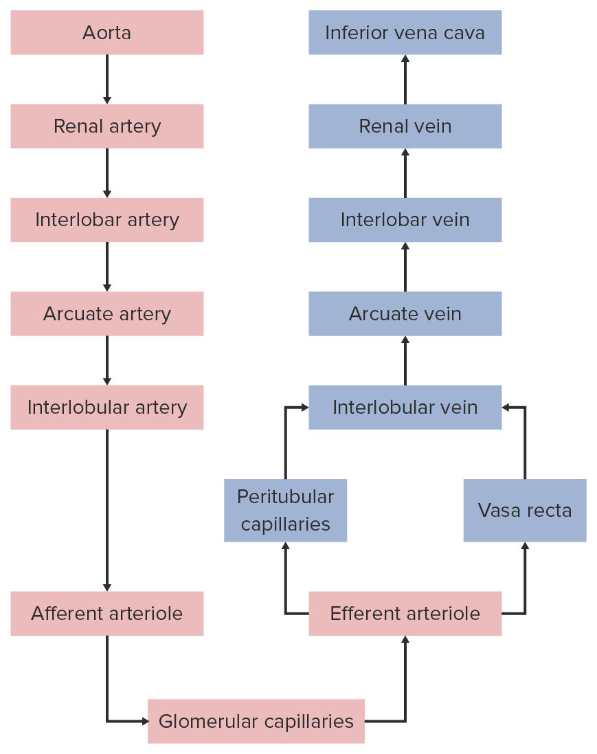
Renal and segmental arteries Arteries Arteries are tubular collections of cells that transport oxygenated blood and nutrients from the heart to the tissues of the body. The blood passes through the arteries in order of decreasing luminal diameter, starting in the largest artery (the aorta) and ending in the small arterioles. Arteries are classified into 3 types: large elastic arteries, medium muscular arteries, and small arteries and arterioles. Arteries: Histology

  • Each kidney is perfused by a renal artery Renal artery A branch of the abdominal aorta which supplies the kidneys, adrenal glands and ureters. Glomerular Filtration:
    • Branch off the abdominal aorta Abdominal Aorta The aorta from the diaphragm to the bifurcation into the right and left common iliac arteries. Posterior Abdominal Wall: Anatomy below the superior mesenteric artery Superior mesenteric artery A large vessel supplying the whole length of the small intestine except the superior part of the duodenum. It also supplies the cecum and the ascending part of the colon and about half the transverse part of the colon. It arises from the anterior surface of the aorta below the celiac artery at the level of the first lumbar vertebra. Small Intestine: Anatomy (SMA)
    • Right renal artery Renal artery A branch of the abdominal aorta which supplies the kidneys, adrenal glands and ureters. Glomerular Filtration passes posterior to the vena cava.
  • The renal arteries Arteries Arteries are tubular collections of cells that transport oxygenated blood and nutrients from the heart to the tissues of the body. The blood passes through the arteries in order of decreasing luminal diameter, starting in the largest artery (the aorta) and ending in the small arterioles. Arteries are classified into 3 types: large elastic arteries, medium muscular arteries, and small arteries and arterioles. Arteries: Histology divide into 5 segmental arteries Arteries Arteries are tubular collections of cells that transport oxygenated blood and nutrients from the heart to the tissues of the body. The blood passes through the arteries in order of decreasing luminal diameter, starting in the largest artery (the aorta) and ending in the small arterioles. Arteries are classified into 3 types: large elastic arteries, medium muscular arteries, and small arteries and arterioles. Arteries: Histology, which irrigate separate segments of the kidney (no anastomosis between the segments).
  • Renal segments:
    • Superior
    • Anterior superior
    • Anterior inferior
    • Inferior
    • Posterior
Segments of the kidney

Segments of the kidney

Image by Lecturio.
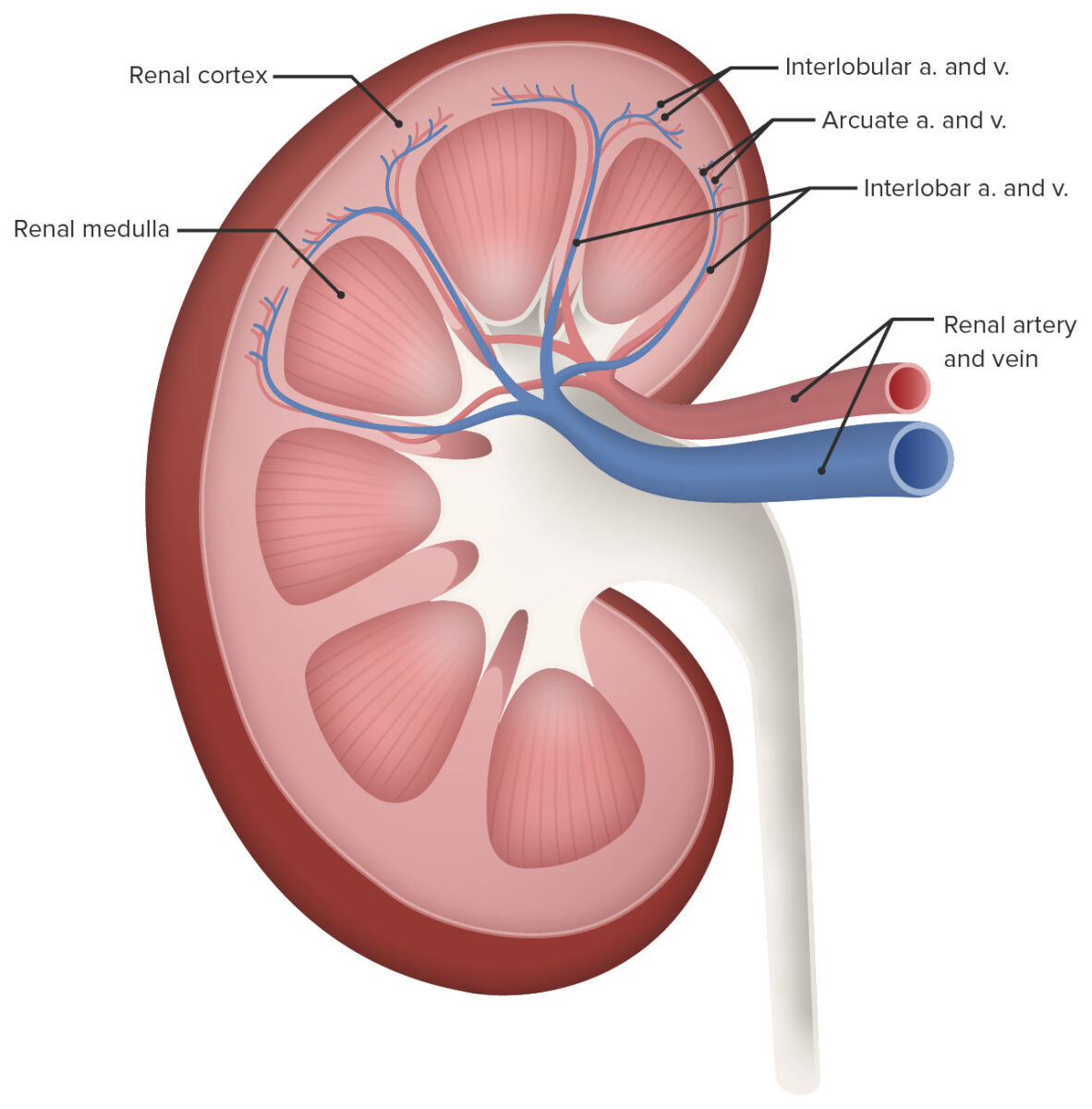
Smaller arteries Arteries Arteries are tubular collections of cells that transport oxygenated blood and nutrients from the heart to the tissues of the body. The blood passes through the arteries in order of decreasing luminal diameter, starting in the largest artery (the aorta) and ending in the small arterioles. Arteries are classified into 3 types: large elastic arteries, medium muscular arteries, and small arteries and arterioles. Arteries: Histology, capillaries Capillaries Capillaries are the primary structures in the circulatory system that allow the exchange of gas, nutrients, and other materials between the blood and the extracellular fluid (ECF). Capillaries are the smallest of the blood vessels. Because a capillary diameter is so small, only 1 RBC may pass through at a time. Capillaries: Histology, and veins Veins Veins are tubular collections of cells, which transport deoxygenated blood and waste from the capillary beds back to the heart. Veins are classified into 3 types: small veins/venules, medium veins, and large veins. Each type contains 3 primary layers: tunica intima, tunica media, and tunica adventitia. Veins: Histology

  • Segmental arteries Arteries Arteries are tubular collections of cells that transport oxygenated blood and nutrients from the heart to the tissues of the body. The blood passes through the arteries in order of decreasing luminal diameter, starting in the largest artery (the aorta) and ending in the small arterioles. Arteries are classified into 3 types: large elastic arteries, medium muscular arteries, and small arteries and arterioles. Arteries: Histology branch into interlobar arteries Arteries Arteries are tubular collections of cells that transport oxygenated blood and nutrients from the heart to the tissues of the body. The blood passes through the arteries in order of decreasing luminal diameter, starting in the largest artery (the aorta) and ending in the small arterioles. Arteries are classified into 3 types: large elastic arteries, medium muscular arteries, and small arteries and arterioles. Arteries: Histology, which run between renal pyramids.
  • Interlobar arteries Arteries Arteries are tubular collections of cells that transport oxygenated blood and nutrients from the heart to the tissues of the body. The blood passes through the arteries in order of decreasing luminal diameter, starting in the largest artery (the aorta) and ending in the small arterioles. Arteries are classified into 3 types: large elastic arteries, medium muscular arteries, and small arteries and arterioles. Arteries: Histology branch into arcuate arteries Arteries Arteries are tubular collections of cells that transport oxygenated blood and nutrients from the heart to the tissues of the body. The blood passes through the arteries in order of decreasing luminal diameter, starting in the largest artery (the aorta) and ending in the small arterioles. Arteries are classified into 3 types: large elastic arteries, medium muscular arteries, and small arteries and arterioles. Arteries: Histology, which run along the base of the renal pyramids in the renal cortex.
  • Arcuate arteries Arteries Arteries are tubular collections of cells that transport oxygenated blood and nutrients from the heart to the tissues of the body. The blood passes through the arteries in order of decreasing luminal diameter, starting in the largest artery (the aorta) and ending in the small arterioles. Arteries are classified into 3 types: large elastic arteries, medium muscular arteries, and small arteries and arterioles. Arteries: Histology send off small branches called interlobular arteries Arteries Arteries are tubular collections of cells that transport oxygenated blood and nutrients from the heart to the tissues of the body. The blood passes through the arteries in order of decreasing luminal diameter, starting in the largest artery (the aorta) and ending in the small arterioles. Arteries are classified into 3 types: large elastic arteries, medium muscular arteries, and small arteries and arterioles. Arteries: Histology.
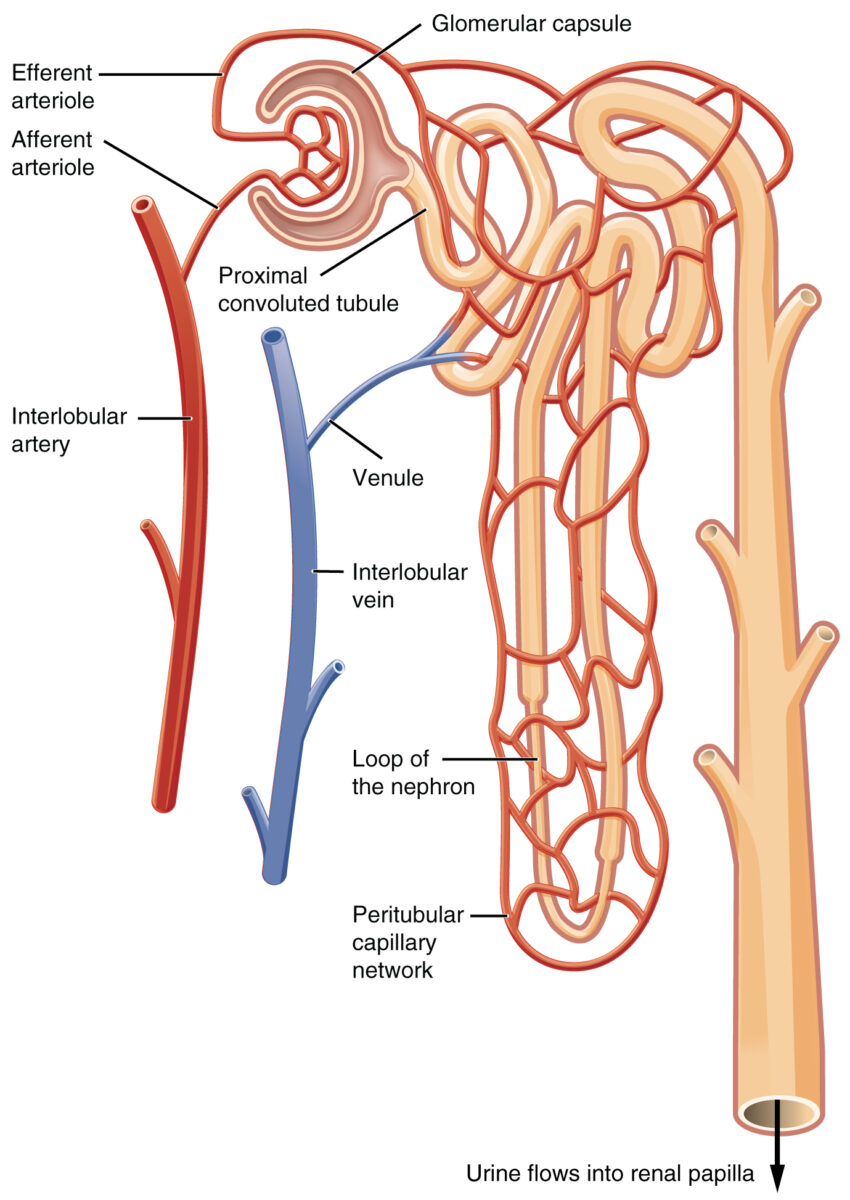
  • Interlobular arteries Arteries Arteries are tubular collections of cells that transport oxygenated blood and nutrients from the heart to the tissues of the body. The blood passes through the arteries in order of decreasing luminal diameter, starting in the largest artery (the aorta) and ending in the small arterioles. Arteries are classified into 3 types: large elastic arteries, medium muscular arteries, and small arteries and arterioles. Arteries: Histology branch into afferent Afferent Neurons which conduct nerve impulses to the central nervous system. Nervous System: Histology arterioles Arterioles The smallest divisions of the arteries located between the muscular arteries and the capillaries. Arteries: Histology, which lead into Bowman capsules.
  • After blood leaves the glomerulus via the efferent Efferent Neurons which send impulses peripherally to activate muscles or secretory cells. Nervous System: Histology arterioles Arterioles The smallest divisions of the arteries located between the muscular arteries and the capillaries. Arteries: Histology, it travels to either the peritubular capillaries Peritubular capillaries Glomerular Filtration or the vasa recta Vasa recta Glomerular Filtration.
  • Peritubular capillaries Peritubular capillaries Glomerular Filtration: supply the cortical tubules (e.g., PCT PCT The renal tubule portion that extends from the bowman capsule in the kidney cortex into the kidney medulla. The proximal tubule consists of a convoluted proximal segment in the cortex, and a distal straight segment descending into the medulla where it forms the u-shaped loop of henle. Osmotic Diuretics and DCT)
  • Vasa recta Vasa recta Glomerular Filtration:
    • Capillary networks that surround the loops of Henle in the medulla
    • Highly permeable to solute and water
    • Vital in maintaining the osmotic gradient in the medulla
  • Blood from the peritubular and vasa recta Vasa recta Glomerular Filtration capillaries Capillaries Capillaries are the primary structures in the circulatory system that allow the exchange of gas, nutrients, and other materials between the blood and the extracellular fluid (ECF). Capillaries are the smallest of the blood vessels. Because a capillary diameter is so small, only 1 RBC may pass through at a time. Capillaries: Histology drain into interlobular veins Veins Veins are tubular collections of cells, which transport deoxygenated blood and waste from the capillary beds back to the heart. Veins are classified into 3 types: small veins/venules, medium veins, and large veins. Each type contains 3 primary layers: tunica intima, tunica media, and tunica adventitia. Veins: Histology.
  • Interlobular veins Veins Veins are tubular collections of cells, which transport deoxygenated blood and waste from the capillary beds back to the heart. Veins are classified into 3 types: small veins/venules, medium veins, and large veins. Each type contains 3 primary layers: tunica intima, tunica media, and tunica adventitia. Veins: Histology → arcuate veins Veins Veins are tubular collections of cells, which transport deoxygenated blood and waste from the capillary beds back to the heart. Veins are classified into 3 types: small veins/venules, medium veins, and large veins. Each type contains 3 primary layers: tunica intima, tunica media, and tunica adventitia. Veins: Histology → interlobar veins Veins Veins are tubular collections of cells, which transport deoxygenated blood and waste from the capillary beds back to the heart. Veins are classified into 3 types: small veins/venules, medium veins, and large veins. Each type contains 3 primary layers: tunica intima, tunica media, and tunica adventitia. Veins: Histology → segmental veins Veins Veins are tubular collections of cells, which transport deoxygenated blood and waste from the capillary beds back to the heart. Veins are classified into 3 types: small veins/venules, medium veins, and large veins. Each type contains 3 primary layers: tunica intima, tunica media, and tunica adventitia. Veins: Histology renal vein Renal vein Short thick veins which return blood from the kidneys to the vena cava. Glomerular Filtration
  • Each kidney has a single renal vein Renal vein Short thick veins which return blood from the kidneys to the vena cava. Glomerular Filtration. These veins Veins Veins are tubular collections of cells, which transport deoxygenated blood and waste from the capillary beds back to the heart. Veins are classified into 3 types: small veins/venules, medium veins, and large veins. Each type contains 3 primary layers: tunica intima, tunica media, and tunica adventitia. Veins: Histology: 
    • Drain directly into the inferior vena cava Inferior vena cava The venous trunk which receives blood from the lower extremities and from the pelvic and abdominal organs. Mediastinum and Great Vessels: Anatomy
    • Run anterior to the arteries Arteries Arteries are tubular collections of cells that transport oxygenated blood and nutrients from the heart to the tissues of the body. The blood passes through the arteries in order of decreasing luminal diameter, starting in the largest artery (the aorta) and ending in the small arterioles. Arteries are classified into 3 types: large elastic arteries, medium muscular arteries, and small arteries and arterioles. Arteries: Histology 
    • The left renal vein Renal vein Short thick veins which return blood from the kidneys to the vena cava. Glomerular Filtration passes anterior to the abdominal aorta Abdominal Aorta The aorta from the diaphragm to the bifurcation into the right and left common iliac arteries. Posterior Abdominal Wall: Anatomy.

Summary of blood flow Blood flow Blood flow refers to the movement of a certain volume of blood through the vasculature over a given unit of time (e.g., mL per minute). Vascular Resistance, Flow, and Mean Arterial Pressure through the kidney

Renal circulation

Renal circulation

Image by Lecturio.

Nerves

Renal innervation includes both afferent Afferent Neurons which conduct nerve impulses to the central nervous system. Nervous System: Histology and efferent Efferent Neurons which send impulses peripherally to activate muscles or secretory cells. Nervous System: Histology nerves through the renal nerve plexus. Innervation is via the autonomic nervous system Autonomic nervous system The ANS is a component of the peripheral nervous system that uses both afferent (sensory) and efferent (effector) neurons, which control the functioning of the internal organs and involuntary processes via connections with the CNS. The ANS consists of the sympathetic and parasympathetic nervous systems. Autonomic Nervous System: Anatomy, primarily through sympathetic fibers:

  • Sympathetic efferent Efferent Neurons which send impulses peripherally to activate muscles or secretory cells. Nervous System: Histology nerves:
    • Primarily via splanchnic nerves
    • Only efferent Efferent Neurons which send impulses peripherally to activate muscles or secretory cells. Nervous System: Histology innervation to the nephrons and renal vasculature
    • Concentrated most heavily around the afferent Afferent Neurons which conduct nerve impulses to the central nervous system. Nervous System: Histology arterioles Arterioles The smallest divisions of the arteries located between the muscular arteries and the capillaries. Arteries: Histology, TAL TAL Renal Sodium and Water Regulation, and DCT
    • Stimulation can activate the RAAS RAAS A blood pressure regulating system of interacting components that include renin; angiotensinogen; angiotensin converting enzyme; angiotensin i; angiotensin ii; and angiotensinase. Renin, an enzyme produced in the kidney, acts on angiotensinogen, an alpha-2 globulin produced by the liver, forming angiotensin I. Angiotensin-converting enzyme, contained in the lung, acts on angiotensin I in the plasma converting it to angiotensin II, an extremely powerful vasoconstrictor. Angiotensin II causes contraction of the arteriolar and renal vascular smooth muscle, leading to retention of salt and water in the kidney and increased arterial blood pressure. In addition, angiotensin II stimulates the release of aldosterone from the adrenal cortex, which in turn also increases salt and water retention in the kidney. Angiotensin-converting enzyme also breaks down bradykinin, a powerful vasodilator and component of the kallikrein-kinin system. Adrenal Hormones.
  • Sensory Sensory Neurons which conduct nerve impulses to the central nervous system. Nervous System: Histology afferent Afferent Neurons which conduct nerve impulses to the central nervous system. Nervous System: Histology nerves: involved in blood pressure regulation
  • Visceral afferent Afferent Neurons which conduct nerve impulses to the central nervous system. Nervous System: Histology nerves: transmit pain Pain An unpleasant sensation induced by noxious stimuli which are detected by nerve endings of nociceptive neurons. Pain: Types and Pathways signals to spinal cord Spinal cord The spinal cord is the major conduction pathway connecting the brain to the body; it is part of the CNS. In cross section, the spinal cord is divided into an H-shaped area of gray matter (consisting of synapsing neuronal cell bodies) and a surrounding area of white matter (consisting of ascending and descending tracts of myelinated axons). Spinal Cord: Anatomy segments T11–L2 

Functions

Functions of the kidneys include:

  • Filter the blood and excrete water-soluble waste in the urine
  • Regulate total body water Total body water Body Fluid Compartments by appropriately concentrating the urine according to the body’s osmotic status
  • Regulate hemodynamic/blood pressure via the RAAS RAAS A blood pressure regulating system of interacting components that include renin; angiotensinogen; angiotensin converting enzyme; angiotensin i; angiotensin ii; and angiotensinase. Renin, an enzyme produced in the kidney, acts on angiotensinogen, an alpha-2 globulin produced by the liver, forming angiotensin I. Angiotensin-converting enzyme, contained in the lung, acts on angiotensin I in the plasma converting it to angiotensin II, an extremely powerful vasoconstrictor. Angiotensin II causes contraction of the arteriolar and renal vascular smooth muscle, leading to retention of salt and water in the kidney and increased arterial blood pressure. In addition, angiotensin II stimulates the release of aldosterone from the adrenal cortex, which in turn also increases salt and water retention in the kidney. Angiotensin-converting enzyme also breaks down bradykinin, a powerful vasodilator and component of the kallikrein-kinin system. Adrenal Hormones
  • Regulate acid–base status
  • Maintain bone metabolism Bone metabolism Bone is the primary storage site of calcium in the body; thus, bone metabolism plays a critical role in maintaining normal calcium levels. Bone metabolism (and thus calcium levels) are primarily regulated by 3 hormones, namely, calcitonin, parathyroid hormone (PTH), and vitamin D. Calcium Hemostasis and Bone Metabolism via selective excretion/reabsorption of calcium Calcium A basic element found in nearly all tissues. It is a member of the alkaline earth family of metals with the atomic symbol ca, atomic number 20, and atomic weight 40. Calcium is the most abundant mineral in the body and combines with phosphorus to form calcium phosphate in the bones and teeth. It is essential for the normal functioning of nerves and muscles and plays a role in blood coagulation (as factor IV) and in many enzymatic processes. Electrolytes and phosphate Phosphate Inorganic salts of phosphoric acid. Electrolytes
  • Produce RBCs RBCs Erythrocytes, or red blood cells (RBCs), are the most abundant cells in the blood. While erythrocytes in the fetus are initially produced in the yolk sac then the liver, the bone marrow eventually becomes the main site of production. Erythrocytes: Histology via secretion Secretion Coagulation Studies of erythropoietin Erythropoietin Glycoprotein hormone, secreted chiefly by the kidney in the adult and the liver in the fetus, that acts on erythroid stem cells of the bone marrow to stimulate proliferation and differentiation. Erythrocytes: Histology

Clinical Relevance

  • Duplications of the collecting system: known as a “duplex system.” Duplications of the collecting system are the most common congenital anomaly of the urinary tract Urinary tract The urinary tract is located in the abdomen and pelvis and consists of the kidneys, ureters, urinary bladder, and urethra. The structures permit the excretion of urine from the body. Urine flows from the kidneys through the ureters to the urinary bladder and out through the urethra. Urinary Tract: Anatomy. In these cases, a kidney will have 2 separate pelvicaliceal systems and 2 ureters Ureters One of a pair of thick-walled tubes that transports urine from the kidney pelvis to the urinary bladder. Urinary Tract: Anatomy. Ureteral insertion into the bladder Bladder A musculomembranous sac along the urinary tract. Urine flows from the kidneys into the bladder via the ureters, and is held there until urination. Pyelonephritis and Perinephric Abscess from the duplicated system is often abnormal as well. Most affected individuals are asymptomatic, though recurrent urinary tract Urinary tract The urinary tract is located in the abdomen and pelvis and consists of the kidneys, ureters, urinary bladder, and urethra. The structures permit the excretion of urine from the body. Urine flows from the kidneys through the ureters to the urinary bladder and out through the urethra. Urinary Tract: Anatomy infection (UTIs) or obstruction may occur.
  • Renal agenesis Agenesis Teratogenic Birth Defects: congenital absence of a kidney, specifically, renal parenchymal tissue. Renal agenesis Agenesis Teratogenic Birth Defects results from disruption of metanephric development. Most affected individuals are asymptomatic and diagnosed incidentally on imaging. Renal agenesis Agenesis Teratogenic Birth Defects is often associated with additional congenital anomalies.
  • Polycystic kidney disease (PKD): genetic condition caused by either an autosomal recessive Autosomal recessive Autosomal inheritance, both dominant and recessive, refers to the transmission of genes from the 22 autosomal chromosomes. Autosomal recessive diseases are only expressed when 2 copies of the recessive allele are inherited. Autosomal Recessive and Autosomal Dominant Inheritance mutation Mutation Genetic mutations are errors in DNA that can cause protein misfolding and dysfunction. There are various types of mutations, including chromosomal, point, frameshift, and expansion mutations. Types of Mutations in the PKHD1 PKHD1 Autosomal Recessive Polycystic Kidney Disease (ARPKD) gene Gene A category of nucleic acid sequences that function as units of heredity and which code for the basic instructions for the development, reproduction, and maintenance of organisms. Basic Terms of Genetics (fibrocystin) or an autosomal dominant Autosomal dominant Autosomal inheritance, both dominant and recessive, refers to the transmission of genes from the 22 autosomal chromosomes. Autosomal dominant diseases are expressed when only 1 copy of the dominant allele is inherited. Autosomal Recessive and Autosomal Dominant Inheritance mutation Mutation Genetic mutations are errors in DNA that can cause protein misfolding and dysfunction. There are various types of mutations, including chromosomal, point, frameshift, and expansion mutations. Types of Mutations in either the PKD1 or PKD2 gene Gene A category of nucleic acid sequences that function as units of heredity and which code for the basic instructions for the development, reproduction, and maintenance of organisms. Basic Terms of Genetics ( polycystin Polycystin Autosomal dominant polycystic kidney disease (ADPKD) proteins Proteins Linear polypeptides that are synthesized on ribosomes and may be further modified, crosslinked, cleaved, or assembled into complex proteins with several subunits. The specific sequence of amino acids determines the shape the polypeptide will take, during protein folding, and the function of the protein. Energy Homeostasis). Autosomal recessive Autosomal recessive Autosomal inheritance, both dominant and recessive, refers to the transmission of genes from the 22 autosomal chromosomes. Autosomal recessive diseases are only expressed when 2 copies of the recessive allele are inherited. Autosomal Recessive and Autosomal Dominant Inheritance PKD is characterized by multiple microscopic cysts Cysts Any fluid-filled closed cavity or sac that is lined by an epithelium. Cysts can be of normal, abnormal, non-neoplastic, or neoplastic tissues. Fibrocystic Change and can have serious effects in utero. Autosomal dominant Autosomal dominant Autosomal inheritance, both dominant and recessive, refers to the transmission of genes from the 22 autosomal chromosomes. Autosomal dominant diseases are expressed when only 1 copy of the dominant allele is inherited. Autosomal Recessive and Autosomal Dominant Inheritance PKD is characterized by multiple larger cysts Cysts Any fluid-filled closed cavity or sac that is lined by an epithelium. Cysts can be of normal, abnormal, non-neoplastic, or neoplastic tissues. Fibrocystic Change and most commonly presents in adulthood with hematuria Hematuria Presence of blood in the urine. Renal Cell Carcinoma and hypertension Hypertension Hypertension, or high blood pressure, is a common disease that manifests as elevated systemic arterial pressures. Hypertension is most often asymptomatic and is found incidentally as part of a routine physical examination or during triage for an unrelated medical encounter. Hypertension.
  • Goodpasture syndrome Goodpasture Syndrome Goodpasture syndrome, also known as anti-glomerular basement membrane (GBM) disease, is an autoimmune disease characterized by circulating antibodies directed against glomerular and alveolar basement membranes. Affected individuals present with symptoms of rapidly progressive glomerulonephritis and alveolar hemorrhage. Goodpasture Syndrome: also known as anti–glomerular basement membrane Basement membrane A darkly stained mat-like extracellular matrix (ecm) that separates cell layers, such as epithelium from endothelium or a layer of connective tissue. The ecm layer that supports an overlying epithelium or endothelium is called basal lamina. Basement membrane (bm) can be formed by the fusion of either two adjacent basal laminae or a basal lamina with an adjacent reticular lamina of connective tissue. Bm, composed mainly of type IV collagen; glycoprotein laminin; and proteoglycan, provides barriers as well as channels between interacting cell layers. Thin Basement Membrane Nephropathy (TBMN) (anti-GBM) disease. Goodpasture syndrome Goodpasture Syndrome Goodpasture syndrome, also known as anti-glomerular basement membrane (GBM) disease, is an autoimmune disease characterized by circulating antibodies directed against glomerular and alveolar basement membranes. Affected individuals present with symptoms of rapidly progressive glomerulonephritis and alveolar hemorrhage. Goodpasture Syndrome is an autoimmune disease characterized by circulating antibodies Antibodies Immunoglobulins (Igs), also known as antibodies, are glycoprotein molecules produced by plasma cells that act in immune responses by recognizing and binding particular antigens. The various Ig classes are IgG (the most abundant), IgM, IgE, IgD, and IgA, which differ in their biologic features, structure, target specificity, and distribution. Immunoglobulins: Types and Functions directed against glomerular and alveolar basement membranes. Presentation is with signs and symptoms of rapidly progressive glomerulonephritis Rapidly Progressive Glomerulonephritis Rapidly progressive glomerulonephritis (RPGN) is a syndrome of severe glomerular disease with progressive loss of kidney function within weeks to months. Histologically, crescents (the proliferation of epithelial cells and the infiltration of monocytes/macrophages in the Bowman space) are found in the glomeruli and arise from immunologic injury. Rapidly Progressive Glomerulonephritis and alveolar hemorrhage. Management includes plasmapheresis Plasmapheresis Procedure whereby plasma is separated and extracted from anticoagulated whole blood and the red cells retransfused to the donor. Plasmapheresis is also employed for therapeutic use. Stevens-Johnson Syndrome and immunosuppressants Immunosuppressants Immunosuppressants are a class of drugs widely used in the management of autoimmune conditions and organ transplant rejection. The general effect is dampening of the immune response. Immunosuppressants. Renal transplantation is an option in individuals who develop end-stage renal failure Renal failure Conditions in which the kidneys perform below the normal level in the ability to remove wastes, concentrate urine, and maintain electrolyte balance; blood pressure; and calcium metabolism. Renal insufficiency can be classified by the degree of kidney damage (as measured by the level of proteinuria) and reduction in glomerular filtration rate. Crush Syndrome
  • Alport syndrome Alport Syndrome Alport syndrome, also called hereditary nephritis, is a genetic disorder caused by a mutation in the genes encoding for the alpha chains of type IV collagen, resulting in the production of abnormal type IV collagen strands. Patients present with glomerulonephritis, hypertension, edema, hematuria, and proteinuria, as well as with ocular and auditory findings. Alport Syndrome: also called hereditary nephritis. Alport syndrome Alport Syndrome Alport syndrome, also called hereditary nephritis, is a genetic disorder caused by a mutation in the genes encoding for the alpha chains of type IV collagen, resulting in the production of abnormal type IV collagen strands. Patients present with glomerulonephritis, hypertension, edema, hematuria, and proteinuria, as well as with ocular and auditory findings. Alport Syndrome is a genetic disorder caused by a mutation Mutation Genetic mutations are errors in DNA that can cause protein misfolding and dysfunction. There are various types of mutations, including chromosomal, point, frameshift, and expansion mutations. Types of Mutations in the genes Genes A category of nucleic acid sequences that function as units of heredity and which code for the basic instructions for the development, reproduction, and maintenance of organisms. DNA Types and Structure coding for the alpha chains of type IV collagen Type IV collagen A non-fibrillar collagen found in the structure of basement membrane. Collagen type IV molecules assemble to form a sheet-like network which is involved in maintaining the structural integrity of basement membranes. The predominant form of the protein is comprised of two alpha1(IV) subunits and one alpha2(IV) subunit, however, at least six different alpha subunits can be incorporated into the heterotrimer. Alport Syndrome, resulting in the production of abnormal type IV collagen Type IV collagen A non-fibrillar collagen found in the structure of basement membrane. Collagen type IV molecules assemble to form a sheet-like network which is involved in maintaining the structural integrity of basement membranes. The predominant form of the protein is comprised of two alpha1(IV) subunits and one alpha2(IV) subunit, however, at least six different alpha subunits can be incorporated into the heterotrimer. Alport Syndrome strands. Presentation is with glomerulonephritis, hypertension Hypertension Hypertension, or high blood pressure, is a common disease that manifests as elevated systemic arterial pressures. Hypertension is most often asymptomatic and is found incidentally as part of a routine physical examination or during triage for an unrelated medical encounter. Hypertension, edema Edema Edema is a condition in which excess serous fluid accumulates in the body cavity or interstitial space of connective tissues. Edema is a symptom observed in several medical conditions. It can be categorized into 2 types, namely, peripheral (in the extremities) and internal (in an organ or body cavity). Edema, hematuria Hematuria Presence of blood in the urine. Renal Cell Carcinoma, and proteinuria Proteinuria The presence of proteins in the urine, an indicator of kidney diseases. Nephrotic Syndrome in Children, as well as with ocular and auditory findings. A renal biopsy Renal Biopsy Antineutrophil Cytoplasmic Antibody (ANCA)-Associated Vasculitis will show characteristic GBM splitting Splitting Defense Mechanisms.
  • Hydronephrosis Hydronephrosis Hydronephrosis is dilation of the renal collecting system as a result of the obstruction of urine outflow. Hydronephrosis can be unilateral or bilateral. Nephrolithiasis is the most common cause of hydronephrosis in young adults, while prostatic hyperplasia and neoplasm are seen in older patients. Hydronephrosis: dilation of the renal collecting system as a result of the obstruction of urine outflow. Hydronephrosis Hydronephrosis Hydronephrosis is dilation of the renal collecting system as a result of the obstruction of urine outflow. Hydronephrosis can be unilateral or bilateral. Nephrolithiasis is the most common cause of hydronephrosis in young adults, while prostatic hyperplasia and neoplasm are seen in older patients. Hydronephrosis can be unilateral or bilateral. Nephrolithiasis Nephrolithiasis Nephrolithiasis is the formation of a stone, or calculus, anywhere along the urinary tract caused by precipitations of solutes in the urine. The most common type of kidney stone is the calcium oxalate stone, but other types include calcium phosphate, struvite (ammonium magnesium phosphate), uric acid, and cystine stones. Nephrolithiasis is the most common cause of hydronephrosis Hydronephrosis Hydronephrosis is dilation of the renal collecting system as a result of the obstruction of urine outflow. Hydronephrosis can be unilateral or bilateral. Nephrolithiasis is the most common cause of hydronephrosis in young adults, while prostatic hyperplasia and neoplasm are seen in older patients. Hydronephrosis in young adults, while prostatic hyperplasia Hyperplasia An increase in the number of cells in a tissue or organ without tumor formation. It differs from hypertrophy, which is an increase in bulk without an increase in the number of cells. Cellular Adaptation and neoplasm are seen in older individuals. Presentation can be with flank pain Flank pain Pain emanating from below the ribs and above the ilium. Renal Cell Carcinoma, dysuria Dysuria Painful urination. It is often associated with infections of the lower urinary tract. Urinary Tract Infections (UTIs), urgency, fever Fever Fever is defined as a measured body temperature of at least 38°C (100.4°F). Fever is caused by circulating endogenous and/or exogenous pyrogens that increase levels of prostaglandin E2 in the hypothalamus. Fever is commonly associated with chills, rigors, sweating, and flushing of the skin. Fever, palpable abdominal mass Mass Three-dimensional lesion that occupies a space within the breast Imaging of the Breast, fever Fever Fever is defined as a measured body temperature of at least 38°C (100.4°F). Fever is caused by circulating endogenous and/or exogenous pyrogens that increase levels of prostaglandin E2 in the hypothalamus. Fever is commonly associated with chills, rigors, sweating, and flushing of the skin. Fever, and hypertension Hypertension Hypertension, or high blood pressure, is a common disease that manifests as elevated systemic arterial pressures. Hypertension is most often asymptomatic and is found incidentally as part of a routine physical examination or during triage for an unrelated medical encounter. Hypertension. Diagnosis includes imaging with ultrasonography, CT, or IV pyelography Pyelography Hydronephrosis.
  • Nutcracker syndrome: occurs when the left renal vein Renal vein Short thick veins which return blood from the kidneys to the vena cava. Glomerular Filtration is compressed, affecting the venous drainage (and subsequently the arterial supply) of the left kidney, the left adrenal gland, and the left testis (in men) or the left ovary (in women). Symptoms are often vague but may include intermittent flank pain Flank pain Pain emanating from below the ribs and above the ilium. Renal Cell Carcinoma, hematuria Hematuria Presence of blood in the urine. Renal Cell Carcinoma, pelvic pain Pain An unpleasant sensation induced by noxious stimuli which are detected by nerve endings of nociceptive neurons. Pain: Types and Pathways, scrotal edema Scrotal Edema Minimal Change Disease, varicocele Varicocele A condition characterized by the dilated tortuous veins of the spermatic cord with a marked left-sided predominance. Adverse effect on male fertility occurs when varicocele leads to an increased scrotal (and testicular) temperature and reduced testicular volume. Varicocele, Hydrocele, and Spermatocele, or pelvic congestion syndrome. 
  • Renal cell carcinoma Renal cell carcinoma Renal cell carcinoma (RCC) is a tumor that arises from the lining of the renal tubular system within the renal cortex. Renal cell carcinoma is responsible for 80%-85% of all primary renal neoplasms. Most RCCs arise sporadically, but smoking, hypertension, and obesity are linked to its development. Renal Cell Carcinoma ( RCC RCC Renal cell carcinoma (RCC) is a tumor that arises from the lining of the renal tubular system within the renal cortex. Renal cell carcinoma is responsible for 80%-85% of all primary renal neoplasms. Most RCCs arise sporadically, but smoking, hypertension, and obesity are linked to its development. Renal Cell Carcinoma): tumor Tumor Inflammation that arises from the lining of the renal tubular system within the renal cortex. Renal cell carcinoma Renal cell carcinoma Renal cell carcinoma (RCC) is a tumor that arises from the lining of the renal tubular system within the renal cortex. Renal cell carcinoma is responsible for 80%-85% of all primary renal neoplasms. Most RCCs arise sporadically, but smoking, hypertension, and obesity are linked to its development. Renal Cell Carcinoma is responsible for 80%–85% of all primary renal neoplasms Neoplasms New abnormal growth of tissue. Malignant neoplasms show a greater degree of anaplasia and have the properties of invasion and metastasis, compared to benign neoplasms. Benign Bone Tumors. Most RCCs arise sporadically, but smoking Smoking Willful or deliberate act of inhaling and exhaling smoke from burning substances or agents held by hand. Interstitial Lung Diseases, hypertension Hypertension Hypertension, or high blood pressure, is a common disease that manifests as elevated systemic arterial pressures. Hypertension is most often asymptomatic and is found incidentally as part of a routine physical examination or during triage for an unrelated medical encounter. Hypertension, and obesity Obesity Obesity is a condition associated with excess body weight, specifically with the deposition of excessive adipose tissue. Obesity is considered a global epidemic. Major influences come from the western diet and sedentary lifestyles, but the exact mechanisms likely include a mixture of genetic and environmental factors. Obesity are linked to the development of the disease. The condition is usually asymptomatic. The classic clinical triad of RCC RCC Renal cell carcinoma (RCC) is a tumor that arises from the lining of the renal tubular system within the renal cortex. Renal cell carcinoma is responsible for 80%-85% of all primary renal neoplasms. Most RCCs arise sporadically, but smoking, hypertension, and obesity are linked to its development. Renal Cell Carcinoma is flank pain Flank pain Pain emanating from below the ribs and above the ilium. Renal Cell Carcinoma, hematuria Hematuria Presence of blood in the urine. Renal Cell Carcinoma, and a palpable abdominal renal mass Mass Three-dimensional lesion that occupies a space within the breast Imaging of the Breast, but only in about 9% of cases. Renal cell carcinoma Renal cell carcinoma Renal cell carcinoma (RCC) is a tumor that arises from the lining of the renal tubular system within the renal cortex. Renal cell carcinoma is responsible for 80%-85% of all primary renal neoplasms. Most RCCs arise sporadically, but smoking, hypertension, and obesity are linked to its development. Renal Cell Carcinoma is usually diagnosed via CT scan.
  • Horseshoe kidney Horseshoe Kidney Congenital Renal Abnormalities: developmental defect of kidneys in which inferior poles are fused together. As the kidney attempts to migrate superiorly during development, it is blocked by the inferior mesenteric artery Inferior mesenteric artery The artery supplying nearly all the left half of the transverse colon, the whole of the descending colon, the sigmoid colon, and the greater part of the rectum. It is smaller than the superior mesenteric artery and arises from the aorta above its bifurcation into the common iliac arteries. Small Intestine: Anatomy. The vascular supplies and collecting system of the kidney also tend to have various degrees of distortion Distortion Defense Mechanisms. Affected individuals are typically asymptomatic; incidental diagnosis is made on imaging. Other presentations include infection, obstruction, hydronephrosis Hydronephrosis Hydronephrosis is dilation of the renal collecting system as a result of the obstruction of urine outflow. Hydronephrosis can be unilateral or bilateral. Nephrolithiasis is the most common cause of hydronephrosis in young adults, while prostatic hyperplasia and neoplasm are seen in older patients. Hydronephrosis, and calculi. 

References

  1. Chalouhy, C. E. (2017). Kidney anatomy. Medscape. Retrieved September 3, 2021, from https://reference.medscape.com/article/1948775-overview
  2. Gulleroglu, K., Gulleroglu, B., Baskin, E. (2014). Nutcracker syndrome. World Journal of Nephrology 3:277–281. Retrieved September 3, 2021, from https://pubmed.ncbi.nlm.nih.gov/25374822/
  3. Soriano, R. M., Penfold, D., Leslie, S. W. (2021). Anatomy, abdomen and pelvis, kidneys. StatPearls. Retrieved September 3, 2021, from https://www.ncbi.nlm.nih.gov/books/NBK482385/
  4. Tadros, N. N. (2020). Horseshoe kidney. Medscape. Retrieved September 3, 2021, from https://reference.medscape.com/article/441510-overview
  5. Wallace, M. A. (1998). Anatomy and physiology of the kidney. AORN Journal 68:800–824. doi: 10.1016/s0001-2092(06)62377-6
  6. Saladin, K. S., Miller, L. (2004). Anatomy and physiology, 3rd ed., pp. 881–887. McGraw-Hill Education.

Create your free account or log in to continue reading!

Sign up now and get free access to Lecturio with concept pages, medical videos, and questions for your medical education.

User Reviews

Unwrap New Skills This Holiday 🎄 Save 30% on all plans now!

Details