Advertisement

Advertisement

Advertisement

Advertisement

Inflammation

Inflammation is a complex set of responses to infection and injury involving leukocytes Leukocytes White blood cells. These include granular leukocytes (basophils; eosinophils; and neutrophils) as well as non-granular leukocytes (lymphocytes and monocytes). White Myeloid Cells: Histology as the principal cellular mediators in the body’s defense against pathogenic organisms. Inflammation is also seen as a response to tissue injury in the process of wound healing Wound healing Wound healing is a physiological process involving tissue repair in response to injury. It involves a complex interaction of various cell types, cytokines, and inflammatory mediators. Wound healing stages include hemostasis, inflammation, granulation, and remodeling. Wound Healing. The 5 cardinal signs of inflammation are pain Pain An unpleasant sensation induced by noxious stimuli which are detected by nerve endings of nociceptive neurons. Pain: Types and Pathways, heat, redness, swelling, and loss of function. The major cellular response involves neutrophils Neutrophils Granular leukocytes having a nucleus with three to five lobes connected by slender threads of chromatin, and cytoplasm containing fine inconspicuous granules and stainable by neutral dyes. Innate Immunity: Phagocytes and Antigen Presentation and macrophages Macrophages The relatively long-lived phagocytic cell of mammalian tissues that are derived from blood monocytes. Main types are peritoneal macrophages; alveolar macrophages; histiocytes; kupffer cells of the liver; and osteoclasts. They may further differentiate within chronic inflammatory lesions to epithelioid cells or may fuse to form foreign body giant cells or langhans giant cells. Innate Immunity: Phagocytes and Antigen Presentation to phagocytose and lyse the injurious organism or repair necrosed tissue after injury. Inflammation can be pathologic if it is prolonged or when normal processes create an excessive response (such as with atherosclerosis Atherosclerosis Atherosclerosis is a common form of arterial disease in which lipid deposition forms a plaque in the blood vessel walls. Atherosclerosis is an incurable disease, for which there are clearly defined risk factors that often can be reduced through a change in lifestyle and behavior of the patient. Atherosclerosis). There are multiple mediators of inflammation that overlap with innate immunity Innate immunity The capacity of a normal organism to remain unaffected by microorganisms and their toxins. It results from the presence of naturally occurring anti-infective agents, constitutional factors such as body temperature and immediate acting immune cells such as natural killer cells. Innate Immunity: Phagocytes and Antigen Presentation when they respond to injurious stimuli Injurious Stimuli Cell Injury and Death. Inflammation can become chronic, resulting in the formation of granulomas Granulomas A relatively small nodular inflammatory lesion containing grouped mononuclear phagocytes, caused by infectious and noninfectious agents. Sarcoidosis, tissue damage, and the loss of organ function.

Last updated: May 17, 2024

Editorial responsibility: Stanley Oiseth, Lindsay Jones, Evelin Maza

Advertisement

Advertisement

Advertisement

Advertisement

Advertisement

Advertisement

Introduction and Classic Signs

Inflammation is a complex set of responses to infection and injury, involving leukocytes Leukocytes White blood cells. These include granular leukocytes (basophils; eosinophils; and neutrophils) as well as non-granular leukocytes (lymphocytes and monocytes). White Myeloid Cells: Histology as the principal cellular mediators in the body’s defense against pathogenic organisms. Inflammation is also seen as a response to tissue injury in the process of wound healing Wound healing Wound healing is a physiological process involving tissue repair in response to injury. It involves a complex interaction of various cell types, cytokines, and inflammatory mediators. Wound healing stages include hemostasis, inflammation, granulation, and remodeling. Wound Healing.

Inflammatory responses

Types:

  • Physiologic (e.g., wound healing Wound healing Wound healing is a physiological process involving tissue repair in response to injury. It involves a complex interaction of various cell types, cytokines, and inflammatory mediators. Wound healing stages include hemostasis, inflammation, granulation, and remodeling. Wound Healing, localizing infection)
  • Pathologic (e.g., malignancy Malignancy Hemothorax, chronic inflammation)

Triggers of acute inflammation:

  • Infections Infections Invasion of the host organism by microorganisms or their toxins or by parasites that can cause pathological conditions or diseases. Chronic Granulomatous Disease/microbial toxins:
    • Bacterial (primarily)
    • Viral
    • Fungal
    • Parasitic
  • Physical or chemical injury:
    • Thermal injury
    • Radiation Radiation Emission or propagation of acoustic waves (sound), electromagnetic energy waves (such as light; radio waves; gamma rays; or x-rays), or a stream of subatomic particles (such as electrons; neutrons; protons; or alpha particles). Osteosarcoma
    • Environmental chemicals
  • Foreign bodies:
    • Splinters
    • Dirt, glass, or rust in a wound
    • Surgical sutures
  • Necrotic tissue due to ischemia Ischemia A hypoperfusion of the blood through an organ or tissue caused by a pathologic constriction or obstruction of its blood vessels, or an absence of blood circulation. Ischemic Cell Damage or trauma
  • Thrombus
  • Uric acid Uric acid An oxidation product, via xanthine oxidase, of oxypurines such as xanthine and hypoxanthine. It is the final oxidation product of purine catabolism in humans and primates, whereas in most other mammals urate oxidase further oxidizes it to allantoin. Nephrolithiasis (from the breakdown of DNA DNA A deoxyribonucleotide polymer that is the primary genetic material of all cells. Eukaryotic and prokaryotic organisms normally contain DNA in a double-stranded state, yet several important biological processes transiently involve single-stranded regions. DNA, which consists of a polysugar-phosphate backbone possessing projections of purines (adenine and guanine) and pyrimidines (thymine and cytosine), forms a double helix that is held together by hydrogen bonds between these purines and pyrimidines (adenine to thymine and guanine to cytosine). DNA Types and Structure in cells)
  • Mediators from mast cells Mast cells Granulated cells that are found in almost all tissues, most abundantly in the skin and the gastrointestinal tract. Like the basophils, mast cells contain large amounts of histamine and heparin. Unlike basophils, mast cells normally remain in the tissues and do not circulate in the blood. Mast cells, derived from the bone marrow stem cells, are regulated by the stem cell factor. Innate Immunity: Phagocytes and Antigen Presentation, macrophages Macrophages The relatively long-lived phagocytic cell of mammalian tissues that are derived from blood monocytes. Main types are peritoneal macrophages; alveolar macrophages; histiocytes; kupffer cells of the liver; and osteoclasts. They may further differentiate within chronic inflammatory lesions to epithelioid cells or may fuse to form foreign body giant cells or langhans giant cells. Innate Immunity: Phagocytes and Antigen Presentation, and fibroblasts Fibroblasts Connective tissue cells which secrete an extracellular matrix rich in collagen and other macromolecules. Sarcoidosis

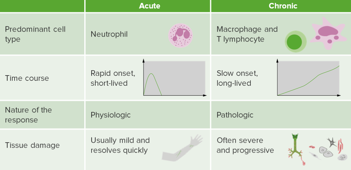
Characteristics of acute inflammation:

  • Immediate onset (minutes)
  • Short duration: Resolves within days
  • Classic signs and symptoms
  • Primary response by neutrophils Neutrophils Granular leukocytes having a nucleus with three to five lobes connected by slender threads of chromatin, and cytoplasm containing fine inconspicuous granules and stainable by neutral dyes. Innate Immunity: Phagocytes and Antigen Presentation:
    • Remove pathogens and necrotic cells/tissues
    • Bridge to innate immunity Innate immunity The capacity of a normal organism to remain unaffected by microorganisms and their toxins. It results from the presence of naturally occurring anti-infective agents, constitutional factors such as body temperature and immediate acting immune cells such as natural killer cells. Innate Immunity: Phagocytes and Antigen Presentation
    • Initiate tissue repair tissue repair The process of generating a scar or less functional tissue with a different form and/or composition of the original tissue. Wound Healing
    • Release proteases Proteases Proteins and Peptides that may damage normal tissues

Characteristics of chronic inflammation:

  • Slow onset (days)
  • Long duration: months or years
  • More specific signs or symptoms
  • Monocytes Monocytes Large, phagocytic mononuclear leukocytes produced in the vertebrate bone marrow and released into the blood; contain a large, oval or somewhat indented nucleus surrounded by voluminous cytoplasm and numerous organelles. Innate Immunity: Phagocytes and Antigen Presentation in the circulation Circulation The movement of the blood as it is pumped through the cardiovascular system. ABCDE Assessment become macrophages Macrophages The relatively long-lived phagocytic cell of mammalian tissues that are derived from blood monocytes. Main types are peritoneal macrophages; alveolar macrophages; histiocytes; kupffer cells of the liver; and osteoclasts. They may further differentiate within chronic inflammatory lesions to epithelioid cells or may fuse to form foreign body giant cells or langhans giant cells. Innate Immunity: Phagocytes and Antigen Presentation in the tissues → degrade debris and orchestrate healing versus scarring

Innate inflammation:

  • Has proinflammatory and antiinflammatory checks and balances
  • Has potential to cause harm
Contrasting acute and chronic inflammation

Contrasting acute and chronic inflammation

Image by Lecturio.
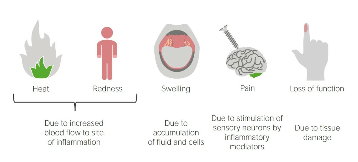
Classic signs (Latin terms)

  • Rubor (redness): due to ↑ blood flow Blood flow Blood flow refers to the movement of a certain volume of blood through the vasculature over a given unit of time (e.g., mL per minute). Vascular Resistance, Flow, and Mean Arterial Pressure and accumulation at the point of injury or inflammation
  • Calor (heat): due to a combination of blood flow Blood flow Blood flow refers to the movement of a certain volume of blood through the vasculature over a given unit of time (e.g., mL per minute). Vascular Resistance, Flow, and Mean Arterial Pressure and local reaction 
  • Tumor (swelling): due to inflammatory mediators →↑ fluid extravasation → edema Edema Edema is a condition in which excess serous fluid accumulates in the body cavity or interstitial space of connective tissues. Edema is a symptom observed in several medical conditions. It can be categorized into 2 types, namely, peripheral (in the extremities) and internal (in an organ or body cavity). Edema
  • Dolor ( pain Pain An unpleasant sensation induced by noxious stimuli which are detected by nerve endings of nociceptive neurons. Pain: Types and Pathways): mediated by cytokines Cytokines Non-antibody proteins secreted by inflammatory leukocytes and some non-leukocytic cells, that act as intercellular mediators. They differ from classical hormones in that they are produced by a number of tissue or cell types rather than by specialized glands. They generally act locally in a paracrine or autocrine rather than endocrine manner. Adaptive Immune Response, bradykinin Bradykinin A nonapeptide messenger that is enzymatically produced from kallidin in the blood where it is a potent but short-lived agent of arteriolar dilation and increased capillary permeability. Bradykinin is also released from mast cells during asthma attacks, from gut walls as a gastrointestinal vasodilator, from damaged tissues as a pain signal, and may be a neurotransmitter. Hereditary Angioedema (C1 Esterase Inhibitor Deficiency), and prostaglandins Prostaglandins A group of compounds derived from unsaturated 20-carbon fatty acids, primarily arachidonic acid, via the cyclooxygenase pathway. They are extremely potent mediators of a diverse group of physiological processes. Eicosanoids
  • Functio laesa (loss of function): results from tissue damage
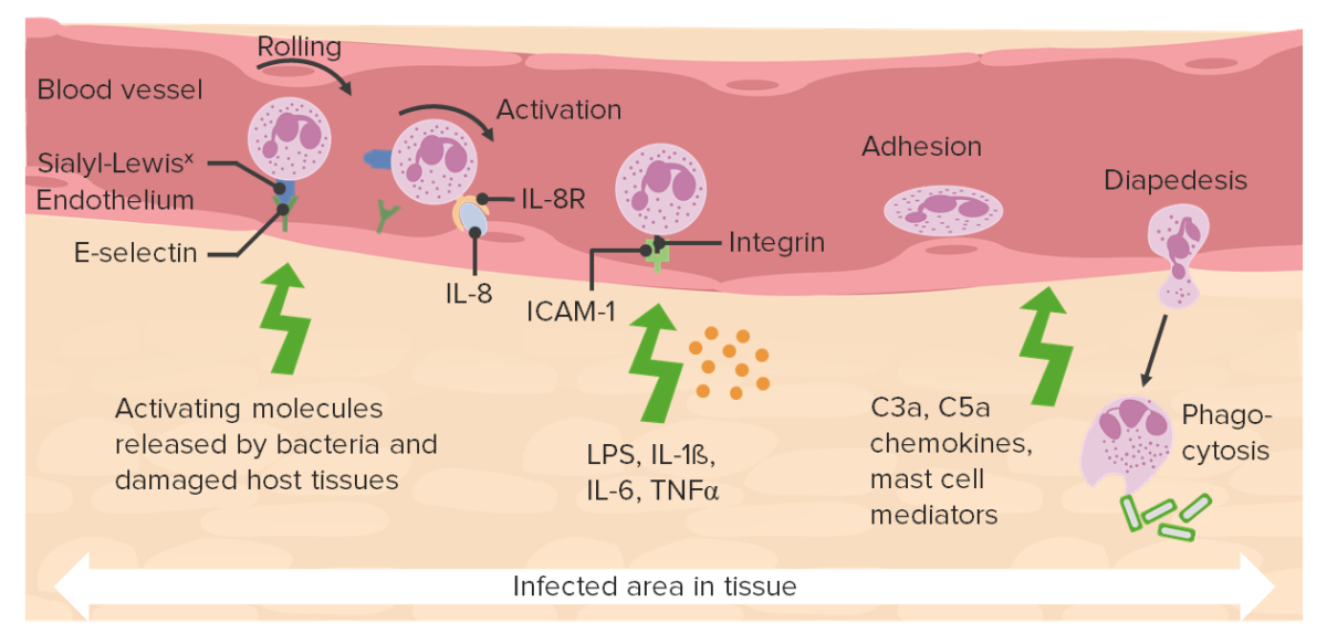
Acute inflammatory response

Acute inflammatory response

Image by Lecturio.

Mediators of Inflammation

There are multiple mediators of inflammation that bring inflammatory cells into the sites of injury and overlap with innate immunity Innate immunity The capacity of a normal organism to remain unaffected by microorganisms and their toxins. It results from the presence of naturally occurring anti-infective agents, constitutional factors such as body temperature and immediate acting immune cells such as natural killer cells. Innate Immunity: Phagocytes and Antigen Presentation when they respond to injurious stimuli Injurious Stimuli Cell Injury and Death.

Primary cellular response (WBCs)

  • Neutrophils Neutrophils Granular leukocytes having a nucleus with three to five lobes connected by slender threads of chromatin, and cytoplasm containing fine inconspicuous granules and stainable by neutral dyes. Innate Immunity: Phagocytes and Antigen Presentation
  • Macrophages Macrophages The relatively long-lived phagocytic cell of mammalian tissues that are derived from blood monocytes. Main types are peritoneal macrophages; alveolar macrophages; histiocytes; kupffer cells of the liver; and osteoclasts. They may further differentiate within chronic inflammatory lesions to epithelioid cells or may fuse to form foreign body giant cells or langhans giant cells. Innate Immunity: Phagocytes and Antigen Presentation and monocytes Monocytes Large, phagocytic mononuclear leukocytes produced in the vertebrate bone marrow and released into the blood; contain a large, oval or somewhat indented nucleus surrounded by voluminous cytoplasm and numerous organelles. Innate Immunity: Phagocytes and Antigen Presentation
  • Mast cells Mast cells Granulated cells that are found in almost all tissues, most abundantly in the skin and the gastrointestinal tract. Like the basophils, mast cells contain large amounts of histamine and heparin. Unlike basophils, mast cells normally remain in the tissues and do not circulate in the blood. Mast cells, derived from the bone marrow stem cells, are regulated by the stem cell factor. Innate Immunity: Phagocytes and Antigen Presentation and basophils Basophils Granular leukocytes characterized by a relatively pale-staining, lobate nucleus and cytoplasm containing coarse dark-staining granules of variable size and stainable by basic dyes. Innate Immunity: Phagocytes and Antigen Presentation

Mediators of inflammation

  • Acute-phase proteins Proteins Linear polypeptides that are synthesized on ribosomes and may be further modified, crosslinked, cleaved, or assembled into complex proteins with several subunits. The specific sequence of amino acids determines the shape the polypeptide will take, during protein folding, and the function of the protein. Energy Homeostasis (e.g., CRP)
  • Cytokines Cytokines Non-antibody proteins secreted by inflammatory leukocytes and some non-leukocytic cells, that act as intercellular mediators. They differ from classical hormones in that they are produced by a number of tissue or cell types rather than by specialized glands. They generally act locally in a paracrine or autocrine rather than endocrine manner. Adaptive Immune Response: signaling proteins Proteins Linear polypeptides that are synthesized on ribosomes and may be further modified, crosslinked, cleaved, or assembled into complex proteins with several subunits. The specific sequence of amino acids determines the shape the polypeptide will take, during protein folding, and the function of the protein. Energy Homeostasis released by immune cells
    • Interleukins Interleukins Interleukins are a type of cytokines (signaling proteins) that communicate messages between different parts of the immune system. The majority of interleukins are synthesized by helper CD4 T lymphocytes along with other cells such as monocytes, macrophages, and endothelial cells. Interleukins:
      • Proinflammatory: IL-1β, IL-6, IL-8, interferon ( IFN IFN Interferon (IFN) is a cytokine with antiviral properties (it interferes with viral infections) and various roles in immunoregulation. The different types are type I IFN (IFN-ɑ and IFN-β), type II IFN (IFN-ɣ), and type III IFN (IFN-ƛ). Interferons)-γ
      • Antiinflammatory: IL-4, IL-10, IL-13
    • Tumor necrosis Necrosis The death of cells in an organ or tissue due to disease, injury or failure of the blood supply. Ischemic Cell Damage factor ( TNF TNF Tumor necrosis factor (TNF) is a major cytokine, released primarily by macrophages in response to stimuli. The presence of microbial products and dead cells and injury are among the stimulating factors. This protein belongs to the TNF superfamily, a group of ligands and receptors performing functions in inflammatory response, morphogenesis, and cell proliferation. Tumor Necrosis Factor (TNF))
  • Reactive oxygen species Reactive oxygen species Molecules or ions formed by the incomplete one-electron reduction of oxygen. These reactive oxygen intermediates include singlet oxygen; superoxides; peroxides; hydroxyl radical; and hypochlorous acid. They contribute to the microbicidal activity of phagocytes, regulation of signal transduction and gene expression, and the oxidative damage to nucleic acids; proteins; and lipids. Nonalcoholic Fatty Liver Disease (ROS)
  • Platelet-activating factor:
    • Made from membrane phospholipids Phospholipids Lipids containing one or more phosphate groups, particularly those derived from either glycerol (phosphoglycerides) or sphingosine (sphingolipids). They are polar lipids that are of great importance for the structure and function of cell membranes and are the most abundant of membrane lipids, although not stored in large amounts in the system. Lipid Metabolism
    • Causes vasodilation Vasodilation The physiological widening of blood vessels by relaxing the underlying vascular smooth muscle. Pulmonary Hypertension Drugs, ↑ vascular permeability, and platelet activation Platelet activation A series of progressive, overlapping events, triggered by exposure of the platelets to subendothelial tissue. These events include shape change, adhesiveness, aggregation, and release reactions. When carried through to completion, these events lead to the formation of a stable hemostatic plug. Hemostasis
    • Also is a cytokine
  • Arachidonic acid Arachidonic Acid An unsaturated, essential fatty acid. It is found in animal and human fat as well as in the liver, brain, and glandular organs, and is a constituent of animal phosphatides. It is formed by the synthesis from dietary linoleic acid and is a precursor in the biosynthesis of prostaglandins, thromboxanes, and leukotrienes. Nonsteroidal Antiinflammatory Drugs (NSAIDs) ( AA AA Amyloidosis) metabolites:
    • Prostaglandins Prostaglandins A group of compounds derived from unsaturated 20-carbon fatty acids, primarily arachidonic acid, via the cyclooxygenase pathway. They are extremely potent mediators of a diverse group of physiological processes. Eicosanoids vasodilation Vasodilation The physiological widening of blood vessels by relaxing the underlying vascular smooth muscle. Pulmonary Hypertension Drugs; increased vascular permeability
    • Prostacyclin Prostacyclin A prostaglandin that is a powerful vasodilator and inhibits platelet aggregation. It is biosynthesized enzymatically from prostaglandin endoperoxides in human vascular tissue. The sodium salt has been also used to treat primary pulmonary hypertension. Eicosanoids (prostaglandin I2 ( PGI2 PGI2 A prostaglandin that is a powerful vasodilator and inhibits platelet aggregation. It is biosynthesized enzymatically from prostaglandin endoperoxides in human vascular tissue. The sodium salt has been also used to treat primary pulmonary hypertension. Eicosanoids)) → vasodilation Vasodilation The physiological widening of blood vessels by relaxing the underlying vascular smooth muscle. Pulmonary Hypertension Drugs; inhibits platelet aggregation Platelet aggregation The attachment of platelets to one another. This clumping together can be induced by a number of agents (e.g., thrombin; collagen) and is part of the mechanism leading to the formation of a thrombus. Hemostasis
    • Thromboxane A2 Thromboxane A2 An unstable intermediate between the prostaglandin endoperoxides and thromboxane B2. The compound has a bicyclic oxaneoxetane structure. It is a potent inducer of platelet aggregation and causes vasoconstriction. It is the principal component of rabbit aorta contracting substance (RCS). Arterial Pressure Regulation vasoconstriction Vasoconstriction The physiological narrowing of blood vessels by contraction of the vascular smooth muscle. Vascular Resistance, Flow, and Mean Arterial Pressure; promotes platelet aggregation Platelet aggregation The attachment of platelets to one another. This clumping together can be induced by a number of agents (e.g., thrombin; collagen) and is part of the mechanism leading to the formation of a thrombus. Hemostasis (opposite effect of prostacyclin Prostacyclin A prostaglandin that is a powerful vasodilator and inhibits platelet aggregation. It is biosynthesized enzymatically from prostaglandin endoperoxides in human vascular tissue. The sodium salt has been also used to treat primary pulmonary hypertension. Eicosanoids
    • Leukotrienes Leukotrienes A family of biologically active compounds derived from arachidonic acid by oxidative metabolism through the 5-lipoxygenase pathway. They participate in host defense reactions and pathophysiological conditions such as immediate hypersensitivity and inflammation. They have potent actions on many essential organs and systems, including the cardiovascular, pulmonary, and central nervous system as well as the gastrointestinal tract and the immune system. Eicosanoids (proinflammatory) → vasoconstriction Vasoconstriction The physiological narrowing of blood vessels by contraction of the vascular smooth muscle. Vascular Resistance, Flow, and Mean Arterial Pressure, bronchospasm Bronchospasm Asthma Drugs, ↑ vascular permeability

Acute-phase reactants (APRs)

  • Under the influence of cytokines Cytokines Non-antibody proteins secreted by inflammatory leukocytes and some non-leukocytic cells, that act as intercellular mediators. They differ from classical hormones in that they are produced by a number of tissue or cell types rather than by specialized glands. They generally act locally in a paracrine or autocrine rather than endocrine manner. Adaptive Immune Response (IL-6), hepatocytes Hepatocytes The main structural component of the liver. They are specialized epithelial cells that are organized into interconnected plates called lobules. Liver: Anatomy produce APRs.
  • > 30 APRs are produced.
  • Used as laboratory markers of acute inflammation
Table: Selected acute-phase reactants (APRs)
Reactant Function
CRP
  • Activates the complement cascade Complement cascade The sequential activation of serum complement proteins to create the complement membrane attack complex. Factors initiating complement activation include antigen-antibody complexes, microbial antigens, or cell surface polysaccharides. C3 Deficiency → promotes phagocytosis Phagocytosis The engulfing and degradation of microorganisms; other cells that are dead, dying, or pathogenic; and foreign particles by phagocytic cells (phagocytes). Innate Immunity: Phagocytes and Antigen Presentation
  • Sensitive, but nonspecific
  • ↑ within 12 hours after onset
  • Half-life Half-Life The time it takes for a substance (drug, radioactive nuclide, or other) to lose half of its pharmacologic, physiologic, or radiologic activity. Pharmacokinetics and Pharmacodynamics: 24 hours
ESR ESR Soft Tissue Abscess
  • The rate at which red blood cells Red blood cells Erythrocytes, or red blood cells (RBCs), are the most abundant cells in the blood. While erythrocytes in the fetus are initially produced in the yolk sac then the liver, the bone marrow eventually becomes the main site of production. Erythrocytes: Histology settle at the bottom of a thin tube
  • Rate ↑ when inflammatory proteins Proteins Linear polypeptides that are synthesized on ribosomes and may be further modified, crosslinked, cleaved, or assembled into complex proteins with several subunits. The specific sequence of amino acids determines the shape the polypeptide will take, during protein folding, and the function of the protein. Energy Homeostasis are present
  • Nonspecific
Haptoglobin
  • Binds iron Iron A metallic element with atomic symbol fe, atomic number 26, and atomic weight 55. 85. It is an essential constituent of hemoglobins; cytochromes; and iron-binding proteins. It plays a role in cellular redox reactions and in the transport of oxygen. Trace Elements to ↓ availability to pathogens
  • Removes free heme from hemolysis (heme is an oxidant → cell damage)
SAA Causes immune cell recruitment Recruitment Skeletal Muscle Contraction at the site of inflammation
ESR: erythrocyte sedimentation rate
SAA: serum amyloid A

Physiology of Acute Inflammation

General process

  • Recognition of injury → vascular response phase → cellular response phase (neutrophil recruitment Recruitment Skeletal Muscle Contraction, phagocytosis Phagocytosis The engulfing and degradation of microorganisms; other cells that are dead, dying, or pathogenic; and foreign particles by phagocytic cells (phagocytes). Innate Immunity: Phagocytes and Antigen Presentation and killing, mediator release) → tissue swelling
  • Lines of defense:
    • 1st: Resident macrophages Macrophages The relatively long-lived phagocytic cell of mammalian tissues that are derived from blood monocytes. Main types are peritoneal macrophages; alveolar macrophages; histiocytes; kupffer cells of the liver; and osteoclasts. They may further differentiate within chronic inflammatory lesions to epithelioid cells or may fuse to form foreign body giant cells or langhans giant cells. Innate Immunity: Phagocytes and Antigen Presentation begin phagocytosis Phagocytosis The engulfing and degradation of microorganisms; other cells that are dead, dying, or pathogenic; and foreign particles by phagocytic cells (phagocytes). Innate Immunity: Phagocytes and Antigen Presentation.
    • 2nd: Chemokine-induced neutrophil extravasation
    • 3rd: Monocyte extravasation → tissue macrophages Macrophages The relatively long-lived phagocytic cell of mammalian tissues that are derived from blood monocytes. Main types are peritoneal macrophages; alveolar macrophages; histiocytes; kupffer cells of the liver; and osteoclasts. They may further differentiate within chronic inflammatory lesions to epithelioid cells or may fuse to form foreign body giant cells or langhans giant cells. Innate Immunity: Phagocytes and Antigen Presentation ( phagocytosis Phagocytosis The engulfing and degradation of microorganisms; other cells that are dead, dying, or pathogenic; and foreign particles by phagocytic cells (phagocytes). Innate Immunity: Phagocytes and Antigen Presentation)
    • 4th: ↑ Cell production by the bone marrow Bone marrow The soft tissue filling the cavities of bones. Bone marrow exists in two types, yellow and red. Yellow marrow is found in the large cavities of large bones and consists mostly of fat cells and a few primitive blood cells. Red marrow is a hematopoietic tissue and is the site of production of erythrocytes and granular leukocytes. Bone marrow is made up of a framework of connective tissue containing branching fibers with the frame being filled with marrow cells. Bone Marrow: Composition and Hematopoiesis

Recognition of tissue injury

  • Tissue damage → stimulates pathogen-associated molecular patterns Pathogen-Associated Molecular Patterns Sepsis and Septic Shock ( PAMPs PAMPs Sepsis and Septic Shock
  • Neutrophils Neutrophils Granular leukocytes having a nucleus with three to five lobes connected by slender threads of chromatin, and cytoplasm containing fine inconspicuous granules and stainable by neutral dyes. Innate Immunity: Phagocytes and Antigen Presentation migrate in response to infection, then recognize these molecules:
    • PAMPs PAMPs Sepsis and Septic Shock (from pathogens such as bacteria Bacteria Bacteria are prokaryotic single-celled microorganisms that are metabolically active and divide by binary fission. Some of these organisms play a significant role in the pathogenesis of diseases. Bacteriology):
      • Double-stranded RNA RNA A polynucleotide consisting essentially of chains with a repeating backbone of phosphate and ribose units to which nitrogenous bases are attached. RNA is unique among biological macromolecules in that it can encode genetic information, serve as an abundant structural component of cells, and also possesses catalytic activity. RNA Types and Structure (dsRNA)
      • Unmethylated islands of DNA DNA A deoxyribonucleotide polymer that is the primary genetic material of all cells. Eukaryotic and prokaryotic organisms normally contain DNA in a double-stranded state, yet several important biological processes transiently involve single-stranded regions. DNA, which consists of a polysugar-phosphate backbone possessing projections of purines (adenine and guanine) and pyrimidines (thymine and cytosine), forms a double helix that is held together by hydrogen bonds between these purines and pyrimidines (adenine to thymine and guanine to cytosine). DNA Types and Structure (found in bacteria Bacteria Bacteria are prokaryotic single-celled microorganisms that are metabolically active and divide by binary fission. Some of these organisms play a significant role in the pathogenesis of diseases. Bacteriology, not humans)
      • Lipopolysaccharides Lipopolysaccharides Lipid-containing polysaccharides which are endotoxins and important group-specific antigens. They are often derived from the cell wall of gram-negative bacteria and induce immunoglobulin secretion. The lipopolysaccharide molecule consists of three parts: lipid a, core polysaccharide, and o-specific chains (o antigens). When derived from Escherichia coli, lipopolysaccharides serve as polyclonal b-cell mitogens commonly used in laboratory immunology. Diarrheagenic E. coli 
      • Mannose
      • Phosphorylcholine
    • Damage-associated molecular patterns Damage-Associated Molecular Patterns Sepsis and Septic Shock (DAMPs):
      • Found in dead tissue; neutrophils Neutrophils Granular leukocytes having a nucleus with three to five lobes connected by slender threads of chromatin, and cytoplasm containing fine inconspicuous granules and stainable by neutral dyes. Innate Immunity: Phagocytes and Antigen Presentation respond to degrade it 
      • Part of the host defense system
      • Can also promote pathologic inflammatory responses in chronic inflammatory diseases (e.g., rheumatoid arthritis Arthritis Acute or chronic inflammation of joints. Osteoarthritis, systemic lupus erythematosus Systemic lupus erythematosus Systemic lupus erythematosus (SLE) is a chronic autoimmune, inflammatory condition that causes immune-complex deposition in organs, resulting in systemic manifestations. Women, particularly those of African American descent, are more commonly affected. Systemic Lupus Erythematosus, atherosclerosis Atherosclerosis Atherosclerosis is a common form of arterial disease in which lipid deposition forms a plaque in the blood vessel walls. Atherosclerosis is an incurable disease, for which there are clearly defined risk factors that often can be reduced through a change in lifestyle and behavior of the patient. Atherosclerosis, Alzheimer disease Alzheimer disease As the most common cause of dementia, Alzheimer disease affects not only many individuals but also their families. Alzheimer disease is a progressive neurodegenerative disease that causes brain atrophy and presents with a decline in memory, cognition, and social skills. Alzheimer Disease, and cancer)
    • Opsonins: proteins Proteins Linear polypeptides that are synthesized on ribosomes and may be further modified, crosslinked, cleaved, or assembled into complex proteins with several subunits. The specific sequence of amino acids determines the shape the polypeptide will take, during protein folding, and the function of the protein. Energy Homeostasis that induce phagocytosis Phagocytosis The engulfing and degradation of microorganisms; other cells that are dead, dying, or pathogenic; and foreign particles by phagocytic cells (phagocytes). Innate Immunity: Phagocytes and Antigen Presentation
      • IgG IgG The major immunoglobulin isotype class in normal human serum. There are several isotype subclasses of igg, for example, igg1, igg2a, and igg2b. Hypersensitivity Pneumonitis
      • Complement fragments
    • Pattern-recognition receptors Receptors Receptors are proteins located either on the surface of or within a cell that can bind to signaling molecules known as ligands (e.g., hormones) and cause some type of response within the cell. Receptors (PRRs):
      • TLR: expressed on sentinel cells such as macrophages Macrophages The relatively long-lived phagocytic cell of mammalian tissues that are derived from blood monocytes. Main types are peritoneal macrophages; alveolar macrophages; histiocytes; kupffer cells of the liver; and osteoclasts. They may further differentiate within chronic inflammatory lesions to epithelioid cells or may fuse to form foreign body giant cells or langhans giant cells. Innate Immunity: Phagocytes and Antigen Presentation
      • Pentraxins Pentraxins Innate Immunity: Barriers, Complement, and Cytokines: proteins Proteins Linear polypeptides that are synthesized on ribosomes and may be further modified, crosslinked, cleaved, or assembled into complex proteins with several subunits. The specific sequence of amino acids determines the shape the polypeptide will take, during protein folding, and the function of the protein. Energy Homeostasis involved in immunologic responses, including CRP and serum amyloid
    • Immune receptors Receptors Receptors are proteins located either on the surface of or within a cell that can bind to signaling molecules known as ligands (e.g., hormones) and cause some type of response within the cell. Receptors: Fc Fc Crystallizable fragments composed of the carboxy-terminal halves of both immunoglobulin heavy chains linked to each other by disulfide bonds. Fc fragments contain the carboxy-terminal parts of the heavy chain constant regions that are responsible for the effector functions of an immunoglobulin (complement fixation, binding to the cell membrane via fc receptors, and placental transport). This fragment can be obtained by digestion of immunoglobulins with the proteolytic enzyme papain. Immunoglobulins: Types and Functions receptors Receptors Receptors are proteins located either on the surface of or within a cell that can bind to signaling molecules known as ligands (e.g., hormones) and cause some type of response within the cell. Receptors, complement receptors Receptors Receptors are proteins located either on the surface of or within a cell that can bind to signaling molecules known as ligands (e.g., hormones) and cause some type of response within the cell. Receptors
  • PAMPs PAMPs Sepsis and Septic Shock and DAMPs → activate proinflammatory cytokines Proinflammatory Cytokines Metabolic Syndrome and chemokines Chemokines Class of pro-inflammatory cytokines that have the ability to attract and activate leukocytes. They can be divided into at least three structural branches: c; cc; and cxc; according to variations in a shared cysteine motif. Adaptive Cell-mediated Immunity
    • Endothelial cells on blood vessels release prostaglandins Prostaglandins A group of compounds derived from unsaturated 20-carbon fatty acids, primarily arachidonic acid, via the cyclooxygenase pathway. They are extremely potent mediators of a diverse group of physiological processes. Eicosanoids and nitric oxide Nitric Oxide A free radical gas produced endogenously by a variety of mammalian cells, synthesized from arginine by nitric oxide synthase. Nitric oxide is one of the endothelium-dependent relaxing factors released by the vascular endothelium and mediates vasodilation. It also inhibits platelet aggregation, induces disaggregation of aggregated platelets, and inhibits platelet adhesion to the vascular endothelium. Nitric oxide activates cytosolic guanylate cyclase and thus elevates intracellular levels of cyclic gmp. Pulmonary Hypertension Drugs.
    • Mast cells Mast cells Granulated cells that are found in almost all tissues, most abundantly in the skin and the gastrointestinal tract. Like the basophils, mast cells contain large amounts of histamine and heparin. Unlike basophils, mast cells normally remain in the tissues and do not circulate in the blood. Mast cells, derived from the bone marrow stem cells, are regulated by the stem cell factor. Innate Immunity: Phagocytes and Antigen Presentation and basophils Basophils Granular leukocytes characterized by a relatively pale-staining, lobate nucleus and cytoplasm containing coarse dark-staining granules of variable size and stainable by basic dyes. Innate Immunity: Phagocytes and Antigen Presentation release histamine.
    • Platelets Platelets Platelets are small cell fragments involved in hemostasis. Thrombopoiesis takes place primarily in the bone marrow through a series of cell differentiation and is influenced by several cytokines. Platelets are formed after fragmentation of the megakaryocyte cytoplasm. Platelets: Histology release prostaglandins Prostaglandins A group of compounds derived from unsaturated 20-carbon fatty acids, primarily arachidonic acid, via the cyclooxygenase pathway. They are extremely potent mediators of a diverse group of physiological processes. Eicosanoids and serotonin Serotonin A biochemical messenger and regulator, synthesized from the essential amino acid l-tryptophan. In humans it is found primarily in the central nervous system, gastrointestinal tract, and blood platelets. Serotonin mediates several important physiological functions including neurotransmission, gastrointestinal motility, hemostasis, and cardiovascular integrity. Receptors and Neurotransmitters of the CNS.

Vascular response phase

Vasodilation Vasodilation The physiological widening of blood vessels by relaxing the underlying vascular smooth muscle. Pulmonary Hypertension Drugs:

  • Brings more blood flow Blood flow Blood flow refers to the movement of a certain volume of blood through the vasculature over a given unit of time (e.g., mL per minute). Vascular Resistance, Flow, and Mean Arterial Pressure to the tissue
  • Allows delivery of cells and mediators to the injured tissue for response
  • Mediators of the vascular response:
    • Mast cell Mast cell Granulated cells that are found in almost all tissues, most abundantly in the skin and the gastrointestinal tract. Like the basophils, mast cells contain large amounts of histamine and heparin. Unlike basophils, mast cells normally remain in the tissues and do not circulate in the blood. Mast cells, derived from the bone marrow stem cells, are regulated by the stem cell factor. Angioedema degranulation → histamine release → relaxation of smooth muscle ( vasodilation Vasodilation The physiological widening of blood vessels by relaxing the underlying vascular smooth muscle. Pulmonary Hypertension Drugs)→ ↑ blood flow Blood flow Blood flow refers to the movement of a certain volume of blood through the vasculature over a given unit of time (e.g., mL per minute). Vascular Resistance, Flow, and Mean Arterial Pressure
    • Nitric oxide Nitric Oxide A free radical gas produced endogenously by a variety of mammalian cells, synthesized from arginine by nitric oxide synthase. Nitric oxide is one of the endothelium-dependent relaxing factors released by the vascular endothelium and mediates vasodilation. It also inhibits platelet aggregation, induces disaggregation of aggregated platelets, and inhibits platelet adhesion to the vascular endothelium. Nitric oxide activates cytosolic guanylate cyclase and thus elevates intracellular levels of cyclic gmp. Pulmonary Hypertension Drugs (synthesized by endothelium Endothelium A layer of epithelium that lines the heart, blood vessels (vascular endothelium), lymph vessels (lymphatic endothelium), and the serous cavities of the body. Arteries: Histology and macrophages Macrophages The relatively long-lived phagocytic cell of mammalian tissues that are derived from blood monocytes. Main types are peritoneal macrophages; alveolar macrophages; histiocytes; kupffer cells of the liver; and osteoclasts. They may further differentiate within chronic inflammatory lesions to epithelioid cells or may fuse to form foreign body giant cells or langhans giant cells. Innate Immunity: Phagocytes and Antigen Presentation) → vasodilation Vasodilation The physiological widening of blood vessels by relaxing the underlying vascular smooth muscle. Pulmonary Hypertension Drugs → ↑ blood flow Blood flow Blood flow refers to the movement of a certain volume of blood through the vasculature over a given unit of time (e.g., mL per minute). Vascular Resistance, Flow, and Mean Arterial Pressure
    • Eicosanoids Eicosanoids Eicosanoids are cell-signaling molecules produced from arachidonic acid. With the action of phospholipase A2, arachidonic acid is released from the plasma membrane. The different families of eicosanoids, which are prostaglandins (PGs), thromboxanes (TXA2s), prostacyclin (PGI2), lipoxins (LXs), and leukotrienes (LTs), emerge from a series of reactions catalyzed by different enzymes. Eicosanoids ( prostaglandins Prostaglandins A group of compounds derived from unsaturated 20-carbon fatty acids, primarily arachidonic acid, via the cyclooxygenase pathway. They are extremely potent mediators of a diverse group of physiological processes. Eicosanoids and leukotrienes Leukotrienes A family of biologically active compounds derived from arachidonic acid by oxidative metabolism through the 5-lipoxygenase pathway. They participate in host defense reactions and pathophysiological conditions such as immediate hypersensitivity and inflammation. They have potent actions on many essential organs and systems, including the cardiovascular, pulmonary, and central nervous system as well as the gastrointestinal tract and the immune system. Eicosanoids): also cause vasodilation Vasodilation The physiological widening of blood vessels by relaxing the underlying vascular smooth muscle. Pulmonary Hypertension Drugs

Increased permeability: 

  • Causes extracellular fluid shift Fluid Shift Translocation of body fluids from one compartment to another, such as from the vascular to the interstitial compartments. Fluid shifts are associated with profound changes in vascular permeability and water-electrolyte imbalance. The shift can also be from the lower body to the upper body as in conditions of weightlessness. Volume Depletion and Dehydration
  • Slows blood flow Blood flow Blood flow refers to the movement of a certain volume of blood through the vasculature over a given unit of time (e.g., mL per minute). Vascular Resistance, Flow, and Mean Arterial Pressure and allows cell recruitment Recruitment Skeletal Muscle Contraction
  • Deposits circulating mediators to the site of injury
  • Early response (in minutes) → direct endothelial injury and driven by:
    • Histamine
    • Bradykinin Bradykinin A nonapeptide messenger that is enzymatically produced from kallidin in the blood where it is a potent but short-lived agent of arteriolar dilation and increased capillary permeability. Bradykinin is also released from mast cells during asthma attacks, from gut walls as a gastrointestinal vasodilator, from damaged tissues as a pain signal, and may be a neurotransmitter. Hereditary Angioedema (C1 Esterase Inhibitor Deficiency)
    • Leukotrienes Leukotrienes A family of biologically active compounds derived from arachidonic acid by oxidative metabolism through the 5-lipoxygenase pathway. They participate in host defense reactions and pathophysiological conditions such as immediate hypersensitivity and inflammation. They have potent actions on many essential organs and systems, including the cardiovascular, pulmonary, and central nervous system as well as the gastrointestinal tract and the immune system. Eicosanoids
  • Late response (hours later) is by leukocyte-mediated damage through:
    • Cytokines Cytokines Non-antibody proteins secreted by inflammatory leukocytes and some non-leukocytic cells, that act as intercellular mediators. They differ from classical hormones in that they are produced by a number of tissue or cell types rather than by specialized glands. They generally act locally in a paracrine or autocrine rather than endocrine manner. Adaptive Immune Response:
      • IL-1
      • TNF TNF Tumor necrosis factor (TNF) is a major cytokine, released primarily by macrophages in response to stimuli. The presence of microbial products and dead cells and injury are among the stimulating factors. This protein belongs to the TNF superfamily, a group of ligands and receptors performing functions in inflammatory response, morphogenesis, and cell proliferation. Tumor Necrosis Factor (TNF)
    • ROS
    • Prostaglandins Prostaglandins A group of compounds derived from unsaturated 20-carbon fatty acids, primarily arachidonic acid, via the cyclooxygenase pathway. They are extremely potent mediators of a diverse group of physiological processes. Eicosanoids
  • Edema Edema Edema is a condition in which excess serous fluid accumulates in the body cavity or interstitial space of connective tissues. Edema is a symptom observed in several medical conditions. It can be categorized into 2 types, namely, peripheral (in the extremities) and internal (in an organ or body cavity). Edema is seen because of:
    • ↑ Endothelial permeability
    • Endothelial damage
    • Vasodilation Vasodilation The physiological widening of blood vessels by relaxing the underlying vascular smooth muscle. Pulmonary Hypertension Drugs and stasis

Cellular release of chemicals causes:

  • Transient vasoconstriction Vasoconstriction The physiological narrowing of blood vessels by contraction of the vascular smooth muscle. Vascular Resistance, Flow, and Mean Arterial Pressure due to endothelial cell contraction
  • Followed by endothelial cell retraction → vasodilation Vasodilation The physiological widening of blood vessels by relaxing the underlying vascular smooth muscle. Pulmonary Hypertension Drugs 
  • ↑ Vascular permeability → exudation of fluid and cells into tissue → fluid shift Fluid Shift Translocation of body fluids from one compartment to another, such as from the vascular to the interstitial compartments. Fluid shifts are associated with profound changes in vascular permeability and water-electrolyte imbalance. The shift can also be from the lower body to the upper body as in conditions of weightlessness. Volume Depletion and Dehydration

Cellular response phase

Neutrophil recruitment Recruitment Skeletal Muscle Contraction:

  • Chemotactic substances:
    • N-formyl methionine (on bacterial cell walls)
    • Leukotriene Leukotriene Asthma Drugs B4
    • Complement C5a
    • Platelet-activating factor
    • Chemokines Chemokines Class of pro-inflammatory cytokines that have the ability to attract and activate leukocytes. They can be divided into at least three structural branches: c; cc; and cxc; according to variations in a shared cysteine motif. Adaptive Cell-mediated Immunity:
      • Activate leukocyte integrins Integrins A family of transmembrane glycoproteins (membrane glycoproteins) consisting of noncovalent heterodimers. They interact with a wide variety of ligands including extracellular matrix proteins; complement, and other cells, while their intracellular domains interact with the cytoskeleton. The integrins consist of at least three identified families: the cytoadhesin receptors(receptors, cytoadhesin), the leukocyte adhesion receptors (receptors, leukocyte adhesion), and the very late antigen receptors. Each family contains a common beta-subunit (integrin beta chains) combined with one or more distinct alpha-subunits (integrin alpha chains). These receptors participate in cell-matrix and cell-cell adhesion in many physiologically important processes, including embryological development; hemostasis; thrombosis; wound healing; immune and nonimmune defense mechanisms; and oncogenic transformation. Leukocyte Adhesion Deficiency Type 1
      • Attract leukocytes Leukocytes White blood cells. These include granular leukocytes (basophils; eosinophils; and neutrophils) as well as non-granular leukocytes (lymphocytes and monocytes). White Myeloid Cells: Histology to move out of the bloodstream
      • Activate antimicrobial functions
  • Rolling and adhesion Adhesion The process whereby platelets adhere to something other than platelets, e.g., collagen; basement membrane; microfibrils; or other ‘foreign’ surfaces. Coagulation Studies:
    • Endothelial cells express E-selectin E-selectin Cell adhesion molecule and cd antigen that mediates neutrophil, monocyte, and memory t-cell adhesion to cytokine-activated endothelial cells. E-selectin recognizes sialylated carbohydrate groups related to the lewis X or lewis a family. Tumor Necrosis Factor (TNF) and P-selectin P-selectin Cell adhesion molecule and cd antigen that mediates the adhesion of neutrophils and monocytes to activated platelets and endothelial cells. Tumor Necrosis Factor (TNF) under the influence of TNF TNF Tumor necrosis factor (TNF) is a major cytokine, released primarily by macrophages in response to stimuli. The presence of microbial products and dead cells and injury are among the stimulating factors. This protein belongs to the TNF superfamily, a group of ligands and receptors performing functions in inflammatory response, morphogenesis, and cell proliferation. Tumor Necrosis Factor (TNF) and IL-1 →
    • Bind BIND Hyperbilirubinemia of the Newborn to the sialyl-Lewis X glycoprotein on neutrophils Neutrophils Granular leukocytes having a nucleus with three to five lobes connected by slender threads of chromatin, and cytoplasm containing fine inconspicuous granules and stainable by neutral dyes. Innate Immunity: Phagocytes and Antigen Presentation → 
    • Endothelial cells express ICAM-1 (intercellular adhesion Adhesion The process whereby platelets adhere to something other than platelets, e.g., collagen; basement membrane; microfibrils; or other ‘foreign’ surfaces. Coagulation Studies molecule 1, a glycoprotein also known as CD54) and chemokines Chemokines Class of pro-inflammatory cytokines that have the ability to attract and activate leukocytes. They can be divided into at least three structural branches: c; cc; and cxc; according to variations in a shared cysteine motif. Adaptive Cell-mediated Immunity activate the integrins Integrins A family of transmembrane glycoproteins (membrane glycoproteins) consisting of noncovalent heterodimers. They interact with a wide variety of ligands including extracellular matrix proteins; complement, and other cells, while their intracellular domains interact with the cytoskeleton. The integrins consist of at least three identified families: the cytoadhesin receptors(receptors, cytoadhesin), the leukocyte adhesion receptors (receptors, leukocyte adhesion), and the very late antigen receptors. Each family contains a common beta-subunit (integrin beta chains) combined with one or more distinct alpha-subunits (integrin alpha chains). These receptors participate in cell-matrix and cell-cell adhesion in many physiologically important processes, including embryological development; hemostasis; thrombosis; wound healing; immune and nonimmune defense mechanisms; and oncogenic transformation. Leukocyte Adhesion Deficiency Type 1 on the neutrophil →
    • Activation and adhesion Adhesion The process whereby platelets adhere to something other than platelets, e.g., collagen; basement membrane; microfibrils; or other ‘foreign’ surfaces. Coagulation Studies of the neutrophil 
  • Transmigration Transmigration Leukocyte Adhesion Deficiency Type 1:

Phagocytosis Phagocytosis The engulfing and degradation of microorganisms; other cells that are dead, dying, or pathogenic; and foreign particles by phagocytic cells (phagocytes). Innate Immunity: Phagocytes and Antigen Presentation and killing: 

  • Neutrophils Neutrophils Granular leukocytes having a nucleus with three to five lobes connected by slender threads of chromatin, and cytoplasm containing fine inconspicuous granules and stainable by neutral dyes. Innate Immunity: Phagocytes and Antigen Presentation have transmigrated →
  • Phagocytosis Phagocytosis The engulfing and degradation of microorganisms; other cells that are dead, dying, or pathogenic; and foreign particles by phagocytic cells (phagocytes). Innate Immunity: Phagocytes and Antigen Presentation: Neutrophils Neutrophils Granular leukocytes having a nucleus with three to five lobes connected by slender threads of chromatin, and cytoplasm containing fine inconspicuous granules and stainable by neutral dyes. Innate Immunity: Phagocytes and Antigen Presentation bind BIND Hyperbilirubinemia of the Newborn to PRRs → membrane wraps around and engulfs microorganism →
  • Leukocyte killing→ destruction of the engulfed microorganisms by:
  • The complement pathway → cell death Cell death Injurious stimuli trigger the process of cellular adaptation, whereby cells respond to withstand the harmful changes in their environment. Overwhelmed adaptive mechanisms lead to cell injury. Mild stimuli produce reversible injury. If the stimulus is severe or persistent, injury becomes irreversible. Apoptosis is programmed cell death, a mechanism with both physiologic and pathologic effects. Cell Injury and Death by formation of a membrane attack complex Membrane attack complex A product of complement activation cascade, regardless of the pathways, that forms transmembrane channels causing disruption of the target cell membrane and cell lysis. It is formed by the sequential assembly of terminal complement components (complement C5b; complement C6; complement C7; complement C8; and complement C9) into the target membrane. The resultant C5b-8-poly-c9 is the ‘membrane attack complex’ or MAC. Type II Hypersensitivity Reaction (MAC)
    • Definition of complement:
      • A collection of circulating proteins Proteins Linear polypeptides that are synthesized on ribosomes and may be further modified, crosslinked, cleaved, or assembled into complex proteins with several subunits. The specific sequence of amino acids determines the shape the polypeptide will take, during protein folding, and the function of the protein. Energy Homeostasis from the liver Liver The liver is the largest gland in the human body. The liver is found in the superior right quadrant of the abdomen and weighs approximately 1.5 kilograms. Its main functions are detoxification, metabolism, nutrient storage (e.g., iron and vitamins), synthesis of coagulation factors, formation of bile, filtration, and storage of blood. Liver: Anatomy and macrophage-synthesized proteins Proteins Linear polypeptides that are synthesized on ribosomes and may be further modified, crosslinked, cleaved, or assembled into complex proteins with several subunits. The specific sequence of amino acids determines the shape the polypeptide will take, during protein folding, and the function of the protein. Energy Homeostasis
      • A cascade of activation triggered by antigen Antigen Substances that are recognized by the immune system and induce an immune reaction. Vaccination–antibody complexes, bacterial polysaccharides Polysaccharides Basics of Carbohydrates, aggregated IgA IgA Represents 15-20% of the human serum immunoglobulins, mostly as the 4-chain polymer in humans or dimer in other mammals. Secretory iga is the main immunoglobulin in secretions. Immunoglobulins: Types and Functions
    • Activated complement fragments cause:
    • Complement fragments are proinflammatory:
      • C3a and C5a are potent chemotactic agents (recruit and activate neutrophils Neutrophils Granular leukocytes having a nucleus with three to five lobes connected by slender threads of chromatin, and cytoplasm containing fine inconspicuous granules and stainable by neutral dyes. Innate Immunity: Phagocytes and Antigen Presentation).
      • C3b induces opsonization (recognizing and targeting invading particles for phagocytosis Phagocytosis The engulfing and degradation of microorganisms; other cells that are dead, dying, or pathogenic; and foreign particles by phagocytic cells (phagocytes). Innate Immunity: Phagocytes and Antigen Presentation) → neutrophils Neutrophils Granular leukocytes having a nucleus with three to five lobes connected by slender threads of chromatin, and cytoplasm containing fine inconspicuous granules and stainable by neutral dyes. Innate Immunity: Phagocytes and Antigen Presentation and macrophages Macrophages The relatively long-lived phagocytic cell of mammalian tissues that are derived from blood monocytes. Main types are peritoneal macrophages; alveolar macrophages; histiocytes; kupffer cells of the liver; and osteoclasts. They may further differentiate within chronic inflammatory lesions to epithelioid cells or may fuse to form foreign body giant cells or langhans giant cells. Innate Immunity: Phagocytes and Antigen Presentation bind BIND Hyperbilirubinemia of the Newborn and kill pathogens
      • C3b and cleavage of other complement factors form a MAC → lysis of microbes

Mediator release:

  • Inflammatory stimuli on membrane phospholipids Phospholipids Lipids containing one or more phosphate groups, particularly those derived from either glycerol (phosphoglycerides) or sphingosine (sphingolipids). They are polar lipids that are of great importance for the structure and function of cell membranes and are the most abundant of membrane lipids, although not stored in large amounts in the system. Lipid Metabolism → cascade of AA AA Amyloidosis metabolites:
    • COX pathway COX pathway Eicosanoids is proinflammatory and produces:
      • Prostaglandins Prostaglandins A group of compounds derived from unsaturated 20-carbon fatty acids, primarily arachidonic acid, via the cyclooxygenase pathway. They are extremely potent mediators of a diverse group of physiological processes. Eicosanoids vasodilation Vasodilation The physiological widening of blood vessels by relaxing the underlying vascular smooth muscle. Pulmonary Hypertension Drugs, ↑ vascular permeability
      • PGI2 PGI2 A prostaglandin that is a powerful vasodilator and inhibits platelet aggregation. It is biosynthesized enzymatically from prostaglandin endoperoxides in human vascular tissue. The sodium salt has been also used to treat primary pulmonary hypertension. Eicosanoids vasodilation Vasodilation The physiological widening of blood vessels by relaxing the underlying vascular smooth muscle. Pulmonary Hypertension Drugs, inhibits platelet aggregation Platelet aggregation The attachment of platelets to one another. This clumping together can be induced by a number of agents (e.g., thrombin; collagen) and is part of the mechanism leading to the formation of a thrombus. Hemostasis
      • Thromboxane A2 Thromboxane A2 An unstable intermediate between the prostaglandin endoperoxides and thromboxane B2. The compound has a bicyclic oxaneoxetane structure. It is a potent inducer of platelet aggregation and causes vasoconstriction. It is the principal component of rabbit aorta contracting substance (RCS). Arterial Pressure Regulation vasoconstriction Vasoconstriction The physiological narrowing of blood vessels by contraction of the vascular smooth muscle. Vascular Resistance, Flow, and Mean Arterial Pressure, promotes platelet aggregation Platelet aggregation The attachment of platelets to one another. This clumping together can be induced by a number of agents (e.g., thrombin; collagen) and is part of the mechanism leading to the formation of a thrombus. Hemostasis (opposite effect of prostacyclin Prostacyclin A prostaglandin that is a powerful vasodilator and inhibits platelet aggregation. It is biosynthesized enzymatically from prostaglandin endoperoxides in human vascular tissue. The sodium salt has been also used to treat primary pulmonary hypertension. Eicosanoids
    • Lipoxygenase pathway Lipoxygenase Pathway Eicosanoids produces:
      • Leukotrienes Leukotrienes A family of biologically active compounds derived from arachidonic acid by oxidative metabolism through the 5-lipoxygenase pathway. They participate in host defense reactions and pathophysiological conditions such as immediate hypersensitivity and inflammation. They have potent actions on many essential organs and systems, including the cardiovascular, pulmonary, and central nervous system as well as the gastrointestinal tract and the immune system. Eicosanoids (proinflammatory) → vasoconstriction Vasoconstriction The physiological narrowing of blood vessels by contraction of the vascular smooth muscle. Vascular Resistance, Flow, and Mean Arterial Pressure, bronchospasm Bronchospasm Asthma Drugs, ↑ vascular permeability 
      • Lipoxins Lipoxins Trihydroxy derivatives of eicosanoic acids. They are primarily derived from arachidonic acid, however eicosapentaenoic acid derivatives also exist. Many of them are naturally occurring mediators of immune regulation. Eicosanoids (antiinflammatory) → inhibit neutrophil adhesion Adhesion The process whereby platelets adhere to something other than platelets, e.g., collagen; basement membrane; microfibrils; or other ‘foreign’ surfaces. Coagulation Studies and chemotaxis Chemotaxis The movement of leukocytes in response to a chemical concentration gradient or to products formed in an immunologic reaction. Leukocyte Adhesion Deficiency Type 1 
  • ROS: involved in leukocyte killing
  • Platelet-activating factor: activated platelets Platelets Platelets are small cell fragments involved in hemostasis. Thrombopoiesis takes place primarily in the bone marrow through a series of cell differentiation and is influenced by several cytokines. Platelets are formed after fragmentation of the megakaryocyte cytoplasm. Platelets: Histology set off cascades of kinins and coagulation factors Coagulation factors Endogenous substances, usually proteins, that are involved in the blood coagulation process. Hemostasis pain Pain An unpleasant sensation induced by noxious stimuli which are detected by nerve endings of nociceptive neurons. Pain: Types and Pathways and clotting
Interplay of various chemokines

Interplay of various chemokines:
Various ligands and receptors bind and promote neutrophil extravasation.
C3a and C5a: complement
ICAM-1: intercellular adhesion molecule 1
LPS: lipopolysaccharides
TNF-α: tumor necrosis factor alpha

Image by Lecturio.

Tissue swelling

  • Fluid in the interstitial space has ↑ amounts of protein and fibrinogen Fibrinogen Plasma glycoprotein clotted by thrombin, composed of a dimer of three non-identical pairs of polypeptide chains (alpha, beta, gamma) held together by disulfide bonds. Fibrinogen clotting is a sol-gel change involving complex molecular arrangements: whereas fibrinogen is cleaved by thrombin to form polypeptides a and b, the proteolytic action of other enzymes yields different fibrinogen degradation products. Hemostasis:
    • Fibrinogen Fibrinogen Plasma glycoprotein clotted by thrombin, composed of a dimer of three non-identical pairs of polypeptide chains (alpha, beta, gamma) held together by disulfide bonds. Fibrinogen clotting is a sol-gel change involving complex molecular arrangements: whereas fibrinogen is cleaved by thrombin to form polypeptides a and b, the proteolytic action of other enzymes yields different fibrinogen degradation products. Hemostasis promotes clotting to contain the pathogen
    • Migration of granulocytes Granulocytes Leukocytes with abundant granules in the cytoplasm. They are divided into three groups according to the staining properties of the granules: neutrophilic, eosinophilic, and basophilic. Mature granulocytes are the neutrophils; eosinophils; and basophils. White Myeloid Cells: Histology and monocytes Monocytes Large, phagocytic mononuclear leukocytes produced in the vertebrate bone marrow and released into the blood; contain a large, oval or somewhat indented nucleus surrounded by voluminous cytoplasm and numerous organelles. Innate Immunity: Phagocytes and Antigen Presentation → extravasate into the tissue → swelling
  • Factor XIIa (Hageman factor):
    • Activates the kinin cascade → bradykinin Bradykinin A nonapeptide messenger that is enzymatically produced from kallidin in the blood where it is a potent but short-lived agent of arteriolar dilation and increased capillary permeability. Bradykinin is also released from mast cells during asthma attacks, from gut walls as a gastrointestinal vasodilator, from damaged tissues as a pain signal, and may be a neurotransmitter. Hereditary Angioedema (C1 Esterase Inhibitor Deficiency)
      • Pain Pain An unpleasant sensation induced by noxious stimuli which are detected by nerve endings of nociceptive neurons. Pain: Types and Pathways
      • ↑ Vascular permeability → swelling
    • Activates the coagulation pathway → clotting
    • Indirectly activates the fibrinolytic system ( anticoagulation Anticoagulation Pulmonary Hypertension Drugs) at the same time as coagulation →
      • Breaks down fibrin Fibrin A protein derived from fibrinogen in the presence of thrombin, which forms part of the blood clot. Rapidly Progressive Glomerulonephritis to limit Limit A value (e.g., pressure or time) that should not be exceeded and which is specified by the operator to protect the lung Invasive Mechanical Ventilation the amount of clotting
      • Activates the complement cascade Complement cascade The sequential activation of serum complement proteins to create the complement membrane attack complex. Factors initiating complement activation include antigen-antibody complexes, microbial antigens, or cell surface polysaccharides. C3 Deficiency involved in phagocytosis Phagocytosis The engulfing and degradation of microorganisms; other cells that are dead, dying, or pathogenic; and foreign particles by phagocytic cells (phagocytes). Innate Immunity: Phagocytes and Antigen Presentation and killing
  • Factor IIa ( thrombin Thrombin An enzyme formed from prothrombin that converts fibrinogen to fibrin. Hemostasis):
    • Triggers acute inflammation
    • Cleaves fibrinogen Fibrinogen Plasma glycoprotein clotted by thrombin, composed of a dimer of three non-identical pairs of polypeptide chains (alpha, beta, gamma) held together by disulfide bonds. Fibrinogen clotting is a sol-gel change involving complex molecular arrangements: whereas fibrinogen is cleaved by thrombin to form polypeptides a and b, the proteolytic action of other enzymes yields different fibrinogen degradation products. Hemostasis to fibrin Fibrin A protein derived from fibrinogen in the presence of thrombin, which forms part of the blood clot. Rapidly Progressive Glomerulonephritis → fibrin-split products

Outcomes of Acute Inflammation

Inflammation can resolve, form abscesses, or become chronic.

  • Resolution:
    • Mediated by IL-10 and transforming growth factor beta Transforming growth factor beta Cell-surface proteins that bind transforming growth factor beta and trigger changes influencing the behavior of cells. Two types of transforming growth factor receptors have been recognized. They differ in affinity for different members of the transforming growth factor beta family and in cellular mechanisms of action. Pulmonary Fibrosis (TGF-beta; antiinflammatory cytokines Cytokines Non-antibody proteins secreted by inflammatory leukocytes and some non-leukocytic cells, that act as intercellular mediators. They differ from classical hormones in that they are produced by a number of tissue or cell types rather than by specialized glands. They generally act locally in a paracrine or autocrine rather than endocrine manner. Adaptive Immune Response)
    • Most inflammatory mediators will break down on their own (e.g., eicosanoids Eicosanoids Eicosanoids are cell-signaling molecules produced from arachidonic acid. With the action of phospholipase A2, arachidonic acid is released from the plasma membrane. The different families of eicosanoids, which are prostaglandins (PGs), thromboxanes (TXA2s), prostacyclin (PGI2), lipoxins (LXs), and leukotrienes (LTs), emerge from a series of reactions catalyzed by different enzymes. Eicosanoids).
    • Neutrophils Neutrophils Granular leukocytes having a nucleus with three to five lobes connected by slender threads of chromatin, and cytoplasm containing fine inconspicuous granules and stainable by neutral dyes. Innate Immunity: Phagocytes and Antigen Presentation:
      • Die approximately 10 hours after extravasation
      • Undergo apoptosis Apoptosis A regulated cell death mechanism characterized by distinctive morphologic changes in the nucleus and cytoplasm, including the endonucleolytic cleavage of genomic DNA, at regularly spaced, internucleosomal sites, I.e., DNA fragmentation. It is genetically-programmed and serves as a balance to mitosis in regulating the size of animal tissues and in mediating pathologic processes associated with tumor growth. Ischemic Cell Damage
      • Digested by macrophages Macrophages The relatively long-lived phagocytic cell of mammalian tissues that are derived from blood monocytes. Main types are peritoneal macrophages; alveolar macrophages; histiocytes; kupffer cells of the liver; and osteoclasts. They may further differentiate within chronic inflammatory lesions to epithelioid cells or may fuse to form foreign body giant cells or langhans giant cells. Innate Immunity: Phagocytes and Antigen Presentation
    • Tissue macrophages Macrophages The relatively long-lived phagocytic cell of mammalian tissues that are derived from blood monocytes. Main types are peritoneal macrophages; alveolar macrophages; histiocytes; kupffer cells of the liver; and osteoclasts. They may further differentiate within chronic inflammatory lesions to epithelioid cells or may fuse to form foreign body giant cells or langhans giant cells. Innate Immunity: Phagocytes and Antigen Presentation remove pathogens and cellular debris → tissue repair tissue repair The process of generating a scar or less functional tissue with a different form and/or composition of the original tissue. Wound Healing wound healing Wound healing Wound healing is a physiological process involving tissue repair in response to injury. It involves a complex interaction of various cell types, cytokines, and inflammatory mediators. Wound healing stages include hemostasis, inflammation, granulation, and remodeling. Wound Healing
    • Lymphatic system Lymphatic system A system of organs and tissues that process and transport immune cells and lymph. Primary Lymphatic Organs → resolves edema Edema Edema is a condition in which excess serous fluid accumulates in the body cavity or interstitial space of connective tissues. Edema is a symptom observed in several medical conditions. It can be categorized into 2 types, namely, peripheral (in the extremities) and internal (in an organ or body cavity). Edema
  • Scarring may occur owing to:
    • Lack of stem cells 
    • Replacement with fibrotic tissue
  • Abscess Abscess Accumulation of purulent material in tissues, organs, or circumscribed spaces, usually associated with signs of infection. Chronic Granulomatous Disease formation may occur:
    • Owing to pyogenic organisms
    • Mediated by IL-1 and TNF-alpha
    • Dead neutrophils Neutrophils Granular leukocytes having a nucleus with three to five lobes connected by slender threads of chromatin, and cytoplasm containing fine inconspicuous granules and stainable by neutral dyes. Innate Immunity: Phagocytes and Antigen Presentation, macrophages Macrophages The relatively long-lived phagocytic cell of mammalian tissues that are derived from blood monocytes. Main types are peritoneal macrophages; alveolar macrophages; histiocytes; kupffer cells of the liver; and osteoclasts. They may further differentiate within chronic inflammatory lesions to epithelioid cells or may fuse to form foreign body giant cells or langhans giant cells. Innate Immunity: Phagocytes and Antigen Presentation, and necrosed tissue result in pus. 
    • Pus that is isolated by fibrosis Fibrosis Any pathological condition where fibrous connective tissue invades any organ, usually as a consequence of inflammation or other injury. Bronchiolitis Obliterans into a fibrous capsule Fibrous capsule Hip Joint: Anatomy abscess Abscess Accumulation of purulent material in tissues, organs, or circumscribed spaces, usually associated with signs of infection. Chronic Granulomatous Disease
  • Systemic effects of acute inflammation:
    • Sepsis Sepsis Systemic inflammatory response syndrome with a proven or suspected infectious etiology. When sepsis is associated with organ dysfunction distant from the site of infection, it is called severe sepsis. When sepsis is accompanied by hypotension despite adequate fluid infusion, it is called septic shock. Sepsis and Septic Shock
    • Hypotension Hypotension Hypotension is defined as low blood pressure, specifically < 90/60 mm Hg, and is most commonly a physiologic response. Hypotension may be mild, serious, or life threatening, depending on the cause. Hypotension (due to cytokine-induced vasodilation Vasodilation The physiological widening of blood vessels by relaxing the underlying vascular smooth muscle. Pulmonary Hypertension Drugs)
    • Thrombosis Thrombosis Formation and development of a thrombus or blood clot in the blood vessel. Epidemic Typhus (due to mediators causing activation of platelets Platelets Platelets are small cell fragments involved in hemostasis. Thrombopoiesis takes place primarily in the bone marrow through a series of cell differentiation and is influenced by several cytokines. Platelets are formed after fragmentation of the megakaryocyte cytoplasm. Platelets: Histology and clotting factors)
  • Chronic inflammation:
    • Mediated by IL-6 and IL-12
    • Involves activation of T helper cells → infiltrate tissue
    • Ongoing, unchecked inflammation → tissue damage and necrosis Necrosis The death of cells in an organ or tissue due to disease, injury or failure of the blood supply. Ischemic Cell Damage

Chronic Inflammation

Acute inflammation with persistent reinjury can lead to chronic inflammation. Chronic inflammation can also be seen with viral infections Infections Invasion of the host organism by microorganisms or their toxins or by parasites that can cause pathological conditions or diseases. Chronic Granulomatous Disease and autoimmune disorders.
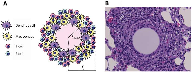
Overview of chronic inflammation

  • Lymphocytes Lymphocytes Lymphocytes are heterogeneous WBCs involved in immune response. Lymphocytes develop from the bone marrow, starting from hematopoietic stem cells (HSCs) and progressing to common lymphoid progenitors (CLPs). B and T lymphocytes and natural killer (NK) cells arise from the lineage. Lymphocytes: Histology and macrophages Macrophages The relatively long-lived phagocytic cell of mammalian tissues that are derived from blood monocytes. Main types are peritoneal macrophages; alveolar macrophages; histiocytes; kupffer cells of the liver; and osteoclasts. They may further differentiate within chronic inflammatory lesions to epithelioid cells or may fuse to form foreign body giant cells or langhans giant cells. Innate Immunity: Phagocytes and Antigen Presentation are key cells involved.
  • Can be prolonged (weeks to years)
  • A central component of various chronic conditions:
    • Autoimmune diseases Autoimmune diseases Disorders that are characterized by the production of antibodies that react with host tissues or immune effector cells that are autoreactive to endogenous peptides. Selective IgA Deficiency, such as:
      • Rheumatoid arthritis Arthritis Acute or chronic inflammation of joints. Osteoarthritis
      • Systemic lupus erythematosus Systemic lupus erythematosus Systemic lupus erythematosus (SLE) is a chronic autoimmune, inflammatory condition that causes immune-complex deposition in organs, resulting in systemic manifestations. Women, particularly those of African American descent, are more commonly affected. Systemic Lupus Erythematosus
      • Multiple sclerosis Sclerosis A pathological process consisting of hardening or fibrosis of an anatomical structure, often a vessel or a nerve. Wilms Tumor
    • Neurodegenerative diseases 
    • Granulomatous inflammation Granulomatous Inflammation Chalazion, such as with TB TB Tuberculosis (TB) is an infectious disease caused by Mycobacterium tuberculosis complex bacteria. The bacteria usually attack the lungs but can also damage other parts of the body. Approximately 30% of people around the world are infected with this pathogen, with the majority harboring a latent infection. Tuberculosis spreads through the air when a person with active pulmonary infection coughs or sneezes. Tuberculosis
    • Cancer
    • Chronic obstructive pulmonary disease Pulmonary disease Diseases involving the respiratory system. Blastomyces/Blastomycosis ( COPD COPD Chronic obstructive pulmonary disease (COPD) is a lung disease characterized by progressive, largely irreversible airflow obstruction. The condition usually presents in middle-aged or elderly persons with a history of cigarette smoking. Signs and symptoms include prolonged expiration, wheezing, diminished breath sounds, progressive dyspnea, and chronic cough. Chronic Obstructive Pulmonary Disease (COPD))
    • Inflammatory bowel disease:
      • Crohn disease
      • Ulcerative colitis Colitis Inflammation of the colon section of the large intestine, usually with symptoms such as diarrhea (often with blood and mucus), abdominal pain, and fever. Pseudomembranous Colitis

Etiology

  • Failure of complete removal of the pathogen (e.g., TB TB Tuberculosis (TB) is an infectious disease caused by Mycobacterium tuberculosis complex bacteria. The bacteria usually attack the lungs but can also damage other parts of the body. Approximately 30% of people around the world are infected with this pathogen, with the majority harboring a latent infection. Tuberculosis spreads through the air when a person with active pulmonary infection coughs or sneezes. Tuberculosis)
  • Persistent stimuli (e.g., an inhaled foreign body Foreign Body Foreign Body Aspiration such as silica)
  • Inflammatory response producing continuous oxidative damage stimulating chronic inflammation (e.g., atherosclerosis Atherosclerosis Atherosclerosis is a common form of arterial disease in which lipid deposition forms a plaque in the blood vessel walls. Atherosclerosis is an incurable disease, for which there are clearly defined risk factors that often can be reduced through a change in lifestyle and behavior of the patient. Atherosclerosis)

Mediators of chronic inflammation

  • Macrophages Macrophages The relatively long-lived phagocytic cell of mammalian tissues that are derived from blood monocytes. Main types are peritoneal macrophages; alveolar macrophages; histiocytes; kupffer cells of the liver; and osteoclasts. They may further differentiate within chronic inflammatory lesions to epithelioid cells or may fuse to form foreign body giant cells or langhans giant cells. Innate Immunity: Phagocytes and Antigen Presentation:
    • Sources of cytokines Cytokines Non-antibody proteins secreted by inflammatory leukocytes and some non-leukocytic cells, that act as intercellular mediators. They differ from classical hormones in that they are produced by a number of tissue or cell types rather than by specialized glands. They generally act locally in a paracrine or autocrine rather than endocrine manner. Adaptive Immune Response and chemokines Chemokines Class of pro-inflammatory cytokines that have the ability to attract and activate leukocytes. They can be divided into at least three structural branches: c; cc; and cxc; according to variations in a shared cysteine motif. Adaptive Cell-mediated Immunity
    • Produce complement and coagulation factors Coagulation factors Endogenous substances, usually proteins, that are involved in the blood coagulation process. Hemostasis
    • Also make:
      • Proteases Proteases Proteins and Peptides
      • ROS
      • Eicosanoids Eicosanoids Eicosanoids are cell-signaling molecules produced from arachidonic acid. With the action of phospholipase A2, arachidonic acid is released from the plasma membrane. The different families of eicosanoids, which are prostaglandins (PGs), thromboxanes (TXA2s), prostacyclin (PGI2), lipoxins (LXs), and leukotrienes (LTs), emerge from a series of reactions catalyzed by different enzymes. Eicosanoids
  • T lymphocytes T lymphocytes Lymphocytes responsible for cell-mediated immunity. Two types have been identified – cytotoxic (t-lymphocytes, cytotoxic) and helper T-lymphocytes (t-lymphocytes, helper-inducer). They are formed when lymphocytes circulate through the thymus gland and differentiate to thymocytes. When exposed to an antigen, they divide rapidly and produce large numbers of new T cells sensitized to that antigen. T cells: Types and Functions
  • B cells B cells Lymphoid cells concerned with humoral immunity. They are short-lived cells resembling bursa-derived lymphocytes of birds in their production of immunoglobulin upon appropriate stimulation. B cells: Types and Functions ( plasma Plasma The residual portion of blood that is left after removal of blood cells by centrifugation without prior blood coagulation. Transfusion Products cells)
  • Eosinophils Eosinophils Granular leukocytes with a nucleus that usually has two lobes connected by a slender thread of chromatin, and cytoplasm containing coarse, round granules that are uniform in size and stainable by eosin. Innate Immunity: Phagocytes and Antigen Presentation, mast cells Mast cells Granulated cells that are found in almost all tissues, most abundantly in the skin and the gastrointestinal tract. Like the basophils, mast cells contain large amounts of histamine and heparin. Unlike basophils, mast cells normally remain in the tissues and do not circulate in the blood. Mast cells, derived from the bone marrow stem cells, are regulated by the stem cell factor. Innate Immunity: Phagocytes and Antigen Presentation
  • Cytokines Cytokines Non-antibody proteins secreted by inflammatory leukocytes and some non-leukocytic cells, that act as intercellular mediators. They differ from classical hormones in that they are produced by a number of tissue or cell types rather than by specialized glands. They generally act locally in a paracrine or autocrine rather than endocrine manner. Adaptive Immune Response:
    • IL-12
    • TNF TNF Tumor necrosis factor (TNF) is a major cytokine, released primarily by macrophages in response to stimuli. The presence of microbial products and dead cells and injury are among the stimulating factors. This protein belongs to the TNF superfamily, a group of ligands and receptors performing functions in inflammatory response, morphogenesis, and cell proliferation. Tumor Necrosis Factor (TNF)

Types of chronic inflammation

  • Nonspecific:
  • Granulomatous inflammation Granulomatous Inflammation Chalazion:
    • Granulomas Granulomas A relatively small nodular inflammatory lesion containing grouped mononuclear phagocytes, caused by infectious and noninfectious agents. Sarcoidosis are:
      • An aggregation Aggregation The attachment of platelets to one another. This clumping together can be induced by a number of agents (e.g., thrombin; collagen) and is part of the mechanism leading to the formation of a thrombus. Coagulation Studies of macrophages Macrophages The relatively long-lived phagocytic cell of mammalian tissues that are derived from blood monocytes. Main types are peritoneal macrophages; alveolar macrophages; histiocytes; kupffer cells of the liver; and osteoclasts. They may further differentiate within chronic inflammatory lesions to epithelioid cells or may fuse to form foreign body giant cells or langhans giant cells. Innate Immunity: Phagocytes and Antigen Presentation, epithelioid cells, and giant cells Giant cells Multinucleated masses produced by the fusion of many cells; often associated with viral infections. In aids, they are induced when the envelope glycoprotein of the HIV virus binds to the CD4 antigen of uninfected neighboring T4 cells. The resulting syncytium leads to cell death and thus may account for the cytopathic effect of the virus. Giant Cell Arteritis
      • Surrounded by fibroblasts Fibroblasts Connective tissue cells which secrete an extracellular matrix rich in collagen and other macromolecules. Sarcoidosis and lymphocytes Lymphocytes Lymphocytes are heterogeneous WBCs involved in immune response. Lymphocytes develop from the bone marrow, starting from hematopoietic stem cells (HSCs) and progressing to common lymphoid progenitors (CLPs). B and T lymphocytes and natural killer (NK) cells arise from the lineage. Lymphocytes: Histology
    • Maintained by TNF-alpha secreted by epithelioid cells
    • Caseating granuloma: necrotizing core due to infectious material (e.g., TB TB Tuberculosis (TB) is an infectious disease caused by Mycobacterium tuberculosis complex bacteria. The bacteria usually attack the lungs but can also damage other parts of the body. Approximately 30% of people around the world are infected with this pathogen, with the majority harboring a latent infection. Tuberculosis spreads through the air when a person with active pulmonary infection coughs or sneezes. Tuberculosis)
    • Noncaseating granuloma: nonnecrotized core owing to immune-mediated cellular damage (e.g., sarcoidosis Sarcoidosis Sarcoidosis is a multisystem inflammatory disease that causes noncaseating granulomas. The exact etiology is unknown. Sarcoidosis usually affects the lungs and thoracic lymph nodes, but it can also affect almost every system in the body, including the skin, heart, and eyes, most commonly. Sarcoidosis)
Ppd antigen-bead pulmonary granuloma model

(A) Schematic representation and (B) histological appearance of an artificial pulmonary granuloma induced in mouse 4 days after injection of purified protein-derivative (PPD)-coated beads (H&E staining; magnification: ×800)
rbead: radius of the bead
rg: radius of the granuloma

Image: “PPD antigen-bead pulmonary granuloma model” by Fallahi-Sichani, M., et al. License: CC BY 4.0

Outcome of chronic inflammation

  • Scarring and fibrosis Fibrosis Any pathological condition where fibrous connective tissue invades any organ, usually as a consequence of inflammation or other injury. Bronchiolitis Obliterans within the organ → damage and loss of function 
  • Continuous inflammation → neoplasm
  • Amyloidosis Amyloidosis Amyloidosis is a disease caused by abnormal extracellular tissue deposition of fibrils composed of various misfolded low-molecular-weight protein subunits. These proteins are frequently byproducts of other pathological processes (e.g., multiple myeloma). Amyloidosis

Clinical Relevance

Examples of inflammation becoming a pathologic response: 

  • Atherosclerosis Atherosclerosis Atherosclerosis is a common form of arterial disease in which lipid deposition forms a plaque in the blood vessel walls. Atherosclerosis is an incurable disease, for which there are clearly defined risk factors that often can be reduced through a change in lifestyle and behavior of the patient. Atherosclerosis: common form of arterial disease in which lipid deposition forms a plaque Plaque Primary Skin Lesions in the blood vessel walls. Atherosclerosis Atherosclerosis Atherosclerosis is a common form of arterial disease in which lipid deposition forms a plaque in the blood vessel walls. Atherosclerosis is an incurable disease, for which there are clearly defined risk factors that often can be reduced through a change in lifestyle and behavior of the patient. Atherosclerosis starts with endothelial injury secondary to turbulent flow Turbulent flow Vascular Resistance, Flow, and Mean Arterial Pressure and is followed by lipid retention through monocytic response, leading to the formation of foam cells Foam cells Lipid-laden macrophages originating from monocytes or from smooth muscle cells. Atherosclerosis. This process ultimately results in the formation of a fibrous Fibrous Fibrocystic Change plaque Plaque Primary Skin Lesions. The plaque Plaque Primary Skin Lesions narrows the lumen and can cause ischemia Ischemia A hypoperfusion of the blood through an organ or tissue caused by a pathologic constriction or obstruction of its blood vessels, or an absence of blood circulation. Ischemic Cell Damage, resulting in MI MI MI is ischemia and death of an area of myocardial tissue due to insufficient blood flow and oxygenation, usually from thrombus formation on a ruptured atherosclerotic plaque in the epicardial arteries. Clinical presentation is most commonly with chest pain, but women and patients with diabetes may have atypical symptoms. Myocardial Infarction, stroke, CKD CKD Chronic kidney disease (CKD) is kidney impairment that lasts for ≥ 3 months, implying that it is irreversible. Hypertension and diabetes are the most common causes; however, there are a multitude of other etiologies. In the early to moderate stages, CKD is usually asymptomatic and is primarily diagnosed by laboratory abnormalities. Chronic Kidney Disease, and other organ compromise.
  • Multiple sclerosis Sclerosis A pathological process consisting of hardening or fibrosis of an anatomical structure, often a vessel or a nerve. Wilms Tumor ( MS MS Multiple sclerosis (MS) is a chronic inflammatory autoimmune disease that leads to demyelination of the nerves in the CNS. Young women are more predominantly affected by this most common demyelinating condition. Multiple Sclerosis): chronic inflammatory autoimmune disorder Autoimmune Disorder Septic Arthritis leading to demyelination Demyelination Multiple Sclerosis of the CNS. Inflammation occurs when the oligodendrocyte Oligodendrocyte A class of large neuroglial (macroglial) cells in the central nervous system. Oligodendroglia may be called interfascicular, perivascular, or perineuronal (not the same as satellite cells, perineuronal of ganglia) according to their location. They form the insulating myelin sheath of axons in the central nervous system. Nervous System: Histology recognizes self myelin as a foreign antigen Antigen Substances that are recognized by the immune system and induce an immune reaction. Vaccination and results in the removal of the myelin sheath. Symptoms arise owing to the swelling plus loss of tissue function and include neurologic symptoms affecting vision Vision Ophthalmic Exam, motor Motor Neurons which send impulses peripherally to activate muscles or secretory cells. Nervous System: Histology functions, sensation, and autonomic function.
  • Pulmonary fibrosis Fibrosis Any pathological condition where fibrous connective tissue invades any organ, usually as a consequence of inflammation or other injury. Bronchiolitis Obliterans: type of interstitial lung disease. Environmental exposure to minute inhalants such as smoke, pollutants, and dust causes an inflammatory process within the lung tissue, resulting in fibrosis Fibrosis Any pathological condition where fibrous connective tissue invades any organ, usually as a consequence of inflammation or other injury. Bronchiolitis Obliterans on injury repair. Individuals often present in a moderate-to-advanced stage with progressive dyspnea Dyspnea Dyspnea is the subjective sensation of breathing discomfort. Dyspnea is a normal manifestation of heavy physical or psychological exertion, but also may be caused by underlying conditions (both pulmonary and extrapulmonary). Dyspnea and nonproductive cough. The diagnosis is made on the basis of imaging findings and pulmonary function testing Pulmonary Function Testing Pulmonary Function Tests. Lung transplantation Lung transplantation The transference of either one or both of the lungs from one human or animal to another. Organ Transplantation is the only curative intervention. 

Disorders due to deficiency in some mediators of the inflammatory response: 

  • Defects in adhesion Adhesion The process whereby platelets adhere to something other than platelets, e.g., collagen; basement membrane; microfibrils; or other ‘foreign’ surfaces. Coagulation Studies: Leukocyte adhesion Leukocyte adhesion Leukocyte Adhesion Deficiency Type 1 deficiency I (LAD I) is due to defective synthesis Synthesis Polymerase Chain Reaction (PCR) of integrin in the cellular response phase and deficient binding to immunoglobulins Immunoglobulins Immunoglobulins (Igs), also known as antibodies, are glycoprotein molecules produced by plasma cells that act in immune responses by recognizing and binding particular antigens. The various Ig classes are IgG (the most abundant), IgM, IgE, IgD, and IgA, which differ in their biologic features, structure, target specificity, and distribution. Immunoglobulins: Types and Functions, which result in recurrent infections Recurrent infections Common Variable Immunodeficiency (CVID). Leukocyte adhesion Leukocyte adhesion Leukocyte Adhesion Deficiency Type 1 deficiency II (LAD II) is a rare disorder in which neutrophils Neutrophils Granular leukocytes having a nucleus with three to five lobes connected by slender threads of chromatin, and cytoplasm containing fine inconspicuous granules and stainable by neutral dyes. Innate Immunity: Phagocytes and Antigen Presentation are deficient in the expression of sialyl-Lewis X, which is needed for rolling in the cellular response phase of inflammation. Leukocyte adhesion Leukocyte adhesion Leukocyte Adhesion Deficiency Type 1 deficiency II is characterized by recurrent infections Recurrent infections Common Variable Immunodeficiency (CVID), persistent leukocytosis Leukocytosis A transient increase in the number of leukocytes in a body fluid. West Nile Virus, and severe deficits in development and growth.
  • Defect in phagolysosome Phagolysosome Chédiak-Higashi Syndrome formation: Chediak-Higashi syndrome is an autosomal recessive Autosomal recessive Autosomal inheritance, both dominant and recessive, refers to the transmission of genes from the 22 autosomal chromosomes. Autosomal recessive diseases are only expressed when 2 copies of the recessive allele are inherited. Autosomal Recessive and Autosomal Dominant Inheritance disorder caused by mutations affecting a lysosomal trafficking regulator Lysosomal trafficking regulator Chédiak-Higashi Syndrome protein, whereby phagocytosed bacteria Bacteria Bacteria are prokaryotic single-celled microorganisms that are metabolically active and divide by binary fission. Some of these organisms play a significant role in the pathogenesis of diseases. Bacteriology are not destroyed by lysosomal enzymes Enzymes Enzymes are complex protein biocatalysts that accelerate chemical reactions without being consumed by them. Due to the body’s constant metabolic needs, the absence of enzymes would make life unsustainable, as reactions would occur too slowly without these molecules. Basics of Enzymes. Chemotaxis Chemotaxis The movement of leukocytes in response to a chemical concentration gradient or to products formed in an immunologic reaction. Leukocyte Adhesion Deficiency Type 1 is also disrupted, resulting in poor neutrophil recruitment Recruitment Skeletal Muscle Contraction and function. Individuals exhibit recurrent pyogenic infections Infections Invasion of the host organism by microorganisms or their toxins or by parasites that can cause pathological conditions or diseases. Chronic Granulomatous Disease, easy bleeding and bruising, and neurologic manifestations. The diagnosis is based on analysis of the individual’s blood or bone marrow smear Bone marrow smear Chédiak-Higashi Syndrome and genetic testing Genetic Testing Detection of a mutation; genotype; karyotype; or specific alleles associated with genetic traits, heritable diseases, or predisposition to a disease, or that may lead to the disease in descendants. It includes prenatal genetic testing. Myotonic Dystrophies
  • Defect in microbicidal activity: Chronic granulomatous disease Granulomatous disease A defect of leukocyte function in which phagocytic cells ingest but fail to digest bacteria, resulting in recurring bacterial infections with granuloma formation. When chronic granulomatous disease is caused by mutations in the cybb gene, the condition is inherited in an X-linked recessive pattern. When chronic granulomatous disease is caused by cyba, ncf1, ncf2, or ncf4 gene mutations, the condition is inherited in an autosomal recessive pattern. Common Variable Immunodeficiency (CVID) ( CGD CGD Chronic granulomatous disease (CGD), as the name implies, is a chronic disorder that is characterized by granuloma formation. This disorder is a consequence of defective phagocytic cells that are unable to produce bactericidal superoxide because of a defect in nicotinamide adenine dinucleotide phosphate (NADPH), the oxidase responsible for the respiratory burst in phagocytic leukocytes. Chronic Granulomatous Disease), seen in individuals with a genetic mutation Mutation Genetic mutations are errors in DNA that can cause protein misfolding and dysfunction. There are various types of mutations, including chromosomal, point, frameshift, and expansion mutations. Types of Mutations for NADPH NADPH Nicotinamide adenine dinucleotide phosphate. A coenzyme composed of ribosylnicotinamide 5′-phosphate (nmn) coupled by pyrophosphate linkage to the 5′-phosphate adenosine 2. Pentose Phosphate Pathway oxidase Oxidase Neisseria, which is responsible for the respiratory burst Respiratory burst A large increase in oxygen uptake by neutrophils and most types of tissue macrophages through activation of an NADPH-cytochrome b-dependent oxidase that reduces oxygen to a superoxide. Individuals with an inherited defect in which the oxidase that reduces oxygen to superoxide is decreased or absent often die as a result of recurrent bacterial infections. Leukocyte Adhesion Deficiency Type 1 in phagocytic leukocytes Leukocytes White blood cells. These include granular leukocytes (basophils; eosinophils; and neutrophils) as well as non-granular leukocytes (lymphocytes and monocytes). White Myeloid Cells: Histology. Infections Infections Invasion of the host organism by microorganisms or their toxins or by parasites that can cause pathological conditions or diseases. Chronic Granulomatous Disease may be contained; however, lysis does not occur, thus resulting in chronic granuloma formation. Individuals with CGD CGD Chronic granulomatous disease (CGD), as the name implies, is a chronic disorder that is characterized by granuloma formation. This disorder is a consequence of defective phagocytic cells that are unable to produce bactericidal superoxide because of a defect in nicotinamide adenine dinucleotide phosphate (NADPH), the oxidase responsible for the respiratory burst in phagocytic leukocytes. Chronic Granulomatous Disease are at increased risk of life-threatening infections Infections Invasion of the host organism by microorganisms or their toxins or by parasites that can cause pathological conditions or diseases. Chronic Granulomatous Disease with fungi Fungi A kingdom of eukaryotic, heterotrophic organisms that live parasitically as saprobes, including mushrooms; yeasts; smuts, molds, etc. They reproduce either sexually or asexually, and have life cycles that range from simple to complex. Filamentous fungi, commonly known as molds, refer to those that grow as multicellular colonies. Mycology and catalase-positive bacteria Bacteria Bacteria are prokaryotic single-celled microorganisms that are metabolically active and divide by binary fission. Some of these organisms play a significant role in the pathogenesis of diseases. Bacteriology.

References

  1. Hall, J. E., Guyton, A. C. (2011). Resistance of the body to infection I. Chapter 33 of Guyton and Hall Textbook of Medical Physiology, 12th ed. Philadelphia: Saunders Elsevier, pp. 428–430.
  2. Kumar, V., Abbas, A. K., Aster, J. C. (2017). Inflammation and repair. Chapter 3 of Robbins Basic Pathology, 10th ed. Elsevier, pp. 57–70, 79–82.
  3. Johnston, R. B. (2021). An overview of the innate immune system. UpToDate. Retrieved September 14, 2021, from https://www.uptodate.com/contents/an-overview-of-the-innate-immune-system
  4. Stone, W. L., Basit, H., Burns, B. (2020). Pathology, inflammation. StatPearls. Retrieved September 14, 2021, from https://www.ncbi.nlm.nih.gov/books/NBK534820/

Create your free account or log in to continue reading!

Sign up now and get free access to Lecturio with concept pages, medical videos, and questions for your medical education.

User Reviews

Unwrap New Skills This Holiday 🎄 Save 30% on all plans now!

Details