Advertisement

Advertisement

Advertisement

Advertisement

Hypercoagulable States

Hypercoagulable states (also referred to as thrombophilias) are a group of hematologic diseases defined by an increased risk of clot formation (i.e., thrombosis Thrombosis Formation and development of a thrombus or blood clot in the blood vessel. Epidemic Typhus) due to either an increase in procoagulants, a decrease in anticoagulants Anticoagulants Anticoagulants are drugs that retard or interrupt the coagulation cascade. The primary classes of available anticoagulants include heparins, vitamin K-dependent antagonists (e.g., warfarin), direct thrombin inhibitors, and factor Xa inhibitors. Anticoagulants, or a decrease in fibrinolysis. There are both inherited and acquired causes, with factor V Factor V Heat- and storage-labile plasma glycoprotein which accelerates the conversion of prothrombin to thrombin in blood coagulation. Factor V accomplishes this by forming a complex with factor Xa, phospholipid, and calcium (prothrombinase complex). Hemostasis Leiden being the most common inherited cause. Clinically, hypercoagulable states present with thrombotic events, which cause vessel occlusion and can lead to organ damage. Thrombotic disorders can be fatal if not treated, and management usually involves anticoagulants Anticoagulants Anticoagulants are drugs that retard or interrupt the coagulation cascade. The primary classes of available anticoagulants include heparins, vitamin K-dependent antagonists (e.g., warfarin), direct thrombin inhibitors, and factor Xa inhibitors. Anticoagulants.

Last updated: May 17, 2024

Editorial responsibility: Stanley Oiseth, Lindsay Jones, Evelin Maza

Advertisement

Advertisement

Advertisement

Advertisement

Advertisement

Advertisement

Overview

Definition

Hypercoagulability, also referred to as a thrombophilia, refers to the increased tendency for blood to form clots, known as thrombi. Hypercoagulable states can be inherited or acquired.

Epidemiology

Prevalence Prevalence The total number of cases of a given disease in a specified population at a designated time. It is differentiated from incidence, which refers to the number of new cases in the population at a given time. Measures of Disease Frequency of inherited thrombophilias:

Table: Prevalence Prevalence The total number of cases of a given disease in a specified population at a designated time. It is differentiated from incidence, which refers to the number of new cases in the population at a given time. Measures of Disease Frequency and risk of venous thromboemolism (VTE) in inherited thrombophilias
Condition Prevalence Prevalence The total number of cases of a given disease in a specified population at a designated time. It is differentiated from incidence, which refers to the number of new cases in the population at a given time. Measures of Disease Frequency Risk of VTE
Factor V Factor V Heat- and storage-labile plasma glycoprotein which accelerates the conversion of prothrombin to thrombin in blood coagulation. Factor V accomplishes this by forming a complex with factor Xa, phospholipid, and calcium (prothrombinase complex). Hemostasis Leiden 3%–7% 4.3%
Prothrombin Prothrombin A plasma protein that is the inactive precursor of thrombin. It is converted to thrombin by a prothrombin activator complex consisting of factor Xa, factor V, phospholipid, and calcium ions. Hemostasis G20210A mutation Mutation Genetic mutations are errors in DNA that can cause protein misfolding and dysfunction. There are various types of mutations, including chromosomal, point, frameshift, and expansion mutations. Types of Mutations 1%–3% 1.9%
Protein C deficiency (heterozygous) 0.02%–0.05% 11.3%
Protein S Protein S Protein S augments the activity of protein C. Hemostasis deficiency (heterozygous) 0.01% 32.4%
Antithrombin deficiency Antithrombin deficiency An absence or reduced level of antithrombin III leading to an increased risk for thrombosis. Budd-Chiari Syndrome 0.02%–0.04% 17.5%

Most common acquired hypercoagulable states:

  • Antiphospholipid syndrome Antiphospholipid syndrome Antiphospholipid syndrome (APLS) is an acquired autoimmune disorder characterized by the persistent presence of antiphospholipid antibodies, which create a hypercoagulable state. These antibodies are most commonly discovered during a workup for a thrombotic event or recurrent pregnancy loss, which are the 2 most common clinical manifestations. Antiphospholipid Syndrome: most common
  • Malignancy Malignancy Hemothorax: 2nd most common

Clinical impact:

  • VTE is 2nd only to MI MI MI is ischemia and death of an area of myocardial tissue due to insufficient blood flow and oxygenation, usually from thrombus formation on a ruptured atherosclerotic plaque in the epicardial arteries. Clinical presentation is most commonly with chest pain, but women and patients with diabetes may have atypical symptoms. Myocardial Infarction as the most common cardiovascular disorder.
  • Up to 4% of strokes are attributable to hypercoagulability disorders.
  • Thrombosis Thrombosis Formation and development of a thrombus or blood clot in the blood vessel. Epidemic Typhus risk factors are identified in over 80% of patients Patients Individuals participating in the health care system for the purpose of receiving therapeutic, diagnostic, or preventive procedures. Clinician–Patient Relationship with thrombotic events.

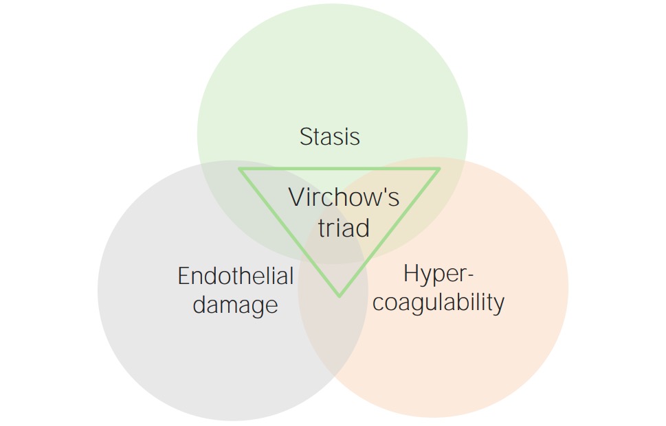
Virchow’s triad Virchow’s triad Deep Vein Thrombosis

Thrombotic events occur under 3 primary conditions, which make up Virchow’s triad Virchow’s triad Deep Vein Thrombosis. These 3 conditions are:
  1. Endothelial injury/ subendothelial Subendothelial Membranoproliferative Glomerulonephritis exposure:
    • Initiates formation of the platelet plug Platelet plug Hemostasis and the coagulation cascade Coagulation cascade The coagulation cascade is a series of reactions that ultimately generates a strong, cross-linked fibrin clot. Hemostasis
    • Factors involved in initiating thrombosis Thrombosis Formation and development of a thrombus or blood clot in the blood vessel. Epidemic Typhus:
      • Tissue factor
      • von Willebrand factor von Willebrand factor A high-molecular-weight plasma protein, produced by endothelial cells and megakaryocytes, that is part of the factor VIII/von Willebrand factor complex. The von Willebrand factor has receptors for collagen, platelets, and ristocetin activity as well as the immunologically distinct antigenic determinants. It functions in adhesion of platelets to collagen and hemostatic plug formation. The prolonged bleeding time in von Willebrand diseases is due to the deficiency of this factor. Hemostasis
      • Collagen Collagen A polypeptide substance comprising about one third of the total protein in mammalian organisms. It is the main constituent of skin; connective tissue; and the organic substance of bones (bone and bones) and teeth (tooth). Connective Tissue: Histology 
  2. Stasis of blood flow Blood flow Blood flow refers to the movement of a certain volume of blood through the vasculature over a given unit of time (e.g., mL per minute). Vascular Resistance, Flow, and Mean Arterial Pressure:
    • Allows more time for platelet aggregation Platelet aggregation The attachment of platelets to one another. This clumping together can be induced by a number of agents (e.g., thrombin; collagen) and is part of the mechanism leading to the formation of a thrombus. Hemostasis and clot formation
    • Slows the removal of coagulation factors Coagulation factors Endogenous substances, usually proteins, that are involved in the blood coagulation process. Hemostasis
  3. Hypercoagulable states (alteration in blood constituents):
    • May result from:
      • An increase in procoagulants
      • A decrease in anticoagulants Anticoagulants Anticoagulants are drugs that retard or interrupt the coagulation cascade. The primary classes of available anticoagulants include heparins, vitamin K-dependent antagonists (e.g., warfarin), direct thrombin inhibitors, and factor Xa inhibitors. Anticoagulants
      • A decrease in fibrinolysis 
    • Balance is tipped to favor thrombosis Thrombosis Formation and development of a thrombus or blood clot in the blood vessel. Epidemic Typhus.
Vichows triad

Virchow’s triad

Image by Lecturio.

Etiology and Pathophysiology

Hypercoagulable states may be primary (inherited) or secondary (acquired).

Primary (inherited) causes of hypercoagulable states

Table: Etiology and pathophysiology of primary (inherited) hypercoagulable states
Condition Inheritance pattern Pathophysiology
Factor V Factor V Heat- and storage-labile plasma glycoprotein which accelerates the conversion of prothrombin to thrombin in blood coagulation. Factor V accomplishes this by forming a complex with factor Xa, phospholipid, and calcium (prothrombinase complex). Hemostasis Leiden Autosomal dominant Autosomal dominant Autosomal inheritance, both dominant and recessive, refers to the transmission of genes from the 22 autosomal chromosomes. Autosomal dominant diseases are expressed when only 1 copy of the dominant allele is inherited. Autosomal Recessive and Autosomal Dominant Inheritance with incomplete penetrance Penetrance The percent frequency with which a dominant or homozygous recessive gene or gene combination manifests itself in the phenotype of the carriers. Familial Juvenile Polyposis
  • Point mutation Point Mutation A mutation caused by the substitution of one nucleotide for another. This results in the DNA molecule having a change in a single base pair. Types of Mutations substitutes glutamine Glutamine A non-essential amino acid present abundantly throughout the body and is involved in many metabolic processes. It is synthesized from glutamic acid and ammonia. It is the principal carrier of nitrogen in the body and is an important energy source for many cells. Synthesis of Nonessential Amino Acids for arginine Arginine An essential amino acid that is physiologically active in the l-form. Urea Cycle at position 506 in the F5 gene Gene A category of nucleic acid sequences that function as units of heredity and which code for the basic instructions for the development, reproduction, and maintenance of organisms. Basic Terms of Genetics, which codes for factor V Factor V Heat- and storage-labile plasma glycoprotein which accelerates the conversion of prothrombin to thrombin in blood coagulation. Factor V accomplishes this by forming a complex with factor Xa, phospholipid, and calcium (prothrombinase complex). Hemostasis.
  • Results in resistance Resistance Physiologically, the opposition to flow of air caused by the forces of friction. As a part of pulmonary function testing, it is the ratio of driving pressure to the rate of air flow. Ventilation: Mechanics of Breathing to factor Va VA Ventilation: Mechanics of Breathing degradation by protein C
  • ↑ Factor Va VA Ventilation: Mechanics of Breathing available → enhanced thrombin Thrombin An enzyme formed from prothrombin that converts fibrinogen to fibrin. Hemostasis formation
Prothrombin Prothrombin A plasma protein that is the inactive precursor of thrombin. It is converted to thrombin by a prothrombin activator complex consisting of factor Xa, factor V, phospholipid, and calcium ions. Hemostasis G20210A (also known as factor II mutation Mutation Genetic mutations are errors in DNA that can cause protein misfolding and dysfunction. There are various types of mutations, including chromosomal, point, frameshift, and expansion mutations. Types of Mutations) Autosomal recessive Autosomal recessive Autosomal inheritance, both dominant and recessive, refers to the transmission of genes from the 22 autosomal chromosomes. Autosomal recessive diseases are only expressed when 2 copies of the recessive allele are inherited. Autosomal Recessive and Autosomal Dominant Inheritance
  • Transposition at the promoter region of the prothrombin Prothrombin A plasma protein that is the inactive precursor of thrombin. It is converted to thrombin by a prothrombin activator complex consisting of factor Xa, factor V, phospholipid, and calcium ions. Hemostasis gene Gene A category of nucleic acid sequences that function as units of heredity and which code for the basic instructions for the development, reproduction, and maintenance of organisms. Basic Terms of Genetics
  • Results in overtranslation of prothrombin Prothrombin A plasma protein that is the inactive precursor of thrombin. It is converted to thrombin by a prothrombin activator complex consisting of factor Xa, factor V, phospholipid, and calcium ions. Hemostasis
  • Thrombin Thrombin An enzyme formed from prothrombin that converts fibrinogen to fibrin. Hemostasis precursor → enhanced thrombin Thrombin An enzyme formed from prothrombin that converts fibrinogen to fibrin. Hemostasis formation
Antithrombin deficiency Antithrombin deficiency An absence or reduced level of antithrombin III leading to an increased risk for thrombosis. Budd-Chiari Syndrome Autosomal dominant Autosomal dominant Autosomal inheritance, both dominant and recessive, refers to the transmission of genes from the 22 autosomal chromosomes. Autosomal dominant diseases are expressed when only 1 copy of the dominant allele is inherited. Autosomal Recessive and Autosomal Dominant Inheritance
  • Antithrombin Antithrombin Endogenous factors and drugs that directly inhibit the action of thrombin, usually by blocking its enzymatic activity. They are distinguished from indirect thrombin inhibitors, such as heparin, which act by enhancing the inhibitory effects of antithrombins. Anticoagulants inactivates thrombin Thrombin An enzyme formed from prothrombin that converts fibrinogen to fibrin. Hemostasis (and is augmented by heparin)
  • Deficiency of antithrombin Antithrombin Endogenous factors and drugs that directly inhibit the action of thrombin, usually by blocking its enzymatic activity. They are distinguished from indirect thrombin inhibitors, such as heparin, which act by enhancing the inhibitory effects of antithrombins. Anticoagulants leads to:
    • ↑ Levels of thrombin Thrombin An enzyme formed from prothrombin that converts fibrinogen to fibrin. Hemostasis and factor Xa
    • Resistance Resistance Physiologically, the opposition to flow of air caused by the forces of friction. As a part of pulmonary function testing, it is the ratio of driving pressure to the rate of air flow. Ventilation: Mechanics of Breathing to heparins
  • Can also be acquired through antithrombin Antithrombin Endogenous factors and drugs that directly inhibit the action of thrombin, usually by blocking its enzymatic activity. They are distinguished from indirect thrombin inhibitors, such as heparin, which act by enhancing the inhibitory effects of antithrombins. Anticoagulants protein loss ( nephrotic syndrome Nephrotic syndrome Nephrotic syndrome is characterized by severe proteinuria, hypoalbuminemia, and peripheral edema. In contrast, the nephritic syndromes present with hematuria, variable loss of renal function, and hypertension, although there is sometimes overlap of > 1 glomerular disease in the same individual. Nephrotic Syndrome) or decreased antithrombin Antithrombin Endogenous factors and drugs that directly inhibit the action of thrombin, usually by blocking its enzymatic activity. They are distinguished from indirect thrombin inhibitors, such as heparin, which act by enhancing the inhibitory effects of antithrombins. Anticoagulants production ( liver Liver The liver is the largest gland in the human body. The liver is found in the superior right quadrant of the abdomen and weighs approximately 1.5 kilograms. Its main functions are detoxification, metabolism, nutrient storage (e.g., iron and vitamins), synthesis of coagulation factors, formation of bile, filtration, and storage of blood. Liver: Anatomy disease)
Protein C or S deficiency Autosomal dominant Autosomal dominant Autosomal inheritance, both dominant and recessive, refers to the transmission of genes from the 22 autosomal chromosomes. Autosomal dominant diseases are expressed when only 1 copy of the dominant allele is inherited. Autosomal Recessive and Autosomal Dominant Inheritance (rare; can be recessive)
  • Protein C inactivates factors Va VA Ventilation: Mechanics of Breathing and VIIIa using protein S Protein S Protein S augments the activity of protein C. Hemostasis as a cofactor.
  • Deficiencies of either result in overactivity of factors Va VA Ventilation: Mechanics of Breathing and VIIIa.
  • ↑ Activity of Va VA Ventilation: Mechanics of Breathing and VIIIa → enhances thrombin Thrombin An enzyme formed from prothrombin that converts fibrinogen to fibrin. Hemostasis formation
  • Can also be acquired in nephrotic syndrome Nephrotic syndrome Nephrotic syndrome is characterized by severe proteinuria, hypoalbuminemia, and peripheral edema. In contrast, the nephritic syndromes present with hematuria, variable loss of renal function, and hypertension, although there is sometimes overlap of > 1 glomerular disease in the same individual. Nephrotic Syndrome and liver Liver The liver is the largest gland in the human body. The liver is found in the superior right quadrant of the abdomen and weighs approximately 1.5 kilograms. Its main functions are detoxification, metabolism, nutrient storage (e.g., iron and vitamins), synthesis of coagulation factors, formation of bile, filtration, and storage of blood. Liver: Anatomy disease
Sticky platelet syndrome Autosomal dominant Autosomal dominant Autosomal inheritance, both dominant and recessive, refers to the transmission of genes from the 22 autosomal chromosomes. Autosomal dominant diseases are expressed when only 1 copy of the dominant allele is inherited. Autosomal Recessive and Autosomal Dominant Inheritance
  • Platelets Platelets Platelets are small cell fragments involved in hemostasis. Thrombopoiesis takes place primarily in the bone marrow through a series of cell differentiation and is influenced by several cytokines. Platelets are formed after fragmentation of the megakaryocyte cytoplasm. Platelets: Histology are hyperaggregable.
  • When platelets Platelets Platelets are small cell fragments involved in hemostasis. Thrombopoiesis takes place primarily in the bone marrow through a series of cell differentiation and is influenced by several cytokines. Platelets are formed after fragmentation of the megakaryocyte cytoplasm. Platelets: Histology are activated, they induce a hypercoagulable state.
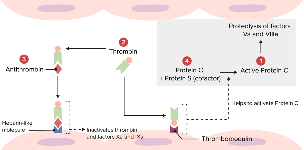
Overview of the physiologic thrombolytic pathway

Overview of the physiologic thrombolytic pathway:
Several hypercoagulable disorders occur because of abnormalities at the following locations.
Mutations at 1: Factor V Leiden makes factor Va resistant to degradation by activated protein C.
Mutations at 2: Prothrombin mutation produces an increased amount of prothrombin, leading to excess thrombin formation.
Mutations at 3: Antithrombin deficiency leads to reduced inactivation of thrombin and factors Xa and IXa.
Mutations at 4: Protein C or S deficiency leads to reduced inactivation of factors Va and VIIIa.

Image by Lecturio.

Secondary (acquired) causes of hypercoagulable states

  • Antiphospholipid syndrome Antiphospholipid syndrome Antiphospholipid syndrome (APLS) is an acquired autoimmune disorder characterized by the persistent presence of antiphospholipid antibodies, which create a hypercoagulable state. These antibodies are most commonly discovered during a workup for a thrombotic event or recurrent pregnancy loss, which are the 2 most common clinical manifestations. Antiphospholipid Syndrome:
    • Antibodies Antibodies Immunoglobulins (Igs), also known as antibodies, are glycoprotein molecules produced by plasma cells that act in immune responses by recognizing and binding particular antigens. The various Ig classes are IgG (the most abundant), IgM, IgE, IgD, and IgA, which differ in their biologic features, structure, target specificity, and distribution. Immunoglobulins: Types and Functions form against phospholipid-binding proteins Proteins Linear polypeptides that are synthesized on ribosomes and may be further modified, crosslinked, cleaved, or assembled into complex proteins with several subunits. The specific sequence of amino acids determines the shape the polypeptide will take, during protein folding, and the function of the protein. Energy Homeostasis.
    • Antibodies Antibodies Immunoglobulins (Igs), also known as antibodies, are glycoprotein molecules produced by plasma cells that act in immune responses by recognizing and binding particular antigens. The various Ig classes are IgG (the most abundant), IgM, IgE, IgD, and IgA, which differ in their biologic features, structure, target specificity, and distribution. Immunoglobulins: Types and Functions result in:
      • Activation of inflammatory cells, endothelial cells, and platelets Platelets Platelets are small cell fragments involved in hemostasis. Thrombopoiesis takes place primarily in the bone marrow through a series of cell differentiation and is influenced by several cytokines. Platelets are formed after fragmentation of the megakaryocyte cytoplasm. Platelets: Histology, promoting thrombosis Thrombosis Formation and development of a thrombus or blood clot in the blood vessel. Epidemic Typhus
      • Inactivation of anticoagulant factors ( proteins Proteins Linear polypeptides that are synthesized on ribosomes and may be further modified, crosslinked, cleaved, or assembled into complex proteins with several subunits. The specific sequence of amino acids determines the shape the polypeptide will take, during protein folding, and the function of the protein. Energy Homeostasis C and S)
    • Antibodies Antibodies Immunoglobulins (Igs), also known as antibodies, are glycoprotein molecules produced by plasma cells that act in immune responses by recognizing and binding particular antigens. The various Ig classes are IgG (the most abundant), IgM, IgE, IgD, and IgA, which differ in their biologic features, structure, target specificity, and distribution. Immunoglobulins: Types and Functions may form spontaneously due to genetic predispositions or in association with other disorders, such as:
      • Systemic lupus erythematosus Systemic lupus erythematosus Systemic lupus erythematosus (SLE) is a chronic autoimmune, inflammatory condition that causes immune-complex deposition in organs, resulting in systemic manifestations. Women, particularly those of African American descent, are more commonly affected. Systemic Lupus Erythematosus ( SLE SLE Systemic lupus erythematosus (SLE) is a chronic autoimmune, inflammatory condition that causes immune-complex deposition in organs, resulting in systemic manifestations. Women, particularly those of African American descent, are more commonly affected. Systemic Lupus Erythematosus; most common, 35%)
      • Rheumatoid arthritis Arthritis Acute or chronic inflammation of joints. Osteoarthritis
      • Sjögren syndrome Sjögren Syndrome Rheumatoid Arthritis
      • Immune thrombocytopenic purpura Immune thrombocytopenic purpura Immune thrombocytopenic purpura (ITP), formerly known as idiopathic thrombocytopenic purpura, is a condition that develops secondary to immune-mediated destruction of platelets, resulting in thrombocytopenia (platelet count < 100,000/mm³). Immune thrombocytopenic purpura can be either primary or secondary due to drugs or underlying disease. Immune Thrombocytopenic Purpura ( ITP ITP Immune thrombocytopenic purpura (ITP), formerly known as idiopathic thrombocytopenic purpura, is a condition that develops secondary to immune-mediated destruction of platelets, resulting in thrombocytopenia (platelet count < 100,000/mm³). Immune thrombocytopenic purpura can be either primary or secondary due to drugs or underlying disease. Immune Thrombocytopenic Purpura)
      • HIV HIV Anti-HIV Drugs
  • Antithrombin deficiency Antithrombin deficiency An absence or reduced level of antithrombin III leading to an increased risk for thrombosis. Budd-Chiari Syndrome
  • Protein C or S deficiencies
  • Malignancy Malignancy Hemothorax:
    • Tumor Tumor Inflammation cells can release procoagulants: tissue factor and cancer procoagulant
    • Tumors may also cause vessel compression Compression Blunt Chest Trauma → stasis
    • Most common with adenocarcinomas (especially lung, pancreatic, and colorectal)
  • Increased estrogen Estrogen Compounds that interact with estrogen receptors in target tissues to bring about the effects similar to those of estradiol. Estrogens stimulate the female reproductive organs, and the development of secondary female sex characteristics. Estrogenic chemicals include natural, synthetic, steroidal, or non-steroidal compounds. Ovaries: Anatomy exposure:
    • Estrogens cause a hypercoagulable state by:
      • ↑ Concentration of procoagulants: prothrombin Prothrombin A plasma protein that is the inactive precursor of thrombin. It is converted to thrombin by a prothrombin activator complex consisting of factor Xa, factor V, phospholipid, and calcium ions. Hemostasis, fibrinogen Fibrinogen Plasma glycoprotein clotted by thrombin, composed of a dimer of three non-identical pairs of polypeptide chains (alpha, beta, gamma) held together by disulfide bonds. Fibrinogen clotting is a sol-gel change involving complex molecular arrangements: whereas fibrinogen is cleaved by thrombin to form polypeptides a and b, the proteolytic action of other enzymes yields different fibrinogen degradation products. Hemostasis, factor VII Factor VII Heat- and storage-stable plasma protein that is activated by tissue thromboplastin to form factor viia in the extrinsic pathway of blood coagulation. The activated form then catalyzes the activation of factor X to factor Xa. Hemostasis, and factor X Factor X Storage-stable glycoprotein blood coagulation factor that can be activated to factor Xa by both the intrinsic and extrinsic pathways. A deficiency of factor X, sometimes called stuart-prower factor deficiency, may lead to a systemic coagulation disorder. Hemostasis
      • Antithrombin Antithrombin Endogenous factors and drugs that directly inhibit the action of thrombin, usually by blocking its enzymatic activity. They are distinguished from indirect thrombin inhibitors, such as heparin, which act by enhancing the inhibitory effects of antithrombins. Anticoagulants levels
    • States associated with ↑ estrogen Estrogen Compounds that interact with estrogen receptors in target tissues to bring about the effects similar to those of estradiol. Estrogens stimulate the female reproductive organs, and the development of secondary female sex characteristics. Estrogenic chemicals include natural, synthetic, steroidal, or non-steroidal compounds. Ovaries: Anatomy:
      • Pregnancy Pregnancy The status during which female mammals carry their developing young (embryos or fetuses) in utero before birth, beginning from fertilization to birth. Pregnancy: Diagnosis, Physiology, and Care
      • Use of estrogen-containing contraceptives
      • Hormone replacement therapy Hormone Replacement Therapy Hormone replacement therapy (HRT) is used to treat symptoms associated with female menopause and in combination to suppress ovulation. Risks and side effects include uterine bleeding, predisposition to cancer, breast tenderness, hyperpigmentation, migraine headaches, hypertension, bloating, and mood changes. Noncontraceptive Estrogen and Progestins

Other conditions that increase thrombotic risk

Many other conditions and states increase thrombotic risk by affecting components of Virchow’s triad Virchow’s triad Deep Vein Thrombosis (typically, stasis, endothelial injury, or both) in ways that promote thrombosis Thrombosis Formation and development of a thrombus or blood clot in the blood vessel. Epidemic Typhus. These conditions include:

  • General risk factors (many are cardiovascular risk factors):
    • Prior history of a VTE
    • Older age (≥ 65 years)
    • Obesity Obesity Obesity is a condition associated with excess body weight, specifically with the deposition of excessive adipose tissue. Obesity is considered a global epidemic. Major influences come from the western diet and sedentary lifestyles, but the exact mechanisms likely include a mixture of genetic and environmental factors. Obesity
    • Diabetes Diabetes Diabetes mellitus (DM) is a metabolic disease characterized by hyperglycemia and dysfunction of the regulation of glucose metabolism by insulin. Type 1 DM is diagnosed mostly in children and young adults as the result of autoimmune destruction of β cells in the pancreas and the resulting lack of insulin. Type 2 DM has a significant association with obesity and is characterized by insulin resistance. Diabetes Mellitus mellitus
    • Immobilization Immobilization Delirium (e.g., cast, stroke patient, post–knee surgery, long flight)
    • Current/recent hospitalization Hospitalization The confinement of a patient in a hospital. Delirium
    • Family history Family History Adult Health Maintenance
    • Smoking Smoking Willful or deliberate act of inhaling and exhaling smoke from burning substances or agents held by hand. Interstitial Lung Diseases
  • Related to surgery or procedures:
    • Surgery, especially orthopedic, vascular, neurologic, and cancer surgeries
    • Recent trauma
    • Presence of a vascular prosthesis (e.g., central venous catheter Central Venous Catheter Central venous catheters are IV lines placed into the large central veins for monitoring of central venous pressure (CVP), prolonged drug administration, or administration of parenteral nutrition. The most common sites of insertion are the internal jugular and subclavian veins. Central Venous Catheter)
  • Cardiovascular conditions:
    • Heart failure Heart Failure A heterogeneous condition in which the heart is unable to pump out sufficient blood to meet the metabolic need of the body. Heart failure can be caused by structural defects, functional abnormalities (ventricular dysfunction), or a sudden overload beyond its capacity. Chronic heart failure is more common than acute heart failure which results from sudden insult to cardiac function, such as myocardial infarction. Total Anomalous Pulmonary Venous Return (TAPVR)
    • Chronic venous insufficiency Chronic venous insufficiency Chronic venous disease is a spectrum of disorders characterized by venous dilation and/or abnormal vein function in the lower extremities resulting from venous hypertension. “Chronic venous insufficiency” refers to the more severe forms of chronic venous disease. Skin changes typically distinguish chronic venous insufficiency from milder forms of venous disease. Chronic Venous Insufficiency
    • Congenital Congenital Chorioretinitis heart disease
    • Vasculitis Vasculitis Inflammation of any one of the blood vessels, including the arteries; veins; and rest of the vasculature system in the body. Systemic Lupus Erythematosus
    • Atherosclerosis Atherosclerosis Atherosclerosis is a common form of arterial disease in which lipid deposition forms a plaque in the blood vessel walls. Atherosclerosis is an incurable disease, for which there are clearly defined risk factors that often can be reduced through a change in lifestyle and behavior of the patient. Atherosclerosis
  • Renal conditions:
    • Chronic renal disease, especially end-stage renal disease
    • Nephrotic syndrome Nephrotic syndrome Nephrotic syndrome is characterized by severe proteinuria, hypoalbuminemia, and peripheral edema. In contrast, the nephritic syndromes present with hematuria, variable loss of renal function, and hypertension, although there is sometimes overlap of > 1 glomerular disease in the same individual. Nephrotic Syndrome
    • Renal transplantation
  • Hematologic/oncologic conditions:
    • Myeloproliferative neoplasms Neoplasms New abnormal growth of tissue. Malignant neoplasms show a greater degree of anaplasia and have the properties of invasion and metastasis, compared to benign neoplasms. Benign Bone Tumors:
      • Essential thrombocythemia Essential thrombocythemia Essential thrombocythemia (ET) is a type of myeloproliferative neoplasm characterized by the clonal thrombocytosis linked to somatic mutations involving Janus kinase 2 (JAK2), calreticulin (CALR), and myeloproliferative leukemia virus oncogene (MPL). Patients can be asymptomatic or present with vasomotor symptoms. Essential Thrombocythemia
      • Polycythemia Polycythemia An increase in the total red cell mass of the blood. Renal Cell Carcinoma vera
    • Paroxysmal nocturnal hemoglobinuria Hemoglobinuria The presence of free hemoglobin in the urine, indicating hemolysis of erythrocytes within the vascular system. After saturating the hemoglobin-binding proteins (haptoglobins), free hemoglobin begins to appear in the urine. Transfusion Reactions ( PNH PNH Paroxysmal nocturnal hemoglobinuria (PNH) is a rare but serious acquired hemolytic anemia with periodic exacerbations. This anemia is caused by nonmalignant clonal expansion of ≥ 1 hematopoietic stem cells that have acquired a somatic mutation of the phosphatidylinositol n-acetylglucosaminyltransferase subunit a (pig-a) gene. Paroxysmal Nocturnal Hemoglobinuria)
    • Hypogammaglobulinemias associated with hyperviscosity:
      • Waldenström’s macroglobulinemia
      • Multiple myeloma
  • Autoimmune and inflammatory conditions:
    • SLE SLE Systemic lupus erythematosus (SLE) is a chronic autoimmune, inflammatory condition that causes immune-complex deposition in organs, resulting in systemic manifestations. Women, particularly those of African American descent, are more commonly affected. Systemic Lupus Erythematosus
    • Rheumatoid arthritis Arthritis Acute or chronic inflammation of joints. Osteoarthritis
    • Inflammatory bowel disease (Crohn’s disease and ulcerative colitis Colitis Inflammation of the colon section of the large intestine, usually with symptoms such as diarrhea (often with blood and mucus), abdominal pain, and fever. Pseudomembranous Colitis)
    • Infection/ sepsis Sepsis Systemic inflammatory response syndrome with a proven or suspected infectious etiology. When sepsis is associated with organ dysfunction distant from the site of infection, it is called severe sepsis. When sepsis is accompanied by hypotension despite adequate fluid infusion, it is called septic shock. Sepsis and Septic Shock
  • Medications:
    • Estrogen- and testosterone-containing drugs
    • Tamoxifen Tamoxifen One of the selective estrogen receptor modulators with tissue-specific activities. Tamoxifen acts as an anti-estrogen (inhibiting agent) in the mammary tissue, but as an estrogen (stimulating agent) in cholesterol metabolism, bone density, and cell proliferation in the endometrium. Antiestrogens
    • Bevacizumab Bevacizumab An anti-vegf humanized murine monoclonal antibody. It inhibits vegf receptors and helps to prevent pathologic angiogenesis. Targeted and Other Nontraditional Antineoplastic Therapy
    • Glucocorticoids Glucocorticoids Glucocorticoids are a class within the corticosteroid family. Glucocorticoids are chemically and functionally similar to endogenous cortisol. There are a wide array of indications, which primarily benefit from the antiinflammatory and immunosuppressive effects of this class of drugs. Glucocorticoids
    • Antidepressants
    • Drugs known to induce antiphospholipid antibodies Antiphospholipid antibodies Autoantibodies directed against phospholipids. These antibodies are characteristically found in patients with systemic lupus erythematosus, antiphospholipid syndrome; related autoimmune diseases, some non-autoimmune diseases, and also in healthy individuals. Antiphospholipid Syndrome (e.g., hydralazine Hydralazine A direct-acting vasodilator that is used as an antihypertensive agent. Heart Failure and Angina Medication, procainamide Procainamide A class ia antiarrhythmic drug that is structurally-related to procaine. Class 1 Antiarrhythmic Drugs (Sodium Channel Blockers), phenothiazines)

Clinical Presentation

The primary clinical presentation of a hypercoagulable state will be a thrombotic event or an asymptomatic family member of a patient with a known primary hypercoagulable condition presenting for evaluation.
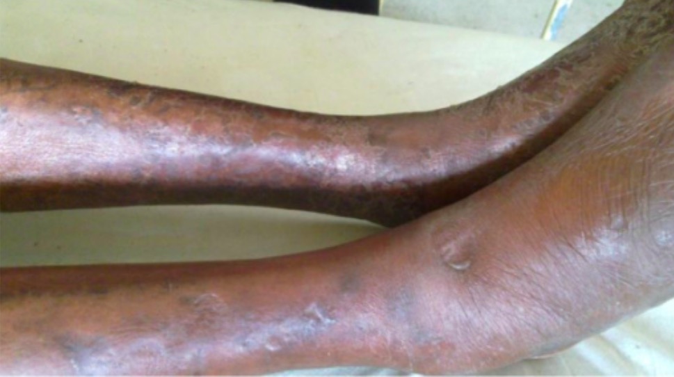
Thromboembolic presentations

  • Superficial vein thrombosis Thrombosis Formation and development of a thrombus or blood clot in the blood vessel. Epidemic Typhus (rarely severe)
  • Deep venous thrombosis Venous thrombosis The formation or presence of a blood clot (thrombus) within a vein. Budd-Chiari Syndrome ( DVT DVT Deep vein thrombosis (DVT) usually occurs in the deep veins of the lower extremities. The affected veins include the femoral, popliteal, iliofemoral, and pelvic veins. Proximal DVT is more likely to cause a pulmonary embolism (PE) and is generally considered more serious. Deep Vein Thrombosis):
    • DVTs of the lower extremities (most common DVTs):
      • Unilateral swelling Swelling Inflammation, redness Redness Inflammation, and/or pain Pain An unpleasant sensation induced by noxious stimuli which are detected by nerve endings of nociceptive neurons. Pain: Types and Pathways in the extremity
      • Pain Pain An unpleasant sensation induced by noxious stimuli which are detected by nerve endings of nociceptive neurons. Pain: Types and Pathways with dorsiflexion of the foot Foot The foot is the terminal portion of the lower limb, whose primary function is to bear weight and facilitate locomotion. The foot comprises 26 bones, including the tarsal bones, metatarsal bones, and phalanges. The bones of the foot form longitudinal and transverse arches and are supported by various muscles, ligaments, and tendons. Foot: Anatomy distal to a DVT DVT Deep vein thrombosis (DVT) usually occurs in the deep veins of the lower extremities. The affected veins include the femoral, popliteal, iliofemoral, and pelvic veins. Proximal DVT is more likely to cause a pulmonary embolism (PE) and is generally considered more serious. Deep Vein Thrombosis
    • DVTs of the upper extremities
    • Cerebral sinus thrombosis Thrombosis Formation and development of a thrombus or blood clot in the blood vessel. Epidemic Typhus
    • Hepatic and portal vein Portal vein A short thick vein formed by union of the superior mesenteric vein and the splenic vein. Liver: Anatomy thrombosis Thrombosis Formation and development of a thrombus or blood clot in the blood vessel. Epidemic Typhus ( Budd-Chiari syndrome Budd-Chiari syndrome Budd-Chiari syndrome is a condition resulting from the interruption of the normal outflow of blood from the liver. The primary type arises from a venous process (affecting the hepatic veins or inferior vena cava) such as thrombosis, but can also be from a lesion compressing or invading the veins (secondary type). The patient typically presents with hepatomegaly, ascites, and abdominal discomfort. Budd-Chiari Syndrome)
    • Renal vein Renal vein Short thick veins which return blood from the kidneys to the vena cava. Glomerular Filtration thrombosis Thrombosis Formation and development of a thrombus or blood clot in the blood vessel. Epidemic Typhus
    • Adrenal vein thrombosis Thrombosis Formation and development of a thrombus or blood clot in the blood vessel. Epidemic Typhus
  • Pulmonary embolism Pulmonary Embolism Pulmonary embolism (PE) is a potentially fatal condition that occurs as a result of intraluminal obstruction of the main pulmonary artery or its branches. The causative factors include thrombi, air, amniotic fluid, and fat. In PE, gas exchange is impaired due to the decreased return of deoxygenated blood to the lungs. Pulmonary Embolism (PE)
    • Dyspnea Dyspnea Dyspnea is the subjective sensation of breathing discomfort. Dyspnea is a normal manifestation of heavy physical or psychological exertion, but also may be caused by underlying conditions (both pulmonary and extrapulmonary). Dyspnea
    • Chest pain Pain An unpleasant sensation induced by noxious stimuli which are detected by nerve endings of nociceptive neurons. Pain: Types and Pathways
    • Acute respiratory distress
    • Pulmonary hypertension Pulmonary Hypertension Pulmonary hypertension (PH) or pulmonary arterial hypertension (PAH) is characterized by elevated pulmonary arterial pressure, which can lead to chronic progressive right heart failure. Pulmonary hypertension is grouped into 5 categories based on etiology, which include primary PAH, and PH due to cardiac disease, lung or hypoxic disease, chronic thromboembolic disease, and multifactorial or unclear etiologies. Pulmonary Hypertension
  • Arterial thrombosis Thrombosis Formation and development of a thrombus or blood clot in the blood vessel. Epidemic Typhus:
    • Stroke (most common arterial thrombosis Thrombosis Formation and development of a thrombus or blood clot in the blood vessel. Epidemic Typhus) and transient ischemic attacks (TIAs):
      • Focal neurologic findings
      • Cognitive deficits
    • MI MI MI is ischemia and death of an area of myocardial tissue due to insufficient blood flow and oxygenation, usually from thrombus formation on a ruptured atherosclerotic plaque in the epicardial arteries. Clinical presentation is most commonly with chest pain, but women and patients with diabetes may have atypical symptoms. Myocardial Infarction:
      • Chest pain Pain An unpleasant sensation induced by noxious stimuli which are detected by nerve endings of nociceptive neurons. Pain: Types and Pathways
      • Dyspnea Dyspnea Dyspnea is the subjective sensation of breathing discomfort. Dyspnea is a normal manifestation of heavy physical or psychological exertion, but also may be caused by underlying conditions (both pulmonary and extrapulmonary). Dyspnea on exertion
    • Retinal thrombosis Thrombosis Formation and development of a thrombus or blood clot in the blood vessel. Epidemic Typhus
    • Nephropathy due to vaso-occlusion in the small renal vessels
  • Recurrent thromboembolic events
Pitting oedema of right leg

Pitting edema with swelling of the right leg due to a deep venous thrombosis

Image: “Pitting oedema of right leg” by Department of medicine (ward 45), the National hospital of Sri Lanka, (Regent Street), Colombo, (00800), Sri Lanka. License: CC BY 2.0

Additional specific findings associated with hypercoagulable states

Factor V Factor V Heat- and storage-labile plasma glycoprotein which accelerates the conversion of prothrombin to thrombin in blood coagulation. Factor V accomplishes this by forming a complex with factor Xa, phospholipid, and calcium (prothrombinase complex). Hemostasis Leiden and prothrombin Prothrombin A plasma protein that is the inactive precursor of thrombin. It is converted to thrombin by a prothrombin activator complex consisting of factor Xa, factor V, phospholipid, and calcium ions. Hemostasis G20210A mutations do not have any unique presentations beyond recurrent thrombotic events. Several specific findings may be noted in:
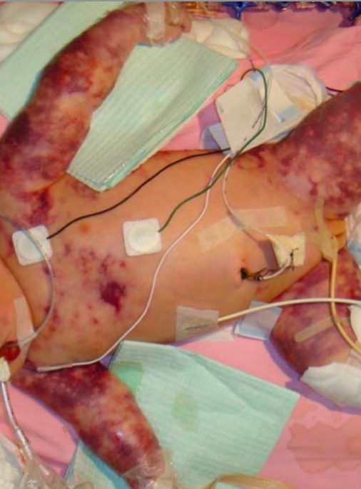
  • Protein C or S deficiency:
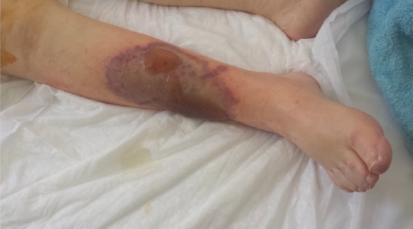
    • Neonatal purpura fulminans Purpura fulminans A severe, rapidly fatal reaction occurring most commonly in children following an infectious illness. It is characterized by large, rapidly spreading skin hemorrhages, fever, or shock. Purpura fulminans often accompanies or is triggered by disseminated intravascular coagulation. Disseminated Intravascular Coagulation:
      • Purpuric lesions developing at many different sites during the 1st 72 hours of life
      • Lesions enlarge and produce hemorrhagic bullae Bullae Erythema Multiforme with subsequent necrosis Necrosis The death of cells in an organ or tissue due to disease, injury or failure of the blood supply. Ischemic Cell Damage and black eschar formation.
    • Warfarin-induced skin necrosis Skin Necrosis Rocky Mountain Spotted Fever:
      • Diffuse necrosis Necrosis The death of cells in an organ or tissue due to disease, injury or failure of the blood supply. Ischemic Cell Damage of the skin Skin The skin, also referred to as the integumentary system, is the largest organ of the body. The skin is primarily composed of the epidermis (outer layer) and dermis (deep layer). The epidermis is primarily composed of keratinocytes that undergo rapid turnover, while the dermis contains dense layers of connective tissue. Skin: Structure and Functions and/or subcutaneous tissue Subcutaneous tissue Loose connective tissue lying under the dermis, which binds skin loosely to subjacent tissues. It may contain a pad of adipocytes, which vary in number according to the area of the body and vary in size according to the nutritional state. Soft Tissue Abscess due to thrombosis Thrombosis Formation and development of a thrombus or blood clot in the blood vessel. Epidemic Typhus
      • Occurs during the 1st few days of warfarin Warfarin An anticoagulant that acts by inhibiting the synthesis of vitamin K-dependent coagulation factors. Warfarin is indicated for the prophylaxis and/or treatment of venous thrombosis and its extension, pulmonary embolism, and atrial fibrillation with embolization. It is also used as an adjunct in the prophylaxis of systemic embolism after myocardial infarction. Warfarin is also used as a rodenticide. Anticoagulants administration
  • Antithrombin deficiency Antithrombin deficiency An absence or reduced level of antithrombin III leading to an increased risk for thrombosis. Budd-Chiari Syndrome:
    • Heparin resistance Resistance Physiologically, the opposition to flow of air caused by the forces of friction. As a part of pulmonary function testing, it is the ratio of driving pressure to the rate of air flow. Ventilation: Mechanics of Breathing: There is no ↑ in the aPTT with heparin administration.
  • Antiphospholipid syndrome Antiphospholipid syndrome Antiphospholipid syndrome (APLS) is an acquired autoimmune disorder characterized by the persistent presence of antiphospholipid antibodies, which create a hypercoagulable state. These antibodies are most commonly discovered during a workup for a thrombotic event or recurrent pregnancy loss, which are the 2 most common clinical manifestations. Antiphospholipid Syndrome may present with obstetric complications, including:
    • Recurrent pregnancy Pregnancy The status during which female mammals carry their developing young (embryos or fetuses) in utero before birth, beginning from fertilization to birth. Pregnancy: Diagnosis, Physiology, and Care loss (i.e., miscarriage Miscarriage Spontaneous abortion, also known as miscarriage, is the loss of a pregnancy before 20 weeks’ gestation. However, the layperson use of the term “abortion” is often intended to refer to induced termination of a pregnancy, whereas “miscarriage” is preferred for spontaneous loss. Spontaneous Abortion)
    • Premature Premature Childbirth before 37 weeks of pregnancy (259 days from the first day of the mother’s last menstrual period, or 245 days after fertilization). Necrotizing Enterocolitis birth
    • Placental insufficiency Placental Insufficiency Failure of the placenta to deliver an adequate supply of nutrients and oxygen to the fetus. Neonatal Polycythemia
  • Other medical conditions:
    • Many additional clinical findings related to an underlying condition associated with hypercoagulability may be present.
    • For example:
      • Weight loss Weight loss Decrease in existing body weight. Bariatric Surgery and loss of appetite → can be seen in malignancy Malignancy Hemothorax
      • Rash Rash Rocky Mountain Spotted Fever, joint pain Pain An unpleasant sensation induced by noxious stimuli which are detected by nerve endings of nociceptive neurons. Pain: Types and Pathways, and fatigue Fatigue The state of weariness following a period of exertion, mental or physical, characterized by a decreased capacity for work and reduced efficiency to respond to stimuli. Fibromyalgia SLE SLE Systemic lupus erythematosus (SLE) is a chronic autoimmune, inflammatory condition that causes immune-complex deposition in organs, resulting in systemic manifestations. Women, particularly those of African American descent, are more commonly affected. Systemic Lupus Erythematosus
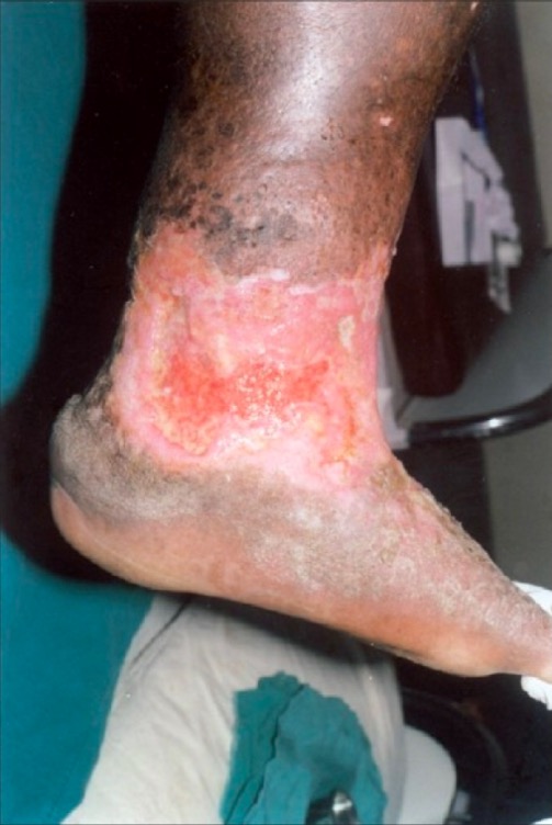
      • Characteristic skin Skin The skin, also referred to as the integumentary system, is the largest organ of the body. The skin is primarily composed of the epidermis (outer layer) and dermis (deep layer). The epidermis is primarily composed of keratinocytes that undergo rapid turnover, while the dermis contains dense layers of connective tissue. Skin: Structure and Functions changes associated with chronic venous insufficiency Chronic venous insufficiency Chronic venous disease is a spectrum of disorders characterized by venous dilation and/or abnormal vein function in the lower extremities resulting from venous hypertension. “Chronic venous insufficiency” refers to the more severe forms of chronic venous disease. Skin changes typically distinguish chronic venous insufficiency from milder forms of venous disease. Chronic Venous Insufficiency
      • Splenomegaly Splenomegaly Splenomegaly is pathologic enlargement of the spleen that is attributable to numerous causes, including infections, hemoglobinopathies, infiltrative processes, and outflow obstruction of the portal vein. Splenomegaly → myeloproliferative disorders

Diagnosis

Basic evaluation

These studies should be performed in most patients Patients Individuals participating in the health care system for the purpose of receiving therapeutic, diagnostic, or preventive procedures. Clinician–Patient Relationship with suspected thrombotic events.
  • CBC and peripheral blood smear Peripheral Blood Smear Anemia: Overview and Types may reveal clues to the underlying etiology, for example:
    • Hematocrit Hematocrit The volume of packed red blood cells in a blood specimen. The volume is measured by centrifugation in a tube with graduated markings, or with automated blood cell counters. It is an indicator of erythrocyte status in disease. For example, anemia shows a low value; polycythemia, a high value. Neonatal Polycythemia polycythemia Polycythemia An increase in the total red cell mass of the blood. Renal Cell Carcinoma
    • Hematocrit Hematocrit The volume of packed red blood cells in a blood specimen. The volume is measured by centrifugation in a tube with graduated markings, or with automated blood cell counters. It is an indicator of erythrocyte status in disease. For example, anemia shows a low value; polycythemia, a high value. Neonatal Polycythemia PNH PNH Paroxysmal nocturnal hemoglobinuria (PNH) is a rare but serious acquired hemolytic anemia with periodic exacerbations. This anemia is caused by nonmalignant clonal expansion of ≥ 1 hematopoietic stem cells that have acquired a somatic mutation of the phosphatidylinositol n-acetylglucosaminyltransferase subunit a (pig-a) gene. Paroxysmal Nocturnal Hemoglobinuria
    • Platelets Platelets Platelets are small cell fragments involved in hemostasis. Thrombopoiesis takes place primarily in the bone marrow through a series of cell differentiation and is influenced by several cytokines. Platelets are formed after fragmentation of the megakaryocyte cytoplasm. Platelets: Histology → thrombocytosis/thrombocythemia
    • Abnormal lymphocytes Lymphocytes Lymphocytes are heterogeneous WBCs involved in immune response. Lymphocytes develop from the bone marrow, starting from hematopoietic stem cells (HSCs) and progressing to common lymphoid progenitors (CLPs). B and T lymphocytes and natural killer (NK) cells arise from the lineage. Lymphocytes: Histology on smear → bone marrow Bone marrow The soft tissue filling the cavities of bones. Bone marrow exists in two types, yellow and red. Yellow marrow is found in the large cavities of large bones and consists mostly of fat cells and a few primitive blood cells. Red marrow is a hematopoietic tissue and is the site of production of erythrocytes and granular leukocytes. Bone marrow is made up of a framework of connective tissue containing branching fibers with the frame being filled with marrow cells. Bone Marrow: Composition and Hematopoiesis malignancy Malignancy Hemothorax
  • D-dimer D-dimer Deep Vein Thrombosis:
    • A fibrin-degradation product that will be ↑ in the presence of a DVT DVT Deep vein thrombosis (DVT) usually occurs in the deep veins of the lower extremities. The affected veins include the femoral, popliteal, iliofemoral, and pelvic veins. Proximal DVT is more likely to cause a pulmonary embolism (PE) and is generally considered more serious. Deep Vein Thrombosis
    • Multiple conditions can ↑ the D-dimer D-dimer Deep Vein Thrombosis level, but a DVT DVT Deep vein thrombosis (DVT) usually occurs in the deep veins of the lower extremities. The affected veins include the femoral, popliteal, iliofemoral, and pelvic veins. Proximal DVT is more likely to cause a pulmonary embolism (PE) and is generally considered more serious. Deep Vein Thrombosis is highly unlikely if the D-dimer D-dimer Deep Vein Thrombosis level is normal.
  • Coagulation studies Coagulation studies Coagulation studies are a group of hematologic laboratory studies that reflect the function of blood vessels, platelets, and coagulation factors, which all interact with one another to achieve hemostasis. Coagulation studies are usually ordered to evaluate patients with bleeding or hypercoagulation disorders. Coagulation Studies:
    • ↑ PT/INR may suggest underlying liver Liver The liver is the largest gland in the human body. The liver is found in the superior right quadrant of the abdomen and weighs approximately 1.5 kilograms. Its main functions are detoxification, metabolism, nutrient storage (e.g., iron and vitamins), synthesis of coagulation factors, formation of bile, filtration, and storage of blood. Liver: Anatomy disease
    • ↑ aPTT in antiphospholipid syndrome Antiphospholipid syndrome Antiphospholipid syndrome (APLS) is an acquired autoimmune disorder characterized by the persistent presence of antiphospholipid antibodies, which create a hypercoagulable state. These antibodies are most commonly discovered during a workup for a thrombotic event or recurrent pregnancy loss, which are the 2 most common clinical manifestations. Antiphospholipid Syndrome (1 of the antibodies Antibodies Immunoglobulins (Igs), also known as antibodies, are glycoprotein molecules produced by plasma cells that act in immune responses by recognizing and binding particular antigens. The various Ig classes are IgG (the most abundant), IgM, IgE, IgD, and IgA, which differ in their biologic features, structure, target specificity, and distribution. Immunoglobulins: Types and Functions, lupus anticoagulant Lupus anticoagulant An antiphospholipid antibody found in association with systemic lupus erythematosus, antiphospholipid syndrome; and in a variety of other diseases as well as in healthy individuals. In vitro, the antibody interferes with the conversion of prothrombin to thrombin and prolongs the partial thromboplastin time. In vivo, it exerts a procoagulant effect resulting in thrombosis mainly in the larger veins and arteries. It further causes obstetrical complications, including fetal death and spontaneous abortion, as well as a variety of hematologic and neurologic complications. Antiphospholipid Syndrome, acts as an anticoagulant in vitro, but it promotes thrombosis Thrombosis Formation and development of a thrombus or blood clot in the blood vessel. Epidemic Typhus in vivo)
  • Chemistries: to evaluate liver Liver The liver is the largest gland in the human body. The liver is found in the superior right quadrant of the abdomen and weighs approximately 1.5 kilograms. Its main functions are detoxification, metabolism, nutrient storage (e.g., iron and vitamins), synthesis of coagulation factors, formation of bile, filtration, and storage of blood. Liver: Anatomy and kidney function
  • Age-appropriate screening Screening Preoperative Care for malignancy Malignancy Hemothorax: 
    • Pap smear Pap smear Cytological preparation of cells collected from a mucosal surface and stained with Papanicolaou stain. Cervical Cancer Screening test
    • Mammography Mammography Radiographic examination of the breast. Breast Cancer Screening
    • Prostate specific antigen Prostate Specific Antigen A glycoprotein that is a kallikrein-like serine proteinase and an esterase, produced by epithelial cells of both normal and malignant prostate tissue. It is an important marker for the diagnosis of prostate cancer. Prostate Cancer ( PSA PSA A glycoprotein that is a kallikrein-like serine proteinase and an esterase, produced by epithelial cells of both normal and malignant prostate tissue. It is an important marker for the diagnosis of prostate cancer. Prostate Cancer) test
    • Stool Hemoccult and/or colonoscopy Colonoscopy Endoscopic examination, therapy or surgery of the luminal surface of the colon. Colorectal Cancer Screening
  • Imaging:
    • CT pulmonary angiography Angiography Radiography of blood vessels after injection of a contrast medium. Cardiac Surgery and/or V/Q for suspected PE
    • Duplex ultrasonography of the limbs for suspected DVT DVT Deep vein thrombosis (DVT) usually occurs in the deep veins of the lower extremities. The affected veins include the femoral, popliteal, iliofemoral, and pelvic veins. Proximal DVT is more likely to cause a pulmonary embolism (PE) and is generally considered more serious. Deep Vein Thrombosis; findings of DVT DVT Deep vein thrombosis (DVT) usually occurs in the deep veins of the lower extremities. The affected veins include the femoral, popliteal, iliofemoral, and pelvic veins. Proximal DVT is more likely to cause a pulmonary embolism (PE) and is generally considered more serious. Deep Vein Thrombosis include:
      • Noncompressible lumen
      • Hyperechoic Hyperechoic A structure that produces a high-amplitude echo (lighter grays and white) Ultrasound (Sonography) mass Mass Three-dimensional lesion that occupies a space within the breast Imaging of the Breast
      • Decreased/absent flow Flow Blood flows through the heart, arteries, capillaries, and veins in a closed, continuous circuit. Flow is the movement of volume per unit of time. Flow is affected by the pressure gradient and the resistance fluid encounters between 2 points. Vascular resistance is the opposition to flow, which is caused primarily by blood friction against vessel walls. Vascular Resistance, Flow, and Mean Arterial Pressure
    • Other imaging may be indicated based on clinical presentation (e.g., chest X-ray X-ray Penetrating electromagnetic radiation emitted when the inner orbital electrons of an atom are excited and release radiant energy. X-ray wavelengths range from 1 pm to 10 nm. Hard x-rays are the higher energy, shorter wavelength x-rays. Soft x-rays or grenz rays are less energetic and longer in wavelength. The short wavelength end of the x-ray spectrum overlaps the gamma rays wavelength range. The distinction between gamma rays and x-rays is based on their radiation source. Pulmonary Function Tests for suspected malignancy Malignancy Hemothorax).
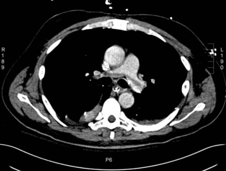
Ct of saddle pulmonary embolism

CT angiogram of the chest showing a saddle pulmonary embolus

Image: “Large saddle pulmonary embolism” by Rhode Island Hospital, Brown University School of Medicine, 2 Dudley Street, Providence, RI, USA. License: CC BY 2.0

Indications for a thrombophilia workup

Consider ordering additional specific tests to screen for an inherited thrombophilia if patients Patients Individuals participating in the health care system for the purpose of receiving therapeutic, diagnostic, or preventive procedures. Clinician–Patient Relationship meet any of the following criteria:

Testing for specific conditions

If patients Patients Individuals participating in the health care system for the purpose of receiving therapeutic, diagnostic, or preventive procedures. Clinician–Patient Relationship meet any of the above criteria for a thrombophilia workup, the following tests can be ordered to screen for specific inherited thrombophilias:

  • Factor V Factor V Heat- and storage-labile plasma glycoprotein which accelerates the conversion of prothrombin to thrombin in blood coagulation. Factor V accomplishes this by forming a complex with factor Xa, phospholipid, and calcium (prothrombinase complex). Hemostasis Leiden: activated protein C resistance Resistance Physiologically, the opposition to flow of air caused by the forces of friction. As a part of pulmonary function testing, it is the ratio of driving pressure to the rate of air flow. Ventilation: Mechanics of Breathing assay
  • Prothrombin Prothrombin A plasma protein that is the inactive precursor of thrombin. It is converted to thrombin by a prothrombin activator complex consisting of factor Xa, factor V, phospholipid, and calcium ions. Hemostasis gene mutation Gene Mutation Myotonic Dystrophies: molecular analysis of the prothrombin Prothrombin A plasma protein that is the inactive precursor of thrombin. It is converted to thrombin by a prothrombin activator complex consisting of factor Xa, factor V, phospholipid, and calcium ions. Hemostasis gene Gene A category of nucleic acid sequences that function as units of heredity and which code for the basic instructions for the development, reproduction, and maintenance of organisms. Basic Terms of Genetics
  • Antithrombin Antithrombin Endogenous factors and drugs that directly inhibit the action of thrombin, usually by blocking its enzymatic activity. They are distinguished from indirect thrombin inhibitors, such as heparin, which act by enhancing the inhibitory effects of antithrombins. Anticoagulants: antithrombin Antithrombin Endogenous factors and drugs that directly inhibit the action of thrombin, usually by blocking its enzymatic activity. They are distinguished from indirect thrombin inhibitors, such as heparin, which act by enhancing the inhibitory effects of antithrombins. Anticoagulants–heparin cofactor assay 
  • Proteins Proteins Linear polypeptides that are synthesized on ribosomes and may be further modified, crosslinked, cleaved, or assembled into complex proteins with several subunits. The specific sequence of amino acids determines the shape the polypeptide will take, during protein folding, and the function of the protein. Energy Homeostasis C: functional assay to detect quantitative and qualitative defects
  • Protein S Protein S Protein S augments the activity of protein C. Hemostasis:
    • Free protein S Protein S Protein S augments the activity of protein C. Hemostasis antigen Antigen Substances that are recognized by the immune system and induce an immune reaction. Vaccination assay
    • Protein S Protein S Protein S augments the activity of protein C. Hemostasis functional assay
  • Antiphospholipid syndrome Antiphospholipid syndrome Antiphospholipid syndrome (APLS) is an acquired autoimmune disorder characterized by the persistent presence of antiphospholipid antibodies, which create a hypercoagulable state. These antibodies are most commonly discovered during a workup for a thrombotic event or recurrent pregnancy loss, which are the 2 most common clinical manifestations. Antiphospholipid Syndrome: serologies to detect the presence of antibodies Antibodies Immunoglobulins (Igs), also known as antibodies, are glycoprotein molecules produced by plasma cells that act in immune responses by recognizing and binding particular antigens. The various Ig classes are IgG (the most abundant), IgM, IgE, IgD, and IgA, which differ in their biologic features, structure, target specificity, and distribution. Immunoglobulins: Types and Functions
    • Anticardiolipin antibodies Anticardiolipin antibodies Antiphospholipid antibodies found in association with systemic lupus erythematosus, antiphospholipid syndrome; and in a variety of other diseases as well as in healthy individuals. The antibodies are detected by solid-phase immunoassay employing the purified phospholipid antigen cardiolipin. Antiphospholipid Syndrome
    • β2-glycoprotein antibodies Antibodies Immunoglobulins (Igs), also known as antibodies, are glycoprotein molecules produced by plasma cells that act in immune responses by recognizing and binding particular antigens. The various Ig classes are IgG (the most abundant), IgM, IgE, IgD, and IgA, which differ in their biologic features, structure, target specificity, and distribution. Immunoglobulins: Types and Functions
    • Lupus anticoagulant Lupus anticoagulant An antiphospholipid antibody found in association with systemic lupus erythematosus, antiphospholipid syndrome; and in a variety of other diseases as well as in healthy individuals. In vitro, the antibody interferes with the conversion of prothrombin to thrombin and prolongs the partial thromboplastin time. In vivo, it exerts a procoagulant effect resulting in thrombosis mainly in the larger veins and arteries. It further causes obstetrical complications, including fetal death and spontaneous abortion, as well as a variety of hematologic and neurologic complications. Antiphospholipid Syndrome

Note: Acute thrombosis Thrombosis Formation and development of a thrombus or blood clot in the blood vessel. Epidemic Typhus and/or anticoagulants Anticoagulants Anticoagulants are drugs that retard or interrupt the coagulation cascade. The primary classes of available anticoagulants include heparins, vitamin K-dependent antagonists (e.g., warfarin), direct thrombin inhibitors, and factor Xa inhibitors. Anticoagulants may reduce plasma Plasma The residual portion of blood that is left after removal of blood cells by centrifugation without prior blood coagulation. Transfusion Products concentrations of antithrombin Antithrombin Endogenous factors and drugs that directly inhibit the action of thrombin, usually by blocking its enzymatic activity. They are distinguished from indirect thrombin inhibitors, such as heparin, which act by enhancing the inhibitory effects of antithrombins. Anticoagulants, protein C, and protein S Protein S Protein S augments the activity of protein C. Hemostasis.

Management

Management prior to a 1st thrombotic event

In patients Patients Individuals participating in the health care system for the purpose of receiving therapeutic, diagnostic, or preventive procedures. Clinician–Patient Relationship with known inherited thrombophilias or who have risk factors for thrombosis Thrombosis Formation and development of a thrombus or blood clot in the blood vessel. Epidemic Typhus:

  • Encourage smoking Smoking Willful or deliberate act of inhaling and exhaling smoke from burning substances or agents held by hand. Interstitial Lung Diseases cessation.
  • Avoid estrogen-containing contraceptives.
  • Early mobilization after surgery
  • Manage underlying medical conditions (e.g., hyperlipidemia, hypertension Hypertension Hypertension, or high blood pressure, is a common disease that manifests as elevated systemic arterial pressures. Hypertension is most often asymptomatic and is found incidentally as part of a routine physical examination or during triage for an unrelated medical encounter. Hypertension, nephrotic syndrome Nephrotic syndrome Nephrotic syndrome is characterized by severe proteinuria, hypoalbuminemia, and peripheral edema. In contrast, the nephritic syndromes present with hematuria, variable loss of renal function, and hypertension, although there is sometimes overlap of > 1 glomerular disease in the same individual. Nephrotic Syndrome).
  • Consider low-dose aspirin Aspirin The prototypical analgesic used in the treatment of mild to moderate pain. It has anti-inflammatory and antipyretic properties and acts as an inhibitor of cyclooxygenase which results in the inhibition of the biosynthesis of prostaglandins. Aspirin also inhibits platelet aggregation and is used in the prevention of arterial and venous thrombosis. Nonsteroidal Antiinflammatory Drugs (NSAIDs) (evidence is limited; consider entire clinical picture).
  • For patients Patients Individuals participating in the health care system for the purpose of receiving therapeutic, diagnostic, or preventive procedures. Clinician–Patient Relationship with deficiencies of anticoagulants Anticoagulants Anticoagulants are drugs that retard or interrupt the coagulation cascade. The primary classes of available anticoagulants include heparins, vitamin K-dependent antagonists (e.g., warfarin), direct thrombin inhibitors, and factor Xa inhibitors. Anticoagulants ( antithrombin Antithrombin Endogenous factors and drugs that directly inhibit the action of thrombin, usually by blocking its enzymatic activity. They are distinguished from indirect thrombin inhibitors, such as heparin, which act by enhancing the inhibitory effects of antithrombins. Anticoagulants or protein C or S): Replace missing factors.
  • For patients Patients Individuals participating in the health care system for the purpose of receiving therapeutic, diagnostic, or preventive procedures. Clinician–Patient Relationship with a temporary ↑ risk of thrombosis Thrombosis Formation and development of a thrombus or blood clot in the blood vessel. Epidemic Typhus (e.g., postsurgery, pregnancy Pregnancy The status during which female mammals carry their developing young (embryos or fetuses) in utero before birth, beginning from fertilization to birth. Pregnancy: Diagnosis, Physiology, and Care): anticoagulation Anticoagulation Pulmonary Hypertension Drugs for 3–6 months or until the risk factor is no longer present

Management for acute thrombotic events

Anticoagulation Anticoagulation Pulmonary Hypertension Drugs is the mainstay of therapy for thrombotic events. Options for initial anticoagulation Anticoagulation Pulmonary Hypertension Drugs include:

  • Unfractionated heparin Unfractionated heparin A highly acidic mucopolysaccharide formed of equal parts of sulfated d-glucosamine and d-glucuronic acid with sulfaminic bridges. The molecular weight ranges from six to twenty thousand. Heparin occurs in and is obtained from liver, lung, mast cells, etc. , of vertebrates. Its function is unknown, but it is used to prevent blood clotting in vivo and vitro, in the form of many different salts. Anticoagulants ( UFH UFH A highly acidic mucopolysaccharide formed of equal parts of sulfated d-glucosamine and d-glucuronic acid with sulfaminic bridges. The molecular weight ranges from six to twenty thousand. Heparin occurs in and is obtained from liver, lung, mast cells, etc. , of vertebrates. Its function is unknown, but it is used to prevent blood clotting in vivo and vitro, in the form of many different salts. Anticoagulants)
  • Subcutaneous low-molecular-weight heparin (LMWH)
  • Subcutaneous fondaparinux Fondaparinux Synthetic pentasaccharide that mediates the interaction of heparin with antithrombins and inhibits factor Xa; it is used for prevention of venous thromboembolism after surgery. Anticoagulants ( indirect factor Xa inhibitor Indirect Factor Xa Inhibitor Anticoagulants)
  • Oral rivaroxaban Rivaroxaban A morpholine and thiophene derivative that functions as a factor Xa inhibitor and is used in the treatment and prevention of deep-vein thrombosis and pulmonary embolism. It is also used for the prevention of stroke and systemic embolization in patients with non-valvular atrial fibrillation, and for the prevention of atherothrombotic events in patients after an acute coronary syndrome. Anticoagulants or apixaban Apixaban Anticoagulants (direct factor Xa inhibitors)
  • Note: Warfarin Warfarin An anticoagulant that acts by inhibiting the synthesis of vitamin K-dependent coagulation factors. Warfarin is indicated for the prophylaxis and/or treatment of venous thrombosis and its extension, pulmonary embolism, and atrial fibrillation with embolization. It is also used as an adjunct in the prophylaxis of systemic embolism after myocardial infarction. Warfarin is also used as a rodenticide. Anticoagulants ( vitamin K Vitamin K A lipid cofactor that is required for normal blood clotting. Several forms of vitamin K have been identified: vitamin K 1 (phytomenadione) derived from plants, vitamin K 2 (menaquinone) from bacteria, and synthetic naphthoquinone provitamins, vitamin K 3 (menadione). Vitamin k 3 provitamins, after being alkylated in vivo, exhibit the antifibrinolytic activity of vitamin k. Green leafy vegetables, liver, cheese, butter, and egg yolk are good sources of vitamin k. Fat-soluble Vitamins and their Deficiencies antagonist) should not be used as monotherapy for initial treatment:
    • The half-lives of the vitamin K Vitamin K A lipid cofactor that is required for normal blood clotting. Several forms of vitamin K have been identified: vitamin K 1 (phytomenadione) derived from plants, vitamin K 2 (menaquinone) from bacteria, and synthetic naphthoquinone provitamins, vitamin K 3 (menadione). Vitamin k 3 provitamins, after being alkylated in vivo, exhibit the antifibrinolytic activity of vitamin k. Green leafy vegetables, liver, cheese, butter, and egg yolk are good sources of vitamin k. Fat-soluble Vitamins and their Deficiencies–dependent anticoagulants Anticoagulants Anticoagulants are drugs that retard or interrupt the coagulation cascade. The primary classes of available anticoagulants include heparins, vitamin K-dependent antagonists (e.g., warfarin), direct thrombin inhibitors, and factor Xa inhibitors. Anticoagulants are much shorter than those of the vitamin K Vitamin K A lipid cofactor that is required for normal blood clotting. Several forms of vitamin K have been identified: vitamin K 1 (phytomenadione) derived from plants, vitamin K 2 (menaquinone) from bacteria, and synthetic naphthoquinone provitamins, vitamin K 3 (menadione). Vitamin k 3 provitamins, after being alkylated in vivo, exhibit the antifibrinolytic activity of vitamin k. Green leafy vegetables, liver, cheese, butter, and egg yolk are good sources of vitamin k. Fat-soluble Vitamins and their Deficiencies–dependent procoagulants → warfarin Warfarin An anticoagulant that acts by inhibiting the synthesis of vitamin K-dependent coagulation factors. Warfarin is indicated for the prophylaxis and/or treatment of venous thrombosis and its extension, pulmonary embolism, and atrial fibrillation with embolization. It is also used as an adjunct in the prophylaxis of systemic embolism after myocardial infarction. Warfarin is also used as a rodenticide. Anticoagulants causes an initial period of hypercoagulability
    • Warfarin Warfarin An anticoagulant that acts by inhibiting the synthesis of vitamin K-dependent coagulation factors. Warfarin is indicated for the prophylaxis and/or treatment of venous thrombosis and its extension, pulmonary embolism, and atrial fibrillation with embolization. It is also used as an adjunct in the prophylaxis of systemic embolism after myocardial infarction. Warfarin is also used as a rodenticide. Anticoagulants may be started in combination with other agents (e.g., LMWH).
    • Useful for maintenance therapy

Duration of therapy:

  • Should be started immediately on diagnosis
  • Typically continue for 3–6 months, sometimes longer.

Prophylaxis Prophylaxis Cephalosporins after a thrombotic event

  • Patients Patients Individuals participating in the health care system for the purpose of receiving therapeutic, diagnostic, or preventive procedures. Clinician–Patient Relationship who typically require ongoing prophylactic anticoagulation Anticoagulation Pulmonary Hypertension Drugs:
    • Inherited thrombophilia and history of a thrombotic event 
    • Nonmodifiable causes of hypercoagulability (e.g., malignancy Malignancy Hemothorax)
  • For patients Patients Individuals participating in the health care system for the purpose of receiving therapeutic, diagnostic, or preventive procedures. Clinician–Patient Relationship who do not desire pregnancy Pregnancy The status during which female mammals carry their developing young (embryos or fetuses) in utero before birth, beginning from fertilization to birth. Pregnancy: Diagnosis, Physiology, and Care, options include:
    • Warfarin Warfarin An anticoagulant that acts by inhibiting the synthesis of vitamin K-dependent coagulation factors. Warfarin is indicated for the prophylaxis and/or treatment of venous thrombosis and its extension, pulmonary embolism, and atrial fibrillation with embolization. It is also used as an adjunct in the prophylaxis of systemic embolism after myocardial infarction. Warfarin is also used as a rodenticide. Anticoagulants therapy (goal INR, 2–3)
    • Rivaroxaban Rivaroxaban A morpholine and thiophene derivative that functions as a factor Xa inhibitor and is used in the treatment and prevention of deep-vein thrombosis and pulmonary embolism. It is also used for the prevention of stroke and systemic embolization in patients with non-valvular atrial fibrillation, and for the prevention of atherothrombotic events in patients after an acute coronary syndrome. Anticoagulants or apixaban Apixaban Anticoagulants
  • For patients Patients Individuals participating in the health care system for the purpose of receiving therapeutic, diagnostic, or preventive procedures. Clinician–Patient Relationship who do desire pregnancy Pregnancy The status during which female mammals carry their developing young (embryos or fetuses) in utero before birth, beginning from fertilization to birth. Pregnancy: Diagnosis, Physiology, and Care:
    • LMWH and aspirin Aspirin The prototypical analgesic used in the treatment of mild to moderate pain. It has anti-inflammatory and antipyretic properties and acts as an inhibitor of cyclooxygenase which results in the inhibition of the biosynthesis of prostaglandins. Aspirin also inhibits platelet aggregation and is used in the prevention of arterial and venous thrombosis. Nonsteroidal Antiinflammatory Drugs (NSAIDs) ( warfarin Warfarin An anticoagulant that acts by inhibiting the synthesis of vitamin K-dependent coagulation factors. Warfarin is indicated for the prophylaxis and/or treatment of venous thrombosis and its extension, pulmonary embolism, and atrial fibrillation with embolization. It is also used as an adjunct in the prophylaxis of systemic embolism after myocardial infarction. Warfarin is also used as a rodenticide. Anticoagulants is teratogenic)
  • For patients Patients Individuals participating in the health care system for the purpose of receiving therapeutic, diagnostic, or preventive procedures. Clinician–Patient Relationship with contraindications Contraindications A condition or factor associated with a recipient that makes the use of a drug, procedure, or physical agent improper or inadvisable. Contraindications may be absolute (life threatening) or relative (higher risk of complications in which benefits may outweigh risks). Noninvasive Ventilation to anticoagulation Anticoagulation Pulmonary Hypertension Drugs (e.g., severe bleeding diathesis Bleeding diathesis Wiskott-Aldrich Syndrome, planned surgeries with high bleeding risk): Place an inferior vena cava Inferior vena cava The venous trunk which receives blood from the lower extremities and from the pelvic and abdominal organs. Mediastinum and Great Vessels: Anatomy ( IVC IVC The venous trunk which receives blood from the lower extremities and from the pelvic and abdominal organs. Mediastinum and Great Vessels: Anatomy) filter.

References

  1. Bauer, K. (2020). Evaluating adult patients with established venous thromboembolism for acquired and inherited risk factors. Retrieved May 1, 2021, from https://www.uptodate.com/contents/evaluating-adult-patients-with-established-venous-thromboembolism-for-acquired-and-inherited-risk-factors
  2. Bauer, K. (2021). Overview of the causes of venous thrombosis. Retrieved May 1, 2021, from  https://www.uptodate.com/contents/overview-of-the-causes-of-venous-thrombosis
  3. Lip, G. (2020). Overview of the treatment of lower extremity deep vein thrombosis (DVT). Retrieved May 1, 2021, from  https://www.uptodate.com/contents/overview-of-the-treatment-of-lower-extremity-deep-vein-thrombosis-dvt
  4. Lip, G. (2021). Selecting adult patients with lower extremity deep venous thrombosis and pulmonary embolism for indefinite anticoagulation. Retrieved May 1, 2021, from https://www.uptodate.com/contents/selecting-adult-patients-with-lower-extremity-deep-venous-thrombosis-and-pulmonary-embolism-for-indefinite-anticoagulation
  5. Thomas, R.H. (2001). Hypercoagulability syndromes. Arch Intern Med 161:2433–2439.
  6. Schick, P. (2020). Hereditary and acquired hypercoagulability. Medscape. Retrieved May 4, 2021, from https://emedicine.medscape.com/article/211039-overview

Create your free account or log in to continue reading!

Sign up now and get free access to Lecturio with concept pages, medical videos, and questions for your medical education.

User Reviews

Unwrap New Skills This Holiday 🎄 Save 30% on all plans now!

Details