Advertisement

Advertisement

Advertisement

Advertisement

Grading, Staging, and Metastasis

Cancer is the 2nd leading cause of death in the US after cardiovascular disease. Many malignancies are treatable or curable, but some may recur. Thus, all malignancies must be assigned a grade and stage in order to guide management and determine prognosis Prognosis A prediction of the probable outcome of a disease based on a individual's condition and the usual course of the disease as seen in similar situations. Non-Hodgkin Lymphomas. The tumor Tumor Inflammation grade classifies a tumor Tumor Inflammation by its histology and is part of the internationally accepted TNM ( tumor Tumor Inflammation, node, metastasis) staging system, which is used to characterize the extent of the disease. Metastatic disease refers to cancer that has spread beyond its primary tumor Tumor Inflammation site.

Last updated: Jan 16, 2024

Editorial responsibility: Stanley Oiseth, Lindsay Jones, Evelin Maza

Advertisement

Advertisement

Advertisement

Advertisement

Advertisement

Advertisement

Advertisement

Advertisement

Grading

Grading is the histologic assessment of tumor Tumor Inflammation cells according to their state of differentiation.

  • Microscopic examination of tumor Tumor Inflammation cells after surgery or biopsy Biopsy Removal and pathologic examination of specimens from the living body. Ewing Sarcoma
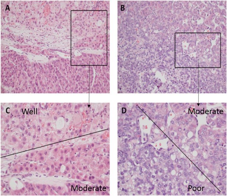
  • Tumor Tumor Inflammation cells are grouped into 4 types based on their resemblance to/difference from healthy cells (G1, G2, G3, G4):
    • Well-differentiated tumors (low grade, G1) generally have a better prognosis Prognosis A prediction of the probable outcome of a disease based on a individual’s condition and the usual course of the disease as seen in similar situations. Non-Hodgkin Lymphomas than poorly differentiated tumors (high grade, G4).
    • Poorly differentiated tumors are called anaplastic.
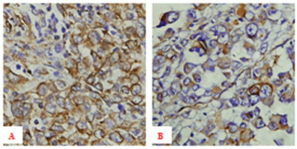
  • Tissue markers are helpful if cells of unknown origin are found:

Grading is performed for most types of tumors, but there are specific exceptions.

  • Prostate Prostate The prostate is a gland in the male reproductive system. The gland surrounds the bladder neck and a portion of the urethra. The prostate is an exocrine gland that produces a weakly acidic secretion, which accounts for roughly 20% of the seminal fluid. cancer: uses the Gleason score ranging from 2–10
    • Based on the growth pattern and degree of differentiation of tumor Tumor Inflammation cells
    • A higher score is associated with disease outside the prostate Prostate The prostate is a gland in the male reproductive system. The gland surrounds the bladder neck and a portion of the urethra. The prostate is an exocrine gland that produces a weakly acidic secretion, which accounts for roughly 20% of the seminal fluid. .
    • A higher score indicates a greater likelihood of a worse prognosis Prognosis A prediction of the probable outcome of a disease based on a individual’s condition and the usual course of the disease as seen in similar situations. Non-Hodgkin Lymphomas.
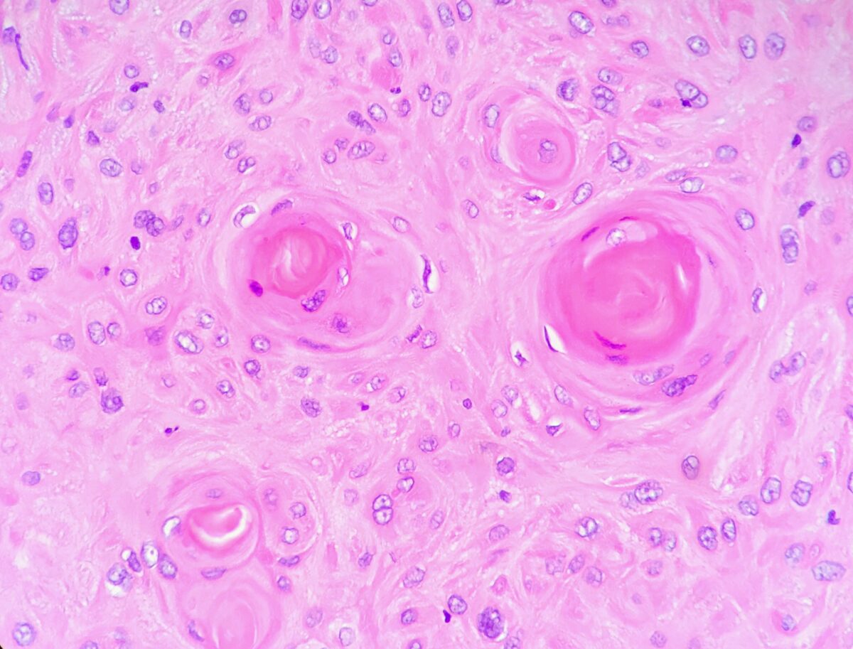
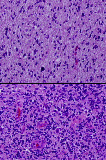
  • Brain Brain The part of central nervous system that is contained within the skull (cranium). Arising from the neural tube, the embryonic brain is comprised of three major parts including prosencephalon (the forebrain); mesencephalon (the midbrain); and rhombencephalon (the hindbrain). The developed brain consists of cerebrum; cerebellum; and other structures in the brain stem. Nervous System: Anatomy, Structure, and Classification tumors: specific WHO classification
    • Uses both genotype Genotype The genetic constitution of the individual, comprising the alleles present at each genetic locus. Basic Terms of Genetics and phenotype Phenotype The complete genetic complement contained in the DNA of a set of chromosomes in a human. The length of the human genome is about 3 billion base pairs. Basic Terms of Genetics for some tumors
    • Immunohistochemical data for molecular genetic alterations are used in addition to histology.
  • Breast cancer Breast cancer Breast cancer is a disease characterized by malignant transformation of the epithelial cells of the breast. Breast cancer is the most common form of cancer and 2nd most common cause of cancer-related death among women. Breast Cancer: uses the Nottingham grading system
    • Based on tubule formation of the tumor Tumor Inflammation, nuclear grade, and mitotic rate
    • Each category is scored between 1 and 3, and a total score is calculated.
    • Total score of 3–5 is G1 (low grade; well differentiated).
    • Total score of 6–7 is G2 (intermediate grade; moderately differentiated).
    • Total score of 8–9 is G3 (high grade; poorly differentiated).

Tumor Tumor Inflammation grading:

  • Gx = grade cannot be assessed (undetermined grade)
  • G1 = well differentiated, close similarity to original tissue (low grade)
  • G2 = somewhat differentiated malignant tissue (intermediate grade)
  • G3 = poorly differentiated malignant tissue (high grade)
  • G4 = undifferentiated malignant tissue: The original tissue that gave rise to the tumor Tumor Inflammation can be determined only by immunohistochemical evaluation or not at all (high grade).

Related videos

Staging

Staging for cancer describes the extent of the disease and is used to help communicate with other members of the medical and surgical team for treatment decisions and prognosis Prognosis A prediction of the probable outcome of a disease based on a individual’s condition and the usual course of the disease as seen in similar situations. Non-Hodgkin Lymphomas. For example, with colon Colon The large intestines constitute the last portion of the digestive system. The large intestine consists of the cecum, appendix, colon (with ascending, transverse, descending, and sigmoid segments), rectum, and anal canal. The primary function of the colon is to remove water and compact the stool prior to expulsion from the body via the rectum and anal canal. Colon, Cecum, and Appendix: Anatomy cancer, after surgery and pathologic staging, chemotherapy Chemotherapy Osteosarcoma given to individuals with stage III disease eradicates micrometastases, reduces the likelihood of disease recurrence, and increases cure rates.
  • Clinical staging (“c” before the stage) happens before surgery.
    • Physical exam
    • Imaging with CT, MRI, or PET PET An imaging technique that combines a positron-emission tomography (PET) scanner and a ct X ray scanner. This establishes a precise anatomic localization in the same session. Nuclear Imaging scans
  • Pathologic staging (“p” before the stage) is done after biopsy Biopsy Removal and pathologic examination of specimens from the living body. Ewing Sarcoma or surgical removal.
  • Stages range from localized to widespread:
    • Stage 0: carcinoma in situ (marked growth of abnormal cells that have not spread to neighboring tissue but have the potential to develop into a tumor Tumor Inflammation)
    • Stage I: localized (beyond the basement membrane Basement membrane A darkly stained mat-like extracellular matrix (ecm) that separates cell layers, such as epithelium from endothelium or a layer of connective tissue. The ecm layer that supports an overlying epithelium or endothelium is called basal lamina. Basement membrane (bm) can be formed by the fusion of either two adjacent basal laminae or a basal lamina with an adjacent reticular lamina of connective tissue. Bm, composed mainly of type IV collagen; glycoprotein laminin; and proteoglycan, provides barriers as well as channels between interacting cell layers. Thin Basement Membrane Nephropathy (TBMN))
    • Stage II: early localized advanced
    • Stage III: late locally advanced
    • Stage IV: metastasized to different organs
  • Stage is applied after a tumor Tumor Inflammation is classified using the TNM system.
    • The American Joint Committee on Cancer (AJCC) updates the staging system periodically; currently, the 8th edition is being followed (effective 2018).
    • Relies on anatomic, macroscopic groupings of disease with similar prognoses
    • T = size and extent of the primary tumor Tumor Inflammation:
      • Tx: Primary tumor Tumor Inflammation cannot be measured.
      • T0: absence of primary tumor Tumor Inflammation
      • T1– T4 T4 The major hormone derived from the thyroid gland. Thyroxine is synthesized via the iodination of tyrosines (monoiodotyrosine) and the coupling of iodotyrosines (diiodotyrosine) in the thyroglobulin. Thyroxine is released from thyroglobulin by proteolysis and secreted into the blood. Thyroxine is peripherally deiodinated to form triiodothyronine which exerts a broad spectrum of stimulatory effects on cell metabolism. Thyroid Hormones: assignment according to the specific type of tumor Tumor Inflammation considering criteria such as size, invasive depth, and infiltration of neighboring tissue and organs
    • N = involvement of lymph Lymph The interstitial fluid that is in the lymphatic system. Secondary Lymphatic Organs nodes:
    • M = status of metastases:
      • Mx: Distant metastases cannot be assessed.
      • M0: no metastases
      • M1: distant metastases observed
  • Staging is specific for each cancer; some deviate from the commonly used TNM system.
    • Lymphoma Lymphoma A general term for various neoplastic diseases of the lymphoid tissue. Imaging of the Mediastinum: Ann Arbor staging
    • Cervical and ovarian cancer Ovarian cancer Ovarian cancer is a malignant tumor arising from the ovarian tissue and is classified according to the type of tissue from which it originates. The 3 major types of ovarian cancer are epithelial ovarian carcinomas (EOCs), ovarian germ cell tumors (OGCTs), and sex cord-stromal tumors (SCSTs). Ovarian Cancer: International Federation of Gynecology and Obstetrics (FIGO) system
    • Breast carcinoma staging uses TNM plus other factors to determine stage:
      • Grade
      • Receptor Receptor Receptors are proteins located either on the surface of or within a cell that can bind to signaling molecules known as ligands (e.g., hormones) and cause some type of response within the cell. Receptors status: human epidermal growth factor receptor Receptor Receptors are proteins located either on the surface of or within a cell that can bind to signaling molecules known as ligands (e.g., hormones) and cause some type of response within the cell. Receptors (HER)-2, estrogen Estrogen Compounds that interact with estrogen receptors in target tissues to bring about the effects similar to those of estradiol. Estrogens stimulate the female reproductive organs, and the development of secondary female sex characteristics. Estrogenic chemicals include natural, synthetic, steroidal, or non-steroidal compounds. Ovaries: Anatomy receptor Receptor Receptors are proteins located either on the surface of or within a cell that can bind to signaling molecules known as ligands (e.g., hormones) and cause some type of response within the cell. Receptors (ER), and progesterone Progesterone The major progestational steroid that is secreted primarily by the corpus luteum and the placenta. Progesterone acts on the uterus, the mammary glands and the brain. It is required in embryo implantation; pregnancy maintenance, and the development of mammary tissue for milk production. Progesterone, converted from pregnenolone, also serves as an intermediate in the biosynthesis of gonadal steroid hormones and adrenal corticosteroids. Gonadal Hormones receptor Receptor Receptors are proteins located either on the surface of or within a cell that can bind to signaling molecules known as ligands (e.g., hormones) and cause some type of response within the cell. Receptors (PR)
    • Lung cancer Lung cancer Lung cancer is the malignant transformation of lung tissue and the leading cause of cancer-related deaths. The majority of cases are associated with long-term smoking. The disease is generally classified histologically as either small cell lung cancer or non-small cell lung cancer. Symptoms include cough, dyspnea, weight loss, and chest discomfort. Lung Cancer:
      • T relates to tumor Tumor Inflammation size but also to whether it has invaded nearby structures.
      • N of the TNM stage uses the number of involved nodal stations.
      • M of the TNM stage is divided according to whether metastatic disease is limited to the chest or single/multiple extrathoracic sites of metastasis.
    • Melanoma Melanoma Melanoma is a malignant tumor arising from melanocytes, the melanin-producing cells of the epidermis. These tumors are most common in fair-skinned individuals with a history of excessive sun exposure and sunburns. Melanoma uses the TNM staging system; however:
    • Colon Colon The large intestines constitute the last portion of the digestive system. The large intestine consists of the cecum, appendix, colon (with ascending, transverse, descending, and sigmoid segments), rectum, and anal canal. The primary function of the colon is to remove water and compact the stool prior to expulsion from the body via the rectum and anal canal. Colon, Cecum, and Appendix: Anatomy cancer staging:
      • Clinical staging is based on exam, CT results, and tumor Tumor Inflammation marker carcinoembryonic ( CEA CEA A glycoprotein that is secreted into the luminal surface of the epithelia in the gastrointestinal tract. It is found in the feces and pancreaticobiliary secretions and is used to monitor the response to colon cancer treatment. Serum Tumor Markers) antigen Antigen Substances that are recognized by the immune system and induce an immune reaction. Vaccination levels.
      • Pathologic staging is based on TNM.
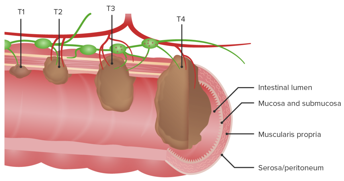
Example of t1–t4 staging for tumor in the colon-01

Example of T1–T4 staging for tumor in the colon
T1: Tumor invades the submucosa (through the muscularis mucosa but not into the muscularis propria).
T2: Tumor invades the muscularis propria.
T3: Tumor invades through the muscularis propria into the surrounding tissues.
T4: Direct extension through the serosa, invading the visceral peritoneum or adhering to adjacent organs

Image by Lecturio. License: CC BY-NC-SA 4.0

Metastasis

Definition

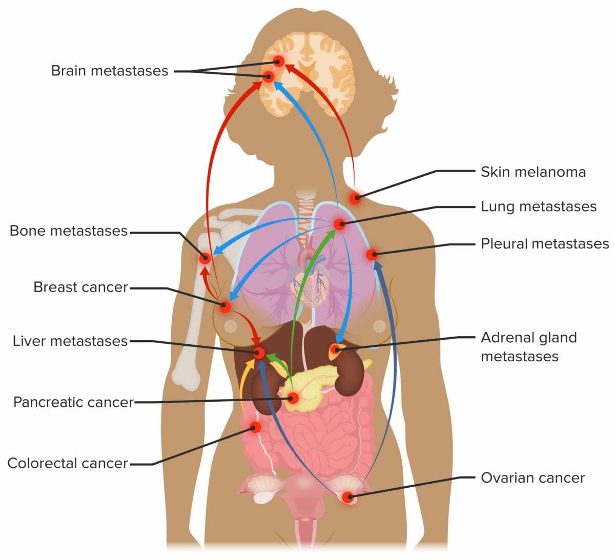
Metastasis is the spread of a tumor Tumor Inflammation from its primary site to nearby or distant places.
Routes of metastasis

  • Local spread: through infiltration in adjacent healthy tissues
  • Seeding: through cavities such as the peritoneum Peritoneum The peritoneum is a serous membrane lining the abdominopelvic cavity. This lining is formed by connective tissue and originates from the mesoderm. The membrane lines both the abdominal walls (as parietal peritoneum) and all of the visceral organs (as visceral peritoneum). Peritoneum: Anatomy, pleura Pleura The pleura is a serous membrane that lines the walls of the thoracic cavity and the surface of the lungs. This structure of mesodermal origin covers both lungs, the mediastinum, the thoracic surface of the diaphragm, and the inner part of the thoracic cage. The pleura is divided into a visceral pleura and parietal pleura. Pleura: Anatomy, and CSF
    • Ovarian cancer Ovarian cancer Ovarian cancer is a malignant tumor arising from the ovarian tissue and is classified according to the type of tissue from which it originates. The 3 major types of ovarian cancer are epithelial ovarian carcinomas (EOCs), ovarian germ cell tumors (OGCTs), and sex cord-stromal tumors (SCSTs). Ovarian Cancer
    • Gastric cancer Gastric cancer Gastric cancer is the 3rd-most common cause of cancer-related deaths worldwide. The majority of cases are from adenocarcinoma. The modifiable risk factors include Helicobacter pylori infection, smoking, and nitrate-rich diets. Gastric Cancer
    • Mesothelioma Mesothelioma Malignant mesothelioma (usually referred to as simply mesothelioma) is the malignant growth of mesothelial cells, most commonly affecting the pleura. The majority of cases are associated with occupational exposure to asbestos that occurred > 20 years before clinical onset, which includes dyspnea, chest pain, coughing, fatigue, and weight loss. Malignant Mesothelioma
    • Glioblastoma multiforme Glioblastoma multiforme Glioblastoma multiforme is a high-grade astrocytoma, an aggressive brain tumor arising from astrocytes, with an unknown cause and a poorly understood link to risk factors. There are two main types: primary, a more aggressive form seen more commonly in older patients, and secondary, developing from lower-grade astrocytomas and seen more commonly in younger patients. Glioblastoma Multiforme (type of brain Brain The part of central nervous system that is contained within the skull (cranium). Arising from the neural tube, the embryonic brain is comprised of three major parts including prosencephalon (the forebrain); mesencephalon (the midbrain); and rhombencephalon (the hindbrain). The developed brain consists of cerebrum; cerebellum; and other structures in the brain stem. Nervous System: Anatomy, Structure, and Classification cancer)
  • Lymphatic spread: through regional lymph nodes Lymph Nodes They are oval or bean shaped bodies (1 – 30 mm in diameter) located along the lymphatic system. Lymphatic Drainage System: Anatomy and lymph Lymph The interstitial fluid that is in the lymphatic system. Secondary Lymphatic Organs vessels
    • Seen with carcinomas, such as breast cancer Breast cancer Breast cancer is a disease characterized by malignant transformation of the epithelial cells of the breast. Breast cancer is the most common form of cancer and 2nd most common cause of cancer-related death among women. Breast Cancer
    • Some exceptions: colon Colon The large intestines constitute the last portion of the digestive system. The large intestine consists of the cecum, appendix, colon (with ascending, transverse, descending, and sigmoid segments), rectum, and anal canal. The primary function of the colon is to remove water and compact the stool prior to expulsion from the body via the rectum and anal canal. Colon, Cecum, and Appendix: Anatomy cancer, which spreads through the portal vein Portal vein A short thick vein formed by union of the superior mesenteric vein and the splenic vein. Liver: Anatomy
  • Hematogenous Hematogenous Hepatocellular Carcinoma (HCC) and Liver Metastases spread: through blood vessels, such as with sarcomas

Mechanism

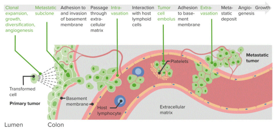
  • Invasion of the extracellular matrix Extracellular matrix A meshwork-like substance found within the extracellular space and in association with the basement membrane of the cell surface. It promotes cellular proliferation and provides a supporting structure to which cells or cell lysates in culture dishes adhere. Hypertrophic and Keloid Scars and basement membrane Basement membrane A darkly stained mat-like extracellular matrix (ecm) that separates cell layers, such as epithelium from endothelium or a layer of connective tissue. The ecm layer that supports an overlying epithelium or endothelium is called basal lamina. Basement membrane (bm) can be formed by the fusion of either two adjacent basal laminae or a basal lamina with an adjacent reticular lamina of connective tissue. Bm, composed mainly of type IV collagen; glycoprotein laminin; and proteoglycan, provides barriers as well as channels between interacting cell layers. Thin Basement Membrane Nephropathy (TBMN) is facilitated by proteases Proteases Proteins and Peptides and hyaluronidases.
  • Malignant cells are transported through the lymph Lymph The interstitial fluid that is in the lymphatic system. Secondary Lymphatic Organs and blood vessels forming a tumor Tumor Inflammation cell embolism.
  • Cells invade other tissues and organs through extravasation:
    • Initially, they lie dormant and excrete signal complexes → angiogenesis Angiogenesis Bartonella
    • Blood vessels form → deliver nutrients

Sites of metastasis

  • Depending on the location of the primary tumor Tumor Inflammation, there are different types of metastases according to blood flow Blood flow Blood flow refers to the movement of a certain volume of blood through the vasculature over a given unit of time (e.g., mL per minute). Vascular Resistance, Flow, and Mean Arterial Pressure.
  • The most common sites are the bones, lungs Lungs Lungs are the main organs of the respiratory system. Lungs are paired viscera located in the thoracic cavity and are composed of spongy tissue. The primary function of the lungs is to oxygenate blood and eliminate CO2. Lungs: Anatomy, liver Liver The liver is the largest gland in the human body. The liver is found in the superior right quadrant of the abdomen and weighs approximately 1.5 kilograms. Its main functions are detoxification, metabolism, nutrient storage (e.g., iron and vitamins), synthesis of coagulation factors, formation of bile, filtration, and storage of blood. Liver: Anatomy, and brain Brain The part of central nervous system that is contained within the skull (cranium). Arising from the neural tube, the embryonic brain is comprised of three major parts including prosencephalon (the forebrain); mesencephalon (the midbrain); and rhombencephalon (the hindbrain). The developed brain consists of cerebrum; cerebellum; and other structures in the brain stem. Nervous System: Anatomy, Structure, and Classification.
  • GI tumors spread through the portal system to the liver Liver The liver is the largest gland in the human body. The liver is found in the superior right quadrant of the abdomen and weighs approximately 1.5 kilograms. Its main functions are detoxification, metabolism, nutrient storage (e.g., iron and vitamins), synthesis of coagulation factors, formation of bile, filtration, and storage of blood. Liver: Anatomy and lungs Lungs Lungs are the main organs of the respiratory system. Lungs are paired viscera located in the thoracic cavity and are composed of spongy tissue. The primary function of the lungs is to oxygenate blood and eliminate CO2. Lungs: Anatomy.

References

  1. Telloni, S.M. (2017). Tumor staging and grading: A primer. Methods Mol Biol. 1606, 1–17. https://pubmed.ncbi.nlm.nih.gov/28501990/
  2. Rosen, R.D., Sapra, A. (2021). TNM Classification. StatPearls. Retrieved May 9, 2021, from https://www.ncbi.nlm.nih.gov/books/NBK553187/
  3. Brierley, J., Gospodarowicz, M., O’Sullivan, B. (2016). The principles of cancer staging. Ecancermedicalscience, 10, ed61. https://pubmed.ncbi.nlm.nih.gov/28101141/
  4. Burstein, H.J. (2020). Tumor, Node, Metastasis (TNM) staging classification for breast cancer. UpToDate. Retrieved July 22, 2021, from https://www.uptodate.com/contents/tumor-node-metastasis-tnm-staging-classification-for-breast-cancer
  5. Yang, X.J. (2021). Interpretation of prostate biopsy. UpToDate. Retrieved July 22, 2021, from https://www.uptodate.com/contents/interpretation-of-prostate-biopsy
  6. Thomas, K.W., Gould, M.K. (2019). Tumor, Node, Metastasis (TNM) staging system for lung cancer. UpToDate. Retrieved July 22, 2021, from https://www.uptodate.com/contents/tumor-node-metastasis-tnm-staging-system-for-lung-cancer
  7. Keung, E.Z., Gershenwald, J.E. (2018). The eighth edition American Joint Committee on Cancer (AJCC) melanoma staging system: Implications for melanoma treatment and care. Expert review of anticancer therapy, 18, 775–84. https://www.ncbi.nlm.nih.gov/pmc/articles/PMC7652033/

Create your free account or log in to continue reading!

Sign up now and get free access to Lecturio with concept pages, medical videos, and questions for your medical education.

User Reviews

Unwrap New Skills This Holiday 🎄 Save 30% on all plans now!

Details