Advertisement

Advertisement

Advertisement

Advertisement

Genitourinary Trauma

Traumatic injuries to the genitourinary (GU) tract include injuries to the kidneys Kidneys The kidneys are a pair of bean-shaped organs located retroperitoneally against the posterior wall of the abdomen on either side of the spine. As part of the urinary tract, the kidneys are responsible for blood filtration and excretion of water-soluble waste in the urine. Kidneys: Anatomy, ureter, bladder Bladder A musculomembranous sac along the urinary tract. Urine flows from the kidneys into the bladder via the ureters, and is held there until urination. Pyelonephritis and Perinephric Abscess, urethra Urethra A tube that transports urine from the urinary bladder to the outside of the body in both the sexes. It also has a reproductive function in the male by providing a passage for sperm. Urinary Tract: Anatomy, or genitals. Typically, injuries to the GU tract alone are not life threatening, but can be associated with other potentially more significant injuries. The GU system is divided into the upper GU tract ( kidneys Kidneys The kidneys are a pair of bean-shaped organs located retroperitoneally against the posterior wall of the abdomen on either side of the spine. As part of the urinary tract, the kidneys are responsible for blood filtration and excretion of water-soluble waste in the urine. Kidneys: Anatomy and ureters Ureters One of a pair of thick-walled tubes that transports urine from the kidney pelvis to the urinary bladder. Urinary Tract: Anatomy) and the lower GU tract ( bladder Bladder A musculomembranous sac along the urinary tract. Urine flows from the kidneys into the bladder via the ureters, and is held there until urination. Pyelonephritis and Perinephric Abscess, urethra Urethra A tube that transports urine from the urinary bladder to the outside of the body in both the sexes. It also has a reproductive function in the male by providing a passage for sperm. Urinary Tract: Anatomy, and external genitalia). Mechanisms include blunt and penetrating injuries. Diagnosis relies on thorough physical exam and imaging. Management depends on the severity of injury and ranges from simple observation and supportive measures to major surgical interventions. Timely diagnosis and intervention are crucial for preventing complications and ensuring optimal outcomes.

Last updated: Jan 24, 2023

Editorial responsibility: Stanley Oiseth, Lindsay Jones, Evelin Maza

Advertisement

Advertisement

Advertisement

Advertisement

Advertisement

Advertisement

Overview

Anatomic divisions

Upper genitourinary (GU) tract:

  • Kidneys Kidneys The kidneys are a pair of bean-shaped organs located retroperitoneally against the posterior wall of the abdomen on either side of the spine. As part of the urinary tract, the kidneys are responsible for blood filtration and excretion of water-soluble waste in the urine. Kidneys: Anatomy
  • Ureters Ureters One of a pair of thick-walled tubes that transports urine from the kidney pelvis to the urinary bladder. Urinary Tract: Anatomy

Lower GU tract:

  • Bladder Bladder A musculomembranous sac along the urinary tract. Urine flows from the kidneys into the bladder via the ureters, and is held there until urination. Pyelonephritis and Perinephric Abscess
  • Urethra Urethra A tube that transports urine from the urinary bladder to the outside of the body in both the sexes. It also has a reproductive function in the male by providing a passage for sperm. Urinary Tract: Anatomy
  • External genitalia
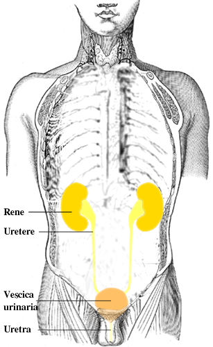
Organs of the urinary tract

Organs of the urinary tract

Image: “Urinary tract it” by Lennert B. License: CC BY 2.5

Epidemiology

  • Approximately 3%–10% of hospitalized trauma patients Patients Individuals participating in the health care system for the purpose of receiving therapeutic, diagnostic, or preventive procedures. Clinician–Patient Relationship have an injury to the GU system.
  • Penetrating injuries (i.e. gunshot and stab wounds Stab wounds Penetrating wounds caused by a pointed object. Penetrating Abdominal Injury) account for 15% of all GU injuries.
  • Uncommon and rarely life threatening, unless kidney vascular structures are involved
  • The kidneys Kidneys The kidneys are a pair of bean-shaped organs located retroperitoneally against the posterior wall of the abdomen on either side of the spine. As part of the urinary tract, the kidneys are responsible for blood filtration and excretion of water-soluble waste in the urine. Kidneys: Anatomy are the most injured GU organs, usually by blunt trauma.
  • Ureteral trauma is very rare, most commonly due to penetrating injury Penetrating Injury Brown-Séquard Syndrome (most cases are iatrogenic Iatrogenic Any adverse condition in a patient occurring as the result of treatment by a physician, surgeon, or other health professional, especially infections acquired by a patient during the course of treatment. Anterior Cord Syndrome).
  • Bladder Bladder A musculomembranous sac along the urinary tract. Urine flows from the kidneys into the bladder via the ureters, and is held there until urination. Pyelonephritis and Perinephric Abscess injuries usually are associated with pelvic fractures Pelvic Fractures Pelvic fractures are a disruption in the cortex of a pelvic bone involving iliac wing fractures, acetabular fractures, or those causing loss of integrity of the pelvic ring (the sacrum and the 2 innominate bones). Patients often present with a history of trauma or a fall, limb length discrepancy, intense pain on palpation, and mechanical instability. Pelvic Fractures
  • Urethral injuries are rare and occur almost exclusively in men.

Etiology

Blunt injuries:

  • Direct blow to the flanks or the back
  • Falling down from a height
  • Car accident (rapid deceleration Deceleration A decrease in the rate of speed. Blunt Chest Trauma injury)

Penetrating injuries:

  • Bullet
  • Stabbing
  • Surgical trauma ( iatrogenic Iatrogenic Any adverse condition in a patient occurring as the result of treatment by a physician, surgeon, or other health professional, especially infections acquired by a patient during the course of treatment. Anterior Cord Syndrome injury)

Related videos

Injuries to the Upper GU Tract

Renal injuries

Pathophysiology:

  • Kidneys Kidneys The kidneys are a pair of bean-shaped organs located retroperitoneally against the posterior wall of the abdomen on either side of the spine. As part of the urinary tract, the kidneys are responsible for blood filtration and excretion of water-soluble waste in the urine. Kidneys: Anatomy are protected by lower ribs Ribs A set of twelve curved bones which connect to the vertebral column posteriorly, and terminate anteriorly as costal cartilage. Together, they form a protective cage around the internal thoracic organs. Chest Wall: Anatomy, back musculature, and perinephric fat.
  • Significant force is required to injure kidneys Kidneys The kidneys are a pair of bean-shaped organs located retroperitoneally against the posterior wall of the abdomen on either side of the spine. As part of the urinary tract, the kidneys are responsible for blood filtration and excretion of water-soluble waste in the urine. Kidneys: Anatomy.
  • Common mechanisms:

Clinical presentation:

  • Flank pain Flank pain Pain emanating from below the ribs and above the ilium. Renal Cell Carcinoma/tenderness
  • Hematuria Hematuria Presence of blood in the urine. Renal Cell Carcinoma (gross or microscopic) 
  • Flank ecchymosis Ecchymosis Extravasation of blood into the skin, resulting in a nonelevated, rounded or irregular, blue or purplish patch, larger than a petechia. Orbital Fractures 
  • Often in association with posterior rib or spine Spine The human spine, or vertebral column, is the most important anatomical and functional axis of the human body. It consists of 7 cervical vertebrae, 12 thoracic vertebrae, and 5 lumbar vertebrae and is limited cranially by the skull and caudally by the sacrum. Vertebral Column: Anatomy fractures
  • May be associated with other intra-abdominal injuries

Diagnosis:

  • Urinalysis Urinalysis Examination of urine by chemical, physical, or microscopic means. Routine urinalysis usually includes performing chemical screening tests, determining specific gravity, observing any unusual color or odor, screening for bacteriuria, and examining the sediment microscopically. Urinary Tract Infections (UTIs) in Children:
  • Imaging:
    • Plain X-ray X-ray Penetrating electromagnetic radiation emitted when the inner orbital electrons of an atom are excited and release radiant energy. X-ray wavelengths range from 1 pm to 10 nm. Hard x-rays are the higher energy, shorter wavelength x-rays. Soft x-rays or grenz rays are less energetic and longer in wavelength. The short wavelength end of the x-ray spectrum overlaps the gamma rays wavelength range. The distinction between gamma rays and x-rays is based on their radiation source. Pulmonary Function Tests:
      • For associated injuries (e.g., rib fractures Rib fractures Fractures of any of the ribs. Flail Chest)
      • Retained bullets/foreign bodies in penetrating trauma
    • Ultrasound: for detection of intraperitoneal Intraperitoneal Peritoneum: Anatomy fluid (blood)
    • Computed tomography (CT) scan:
      • Test of choice in a hemodynamically stable patient
      • Delayed images needed to detect contrast extravasation (bleeding)
      • Also detects other associated injuries
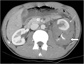
Grade iv left renal injury

Grade IV left renal injury from a motor vehicle accident
Contrast-enhanced CT in arterial phase in axial section showed contrast medium extravasation (black arrow), perirenal hematoma (open arrow), and pararenal hematoma (white arrow).

Image: “The role of interventional radiology for pediatric blunt renal trauma” by Lin WC, Lin CH. License: CC BY 4.0

Management:

  • First step: stabilization, resuscitation Resuscitation The restoration to life or consciousness of one apparently dead. . Neonatal Respiratory Distress Syndrome and identification Identification Defense Mechanisms of other life-threatening injuries
  • Renal injuries themselves are rarely life-threatening except for major avulsions of renal vessels.
  • Management depends on severity:
    • Graded based on American Association for the Surgery of Trauma (AAST) classification (grades Ⅰ through Ⅴ):
      • Grade Ⅰ-Ⅲ injuries do not involve injury to urinary collecting system or vasculature.
      • Grades Ⅳ-Ⅴ injuries are most severe and involve injury to major vessels and/or urinary collecting system.
  • AAST grade Ⅰ and Ⅱ injuries are usually managed nonoperatively.
  • Grade Ⅲ-Ⅳ: may require nephrectomy Nephrectomy Excision of kidney. Renal Cell Carcinoma, angiographic embolization Embolization A method of hemostasis utilizing various agents such as gelfoam, silastic, metal, glass, or plastic pellets, autologous clot, fat, and muscle as emboli. It has been used in the treatment of spinal cord and intracranial arteriovenous malformations, renal arteriovenous fistulas, gastrointestinal bleeding, epistaxis, hypersplenism, certain highly vascular tumors, traumatic rupture of blood vessels, and control of operative hemorrhage. Gastrointestinal Bleeding or stenting for vascular injuries

Ureteral injuries

Mechanisms:

  • Ureteral injury is rare.
  • 75% are iatrogenic Iatrogenic Any adverse condition in a patient occurring as the result of treatment by a physician, surgeon, or other health professional, especially infections acquired by a patient during the course of treatment. Anterior Cord Syndrome (during gynecologic, urologic, or general surgical procedures).
  • Most common blunt trauma is avulsion of uretero-pelvic junction ( deceleration Deceleration A decrease in the rate of speed. Blunt Chest Trauma injury).

Symptoms:

  • May have minimal initial symptoms and often missed
  • Delayed symptoms: fever Fever Fever is defined as a measured body temperature of at least 38°C (100.4°F). Fever is caused by circulating endogenous and/or exogenous pyrogens that increase levels of prostaglandin E2 in the hypothalamus. Fever is commonly associated with chills, rigors, sweating, and flushing of the skin. Fever, flank pain Flank pain Pain emanating from below the ribs and above the ilium. Renal Cell Carcinoma, and palpable mass Mass Three-dimensional lesion that occupies a space within the breast Imaging of the Breast (urinoma)

Diagnosis:

  • Urinalysis Urinalysis Examination of urine by chemical, physical, or microscopic means. Routine urinalysis usually includes performing chemical screening tests, determining specific gravity, observing any unusual color or odor, screening for bacteriuria, and examining the sediment microscopically. Urinary Tract Infections (UTIs) in Children may or may not show hematuria Hematuria Presence of blood in the urine. Renal Cell Carcinoma.
  • Can be picked up on abdominal CT scan
  • Intravenous pyelography Pyelography Hydronephrosis (IVP) if CT is non-diagnostic

Management:

  • Definitive management should be undertaken after other immediately life-threatening injuries have been addressed.
  • Ureteral injuries usually require some reconstruction:
    • Cystoscopic stent placement
    • Operative repair over stent
    • Urinary diversion
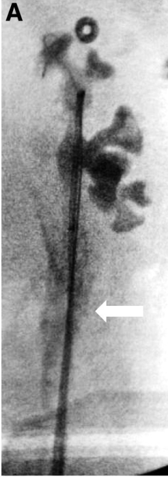
Iatrogenic ureteral trauma

Iatrogenic ureteral trauma sustained during ureter instrumentation
Intra-operative pyelogram shows contrast extravasation (arrow) from left ureter.

Image: “Aftermath of Grade 3 Ureteral Injury from Passage of a Ureteral Access Sheath: Disaster or Deliverance?” by Journal of Endourology Case Reports. License: CC BY 4.0, edited by Lecturio.

Complications

  • Urine extravasation can cause urinomas, abscesses.
  • Hydronephrosis Hydronephrosis Hydronephrosis is dilation of the renal collecting system as a result of the obstruction of urine outflow. Hydronephrosis can be unilateral or bilateral. Nephrolithiasis is the most common cause of hydronephrosis in young adults, while prostatic hyperplasia and neoplasm are seen in older patients. Hydronephrosis
  • Calculus formation
  • Renal hypertension Hypertension Hypertension, or high blood pressure, is a common disease that manifests as elevated systemic arterial pressures. Hypertension is most often asymptomatic and is found incidentally as part of a routine physical examination or during triage for an unrelated medical encounter. Hypertension
  • Loss of renal function

Injuries to the Lower GU Tract

Anatomy

  • Injuries to the lower GU tract include:
    • Bladder Bladder A musculomembranous sac along the urinary tract. Urine flows from the kidneys into the bladder via the ureters, and is held there until urination. Pyelonephritis and Perinephric Abscess 
    • Urethra Urethra A tube that transports urine from the urinary bladder to the outside of the body in both the sexes. It also has a reproductive function in the male by providing a passage for sperm. Urinary Tract: Anatomy
    • Perineum Perineum The body region lying between the genital area and the anus on the surface of the trunk, and to the shallow compartment lying deep to this area that is inferior to the pelvic diaphragm. The surface area is between the vulva and the anus in the female, and between the scrotum and the anus in the male. Vagina, Vulva, and Pelvic Floor: Anatomy 
    • Genitalia 
  • Highly associated with pelvic fractures Pelvic Fractures Pelvic fractures are a disruption in the cortex of a pelvic bone involving iliac wing fractures, acetabular fractures, or those causing loss of integrity of the pelvic ring (the sacrum and the 2 innominate bones). Patients often present with a history of trauma or a fall, limb length discrepancy, intense pain on palpation, and mechanical instability. Pelvic Fractures

Bladder Bladder A musculomembranous sac along the urinary tract. Urine flows from the kidneys into the bladder via the ureters, and is held there until urination. Pyelonephritis and Perinephric Abscess injuries

Extraperitoneal:

  • Confined to extraperitoneal space (the anterior bladder Bladder A musculomembranous sac along the urinary tract. Urine flows from the kidneys into the bladder via the ureters, and is held there until urination. Pyelonephritis and Perinephric Abscess wall or the bladder Bladder A musculomembranous sac along the urinary tract. Urine flows from the kidneys into the bladder via the ureters, and is held there until urination. Pyelonephritis and Perinephric Abscess neck Neck The part of a human or animal body connecting the head to the rest of the body. Peritonsillar Abscess)
  • Symptoms:
  • Diagnosis:
    • Retrograde cystogram 
    • Retrograde CT cystography 
    • CT abdomen and pelvis Pelvis The pelvis consists of the bony pelvic girdle, the muscular and ligamentous pelvic floor, and the pelvic cavity, which contains viscera, vessels, and multiple nerves and muscles. The pelvic girdle, composed of 2 “hip” bones and the sacrum, is a ring-like bony structure of the axial skeleton that links the vertebral column with the lower extremities. Pelvis: Anatomy: shows contrast extravasation from the bladder Bladder A musculomembranous sac along the urinary tract. Urine flows from the kidneys into the bladder via the ureters, and is held there until urination. Pyelonephritis and Perinephric Abscess 
  • Management:
    • Non-operative
    • Continuous prolonged bladder Bladder A musculomembranous sac along the urinary tract. Urine flows from the kidneys into the bladder via the ureters, and is held there until urination. Pyelonephritis and Perinephric Abscess drainage with a Foley catheter 

Intraperitoneal Intraperitoneal Peritoneum: Anatomy

  • Communicating with the peritoneum Peritoneum The peritoneum is a serous membrane lining the abdominopelvic cavity. This lining is formed by connective tissue and originates from the mesoderm. The membrane lines both the abdominal walls (as parietal peritoneum) and all of the visceral organs (as visceral peritoneum). Peritoneum: Anatomy (the dome of the bladder Bladder A musculomembranous sac along the urinary tract. Urine flows from the kidneys into the bladder via the ureters, and is held there until urination. Pyelonephritis and Perinephric Abscess)
  • Symptoms:
    • No desire to urinate
    • Peritonitis Peritonitis Inflammation of the peritoneum lining the abdominal cavity as the result of infectious, autoimmune, or chemical processes. Primary peritonitis is due to infection of the peritoneal cavity via hematogenous or lymphatic spread and without intra-abdominal source. Secondary peritonitis arises from the abdominal cavity itself through rupture or abscess of intra-abdominal organs. Penetrating Abdominal Injury and rebound tenderness Rebound Tenderness Acute Abdomen
    • Referred pain Referred Pain Spinal Disk Herniation to the shoulder due to diaphragmatic irritation
  • Diagnosis:
    • Ultrasound, usually part of focused assessment with sonography Sonography The visualization of deep structures of the body by recording the reflections or echoes of ultrasonic pulses directed into the tissues. Use of ultrasound for imaging or diagnostic purposes employs frequencies ranging from 1. 6 to 10 megahertz. Diagnostic Procedures in Gynecology in trauma (FAST exam)
    • Abdominal CT scan
  • Management: surgical repair 
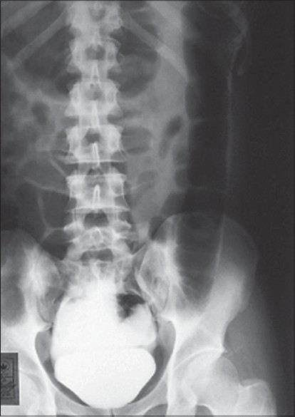
Intraperitoneal bladder rupture

Intraperitoneal bladder rupture: cystogram showing leakage of the contrast into the peritoneal cavity

Image: “Intraperitoneal bladder rupture mimicking acute renal failure” by Arun KG. License: CC BY 2.0

Urethral injuries

Symptoms:

Anterior urethral injuries:

  • Injury to bulbar (spongy) urethra Urethra A tube that transports urine from the urinary bladder to the outside of the body in both the sexes. It also has a reproductive function in the male by providing a passage for sperm. Urinary Tract: Anatomy
  • Associated with perineal saddle injury
  • Blood accumulates in scrotum Scrotum A cutaneous pouch of skin containing the testicles and spermatic cords. Testicles: Anatomy → scrotal hematoma Hematoma A collection of blood outside the blood vessels. Hematoma can be localized in an organ, space, or tissue. Intussusception, ecchymosis Ecchymosis Extravasation of blood into the skin, resulting in a nonelevated, rounded or irregular, blue or purplish patch, larger than a petechia. Orbital Fractures

Posterior urethral injuries:

  • Injury to membranous urethra Membranous urethra Urinary Tract: Anatomy
  • Associated with pelvic fracture Fracture A fracture is a disruption of the cortex of any bone and periosteum and is commonly due to mechanical stress after an injury or accident. Open fractures due to trauma can be a medical emergency. Fractures are frequently associated with automobile accidents, workplace injuries, and trauma. Overview of Bone Fractures
  • Urine leaks into retropubic space.
  • High-riding prostate Prostate The prostate is a gland in the male reproductive system. The gland surrounds the bladder neck and a portion of the urethra. The prostate is an exocrine gland that produces a weakly acidic secretion, which accounts for roughly 20% of the seminal fluid. on digital rectal exam ( DRE DRE A physical examination in which the qualified health care worker inserts a lubricated, gloved finger of one hand into the rectum and may use the other hand to press on the lower abdomen or pelvic area to palpate for abnormalities in the lower rectum, and nearby organs or tissues. The method is commonly used to check the lower rectum, the prostate gland in men, and the uterus and ovaries in women. Prostate Cancer Screening)

Diagnosis: retrograde urethrogram (RUG)

Management:
  • Bladder Bladder A musculomembranous sac along the urinary tract. Urine flows from the kidneys into the bladder via the ureters, and is held there until urination. Pyelonephritis and Perinephric Abscess drainage (suprapubic catheter) with or without delayed repair
  • Urethral catheterization is contraindicated if blood is seen at the urethral meatus.
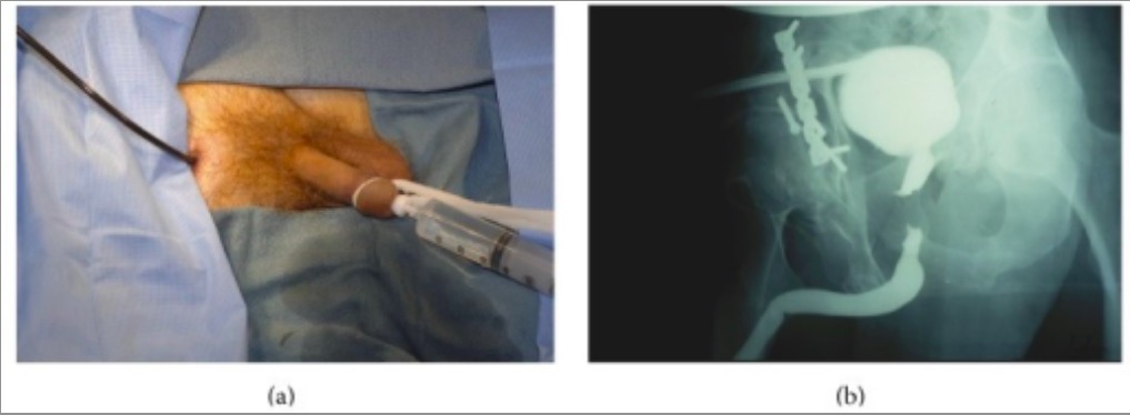
Rug urethral trauma

Individual with a history of pelvic trauma:
(a): A retrograde urethrogram is performed as contrast is simultaneously injected into the posterior urethra through the flexible cystoscope with the tip in the distal prostatic urethra.
(b): Imaging accurately demonstrates the length and location of the defect.
Image: “RUG” by University of California, Irvine, 333 City Boulevard West, Suite 1240, Orange, CA 92868, USA. License: CC BY 3.0

Perineal injury

  • Ecchymosis Ecchymosis Extravasation of blood into the skin, resulting in a nonelevated, rounded or irregular, blue or purplish patch, larger than a petechia. Orbital Fractures with no laceration Laceration Torn, ragged, mangled wounds. Blunt Chest Trauma is managed with perineal pressure (scrotal support with gauze packing).
  • Perineal lacerations with urethral or bladder Bladder A musculomembranous sac along the urinary tract. Urine flows from the kidneys into the bladder via the ureters, and is held there until urination. Pyelonephritis and Perinephric Abscess injury need urology consult to exclude the presence of urethrocutaneous or vesicocutaneous fistula Fistula Abnormal communication most commonly seen between two internal organs, or between an internal organ and the surface of the body. Anal Fistula.
  • Suspected testicular rupture/torsion: surgical management to evaluate the salvageability of the tissue

Penile and scrotal injury

  • Ultrasound is the preferred imaging.
  • Management:
    • Non-operative: if tunica albuginea Tunica albuginea Penis: Anatomy and dartos fascia Fascia Layers of connective tissue of variable thickness. The superficial fascia is found immediately below the skin; the deep fascia invests muscles, nerves, and other organs. Cellulitis are intact
    • Penetration Penetration X-rays requires surgical exploration/repair.

Complications

References

  1. Runyon, M. (2020). Blunt genitourinary trauma: Initial evaluation and management. Retrieved December 19, 2020 from https://www.uptodate.com/contents/blunt-genitourinary-trauma-initial-evaluation-and-management
  2. Voelzke, B. (2020). Overview of traumatic lower genitourinary tract injuries in adults. Retrieved December 19, 2020 from https://www.uptodate.com/contents/overview-of-traumatic-lower-genitourinary-tract-injury
  3. Voelzke, B. (2020). Overview of traumatic upper genitourinary tract injuries in adults. Retrieved December 19, 2020 from https://www.uptodate.com/contents/overview-of-traumatic-upper-genitourinary-tract-injuries-in-adults
  4. Runyon, M. (2020). Penetrating trauma of the upper and lower genitourinary tract: Initial evaluation and management. Retrieved December 19, 2020 from https://www.uptodate.com/contents/penetrating-trauma-of-the-upper-and-lower-genitourinary-tract-initial-evaluation-and-management
  5. Diercks, D., & Clarke, S. (2020). Initial evaluation and management of blunt abdominal trauma in adults. Retrieved December 20, 2020 from https://www.uptodate.com/contents/initial-evaluation-and-management-of-blunt-abdominal-trauma-in-adults
  6. Pereira, B.M.T., de Campos, C.C.C., Calderan, T.R.A. et al. (2013). Bladder injuries after external trauma: 20 years experience report in a population-based cross-sectional view. World J Urol 31: 913-917
  7. Bayne, D., Zaid, U., Alwaal, A., Harris, C., McAninch, J., & Breyer, B. (2016). Lower genitourinary tract trauma. Trauma 18(1):12-20.
  8. Koraitim, M.M., Marzouk, M.E., & Atta, M.A. (1996). Risk factors and mechanism of urethral injury in pelvic fractures. Br J Urol. 77(6):876–80.
  9. Chapple, C., Barbagli, G., & Jordan, G. (2004). Consensus statement on urethral trauma. BJU Int. 93(9):1195–202.

Create your free account or log in to continue reading!

Sign up now and get free access to Lecturio with concept pages, medical videos, and questions for your medical education.

User Reviews

Unwrap New Skills This Holiday 🎄 Save 30% on all plans now!

Details