Advertisement

Advertisement

Advertisement

Advertisement

Esophagus: Anatomy

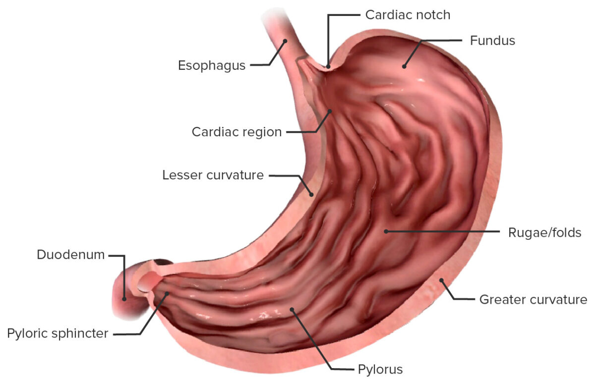
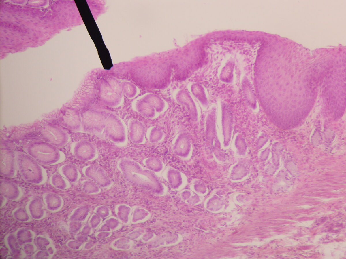
The esophagus is a muscular tube-shaped organ of around 25 centimeters in length that connects the pharynx Pharynx The pharynx is a component of the digestive system that lies posterior to the nasal cavity, oral cavity, and larynx. The pharynx can be divided into the oropharynx, nasopharynx, and laryngopharynx. Pharyngeal muscles play an integral role in vital processes such as breathing, swallowing, and speaking. Pharynx: Anatomy to the stomach Stomach The stomach is a muscular sac in the upper left portion of the abdomen that plays a critical role in digestion. The stomach develops from the foregut and connects the esophagus with the duodenum. Structurally, the stomach is C-shaped and forms a greater and lesser curvature and is divided grossly into regions: the cardia, fundus, body, and pylorus. Stomach: Anatomy. The organ extends from approximately the 6th cervical vertebra to the 11th thoracic vertebra and can be divided grossly into 3 parts: the cervical part, the thoracic part, and the abdominal part. The wall of the esophagus is made up of 4 primary layers: mucosa (lined with squamous epithelium Epithelium The epithelium is a complex of specialized cellular organizations arranged into sheets and lining cavities and covering the surfaces of the body. The cells exhibit polarity, having an apical and a basal pole. Structures important for the epithelial integrity and function involve the basement membrane, the semipermeable sheet on which the cells rest, and interdigitations, as well as cellular junctions. Surface Epithelium: Histology), submucosa, a thicker muscularis layer, and an outer layer of connective tissue Connective tissue Connective tissues originate from embryonic mesenchyme and are present throughout the body except inside the brain and spinal cord. The main function of connective tissues is to provide structural support to organs. Connective tissues consist of cells and an extracellular matrix. Connective Tissue: Histology. The esophagus also has a sphincter at each end, which allows it to help control the passage of food into the stomach Stomach The stomach is a muscular sac in the upper left portion of the abdomen that plays a critical role in digestion. The stomach develops from the foregut and connects the esophagus with the duodenum. Structurally, the stomach is C-shaped and forms a greater and lesser curvature and is divided grossly into regions: the cardia, fundus, body, and pylorus. Stomach: Anatomy.

Last updated: Nov 19, 2024

Editorial responsibility: Stanley Oiseth, Lindsay Jones, Evelin Maza

Advertisement

Advertisement

Advertisement

Advertisement

Advertisement

Advertisement

Development

Embryologic development of the esophagus:
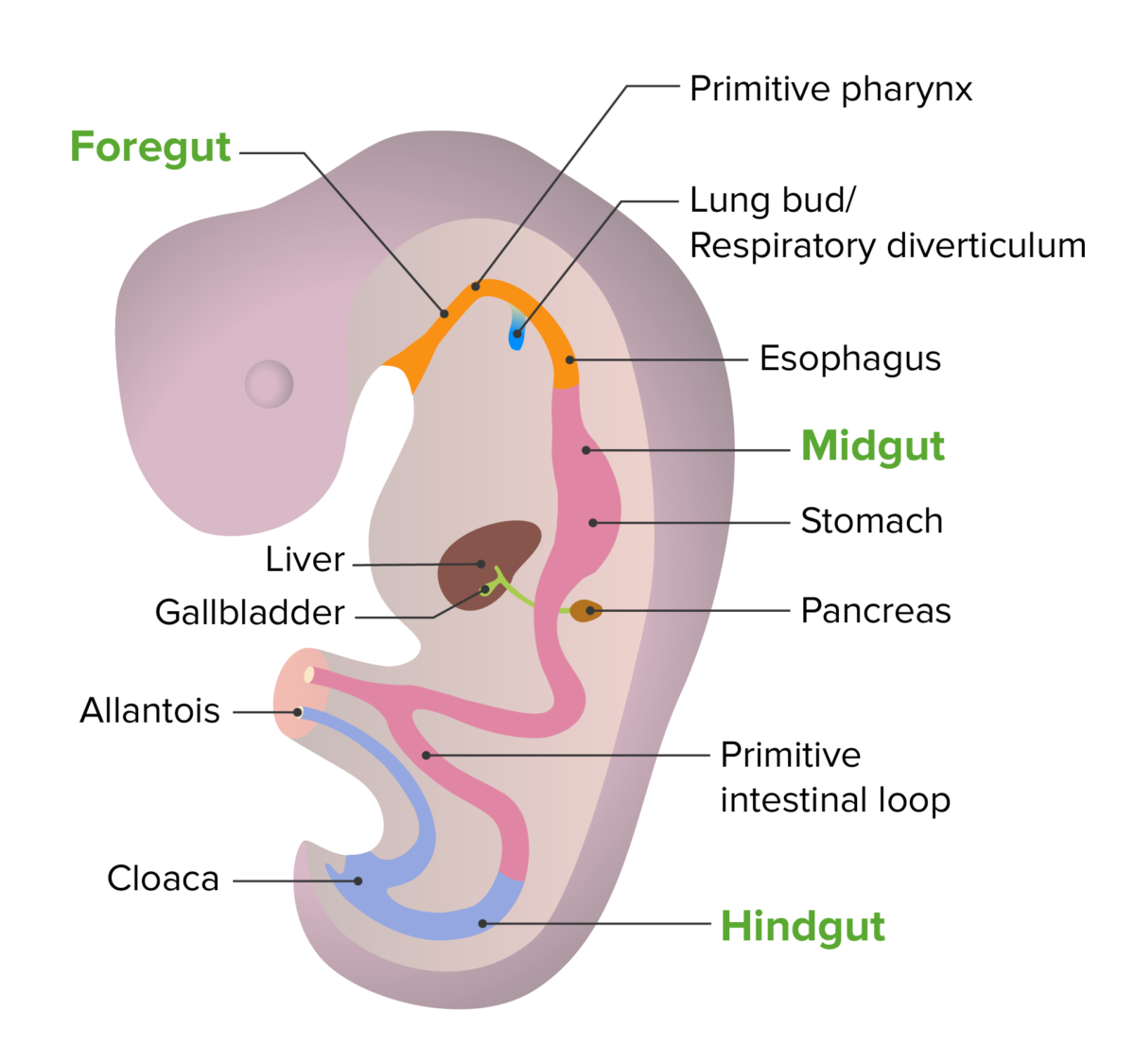
  • Begins forming in the 4th week of development
  • Derived from the foregut Foregut Development of the Abdominal Organs of the primitive gut tube (an invagination of the yolk sac Yolk Sac The first of four extra-embryonic membranes to form during embryogenesis. In reptiles and birds, it arises from endoderm and mesoderm to incorporate the egg yolk into the digestive tract for nourishing the embryo. In placental mammals, its nutritional function is vestigial; however, it is the source of intestinal mucosa; blood cells; and germ cells. It is sometimes called the vitelline sac, which should not be confused with the vitelline membrane of the egg. Embryoblast and Trophoblast Development into the embryo Embryo The entity of a developing mammal, generally from the cleavage of a zygote to the end of embryonic differentiation of basic structures. For the human embryo, this represents the first two months of intrauterine development preceding the stages of the fetus. Fertilization and First Week)
  • Has components of endoderm Endoderm The inner of the three germ layers of an embryo. Gastrulation and Neurulation and mesoderm Mesoderm The middle germ layer of an embryo derived from three paired mesenchymal aggregates along the neural tube. Gastrulation and Neurulation:
Embryonic development of the gut tube

Embryonic development of the gut tube

Image by Lecturio. License: CC BY-NC-SA 4.0
Gross Anatomy

General characteristics and location

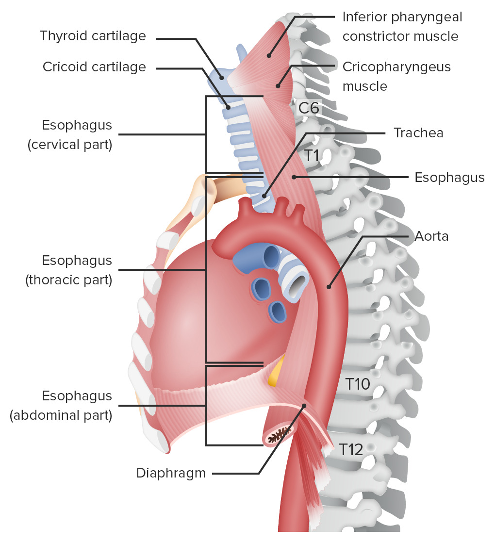
  • A long muscular tube connecting the pharynx Pharynx The pharynx is a component of the digestive system that lies posterior to the nasal cavity, oral cavity, and larynx. The pharynx can be divided into the oropharynx, nasopharynx, and laryngopharynx. Pharyngeal muscles play an integral role in vital processes such as breathing, swallowing, and speaking. Pharynx: Anatomy to the stomach Stomach The stomach is a muscular sac in the upper left portion of the abdomen that plays a critical role in digestion. The stomach develops from the foregut and connects the esophagus with the duodenum. Structurally, the stomach is C-shaped and forms a greater and lesser curvature and is divided grossly into regions: the cardia, fundus, body, and pylorus. Stomach: Anatomy
  • Extends from C6 to T11 vertebra
  • Size:
    • Length: approximately 25 cm 
    • Diameter: approximately 2 cm when relaxed
  • Sections of the esophagus:
    • Cervical: travels through the neck Neck The part of a human or animal body connecting the head to the rest of the body. Peritonsillar Abscess, behind the trachea Trachea The trachea is a tubular structure that forms part of the lower respiratory tract. The trachea is continuous superiorly with the larynx and inferiorly becomes the bronchial tree within the lungs. The trachea consists of a support frame of semicircular, or C-shaped, rings made out of hyaline cartilage and reinforced by collagenous connective tissue. Trachea: Anatomy 
    • Thoracic: located in the mediastinum Mediastinum The mediastinum is the thoracic area between the 2 pleural cavities. The mediastinum contains vital structures of the circulatory, respiratory, digestive, and nervous systems including the heart and esophagus, and major thoracic vessels. Mediastinum and Great Vessels: Anatomy, between the heart and the vertebrae
    • Abdominal: located below the diaphragm Diaphragm The diaphragm is a large, dome-shaped muscle that separates the thoracic cavity from the abdominal cavity. The diaphragm consists of muscle fibers and a large central tendon, which is divided into right and left parts. As the primary muscle of inspiration, the diaphragm contributes 75% of the total inspiratory muscle force. Diaphragm: Anatomy, within the abdominal cavity
Location of the esophagus

Location of the esophagus

Image by Lecturio.
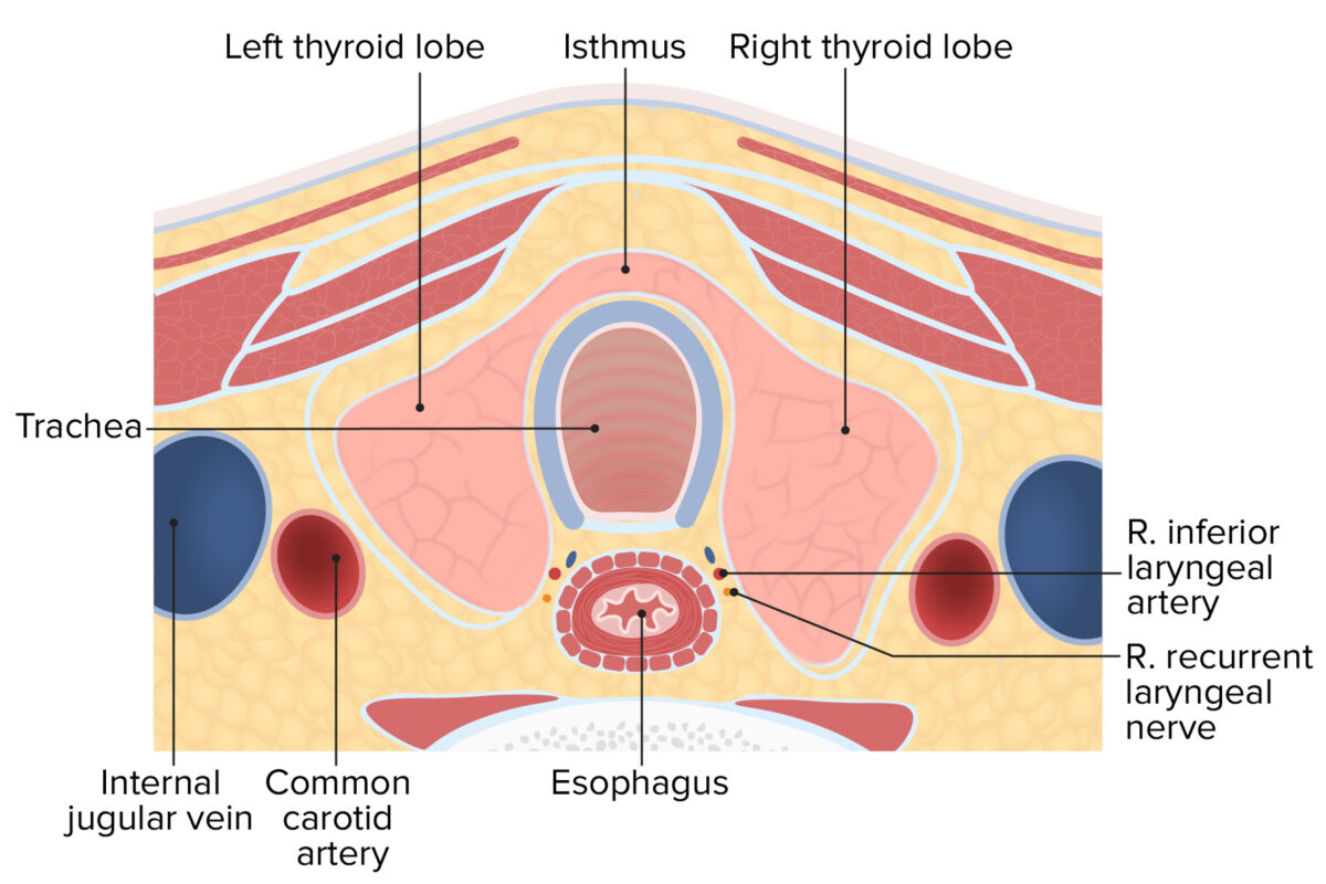
Cervical part (pars cervicalis)

  • Located within the neck Neck The part of a human or animal body connecting the head to the rest of the body. Peritonsillar Abscess, in direct connection with the pharynx Pharynx The pharynx is a component of the digestive system that lies posterior to the nasal cavity, oral cavity, and larynx. The pharynx can be divided into the oropharynx, nasopharynx, and laryngopharynx. Pharyngeal muscles play an integral role in vital processes such as breathing, swallowing, and speaking. Pharynx: Anatomy
  • Length: approximately 5 cm
  • Upper border of the cervical part: lower edge of the cricoid cartilage Cartilage Cartilage is a type of connective tissue derived from embryonic mesenchyme that is responsible for structural support, resilience, and the smoothness of physical actions. Perichondrium (connective tissue membrane surrounding cartilage) compensates for the absence of vasculature in cartilage by providing nutrition and support. Cartilage: Histology (at C6)
    • Known as the pharyngoesophageal junction
    • Upper esophageal sphincter (UES):
      • Made up of several muscles, most importantly the cricopharyngeus muscle
      • Surrounds the pharyngoesophageal junction
      • Helps control the passage of food into the esophagus from the pharynx Pharynx The pharynx is a component of the digestive system that lies posterior to the nasal cavity, oral cavity, and larynx. The pharynx can be divided into the oropharynx, nasopharynx, and laryngopharynx. Pharyngeal muscles play an integral role in vital processes such as breathing, swallowing, and speaking. Pharynx: Anatomy
  • Lower border of the cervical part: the thoracic inlet and sternal notch (at T1)
  • Anatomic relations:
    • Anterior: trachea Trachea The trachea is a tubular structure that forms part of the lower respiratory tract. The trachea is continuous superiorly with the larynx and inferiorly becomes the bronchial tree within the lungs. The trachea consists of a support frame of semicircular, or C-shaped, rings made out of hyaline cartilage and reinforced by collagenous connective tissue. Trachea: Anatomy 
    • Anterolateral: thyroid Thyroid The thyroid gland is one of the largest endocrine glands in the human body. The thyroid gland is a highly vascular, brownish-red gland located in the visceral compartment of the anterior region of the neck. Thyroid Gland: Anatomy 
    • Lateral: carotid sheath
    • Posterior: vertebrae

Thoracic part (pars thoracica)

  • Located in the thorax between the trachea Trachea The trachea is a tubular structure that forms part of the lower respiratory tract. The trachea is continuous superiorly with the larynx and inferiorly becomes the bronchial tree within the lungs. The trachea consists of a support frame of semicircular, or C-shaped, rings made out of hyaline cartilage and reinforced by collagenous connective tissue. Trachea: Anatomy and the aorta Aorta The main trunk of the systemic arteries. Mediastinum and Great Vessels: Anatomy
  • Length: 17–19 centimeters
  • Upper border: the thoracic inlet and sternal notch (at T1)
  • Lower border:
    • Diaphragm Diaphragm The diaphragm is a large, dome-shaped muscle that separates the thoracic cavity from the abdominal cavity. The diaphragm consists of muscle fibers and a large central tendon, which is divided into right and left parts. As the primary muscle of inspiration, the diaphragm contributes 75% of the total inspiratory muscle force. Diaphragm: Anatomy
    • Passes into the upper abdominal cavity through the the esophageal hiatus Esophageal hiatus Diaphragm: Anatomy (a hole in the diaphragm Diaphragm The diaphragm is a large, dome-shaped muscle that separates the thoracic cavity from the abdominal cavity. The diaphragm consists of muscle fibers and a large central tendon, which is divided into right and left parts. As the primary muscle of inspiration, the diaphragm contributes 75% of the total inspiratory muscle force. Diaphragm: Anatomy also called the esophageal foramen Esophageal foramen Diaphragm: Anatomy) at the level of T10
  • Anatomic relations:
    • Structures anterior to the thoracic esophagus:
      • Trachea Trachea The trachea is a tubular structure that forms part of the lower respiratory tract. The trachea is continuous superiorly with the larynx and inferiorly becomes the bronchial tree within the lungs. The trachea consists of a support frame of semicircular, or C-shaped, rings made out of hyaline cartilage and reinforced by collagenous connective tissue. Trachea: Anatomy and left main bronchus
      • Ascending aorta Ascending aorta Mediastinum and Great Vessels: Anatomy and aortic arch Aortic arch Mediastinum and Great Vessels: Anatomy 
      • Left atrium
      • Anterior vagus nerve Vagus nerve The 10th cranial nerve. The vagus is a mixed nerve which contains somatic afferents (from skin in back of the ear and the external auditory meatus), visceral afferents (from the pharynx, larynx, thorax, and abdomen), parasympathetic efferents (to the thorax and abdomen), and efferents to striated muscle (of the larynx and pharynx). Pharynx: Anatomy
    • Structures posterior to the esophagus:
      • Descending aorta Descending aorta Mediastinum and Great Vessels: Anatomy
      • Thoracic vertebra 
      • Thoracic duct Thoracic Duct The largest lymphatic vessel that passes through the chest and drains into the subclavian vein. Lymphatic Drainage System: Anatomy (to the left of the esophagus)
      • Azygos and hemiazygos veins Veins Veins are tubular collections of cells, which transport deoxygenated blood and waste from the capillary beds back to the heart. Veins are classified into 3 types: small veins/venules, medium veins, and large veins. Each type contains 3 primary layers: tunica intima, tunica media, and tunica adventitia. Veins: Histology 
      • Posterior vagal nerve

Abdominal part (pars abdominalis)

  • Located within the abdominal cavity between the diaphragm Diaphragm The diaphragm is a large, dome-shaped muscle that separates the thoracic cavity from the abdominal cavity. The diaphragm consists of muscle fibers and a large central tendon, which is divided into right and left parts. As the primary muscle of inspiration, the diaphragm contributes 75% of the total inspiratory muscle force. Diaphragm: Anatomy and the stomach Stomach The stomach is a muscular sac in the upper left portion of the abdomen that plays a critical role in digestion. The stomach develops from the foregut and connects the esophagus with the duodenum. Structurally, the stomach is C-shaped and forms a greater and lesser curvature and is divided grossly into regions: the cardia, fundus, body, and pylorus. Stomach: Anatomy
  • Length: 1–3 cm
  • Curves sharply to the left to join the cardiac portion of the stomach Stomach The stomach is a muscular sac in the upper left portion of the abdomen that plays a critical role in digestion. The stomach develops from the foregut and connects the esophagus with the duodenum. Structurally, the stomach is C-shaped and forms a greater and lesser curvature and is divided grossly into regions: the cardia, fundus, body, and pylorus. Stomach: Anatomy
  • Upper border: 
    • The esophageal hiatus Esophageal hiatus Diaphragm: Anatomy of the diaphragm Diaphragm The diaphragm is a large, dome-shaped muscle that separates the thoracic cavity from the abdominal cavity. The diaphragm consists of muscle fibers and a large central tendon, which is divided into right and left parts. As the primary muscle of inspiration, the diaphragm contributes 75% of the total inspiratory muscle force. Diaphragm: Anatomy (at T10)
    • Phrenoesophageal membrane: anchors the esophagus to the diaphragm Diaphragm The diaphragm is a large, dome-shaped muscle that separates the thoracic cavity from the abdominal cavity. The diaphragm consists of muscle fibers and a large central tendon, which is divided into right and left parts. As the primary muscle of inspiration, the diaphragm contributes 75% of the total inspiratory muscle force. Diaphragm: Anatomy
  • Lower border: the cardiac orifice of the stomach Stomach The stomach is a muscular sac in the upper left portion of the abdomen that plays a critical role in digestion. The stomach develops from the foregut and connects the esophagus with the duodenum. Structurally, the stomach is C-shaped and forms a greater and lesser curvature and is divided grossly into regions: the cardia, fundus, body, and pylorus. Stomach: Anatomy (at T11)
    • Known as the gastroesophageal (GE) junction
    • Lower esophageal sphincter: 
      • Surrounds the GE junction
      • Muscular ring acts as a functional (physiologic) sphincter
      • Allows food into the stomach Stomach The stomach is a muscular sac in the upper left portion of the abdomen that plays a critical role in digestion. The stomach develops from the foregut and connects the esophagus with the duodenum. Structurally, the stomach is C-shaped and forms a greater and lesser curvature and is divided grossly into regions: the cardia, fundus, body, and pylorus. Stomach: Anatomy
      • Prevents reflux of stomach Stomach The stomach is a muscular sac in the upper left portion of the abdomen that plays a critical role in digestion. The stomach develops from the foregut and connects the esophagus with the duodenum. Structurally, the stomach is C-shaped and forms a greater and lesser curvature and is divided grossly into regions: the cardia, fundus, body, and pylorus. Stomach: Anatomy contents into the esophagus
  • Anatomic relations:
    • Anterior: liver Liver The liver is the largest gland in the human body. The liver is found in the superior right quadrant of the abdomen and weighs approximately 1.5 kilograms. Its main functions are detoxification, metabolism, nutrient storage (e.g., iron and vitamins), synthesis of coagulation factors, formation of bile, filtration, and storage of blood. Liver: Anatomy
    • Posterior: descending aorta Descending aorta Mediastinum and Great Vessels: Anatomy 
    • Right border: continuous with the lesser curvature Lesser curvature Stomach: Anatomy of the stomach Stomach The stomach is a muscular sac in the upper left portion of the abdomen that plays a critical role in digestion. The stomach develops from the foregut and connects the esophagus with the duodenum. Structurally, the stomach is C-shaped and forms a greater and lesser curvature and is divided grossly into regions: the cardia, fundus, body, and pylorus. Stomach: Anatomy
    • Left border: separated from the fundus Fundus The superior portion of the body of the stomach above the level of the cardiac notch. Stomach: Anatomy of the stomach Stomach The stomach is a muscular sac in the upper left portion of the abdomen that plays a critical role in digestion. The stomach develops from the foregut and connects the esophagus with the duodenum. Structurally, the stomach is C-shaped and forms a greater and lesser curvature and is divided grossly into regions: the cardia, fundus, body, and pylorus. Stomach: Anatomy by the cardiac notch Cardiac notch Lungs: Anatomy
  • Relationship to peritoneum Peritoneum The peritoneum is a serous membrane lining the abdominopelvic cavity. This lining is formed by connective tissue and originates from the mesoderm. The membrane lines both the abdominal walls (as parietal peritoneum) and all of the visceral organs (as visceral peritoneum). Peritoneum: Anatomy:
    • Covered by parietal Parietal One of a pair of irregularly shaped quadrilateral bones situated between the frontal bone and occipital bone, which together form the sides of the cranium. Skull: Anatomy peritoneum Peritoneum The peritoneum is a serous membrane lining the abdominopelvic cavity. This lining is formed by connective tissue and originates from the mesoderm. The membrane lines both the abdominal walls (as parietal peritoneum) and all of the visceral organs (as visceral peritoneum). Peritoneum: Anatomy
    • Retroperitoneal Retroperitoneal Peritoneum: Anatomy

Constrictions

Constrictions are normal narrowings in the esophagus tube.

  • Pharyngo-esophageal constriction: 
    • Caused by the cricoid cartilage Cartilage Cartilage is a type of connective tissue derived from embryonic mesenchyme that is responsible for structural support, resilience, and the smoothness of physical actions. Perichondrium (connective tissue membrane surrounding cartilage) compensates for the absence of vasculature in cartilage by providing nutrition and support. Cartilage: Histology
    • Narrowest portion of the esophagus
  • Aortobronchial constriction: caused by proximity to the aortic arch Aortic arch Mediastinum and Great Vessels: Anatomy and the left main bronchus (approximately T4 T4 The major hormone derived from the thyroid gland. Thyroxine is synthesized via the iodination of tyrosines (monoiodotyrosine) and the coupling of iodotyrosines (diiodotyrosine) in the thyroglobulin. Thyroxine is released from thyroglobulin by proteolysis and secreted into the blood. Thyroxine is peripherally deiodinated to form triiodothyronine which exerts a broad spectrum of stimulatory effects on cell metabolism. Thyroid Hormones)
  • Diaphragmatic constriction: caused by the esophagus passing through the esophageal hiatus Esophageal hiatus Diaphragm: Anatomy of the diaphragm Diaphragm The diaphragm is a large, dome-shaped muscle that separates the thoracic cavity from the abdominal cavity. The diaphragm consists of muscle fibers and a large central tendon, which is divided into right and left parts. As the primary muscle of inspiration, the diaphragm contributes 75% of the total inspiratory muscle force. Diaphragm: Anatomy

Microscopic Anatomy

Overview of esophageal wall anatomy

The esophageal wall consists of 4 primary layers: 
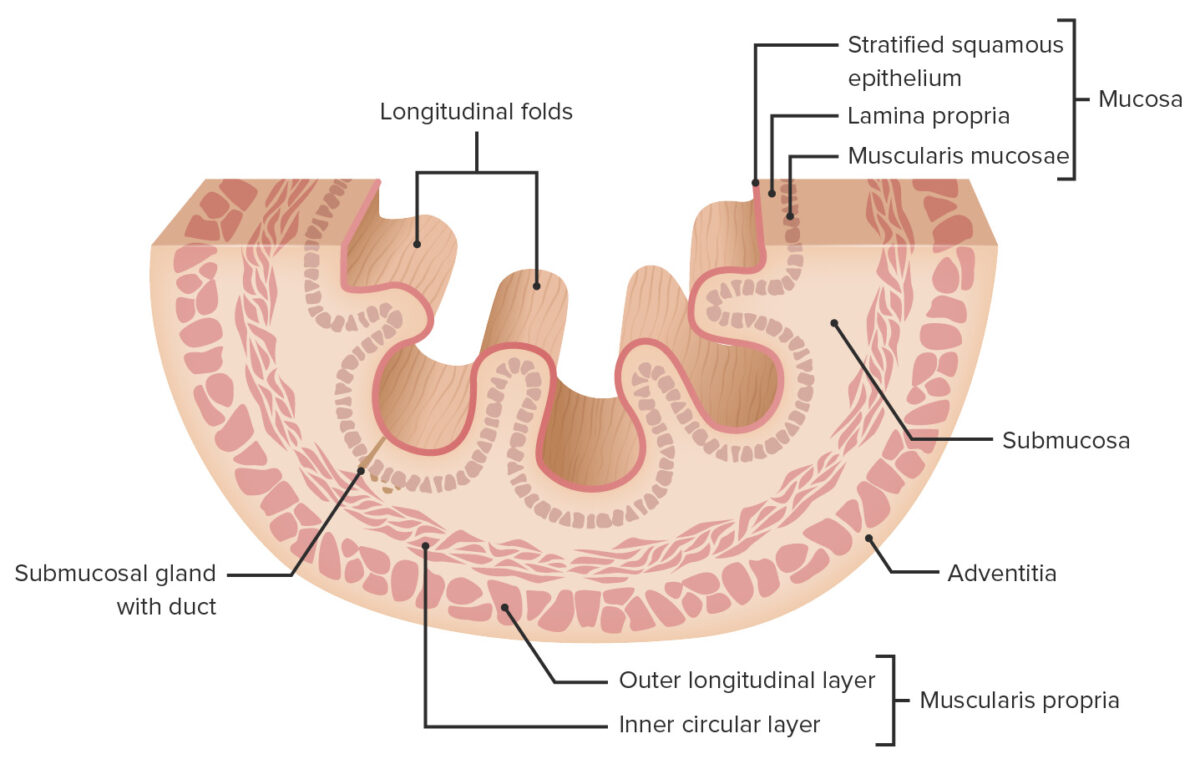
  1. Mucosa
  2. Submucosa
  3. Muscularis propria
  4. Adventitia/serosa
Layers of the esophageal wall

Layers of the esophageal wall

Image by Lecturio.

Mucosal layer

Consists of 3 sublayers:

  1. Non-keratinizing stratified squamous epithelium Stratified squamous epithelium Surface Epithelium: Histology:
    • Lines the lumen
    • Able to resist abrasive forces of food contents and regurgitated stomach Stomach The stomach is a muscular sac in the upper left portion of the abdomen that plays a critical role in digestion. The stomach develops from the foregut and connects the esophagus with the duodenum. Structurally, the stomach is C-shaped and forms a greater and lesser curvature and is divided grossly into regions: the cardia, fundus, body, and pylorus. Stomach: Anatomy acid
    • Z line Z line Skeletal Muscle Contraction (also known as the squamocolumnar junction): transition from squamous to columnar epithelium Epithelium The epithelium is a complex of specialized cellular organizations arranged into sheets and lining cavities and covering the surfaces of the body. The cells exhibit polarity, having an apical and a basal pole. Structures important for the epithelial integrity and function involve the basement membrane, the semipermeable sheet on which the cells rest, and interdigitations, as well as cellular junctions. Surface Epithelium: Histology as the esophagus enters the stomach Stomach The stomach is a muscular sac in the upper left portion of the abdomen that plays a critical role in digestion. The stomach develops from the foregut and connects the esophagus with the duodenum. Structurally, the stomach is C-shaped and forms a greater and lesser curvature and is divided grossly into regions: the cardia, fundus, body, and pylorus. Stomach: Anatomy
  2. Lamina propria Lamina propria Whipple’s Disease, a connective tissue Connective tissue Connective tissues originate from embryonic mesenchyme and are present throughout the body except inside the brain and spinal cord. The main function of connective tissues is to provide structural support to organs. Connective tissues consist of cells and an extracellular matrix. Connective Tissue: Histology layer which contains:
  3. Muscularis mucosae
    • Thin muscular layer
    • Contracts independently of the muscularis externa

Submucosal layer

Located between the mucosal and muscular layers, the submucosal layer is a layer of connective tissue Connective tissue Connective tissues originate from embryonic mesenchyme and are present throughout the body except inside the brain and spinal cord. The main function of connective tissues is to provide structural support to organs. Connective tissues consist of cells and an extracellular matrix. Connective Tissue: Histology that contains:
  • Larger blood vessels
  • Submucosal (Meissner) nerve plexus (ganglia of the ANS ANS The ans is a component of the peripheral nervous system that uses both afferent (sensory) and efferent (effector) neurons, which control the functioning of the internal organs and involuntary processes via connections with the CNS. The ans consists of the sympathetic and parasympathetic nervous systems. Autonomic Nervous System: Anatomy): controls the muscularis mucosa (independent of the muscularis propria)
  • Esophageal glands:
    • Tubuloacinar glands
    • Produce lubricating mucus
  • Longitudinal folds:
    • Thick folds along the length of the esophagus
    • Allow for distension of the esophagus during swallowing Swallowing The act of taking solids and liquids into the gastrointestinal tract through the mouth and throat. Gastrointestinal Motility

Muscularis propria

  • Also called the muscularis externa
  • Responsible for the peristaltic waves that move the food toward the stomach Stomach The stomach is a muscular sac in the upper left portion of the abdomen that plays a critical role in digestion. The stomach develops from the foregut and connects the esophagus with the duodenum. Structurally, the stomach is C-shaped and forms a greater and lesser curvature and is divided grossly into regions: the cardia, fundus, body, and pylorus. Stomach: Anatomy
  • Composed of 2 layers:
    • Inner circular muscle fibers
    • Outer longitudinal muscle fibers
  • Consists of both striated and smooth muscles Smooth muscles Unstriated and unstriped muscle, one of the muscles of the internal organs, blood vessels, hair follicles, etc. Contractile elements are elongated, usually spindle-shaped cells with centrally located nuclei. Smooth muscle fibers are bound together into sheets or bundles by reticular fibers and frequently elastic nets are also abundant. Muscle Tissue: Histology
    • Cranial part (proximal ⅓): striated muscle Striated muscle One of two types of muscle in the body, characterized by the array of bands observed under microscope. Striated muscles can be divided into two subtypes: the cardiac muscle and the skeletal muscle. Muscle Tissue: Histology (under somatic control)
    • Intermediate: mixed
    • Distal part (distal ⅓): smooth muscle (under autonomic control)
  • Contains the myenteric (Auerbach) nerve plexus
    • Ganglia of the ANS ANS The ans is a component of the peripheral nervous system that uses both afferent (sensory) and efferent (effector) neurons, which control the functioning of the internal organs and involuntary processes via connections with the CNS. The ans consists of the sympathetic and parasympathetic nervous systems. Autonomic Nervous System: Anatomy that controls the muscularis externa/stimulates peristalsis Peristalsis A movement, caused by sequential muscle contraction, that pushes the contents of the intestines or other tubular organs in one direction. Gastrointestinal Motility
    • Located between the 2 layers of smooth muscle

Adventitial/serosal layer

  • Connective tissue Connective tissue Connective tissues originate from embryonic mesenchyme and are present throughout the body except inside the brain and spinal cord. The main function of connective tissues is to provide structural support to organs. Connective tissues consist of cells and an extracellular matrix. Connective Tissue: Histology surrounding the esophagus
  • Adventia (more fibrous Fibrous Fibrocystic Change) above the diaphragm Diaphragm The diaphragm is a large, dome-shaped muscle that separates the thoracic cavity from the abdominal cavity. The diaphragm consists of muscle fibers and a large central tendon, which is divided into right and left parts. As the primary muscle of inspiration, the diaphragm contributes 75% of the total inspiratory muscle force. Diaphragm: Anatomy
  • Serosa below the diaphragm Diaphragm The diaphragm is a large, dome-shaped muscle that separates the thoracic cavity from the abdominal cavity. The diaphragm consists of muscle fibers and a large central tendon, which is divided into right and left parts. As the primary muscle of inspiration, the diaphragm contributes 75% of the total inspiratory muscle force. Diaphragm: Anatomy
  • Contains:
    • Large vessels
    • Lymphatic vessels Lymphatic Vessels Tubular vessels that are involved in the transport of lymph and lymphocytes. Lymphatic Drainage System: Anatomy
    • Nerve fascicles:
      • Vagus nerve Vagus nerve The 10th cranial nerve. The vagus is a mixed nerve which contains somatic afferents (from skin in back of the ear and the external auditory meatus), visceral afferents (from the pharynx, larynx, thorax, and abdomen), parasympathetic efferents (to the thorax and abdomen), and efferents to striated muscle (of the larynx and pharynx). Pharynx: Anatomy
      • Esophageal sympathetic plexus
Low magnification cross-sectional image of the esophagus (h&e stain)

Low-magnification cross-sectional image of the esophagus (H&E stain)

Image by Geoffrey Meyer, PhD

Related videos

Neurovasculature

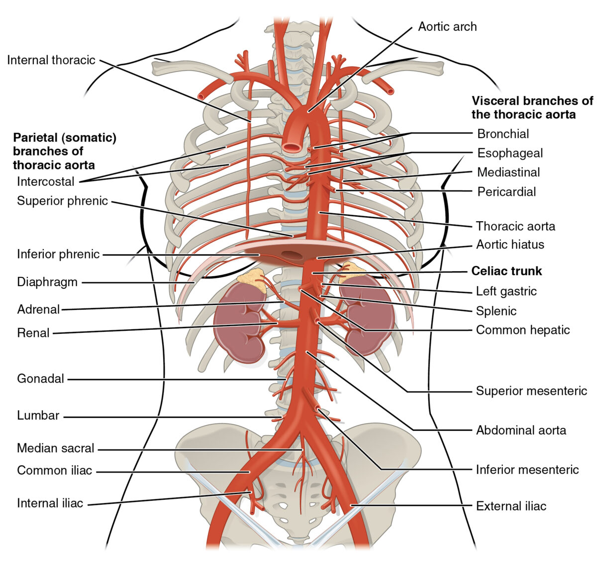
Arterial supply

  • Cervical part: inferior thyroid artery Inferior thyroid artery Thyroid Gland: Anatomy
  • Thoracic part:
    • Branches of the thoracic aorta Aorta The main trunk of the systemic arteries. Mediastinum and Great Vessels: Anatomy:
      • Bronchial branches
      • Esophageal branches
    • Intercostal arteries Arteries Arteries are tubular collections of cells that transport oxygenated blood and nutrients from the heart to the tissues of the body. The blood passes through the arteries in order of decreasing luminal diameter, starting in the largest artery (the aorta) and ending in the small arterioles. Arteries are classified into 3 types: large elastic arteries, medium muscular arteries, and small arteries and arterioles. Arteries: Histology
  • Abdominal part:
Image displaying some of the arteries responsible for the blood supply of the esophagus

Image displaying some of the arteries responsible for the blood supply of the esophagus

Image: “Thoracic Abdominal Arteries” by OpenStax College. License: CC BY 3.0

Venous drainage

  • Cervical part: inferior thyroid Thyroid The thyroid gland is one of the largest endocrine glands in the human body. The thyroid gland is a highly vascular, brownish-red gland located in the visceral compartment of the anterior region of the neck. Thyroid Gland: Anatomy vein
  • Thoracic part: ultimately drains into the inferior vena cava Inferior vena cava The venous trunk which receives blood from the lower extremities and from the pelvic and abdominal organs. Mediastinum and Great Vessels: Anatomy ( IVC IVC The venous trunk which receives blood from the lower extremities and from the pelvic and abdominal organs. Mediastinum and Great Vessels: Anatomy)
    • Azygos veins Veins Veins are tubular collections of cells, which transport deoxygenated blood and waste from the capillary beds back to the heart. Veins are classified into 3 types: small veins/venules, medium veins, and large veins. Each type contains 3 primary layers: tunica intima, tunica media, and tunica adventitia. Veins: Histology
    • Hemiazygos veins Veins Veins are tubular collections of cells, which transport deoxygenated blood and waste from the capillary beds back to the heart. Veins are classified into 3 types: small veins/venules, medium veins, and large veins. Each type contains 3 primary layers: tunica intima, tunica media, and tunica adventitia. Veins: Histology
    • Intercostal veins Veins Veins are tubular collections of cells, which transport deoxygenated blood and waste from the capillary beds back to the heart. Veins are classified into 3 types: small veins/venules, medium veins, and large veins. Each type contains 3 primary layers: tunica intima, tunica media, and tunica adventitia. Veins: Histology
    • Bronchial veins Veins Veins are tubular collections of cells, which transport deoxygenated blood and waste from the capillary beds back to the heart. Veins are classified into 3 types: small veins/venules, medium veins, and large veins. Each type contains 3 primary layers: tunica intima, tunica media, and tunica adventitia. Veins: Histology
  • Abdominal part: ultimately drains into the portal vein Portal vein A short thick vein formed by union of the superior mesenteric vein and the splenic vein. Liver: Anatomy via the left gastric vein
  • Clinical relevance:
    • Veins Veins Veins are tubular collections of cells, which transport deoxygenated blood and waste from the capillary beds back to the heart. Veins are classified into 3 types: small veins/venules, medium veins, and large veins. Each type contains 3 primary layers: tunica intima, tunica media, and tunica adventitia. Veins: Histology surrounding the esophagus form an important anastomosis between the portal system and the systemic veins Veins Veins are tubular collections of cells, which transport deoxygenated blood and waste from the capillary beds back to the heart. Veins are classified into 3 types: small veins/venules, medium veins, and large veins. Each type contains 3 primary layers: tunica intima, tunica media, and tunica adventitia. Veins: Histology.
    • ↑ Pressure in the portal system can lead to esophageal varices, which can cause profuse upper GI bleeding if ruptured

Lymphatic drainage

  • Drained via 2 primary plexuses: dense networks of longitudinal vessels that are continuous with the lymphatic vessels Lymphatic Vessels Tubular vessels that are involved in the transport of lymph and lymphocytes. Lymphatic Drainage System: Anatomy of the pharynx Pharynx The pharynx is a component of the digestive system that lies posterior to the nasal cavity, oral cavity, and larynx. The pharynx can be divided into the oropharynx, nasopharynx, and laryngopharynx. Pharyngeal muscles play an integral role in vital processes such as breathing, swallowing, and speaking. Pharynx: Anatomy (above) and the gastric mucosa Gastric mucosa Lining of the stomach, consisting of an inner epithelium, a middle lamina propria, and an outer muscularis mucosae. The surface cells produce mucus that protects the stomach from attack by digestive acid and enzymes. When the epithelium invaginates into the lamina propria at various region of the stomach (cardia; gastric fundus; and pylorus), different tubular gastric glands are formed. These glands consist of cells that secrete mucus, enzymes, hydrochloric acid, or hormones. Stomach: Anatomy (below)
    • Mucosal plexus
    • Submucosal plexus
  • Cervical part: drains to deep cervical nodes 
  • Thoracic part: drains to posterior mediastinal nodes
  • Abdominal part: drains to left gastric and celiac nodes
  • There is also direct drainage into the thoracic duct Thoracic Duct The largest lymphatic vessel that passes through the chest and drains into the subclavian vein. Lymphatic Drainage System: Anatomy.

Innervation

  • Striated muscles in the upper ⅓: recurrent laryngeal nerve (branch of vagus)
  • Smooth muscles Smooth muscles Unstriated and unstriped muscle, one of the muscles of the internal organs, blood vessels, hair follicles, etc. Contractile elements are elongated, usually spindle-shaped cells with centrally located nuclei. Smooth muscle fibers are bound together into sheets or bundles by reticular fibers and frequently elastic nets are also abundant. Muscle Tissue: Histology: autonomic control via the parasympathetic and sympathetic systems
  • Sympathetic innervation: 
    • Inhibitory:
      • Causes muscle wall relaxation
      • Tonic contraction of the esophageal sphincters
    • Via postganglionic fibers from stellate and thoracic ganglia
    • Includes afferent Afferent Neurons which conduct nerve impulses to the central nervous system. Nervous System: Histology visceral pain Visceral pain Pain originating from internal organs (viscera) associated with autonomic phenomena (pallor; sweating; nausea; and vomiting). It often becomes a referred pain. Pain: Types and Pathways fibers
  • Parasympathetic innervation:
    • Stimulatory:
      • Muscle wall contraction/ peristalsis Peristalsis A movement, caused by sequential muscle contraction, that pushes the contents of the intestines or other tubular organs in one direction. Gastrointestinal Motility waves
      • ↑ Glandular secretions
      • Relaxation of the sphincters to allow passage of food
    • Via branches of the vagus nerve Vagus nerve The 10th cranial nerve. The vagus is a mixed nerve which contains somatic afferents (from skin in back of the ear and the external auditory meatus), visceral afferents (from the pharynx, larynx, thorax, and abdomen), parasympathetic efferents (to the thorax and abdomen), and efferents to striated muscle (of the larynx and pharynx). Pharynx: Anatomy
    • Synapse Synapse The junction between 2 neurons is called a synapse. The synapse allows a neuron to pass an electrical or chemical signal to another neuron or target effector cell. Synapses and Neurotransmission with ganglia in Meissner and Auerbach plexuses
    • Includes afferent Afferent Neurons which conduct nerve impulses to the central nervous system. Nervous System: Histology fibers that can detect pressure

Function

The primary function of the esophagus is to transport the partially digested food from the pharynx Pharynx The pharynx is a component of the digestive system that lies posterior to the nasal cavity, oral cavity, and larynx. The pharynx can be divided into the oropharynx, nasopharynx, and laryngopharynx. Pharyngeal muscles play an integral role in vital processes such as breathing, swallowing, and speaking. Pharynx: Anatomy to the stomach Stomach The stomach is a muscular sac in the upper left portion of the abdomen that plays a critical role in digestion. The stomach develops from the foregut and connects the esophagus with the duodenum. Structurally, the stomach is C-shaped and forms a greater and lesser curvature and is divided grossly into regions: the cardia, fundus, body, and pylorus. Stomach: Anatomy:

  • Wavelike esophageal muscle contractions called peristalsis Peristalsis A movement, caused by sequential muscle contraction, that pushes the contents of the intestines or other tubular organs in one direction. Gastrointestinal Motility move food down into the stomach Stomach The stomach is a muscular sac in the upper left portion of the abdomen that plays a critical role in digestion. The stomach develops from the foregut and connects the esophagus with the duodenum. Structurally, the stomach is C-shaped and forms a greater and lesser curvature and is divided grossly into regions: the cardia, fundus, body, and pylorus. Stomach: Anatomy.
  • Mucus production by esophageal glands → lubricate the bolus and help its transportation
  • Tonic contraction of the sphincters → relax when food is swallowed to allow passage downward

Clinical Relevance

  • Esophageal atresia Atresia Hypoplastic Left Heart Syndrome (HLHS): a congenital anomaly in which the esophagus does not fully develop, creating a blind pouch that prevents swallowed contents from passing into the stomach Stomach The stomach is a muscular sac in the upper left portion of the abdomen that plays a critical role in digestion. The stomach develops from the foregut and connects the esophagus with the duodenum. Structurally, the stomach is C-shaped and forms a greater and lesser curvature and is divided grossly into regions: the cardia, fundus, body, and pylorus. Stomach: Anatomy. Esophageal atresia Atresia Hypoplastic Left Heart Syndrome (HLHS) may cause polyhydramnios Polyhydramnios Polyhydramnios is a pathological excess of amniotic fluid. Common causes of polyhydramnios include fetal anomalies, gestational diabetes, multiple gestations, and congenital infections. Patients are often asymptomatic but may present with dyspnea, extremity swelling, or abdominal distention. Polyhydramnios in utero (because the fetus is unable to normally swallow the amniotic fluid Amniotic fluid A clear, yellowish liquid that envelopes the fetus inside the sac of amnion. In the first trimester, it is likely a transudate of maternal or fetal plasma. In the second trimester, amniotic fluid derives primarily from fetal lung and kidney. Cells or substances in this fluid can be removed for prenatal diagnostic tests (amniocentesis). Placenta, Umbilical Cord, and Amniotic Cavity) and presents at birth with regurgitation Regurgitation Gastroesophageal Reflux Disease (GERD) of all swallowed contents. The condition is diagnosed by an inability to pass a NG tube into the stomach Stomach The stomach is a muscular sac in the upper left portion of the abdomen that plays a critical role in digestion. The stomach develops from the foregut and connects the esophagus with the duodenum. Structurally, the stomach is C-shaped and forms a greater and lesser curvature and is divided grossly into regions: the cardia, fundus, body, and pylorus. Stomach: Anatomy; the tube will be seen coiling in the blind pouch on chest X-ray X-ray Penetrating electromagnetic radiation emitted when the inner orbital electrons of an atom are excited and release radiant energy. X-ray wavelengths range from 1 pm to 10 nm. Hard x-rays are the higher energy, shorter wavelength x-rays. Soft x-rays or grenz rays are less energetic and longer in wavelength. The short wavelength end of the x-ray spectrum overlaps the gamma rays wavelength range. The distinction between gamma rays and x-rays is based on their radiation source. Pulmonary Function Tests. Management is surgical.
  • Tracheoesophageal fistula Fistula Abnormal communication most commonly seen between two internal organs, or between an internal organ and the surface of the body. Anal Fistula (TEF): an abnormal communication Communication The exchange or transmission of ideas, attitudes, or beliefs between individuals or groups. Decision-making Capacity and Legal Competence between the trachea Trachea The trachea is a tubular structure that forms part of the lower respiratory tract. The trachea is continuous superiorly with the larynx and inferiorly becomes the bronchial tree within the lungs. The trachea consists of a support frame of semicircular, or C-shaped, rings made out of hyaline cartilage and reinforced by collagenous connective tissue. Trachea: Anatomy and the esophagus. Tracheoesophageal fistulas are usually associated with other anomalies, especially esophageal atresia Atresia Hypoplastic Left Heart Syndrome (HLHS), VACTERL association VACTERL Association VACTERL association is a rare disorder that affects multiple body systems in fetal development. There is no clear genetic cause or inheritance pattern for the development of this disorder. The acronym VACTERL stands for its characteristic anomalies: Vertebral abnormalities, Anal atresia, Cardiac defects, Tracheoesophageal abnormalities, Renal anomalies, and Limb abnormalities. VACTERL Association anomalies, and CHARGE syndrome CHARGE Syndrome CHARGE syndrome is a rare genetic condition with autosomal dominant inheritance in which almost all body systems are affected. The acronym CHARGE stands for the constellation of clinical features seen with this condition: Coloboma, Heart defects, Atresia choanae, Growth retardation, Genetic Abnormalities, and Ear abnormalities. CHARGE Syndrome. Presentation will depend on the exact anomaly, but symptoms may include inability to feed, vomiting Vomiting The forcible expulsion of the contents of the stomach through the mouth. Hypokalemia/ regurgitation Regurgitation Gastroesophageal Reflux Disease (GERD), excessive secretions, gastric distension from inspired air, and aspiration pneumonia Aspiration pneumonia A type of lung inflammation resulting from the aspiration of food, liquid, or gastric contents into the upper respiratory tract. Pneumonia. Management is surgical.
  • Hiatal hernia Hiatal hernia Stomach herniation located at or near the diaphragmatic opening for the esophagus, the esophageal hiatus. Congenital Diaphragmatic Hernias: protrusion of the abdominal esophagus and/or stomach Stomach The stomach is a muscular sac in the upper left portion of the abdomen that plays a critical role in digestion. The stomach develops from the foregut and connects the esophagus with the duodenum. Structurally, the stomach is C-shaped and forms a greater and lesser curvature and is divided grossly into regions: the cardia, fundus, body, and pylorus. Stomach: Anatomy through the esophageal hiatus Esophageal hiatus Diaphragm: Anatomy. Hiatal hernias may be due to a congenital defect in the diaphragm Diaphragm The diaphragm is a large, dome-shaped muscle that separates the thoracic cavity from the abdominal cavity. The diaphragm consists of muscle fibers and a large central tendon, which is divided into right and left parts. As the primary muscle of inspiration, the diaphragm contributes 75% of the total inspiratory muscle force. Diaphragm: Anatomy, trauma, or iatrogenic Iatrogenic Any adverse condition in a patient occurring as the result of treatment by a physician, surgeon, or other health professional, especially infections acquired by a patient during the course of treatment. Anterior Cord Syndrome disease following surgical dissection of the hiatus during certain procedures (e.g., antireflux procedures). Most hiatal hernias are asymptomatic, but larger hernias may cause heartburn Heartburn Substernal pain or burning sensation, usually associated with regurgitation of gastric juice into the esophagus. Gastroesophageal Reflux Disease (GERD), regurgitation Regurgitation Gastroesophageal Reflux Disease (GERD), or dysphagia Dysphagia Dysphagia is the subjective sensation of difficulty swallowing. Symptoms can range from a complete inability to swallow, to the sensation of solids or liquids becoming “stuck.” Dysphagia is classified as either oropharyngeal or esophageal, with esophageal dysphagia having 2 sub-types: functional and mechanical. Dysphagia.
  • Esophageal diverticula: outpouchings of the esophageal wall forming small sacs. The outpouchings can be classified according to their location: pharyngoesophageal (Zenker’s diverticulum Diverticulum A pouch or sac opening from the colon. Diverticular Disease), midesophageal, or epiphrenic. Symptoms include dysphagia Dysphagia Dysphagia is the subjective sensation of difficulty swallowing. Symptoms can range from a complete inability to swallow, to the sensation of solids or liquids becoming “stuck.” Dysphagia is classified as either oropharyngeal or esophageal, with esophageal dysphagia having 2 sub-types: functional and mechanical. Dysphagia, regurgitation Regurgitation Gastroesophageal Reflux Disease (GERD), and halitosis Halitosis An offensive, foul breath odor resulting from a variety of causes such as poor oral hygiene, dental or oral infections, or the ingestion of certain foods. Oral Cancer ( bad breath Bad breath An offensive, foul breath odor resulting from a variety of causes such as poor oral hygiene, dental or oral infections, or the ingestion of certain foods. Oral Cancer).
  • Achalasia Achalasia Achalasia is a primary esophageal motility disorder that develops from the degeneration of the myenteric plexus. This condition results in impaired lower esophageal sphincter relaxation and absence of normal esophageal peristalsis. Patients typically present with dysphagia to solids and liquids along with regurgitation. Achalasia: a primary esophageal motility Esophageal Motility Gastrointestinal Motility disorder that develops from the degeneration of the myenteric plexus Myenteric plexus One of two ganglionated neural networks which together form the enteric nervous system. The myenteric (Auerbach’s) plexus is located between the longitudinal and circular muscle layers of the gut. Its neurons project to the circular muscle, to other myenteric ganglia, to submucosal ganglia, or directly to the epithelium, and play an important role in regulating and patterning gut motility. Gastrointestinal Neural and Hormonal Signaling, resulting in impaired lower esophageal sphincter relaxation Sphincter relaxation Gastrointestinal Motility and absence of normal esophageal peristalsis Peristalsis A movement, caused by sequential muscle contraction, that pushes the contents of the intestines or other tubular organs in one direction. Gastrointestinal Motility. Patients Patients Individuals participating in the health care system for the purpose of receiving therapeutic, diagnostic, or preventive procedures. Clinician–Patient Relationship typically present with dysphagia Dysphagia Dysphagia is the subjective sensation of difficulty swallowing. Symptoms can range from a complete inability to swallow, to the sensation of solids or liquids becoming “stuck.” Dysphagia is classified as either oropharyngeal or esophageal, with esophageal dysphagia having 2 sub-types: functional and mechanical. Dysphagia to solids and liquids, along with regurgitation Regurgitation Gastroesophageal Reflux Disease (GERD). Diagnosis is established by esophageal manometry Manometry Measurement of the pressure or tension of liquids or gases with a manometer. Achalasia.
  • Esophagitis Esophagitis Esophagitis is the inflammation or irritation of the esophagus. The major types of esophagitis are medication-induced, infectious, eosinophilic, corrosive, and acid reflux. Patients typically present with odynophagia, dysphagia, and retrosternal chest pain. Esophagitis: the inflammation Inflammation Inflammation is a complex set of responses to infection and injury involving leukocytes as the principal cellular mediators in the body’s defense against pathogenic organisms. Inflammation is also seen as a response to tissue injury in the process of wound healing. The 5 cardinal signs of inflammation are pain, heat, redness, swelling, and loss of function. Inflammation or irritation of the esophagus. Esophagitis Esophagitis Esophagitis is the inflammation or irritation of the esophagus. The major types of esophagitis are medication-induced, infectious, eosinophilic, corrosive, and acid reflux. Patients typically present with odynophagia, dysphagia, and retrosternal chest pain. Esophagitis may be due to medications, infections Infections Invasion of the host organism by microorganisms or their toxins or by parasites that can cause pathological conditions or diseases. Chronic Granulomatous Disease, acid reflux, eosinophilia Eosinophilia Abnormal increase of eosinophils in the blood, tissues or organs. Autosomal Dominant Hyperimmunoglobulin E Syndrome, or swallowing Swallowing The act of taking solids and liquids into the gastrointestinal tract through the mouth and throat. Gastrointestinal Motility corrosive substances. Patients Patients Individuals participating in the health care system for the purpose of receiving therapeutic, diagnostic, or preventive procedures. Clinician–Patient Relationship typically present with odynophagia Odynophagia Epiglottitis, dysphagia Dysphagia Dysphagia is the subjective sensation of difficulty swallowing. Symptoms can range from a complete inability to swallow, to the sensation of solids or liquids becoming “stuck.” Dysphagia is classified as either oropharyngeal or esophageal, with esophageal dysphagia having 2 sub-types: functional and mechanical. Dysphagia, and retrosternal chest pain Pain An unpleasant sensation induced by noxious stimuli which are detected by nerve endings of nociceptive neurons. Pain: Types and Pathways. Diagnosis is by endoscopy Endoscopy Procedures of applying endoscopes for disease diagnosis and treatment. Endoscopy involves passing an optical instrument through a small incision in the skin i.e., percutaneous; or through a natural orifice and along natural body pathways such as the digestive tract; and/or through an incision in the wall of a tubular structure or organ, i.e. Transluminal, to examine or perform surgery on the interior parts of the body. Gastroesophageal Reflux Disease (GERD) and biopsy Biopsy Removal and pathologic examination of specimens from the living body. Ewing Sarcoma. Treatment for esophagitis Esophagitis Esophagitis is the inflammation or irritation of the esophagus. The major types of esophagitis are medication-induced, infectious, eosinophilic, corrosive, and acid reflux. Patients typically present with odynophagia, dysphagia, and retrosternal chest pain. Esophagitis depends on the underlying etiology. 
  • Mallory-Weiss syndrome Mallory-Weiss Syndrome Mallory-Weiss syndrome (MWS) is defined by the presence of longitudinal mucosal lacerations in the distal esophagus and proximal stomach, which are usually associated with any action that provokes a sudden rise in intraluminal esophageal pressure, such as forceful or recurrent retching, vomiting, coughing, or straining. Mallory-Weiss Syndrome (Mallory-Weiss Tear): a condition characterized by a longitudinal tear in the mucosa of the esophagus, usually located at the GE junction. The condition is usually caused by forceful vomiting Vomiting The forcible expulsion of the contents of the stomach through the mouth. Hypokalemia and is often associated with alcoholism Alcoholism A primary, chronic disease with genetic, psychosocial, and environmental factors influencing its development and manifestations. The disease is often progressive and fatal. It is characterized by impaired control over drinking, preoccupation with the drug alcohol, use of alcohol despite adverse consequences, and distortions in thinking, most notably denial. Each of these symptoms may be continuous or periodic. Wernicke Encephalopathy and Korsakoff Syndrome. Mallory-Weiss tears can lead to upper GI bleeding, and patients Patients Individuals participating in the health care system for the purpose of receiving therapeutic, diagnostic, or preventive procedures. Clinician–Patient Relationship typically present with epigastric pain Epigastric pain Mallory-Weiss Syndrome (Mallory-Weiss Tear) and/or hematemesis Hematemesis Vomiting of blood that is either fresh bright red, or older ‘coffee-ground’ in character. It generally indicates bleeding of the upper gastrointestinal tract. Mallory-Weiss Syndrome (Mallory-Weiss Tear). Diagnosis and management are via esophageal endoscopy Endoscopy Procedures of applying endoscopes for disease diagnosis and treatment. Endoscopy involves passing an optical instrument through a small incision in the skin i.e., percutaneous; or through a natural orifice and along natural body pathways such as the digestive tract; and/or through an incision in the wall of a tubular structure or organ, i.e. Transluminal, to examine or perform surgery on the interior parts of the body. Gastroesophageal Reflux Disease (GERD)
  • Esophageal varices: dilation of esophageal veins Veins Veins are tubular collections of cells, which transport deoxygenated blood and waste from the capillary beds back to the heart. Veins are classified into 3 types: small veins/venules, medium veins, and large veins. Each type contains 3 primary layers: tunica intima, tunica media, and tunica adventitia. Veins: Histology that form due to increased pressure in the portal venous system. Esophageal varices are seen in approximately ½ of individuals with cirrhosis Cirrhosis Cirrhosis is a late stage of hepatic parenchymal necrosis and scarring (fibrosis) most commonly due to hepatitis C infection and alcoholic liver disease. Patients may present with jaundice, ascites, and hepatosplenomegaly. Cirrhosis can also cause complications such as hepatic encephalopathy, portal hypertension, portal vein thrombosis, and hepatorenal syndrome. Cirrhosis. Risk of variceal rupture is high (occurs in approximately ⅓ of patients Patients Individuals participating in the health care system for the purpose of receiving therapeutic, diagnostic, or preventive procedures. Clinician–Patient Relationship with varices), and significant, life-threatening hemorrhage can result. Management includes surgical options (e.g., banding the varices, shunt placement) and medical options to reduce portal hypertension Portal hypertension Portal hypertension is increased pressure in the portal venous system. This increased pressure can lead to splanchnic vasodilation, collateral blood flow through portosystemic anastomoses, and increased hydrostatic pressure. There are a number of etiologies, including cirrhosis, right-sided congestive heart failure, schistosomiasis, portal vein thrombosis, hepatitis, and Budd-Chiari syndrome. Portal Hypertension (e.g., beta-blockers Beta-blockers Drugs that bind to but do not activate beta-adrenergic receptors thereby blocking the actions of beta-adrenergic agonists. Adrenergic beta-antagonists are used for treatment of hypertension, cardiac arrhythmias, angina pectoris, glaucoma, migraine headaches, and anxiety. Class 2 Antiarrhythmic Drugs (Beta Blockers)).
  • Barrett’s esophagus: a precancerous Precancerous Pathological conditions that tend eventually to become malignant. Barrett Esophagus condition in which the normal squamous epithelium Epithelium The epithelium is a complex of specialized cellular organizations arranged into sheets and lining cavities and covering the surfaces of the body. The cells exhibit polarity, having an apical and a basal pole. Structures important for the epithelial integrity and function involve the basement membrane, the semipermeable sheet on which the cells rest, and interdigitations, as well as cellular junctions. Surface Epithelium: Histology of the esophagus is replaced by columnar epithelium Epithelium The epithelium is a complex of specialized cellular organizations arranged into sheets and lining cavities and covering the surfaces of the body. The cells exhibit polarity, having an apical and a basal pole. Structures important for the epithelial integrity and function involve the basement membrane, the semipermeable sheet on which the cells rest, and interdigitations, as well as cellular junctions. Surface Epithelium: Histology due to long-term reflux esophagitis Esophagitis Esophagitis is the inflammation or irritation of the esophagus. The major types of esophagitis are medication-induced, infectious, eosinophilic, corrosive, and acid reflux. Patients typically present with odynophagia, dysphagia, and retrosternal chest pain. Esophagitis. The condition is associated with an increased risk of esophageal adenocarcinoma Esophageal Adenocarcinoma Esophageal Cancer. The diagnosis is made by endoscopy Endoscopy Procedures of applying endoscopes for disease diagnosis and treatment. Endoscopy involves passing an optical instrument through a small incision in the skin i.e., percutaneous; or through a natural orifice and along natural body pathways such as the digestive tract; and/or through an incision in the wall of a tubular structure or organ, i.e. Transluminal, to examine or perform surgery on the interior parts of the body. Gastroesophageal Reflux Disease (GERD), which reveals proximal displacement Displacement The process by which an emotional or behavioral response that is appropriate for one situation appears in another situation for which it is inappropriate. Defense Mechanisms of the squamocolumnar junction ( Z line Z line Skeletal Muscle Contraction) away from the GE junction. Treatment is primarily with proton pump Pump ACES and RUSH: Resuscitation Ultrasound Protocols inhibitors (PPIs) and lifestyle modifications. 
  • Esophageal cancer Esophageal cancer Esophageal cancer is 1 of the most common causes of cancer-related deaths worldwide. Nearly all esophageal cancers are either adenocarcinoma (commonly affecting the distal esophagus) or squamous cell carcinoma (affecting the proximal two-thirds of the esophagus). Esophageal Cancer: the 2 main types of primary esophageal neoplasms Neoplasms New abnormal growth of tissue. Malignant neoplasms show a greater degree of anaplasia and have the properties of invasion and metastasis, compared to benign neoplasms. Benign Bone Tumors are squamous cell carcinoma Squamous cell carcinoma Cutaneous squamous cell carcinoma (cSCC) is caused by malignant proliferation of atypical keratinocytes. This condition is the 2nd most common skin malignancy and usually affects sun-exposed areas of fair-skinned patients. The cancer presents as a firm, erythematous, keratotic plaque or papule. Squamous Cell Carcinoma (SCC) ((SCC) typically affecting the upper esophagus) and adenocarcinoma (typically affecting the lower esophagus). Risk factors include smoking Smoking Willful or deliberate act of inhaling and exhaling smoke from burning substances or agents held by hand. Interstitial Lung Diseases, obesity Obesity Obesity is a condition associated with excess body weight, specifically with the deposition of excessive adipose tissue. Obesity is considered a global epidemic. Major influences come from the western diet and sedentary lifestyles, but the exact mechanisms likely include a mixture of genetic and environmental factors. Obesity, Barrett’s esophagus, alcohol consumption, and certain dietary factors. Early-stage cancer is often asymptomatic, with dysphagia Dysphagia Dysphagia is the subjective sensation of difficulty swallowing. Symptoms can range from a complete inability to swallow, to the sensation of solids or liquids becoming “stuck.” Dysphagia is classified as either oropharyngeal or esophageal, with esophageal dysphagia having 2 sub-types: functional and mechanical. Dysphagia and weight loss Weight loss Decrease in existing body weight. Bariatric Surgery presenting as the disease progresses. Diagnosis is by endoscopic biopsy Biopsy Removal and pathologic examination of specimens from the living body. Ewing Sarcoma or image-guided biopsy Biopsy Removal and pathologic examination of specimens from the living body. Ewing Sarcoma of the metastatic site.

References

  1. Viswanatha, B. (2015). Esophagus anatomy. Medscape. Retrieved Sep 3, 2021, from https://emedicine.medscape.com/article/1948973-overview#a1 
  2. Mazziotti, M. (2021). Congenital anomalies of esophagus. Medscape. Retrieved Sep 3, 2021, from https://emedicine.medscape.com/article/934420-overview 
  3. Chaudhry, S. (2021). Anatomy, thorax, esophagus. StatPearls. Retrieved Sep 3, 2021, from https://www.statpearls.com/articlelibrary/viewarticle/33963/ 
  4. Saladin, KS, & Miller, L. (Eds.) (2004). The digestive system. In Anatomy and Physiology. (3rd ed., pp. 948–949). 
  5. DeNardi, FG, & Riddell, RH. (1991). The normal esophagus. Am J Surg Pathol, 15(3): 296–309. https://pubmed.ncbi.nlm.nih.gov/1996732/
  6. Nikaki, K, Sawada, A, Ustaoglu, A, & Sifrim, D. (2019). Neuronal control of esophageal peristalsis and its role in esophageal disease. Curr Gastroenterol Rep. 2019, 21(11):59. https://pubmed.ncbi.nlm.nih.gov/31760496/
  7. Sasegbon, A, Hamdy, S. (2017). The anatomy and physiology of normal and abnormal swallowing in oropharyngeal dysphagia. Neurogastroenterol Motil, 29(11). https://pubmed.ncbi.nlm.nih.gov/28547793/

Create your free account or log in to continue reading!

Sign up now and get free access to Lecturio with concept pages, medical videos, and questions for your medical education.

User Reviews

Unwrap New Skills This Holiday 🎄 Save 30% on all plans now!

Details