Advertisement

Advertisement

Advertisement

Advertisement

Esophageal Perforation

Esophageal rupture or perforation Perforation A pathological hole in an organ, blood vessel or other soft part of the body, occurring in the absence of external force. Esophagitis is a transmural defect that occurs in the esophagus Esophagus The esophagus is a muscular tube-shaped organ of around 25 centimeters in length that connects the pharynx to the stomach. The organ extends from approximately the 6th cervical vertebra to the 11th thoracic vertebra and can be divided grossly into 3 parts: the cervical part, the thoracic part, and the abdominal part. Esophagus: Anatomy, exposing the mediastinum Mediastinum The mediastinum is the thoracic area between the 2 pleural cavities. The mediastinum contains vital structures of the circulatory, respiratory, digestive, and nervous systems including the heart and esophagus, and major thoracic vessels. Mediastinum and Great Vessels: Anatomy to GI content. The most common cause of esophageal perforation Perforation A pathological hole in an organ, blood vessel or other soft part of the body, occurring in the absence of external force. Esophagitis is iatrogenic Iatrogenic Any adverse condition in a patient occurring as the result of treatment by a physician, surgeon, or other health professional, especially infections acquired by a patient during the course of treatment. Anterior Cord Syndrome trauma by instrumentation or surgical procedures. Perforation Perforation A pathological hole in an organ, blood vessel or other soft part of the body, occurring in the absence of external force. Esophagitis can also be due to foreign body Foreign Body Foreign Body Aspiration ingestion or non-iatrogenic trauma produced by severe vomiting Vomiting The forcible expulsion of the contents of the stomach through the mouth. Hypokalemia. Esophageal perforation Perforation A pathological hole in an organ, blood vessel or other soft part of the body, occurring in the absence of external force. Esophagitis presents with substernal chest pain Pain An unpleasant sensation induced by noxious stimuli which are detected by nerve endings of nociceptive neurons. Pain: Types and Pathways that can have a sudden or insidious onset. Diagnosis can be achieved through a CT scan of the chest and neck Neck The part of a human or animal body connecting the head to the rest of the body. Peritonsillar Abscess, chest X-ray X-ray Penetrating electromagnetic radiation emitted when the inner orbital electrons of an atom are excited and release radiant energy. X-ray wavelengths range from 1 pm to 10 nm. Hard x-rays are the higher energy, shorter wavelength x-rays. Soft x-rays or grenz rays are less energetic and longer in wavelength. The short wavelength end of the x-ray spectrum overlaps the gamma rays wavelength range. The distinction between gamma rays and x-rays is based on their radiation source. Pulmonary Function Tests, or esophagogram. Management commonly includes surgical repair of the transmural esophageal defect. However, conservative therapy may also be considered for a hemodynamically stable patient with a minor defect. The main complication of esophageal perforation Perforation A pathological hole in an organ, blood vessel or other soft part of the body, occurring in the absence of external force. Esophagitis is acute mediastinitis Acute Mediastinitis Mediastinitis. The mortality Mortality All deaths reported in a given population. Measures of Health Status rate can range from 10%–50%.

Last updated: Jan 16, 2024

Editorial responsibility: Stanley Oiseth, Lindsay Jones, Evelin Maza

Advertisement

Advertisement

Advertisement

Advertisement

Advertisement

Advertisement

Overview

Definition

Esophageal perforation Perforation A pathological hole in an organ, blood vessel or other soft part of the body, occurring in the absence of external force. Esophagitis, also called esophageal rupture, is a transmural defect of the esophageal wall that exposes the mediastinum Mediastinum The mediastinum is the thoracic area between the 2 pleural cavities. The mediastinum contains vital structures of the circulatory, respiratory, digestive, and nervous systems including the heart and esophagus, and major thoracic vessels. Mediastinum and Great Vessels: Anatomy to GI content.

Epidemiology

  • Incidence Incidence The number of new cases of a given disease during a given period in a specified population. It also is used for the rate at which new events occur in a defined population. It is differentiated from prevalence, which refers to all cases in the population at a given time. Measures of Disease Frequency in relation to causes:
    • Most cases are associated with invasive procedures (e.g., endoscopy Endoscopy Procedures of applying endoscopes for disease diagnosis and treatment. Endoscopy involves passing an optical instrument through a small incision in the skin i.e., percutaneous; or through a natural orifice and along natural body pathways such as the digestive tract; and/or through an incision in the wall of a tubular structure or organ, i.e. Transluminal, to examine or perform surgery on the interior parts of the body. Gastroesophageal Reflux Disease (GERD) or surgery).
    • Penetrating injuries Penetrating injuries Wounds caused by objects penetrating the skin. Genitourinary Trauma > blunt injuries Blunt Injuries Genitourinary Trauma
  • Age > 65 years: ↑ risk of perforation Perforation A pathological hole in an organ, blood vessel or other soft part of the body, occurring in the absence of external force. Esophagitis via instrumentation
  • Predilection vary by etiology:
    • Spontaneous esophageal rupture or Boerhaave syndrome: more common in men (3:1)
    • No difference in sex Sex The totality of characteristics of reproductive structure, functions, phenotype, and genotype, differentiating the male from the female organism. Gender Dysphoria predilection in terms of iatrogenic Iatrogenic Any adverse condition in a patient occurring as the result of treatment by a physician, surgeon, or other health professional, especially infections acquired by a patient during the course of treatment. Anterior Cord Syndrome perforation Perforation A pathological hole in an organ, blood vessel or other soft part of the body, occurring in the absence of external force. Esophagitis
  • Mortality Mortality All deaths reported in a given population. Measures of Health Status rate:

Etiology

  • Iatrogenic Iatrogenic Any adverse condition in a patient occurring as the result of treatment by a physician, surgeon, or other health professional, especially infections acquired by a patient during the course of treatment. Anterior Cord Syndrome esophageal perforation Perforation A pathological hole in an organ, blood vessel or other soft part of the body, occurring in the absence of external force. Esophagitis 
  • Noniatrogenic esophageal perforation Perforation A pathological hole in an organ, blood vessel or other soft part of the body, occurring in the absence of external force. Esophagitis:
    • Caustic ingestion Caustic Ingestion Caustic agents are acidic or alkaline substances that damage tissues severely if ingested. Alkali ingestion typically damages the esophagus via liquefactive necrosis, whereas acids cause more severe gastric injury leading to coagulative necrosis. Caustic Ingestion (Cleaning Products)
    • Spontaneous rupture (Boerhaave syndrome)
    • Foreign body Foreign Body Foreign Body Aspiration ingestion
    • Penetrating or blunt trauma
    • Esophageal or mediastinal malignancy Malignancy Hemothorax
    • Intrinsic diseases of the esophagus Esophagus The esophagus is a muscular tube-shaped organ of around 25 centimeters in length that connects the pharynx to the stomach. The organ extends from approximately the 6th cervical vertebra to the 11th thoracic vertebra and can be divided grossly into 3 parts: the cervical part, the thoracic part, and the abdominal part. Esophagus: Anatomy:
      • Crohn’s disease
      • Pill esophagitis Esophagitis Esophagitis is the inflammation or irritation of the esophagus. The major types of esophagitis are medication-induced, infectious, eosinophilic, corrosive, and acid reflux. Patients typically present with odynophagia, dysphagia, and retrosternal chest pain. Esophagitis
      • Infectious esophagitis Esophagitis Esophagitis is the inflammation or irritation of the esophagus. The major types of esophagitis are medication-induced, infectious, eosinophilic, corrosive, and acid reflux. Patients typically present with odynophagia, dysphagia, and retrosternal chest pain. Esophagitis
      • Eosinophilic esophagitis Eosinophilic esophagitis Chronic esophagitis characterized by esophageal mucosal eosinophilia. It is diagnosed when an increase in eosinophils are present over the entire esophagus. The reflux symptoms fail to respond to proton pump inhibitors treatment, unlike in gastroesophageal reflux disease. The symptoms are associated with ige-mediated hypersensitivity to food or inhalant allergens. Esophagitis

Pathophysiology

The pathogenesis of esophageal perforation Perforation A pathological hole in an organ, blood vessel or other soft part of the body, occurring in the absence of external force. Esophagitis depends on the cause.

  • Iatrogenic Iatrogenic Any adverse condition in a patient occurring as the result of treatment by a physician, surgeon, or other health professional, especially infections acquired by a patient during the course of treatment. Anterior Cord Syndrome:
    • Most common cause of esophageal rupture
    • Upper endoscopy Endoscopy Procedures of applying endoscopes for disease diagnosis and treatment. Endoscopy involves passing an optical instrument through a small incision in the skin i.e., percutaneous; or through a natural orifice and along natural body pathways such as the digestive tract; and/or through an incision in the wall of a tubular structure or organ, i.e. Transluminal, to examine or perform surgery on the interior parts of the body. Gastroesophageal Reflux Disease (GERD) and/or dilatation:
    • Esophageal surgery: perforation Perforation A pathological hole in an organ, blood vessel or other soft part of the body, occurring in the absence of external force. Esophagitis more common in the abdominal or lower esophagus Esophagus The esophagus is a muscular tube-shaped organ of around 25 centimeters in length that connects the pharynx to the stomach. The organ extends from approximately the 6th cervical vertebra to the 11th thoracic vertebra and can be divided grossly into 3 parts: the cervical part, the thoracic part, and the abdominal part. Esophagus: Anatomy 
  • Foreign body Foreign Body Foreign Body Aspiration or caustic material ingestion:
    • Acidic or basic solutions can cause thermal reactions that damage the esophageal mucosa Esophageal Mucosa Circular innermost layer of the esophagus wall that mediates esophageal peristalsis which pushes ingested food bolus toward the stomach. Esophagus: Anatomy.
    • Basic solutions: more deleterious for the esophageal mucosa Esophageal Mucosa Circular innermost layer of the esophagus wall that mediates esophageal peristalsis which pushes ingested food bolus toward the stomach. Esophagus: Anatomy than acidic solutions
    • Sharp foreign bodies (e.g., impacted bones) can mechanically damage the esophagus Esophagus The esophagus is a muscular tube-shaped organ of around 25 centimeters in length that connects the pharynx to the stomach. The organ extends from approximately the 6th cervical vertebra to the 11th thoracic vertebra and can be divided grossly into 3 parts: the cervical part, the thoracic part, and the abdominal part. Esophagus: Anatomy.
  • Trauma:
    • Penetrating trauma (gunshot wound: most common in the United States)
    • Blunt trauma (e.g., falling from a great height or a vehicle accident)
  • Malignancy Malignancy Hemothorax:
    • Endophytic Endophytic Retinoblastoma esophageal carcinoma
    • Mediastinal lymphadenopathy Lymphadenopathy Lymphadenopathy is lymph node enlargement (> 1 cm) and is benign and self-limited in most patients. Etiologies include malignancy, infection, and autoimmune disorders, as well as iatrogenic causes such as the use of certain medications. Generalized lymphadenopathy often indicates underlying systemic disease. Lymphadenopathy, in cases of metastatic malignancy Malignancy Hemothorax, can also erode into the esophagus Esophagus The esophagus is a muscular tube-shaped organ of around 25 centimeters in length that connects the pharynx to the stomach. The organ extends from approximately the 6th cervical vertebra to the 11th thoracic vertebra and can be divided grossly into 3 parts: the cervical part, the thoracic part, and the abdominal part. Esophagus: Anatomy.
  • Infection:
    • Infectious etiology can result in esophageal ulcers that lead to perforation Perforation A pathological hole in an organ, blood vessel or other soft part of the body, occurring in the absence of external force. Esophagitis.
    • Examples:
      • Viral esophagitis Esophagitis Esophagitis is the inflammation or irritation of the esophagus. The major types of esophagitis are medication-induced, infectious, eosinophilic, corrosive, and acid reflux. Patients typically present with odynophagia, dysphagia, and retrosternal chest pain. Esophagitis (e.g., herpes simplex Herpes Simplex A group of acute infections caused by herpes simplex virus type 1 or type 2 that is characterized by the development of one or more small fluid-filled vesicles with a raised erythematous base on the skin or mucous membrane. It occurs as a primary infection or recurs due to a reactivation of a latent infection. Congenital TORCH Infections virus Virus Viruses are infectious, obligate intracellular parasites composed of a nucleic acid core surrounded by a protein capsid. Viruses can be either naked (non-enveloped) or enveloped. The classification of viruses is complex and based on many factors, including type and structure of the nucleoid and capsid, the presence of an envelope, the replication cycle, and the host range. Virology, cytomegalovirus Cytomegalovirus CMV is a ubiquitous double-stranded DNA virus belonging to the Herpesviridae family. CMV infections can be transmitted in bodily fluids, such as blood, saliva, urine, semen, and breast milk. The initial infection is usually asymptomatic in the immunocompetent host, or it can present with symptoms of mononucleosis. Cytomegalovirus)
      • Fungal esophagitis Esophagitis Esophagitis is the inflammation or irritation of the esophagus. The major types of esophagitis are medication-induced, infectious, eosinophilic, corrosive, and acid reflux. Patients typically present with odynophagia, dysphagia, and retrosternal chest pain. Esophagitis (e.g., candidiasis Candidiasis Candida is a genus of dimorphic, opportunistic fungi. Candida albicans is part of the normal human flora and is the most common cause of candidiasis. The clinical presentation varies and can include localized mucocutaneous infections (e.g., oropharyngeal, esophageal, intertriginous, and vulvovaginal candidiasis) and invasive disease (e.g., candidemia, intraabdominal abscess, pericarditis, and meningitis). Candida/Candidiasis)
      • Tuberculosis Tuberculosis Tuberculosis (TB) is an infectious disease caused by Mycobacterium tuberculosis complex bacteria. The bacteria usually attack the lungs but can also damage other parts of the body. Approximately 30% of people around the world are infected with this pathogen, with the majority harboring a latent infection. Tuberculosis spreads through the air when a person with active pulmonary infection coughs or sneezes. Tuberculosis
  • Boerhaave syndrome:
    • A sudden increase in intraluminal pressure in the esophagus Esophagus The esophagus is a muscular tube-shaped organ of around 25 centimeters in length that connects the pharynx to the stomach. The organ extends from approximately the 6th cervical vertebra to the 11th thoracic vertebra and can be divided grossly into 3 parts: the cervical part, the thoracic part, and the abdominal part. Esophagus: Anatomy, coupled with negative intrathoracic pressure, can lead to rupture.
    • Intrathoracic pressure can be increased by:
      • Forceful retching and vomiting Vomiting The forcible expulsion of the contents of the stomach through the mouth. Hypokalemia (e.g., after excessive alcohol consumption)
      • Self-induced vomiting Vomiting The forcible expulsion of the contents of the stomach through the mouth. Hypokalemia in bulimia Bulimia Eating an excess amount of food in a short period of time, as seen in the disorder of bulimia nervosa. It is caused by an abnormal craving for food, or insatiable hunger also known as ‘ox hunger’. Bulimia Nervosa nervosa or anorexia Anorexia The lack or loss of appetite accompanied by an aversion to food and the inability to eat. It is the defining characteristic of the disorder anorexia nervosa. Anorexia Nervosa nervosa
      • Childbirth
      • Weightlifting
      • Persistent cough
    • Most common in the posterolateral side of the lower/distal esophagus Esophagus The esophagus is a muscular tube-shaped organ of around 25 centimeters in length that connects the pharynx to the stomach. The organ extends from approximately the 6th cervical vertebra to the 11th thoracic vertebra and can be divided grossly into 3 parts: the cervical part, the thoracic part, and the abdominal part. Esophagus: Anatomy
Candida esophagitis

Candida esophagitis showing white-yellow plaques

Image: “Esophagogastroduodenoscopy” by Department of Medicine (C-HH), and Institute of Traditional Medicine, School of Medicine, National Yang-Ming University, Taipei, Taiwan. License: CC BY 4.0

Clinical Presentation

Clinical features

History:

  • Endoscopy Endoscopy Procedures of applying endoscopes for disease diagnosis and treatment. Endoscopy involves passing an optical instrument through a small incision in the skin i.e., percutaneous; or through a natural orifice and along natural body pathways such as the digestive tract; and/or through an incision in the wall of a tubular structure or organ, i.e. Transluminal, to examine or perform surgery on the interior parts of the body. Gastroesophageal Reflux Disease (GERD) or surgery near the esophagus Esophagus The esophagus is a muscular tube-shaped organ of around 25 centimeters in length that connects the pharynx to the stomach. The organ extends from approximately the 6th cervical vertebra to the 11th thoracic vertebra and can be divided grossly into 3 parts: the cervical part, the thoracic part, and the abdominal part. Esophagus: Anatomy within the last 24 hours
  • Associated conditions: malignancy Malignancy Hemothorax, radiation Radiation Emission or propagation of acoustic waves (sound), electromagnetic energy waves (such as light; radio waves; gamma rays; or x-rays), or a stream of subatomic particles (such as electrons; neutrons; protons; or alpha particles). Osteosarcoma therapy, strictures
  • Severe retching, vomiting Vomiting The forcible expulsion of the contents of the stomach through the mouth. Hypokalemia
  • Caustic or foreign body Foreign Body Foreign Body Aspiration ingestion
  • Traumatic injury
  • Risk factors for infectious esophagitis Esophagitis Esophagitis is the inflammation or irritation of the esophagus. The major types of esophagitis are medication-induced, infectious, eosinophilic, corrosive, and acid reflux. Patients typically present with odynophagia, dysphagia, and retrosternal chest pain. Esophagitis

Manifestations:

  • Acute in onset
  • Symptoms vary by location of injury, which can include:
    • Pharyngeal or neck Neck The part of a human or animal body connecting the head to the rest of the body. Peritonsillar Abscess pain Pain An unpleasant sensation induced by noxious stimuli which are detected by nerve endings of nociceptive neurons. Pain: Types and Pathways
    • Chest pain Pain An unpleasant sensation induced by noxious stimuli which are detected by nerve endings of nociceptive neurons. Pain: Types and Pathways
    • Shortness of breath Shortness of breath Dyspnea is the subjective sensation of breathing discomfort. Dyspnea is a normal manifestation of heavy physical or psychological exertion, but also may be caused by underlying conditions (both pulmonary and extrapulmonary). Dyspnea
    • Dysphagia Dysphagia Dysphagia is the subjective sensation of difficulty swallowing. Symptoms can range from a complete inability to swallow, to the sensation of solids or liquids becoming “stuck.” Dysphagia is classified as either oropharyngeal or esophageal, with esophageal dysphagia having 2 sub-types: functional and mechanical. Dysphagia and/or odynophagia Odynophagia Epiglottitis
    • Abdominal pain Abdominal Pain Acute Abdomen
  • Mackler triad (associated with Boerhaave syndrome):

Exam findings:

  • Tachypnea Tachypnea Increased respiratory rate. Pulmonary Examination
  • Tachycardia Tachycardia Abnormally rapid heartbeat, usually with a heart rate above 100 beats per minute for adults. Tachycardia accompanied by disturbance in the cardiac depolarization (cardiac arrhythmia) is called tachyarrhythmia. Sepsis in Children
  • Crepitus Crepitus Osteoarthritis on the chest wall Chest wall The chest wall consists of skin, fat, muscles, bones, and cartilage. The bony structure of the chest wall is composed of the ribs, sternum, and thoracic vertebrae. The chest wall serves as armor for the vital intrathoracic organs and provides the stability necessary for the movement of the shoulders and arms. Chest Wall: Anatomy (from subcutaneous emphysema Subcutaneous emphysema Presence of air or gas in the subcutaneous tissues of the body. Mallory-Weiss Syndrome (Mallory-Weiss Tear))
  • Hamman’s sign:
    • Mediastinal crunching sound over the precordium synchronous with the heartbeat 
    • From mediastinal emphysema Emphysema Enlargement of air spaces distal to the terminal bronchioles where gas-exchange normally takes place. This is usually due to destruction of the alveolar wall. Pulmonary emphysema can be classified by the location and distribution of the lesions. Chronic Obstructive Pulmonary Disease (COPD)
  • Reduced breath sounds on the side of perforation Perforation A pathological hole in an organ, blood vessel or other soft part of the body, occurring in the absence of external force. Esophagitis
  • Abdominal tenderness (in lower esophageal perforation Perforation A pathological hole in an organ, blood vessel or other soft part of the body, occurring in the absence of external force. Esophagitis)
  • In severe and/or delayed presentation: fever Fever Fever is defined as a measured body temperature of at least 38°C (100.4°F). Fever is caused by circulating endogenous and/or exogenous pyrogens that increase levels of prostaglandin E2 in the hypothalamus. Fever is commonly associated with chills, rigors, sweating, and flushing of the skin. Fever, hypotension Hypotension Hypotension is defined as low blood pressure, specifically < 90/60 mm Hg, and is most commonly a physiologic response. Hypotension may be mild, serious, or life threatening, depending on the cause. Hypotension

Complications

  • Acute mediastinitis Acute Mediastinitis Mediastinitis:
    • Acute inflammation Acute Inflammation Inflammation of the mediastinal tissues due to mediastinal spread of the esophageal and oropharyngeal flora
    • Presents with severe retrosternal chest pain Pain An unpleasant sensation induced by noxious stimuli which are detected by nerve endings of nociceptive neurons. Pain: Types and Pathways, fever Fever Fever is defined as a measured body temperature of at least 38°C (100.4°F). Fever is caused by circulating endogenous and/or exogenous pyrogens that increase levels of prostaglandin E2 in the hypothalamus. Fever is commonly associated with chills, rigors, sweating, and flushing of the skin. Fever, tachypnea Tachypnea Increased respiratory rate. Pulmonary Examination, tachycardia Tachycardia Abnormally rapid heartbeat, usually with a heart rate above 100 beats per minute for adults. Tachycardia accompanied by disturbance in the cardiac depolarization (cardiac arrhythmia) is called tachyarrhythmia. Sepsis in Children, or septic shock Septic shock Sepsis associated with hypotension or hypoperfusion despite adequate fluid resuscitation. Perfusion abnormalities may include, but are not limited to lactic acidosis; oliguria; or acute alteration in mental status. Sepsis and Septic Shock
  • Sepsis Sepsis Systemic inflammatory response syndrome with a proven or suspected infectious etiology. When sepsis is associated with organ dysfunction distant from the site of infection, it is called severe sepsis. When sepsis is accompanied by hypotension despite adequate fluid infusion, it is called septic shock. Sepsis and Septic Shock can develop in cases of delayed presentation.
  • Pleuritis Pleuritis Pleuritis, also known as pleurisy, is an inflammation of the visceral and parietal layers of the pleural membranes of the lungs. The condition can be primary or secondary and results in sudden, sharp, and intense chest pain on inhalation and exhalation. Pleuritis
  • Pericarditis Pericarditis Pericarditis is an inflammation of the pericardium, often with fluid accumulation. It can be caused by infection (often viral), myocardial infarction, drugs, malignancies, metabolic disorders, autoimmune disorders, or trauma. Acute, subacute, and chronic forms exist. Pericarditis
  • Empyema Empyema Presence of pus in a hollow organ or body cavity. Pneumonia: a collection of pus in the pleural cavity Pleural cavity Paired but separate cavity within the thoracic cavity. It consists of the space between the parietal and visceral pleura and normally contains a capillary layer of serous fluid that lubricates the pleural surfaces. Pleura: Anatomy

Diagnosis

  • Cervical X-ray X-ray Penetrating electromagnetic radiation emitted when the inner orbital electrons of an atom are excited and release radiant energy. X-ray wavelengths range from 1 pm to 10 nm. Hard x-rays are the higher energy, shorter wavelength x-rays. Soft x-rays or grenz rays are less energetic and longer in wavelength. The short wavelength end of the x-ray spectrum overlaps the gamma rays wavelength range. The distinction between gamma rays and x-rays is based on their radiation source. Pulmonary Function Tests:
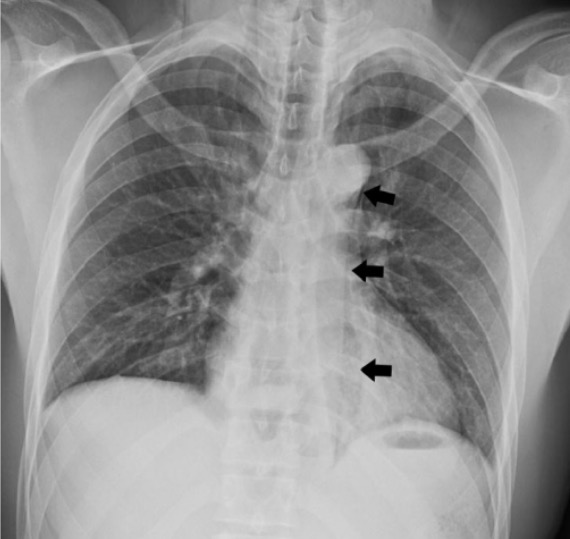
  • Chest X-ray X-ray Penetrating electromagnetic radiation emitted when the inner orbital electrons of an atom are excited and release radiant energy. X-ray wavelengths range from 1 pm to 10 nm. Hard x-rays are the higher energy, shorter wavelength x-rays. Soft x-rays or grenz rays are less energetic and longer in wavelength. The short wavelength end of the x-ray spectrum overlaps the gamma rays wavelength range. The distinction between gamma rays and x-rays is based on their radiation source. Pulmonary Function Tests:
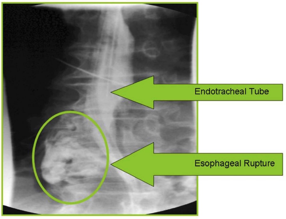
  • Contrast esophagography:
    • Diagnostic
    • Use water-soluble contrast ( Gastrografin Gastrografin Computed Tomography (CT)):
      • For initial study
      • Finding: contrast leakage from the esophagus Esophagus The esophagus is a muscular tube-shaped organ of around 25 centimeters in length that connects the pharynx to the stomach. The organ extends from approximately the 6th cervical vertebra to the 11th thoracic vertebra and can be divided grossly into 3 parts: the cervical part, the thoracic part, and the abdominal part. Esophagus: Anatomy to the mediastinum Mediastinum The mediastinum is the thoracic area between the 2 pleural cavities. The mediastinum contains vital structures of the circulatory, respiratory, digestive, and nervous systems including the heart and esophagus, and major thoracic vessels. Mediastinum and Great Vessels: Anatomy
    • Barium:
      • Not used initially due to the risk of developing acute mediastinitis Acute Mediastinitis Mediastinitis
      • Utilized if water-soluble study is negative (as barium demonstrates small perforation Perforation A pathological hole in an organ, blood vessel or other soft part of the body, occurring in the absence of external force. Esophagitis(s) well)
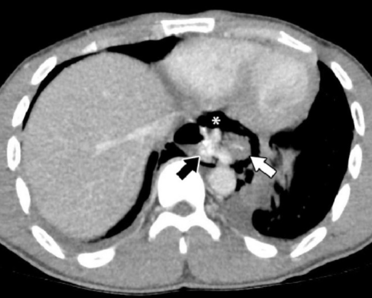
  • CT scan of the chest:
    • Performed when:
      • Chest X-ray X-ray Penetrating electromagnetic radiation emitted when the inner orbital electrons of an atom are excited and release radiant energy. X-ray wavelengths range from 1 pm to 10 nm. Hard x-rays are the higher energy, shorter wavelength x-rays. Soft x-rays or grenz rays are less energetic and longer in wavelength. The short wavelength end of the x-ray spectrum overlaps the gamma rays wavelength range. The distinction between gamma rays and x-rays is based on their radiation source. Pulmonary Function Tests or esophagography is inconclusive
      • Patient is unstable
    • Finding(s):
  • Upper endoscopy Endoscopy Procedures of applying endoscopes for disease diagnosis and treatment. Endoscopy involves passing an optical instrument through a small incision in the skin i.e., percutaneous; or through a natural orifice and along natural body pathways such as the digestive tract; and/or through an incision in the wall of a tubular structure or organ, i.e. Transluminal, to examine or perform surgery on the interior parts of the body. Gastroesophageal Reflux Disease (GERD):
    • Performed when:
      • Location of the perforation Perforation A pathological hole in an organ, blood vessel or other soft part of the body, occurring in the absence of external force. Esophagitis is not clear
      • CT scan is inconclusive
    • Caution must be taken as insufflation of air can lead to extension Extension Examination of the Upper Limbs of the perforation Perforation A pathological hole in an organ, blood vessel or other soft part of the body, occurring in the absence of external force. Esophagitis.

Management

Stabilization

Initial approach:

  • ABCDE survey:
    • Airway: Ensure the patency of the airway Airway ABCDE Assessment.
    • Breathing: Ensure proper ventilation Ventilation The total volume of gas inspired or expired per unit of time, usually measured in liters per minute. Ventilation: Mechanics of Breathing is occurring.
    • Circulation: Measure blood pressure and pulse, and administer IV fluids IV fluids Intravenous fluids are one of the most common interventions administered in medicine to approximate physiologic bodily fluids. Intravenous fluids are divided into 2 categories: crystalloid and colloid solutions. Intravenous fluids have a wide variety of indications, including intravascular volume expansion, electrolyte manipulation, and maintenance fluids. Intravenous Fluids
    • Disability: Perform basic neurologic examination.
    • Exposure: Search for injuries, and perform environmental control. 
  • Nothing by mouth
  • Broad-spectrum Broad-Spectrum Fluoroquinolones IV antibiotics
  • IV analgesic
  • IV proton pump Pump ACES and RUSH: Resuscitation Ultrasound Protocols inhibitor
  • Parenteral nutrition Parenteral nutrition The administering of nutrients for assimilation and utilization by a patient who cannot maintain adequate nutrition by enteral feeding alone. Nutrients are administered by a route other than the alimentary canal (e.g., intravenously, subcutaneously). Central Venous Catheter

Obtain surgical (including cardiothoracic) consult, as even stable patients Stable Patients Blunt Chest Trauma can deteriorate and require surgery.

Further intervention determined by:

  • Size and location of perforation Perforation A pathological hole in an organ, blood vessel or other soft part of the body, occurring in the absence of external force. Esophagitis
  • Comorbidities Comorbidities The presence of co-existing or additional diseases with reference to an initial diagnosis or with reference to the index condition that is the subject of study. Comorbidity may affect the ability of affected individuals to function and also their survival; it may be used as a prognostic indicator for length of hospital stay, cost factors, and outcome or survival. St. Louis Encephalitis Virus

Nonsurgical management

  • Indications:
    • Patient is stable with no signs of sepsis Sepsis Systemic inflammatory response syndrome with a proven or suspected infectious etiology. When sepsis is associated with organ dysfunction distant from the site of infection, it is called severe sepsis. When sepsis is accompanied by hypotension despite adequate fluid infusion, it is called septic shock. Sepsis and Septic Shock.
    • Perforation Perforation A pathological hole in an organ, blood vessel or other soft part of the body, occurring in the absence of external force. Esophagitis is contained:
      • Within the neck Neck The part of a human or animal body connecting the head to the rest of the body. Peritonsillar Abscess OR
      • Between visceral lung pleura Pleura The pleura is a serous membrane that lines the walls of the thoracic cavity and the surface of the lungs. This structure of mesodermal origin covers both lungs, the mediastinum, the thoracic surface of the diaphragm, and the inner part of the thoracic cage. The pleura is divided into a visceral pleura and parietal pleura. Pleura: Anatomy and mediastinum Mediastinum The mediastinum is the thoracic area between the 2 pleural cavities. The mediastinum contains vital structures of the circulatory, respiratory, digestive, and nervous systems including the heart and esophagus, and major thoracic vessels. Mediastinum and Great Vessels: Anatomy
    • Perforation Perforation A pathological hole in an organ, blood vessel or other soft part of the body, occurring in the absence of external force. Esophagitis site is located outside of the abdomen.
    • Does not involve a neoplasm or obstruction
    • Contrast drains back to the esophagus Esophagus The esophagus is a muscular tube-shaped organ of around 25 centimeters in length that connects the pharynx to the stomach. The organ extends from approximately the 6th cervical vertebra to the 11th thoracic vertebra and can be divided grossly into 3 parts: the cervical part, the thoracic part, and the abdominal part. Esophagus: Anatomy
    • Contrast esophagography is available for follow-up assessment.
    • A skilled thoracic surgeon is readily available.
  • Critical care monitoring
  • Any sign of sepsis Sepsis Systemic inflammatory response syndrome with a proven or suspected infectious etiology. When sepsis is associated with organ dysfunction distant from the site of infection, it is called severe sepsis. When sepsis is accompanied by hypotension despite adequate fluid infusion, it is called septic shock. Sepsis and Septic Shock or deterioration → immediate surgery

Surgical management

Indications:

  • Patient is hemodynamically unstable.
  • Patient with intraabdominal esophageal perforation Perforation A pathological hole in an organ, blood vessel or other soft part of the body, occurring in the absence of external force. Esophagitis
  • Patient with esophageal malignancy Malignancy Hemothorax
  • Respiratory complications

Procedure:

  • Surgical repair of perforation Perforation A pathological hole in an organ, blood vessel or other soft part of the body, occurring in the absence of external force. Esophagitis ( standard of care Standard of care The minimum acceptable patient care, based on statutes, court decisions, policies, or professional guidelines. Malpractice)
  • Esophageal stent: may be used in select patients Patients Individuals participating in the health care system for the purpose of receiving therapeutic, diagnostic, or preventive procedures. Clinician–Patient Relationship (with severe comorbidities Comorbidities The presence of co-existing or additional diseases with reference to an initial diagnosis or with reference to the index condition that is the subject of study. Comorbidity may affect the ability of affected individuals to function and also their survival; it may be used as a prognostic indicator for length of hospital stay, cost factors, and outcome or survival. St. Louis Encephalitis Virus and/or cannot undergo surgery)
  • Endoscopic clipping: an option in small perforations that can be corrected with minimal tension
  • Esophagectomy Esophagectomy Excision of part (partial) or all (total) of the esophagus. Esophageal Cancer is used as a last resort.
Esophagotomy

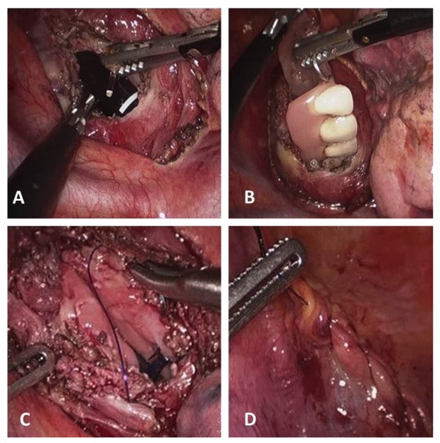
Esophageal perforation by foreign body (dental prosthesis):
A: esophagotomy
B: removal of dental prosthesis
C and D: suture of the esophageal wall and mediastinal pleura

Image: “Thoracoscopic removal of dental prosthesis” by Department of Biomedical Sciences for Health, Division of General Surgery, University of Milano Medical School, IRCCS Policlinico San Donato, Via Morandi 30, 20097, San Donato Milanese, (Milano), Italy. License: CC BY 2.0

Differential Diagnosis

  • Mallory-Weiss syndrome Mallory-Weiss Syndrome Mallory-Weiss syndrome (MWS) is defined by the presence of longitudinal mucosal lacerations in the distal esophagus and proximal stomach, which are usually associated with any action that provokes a sudden rise in intraluminal esophageal pressure, such as forceful or recurrent retching, vomiting, coughing, or straining. Mallory-Weiss Syndrome (Mallory-Weiss Tear): a superficial longitudinal laceration Laceration Torn, ragged, mangled wounds. Blunt Chest Trauma of the esophageal mucosa Esophageal Mucosa Circular innermost layer of the esophagus wall that mediates esophageal peristalsis which pushes ingested food bolus toward the stomach. Esophagus: Anatomy and/or submucosa at the gastroesophageal junction Gastroesophageal junction The area covering the terminal portion of esophagus and the beginning of stomach at the cardiac orifice. Esophagus: Anatomy. As with esophageal perforation Perforation A pathological hole in an organ, blood vessel or other soft part of the body, occurring in the absence of external force. Esophagitis, the condition may be caused by increased intrathoracic pressure (e.g., by vomiting Vomiting The forcible expulsion of the contents of the stomach through the mouth. Hypokalemia). The condition presents with hematemesis Hematemesis Vomiting of blood that is either fresh bright red, or older ‘coffee-ground’ in character. It generally indicates bleeding of the upper gastrointestinal tract. Mallory-Weiss Syndrome (Mallory-Weiss Tear). Diagnosis is mostly clinical and treatment is supportive, with IV fluids IV fluids Intravenous fluids are one of the most common interventions administered in medicine to approximate physiologic bodily fluids. Intravenous fluids are divided into 2 categories: crystalloid and colloid solutions. Intravenous fluids have a wide variety of indications, including intravascular volume expansion, electrolyte manipulation, and maintenance fluids. Intravenous Fluids and analgesics. 
  • Esophageal spasm: also presents with dysphagia Dysphagia Dysphagia is the subjective sensation of difficulty swallowing. Symptoms can range from a complete inability to swallow, to the sensation of solids or liquids becoming “stuck.” Dysphagia is classified as either oropharyngeal or esophageal, with esophageal dysphagia having 2 sub-types: functional and mechanical. Dysphagia to solids and liquids but is associated with sudden onset of chest pain Pain An unpleasant sensation induced by noxious stimuli which are detected by nerve endings of nociceptive neurons. Pain: Types and Pathways that is not exertion related. Two types of this condition are distal esophageal spasm and hypercontractile (jackhammer) esophagus Esophagus The esophagus is a muscular tube-shaped organ of around 25 centimeters in length that connects the pharynx to the stomach. The organ extends from approximately the 6th cervical vertebra to the 11th thoracic vertebra and can be divided grossly into 3 parts: the cervical part, the thoracic part, and the abdominal part. Esophagus: Anatomy. Manometry Manometry Measurement of the pressure or tension of liquids or gases with a manometer. Achalasia shows characteristic esophageal contractions with normal relaxation of the esophagogastric junction Esophagogastric junction The area covering the terminal portion of esophagus and the beginning of stomach at the cardiac orifice. Esophagus: Anatomy. Management involves treating reflux disease (if present) and a trial of calcium Calcium A basic element found in nearly all tissues. It is a member of the alkaline earth family of metals with the atomic symbol ca, atomic number 20, and atomic weight 40. Calcium is the most abundant mineral in the body and combines with phosphorus to form calcium phosphate in the bones and teeth. It is essential for the normal functioning of nerves and muscles and plays a role in blood coagulation (as factor IV) and in many enzymatic processes. Electrolytes channel blocker.
  • Gastroesophageal reflux disease Gastroesophageal Reflux Disease Gastroesophageal reflux disease (GERD) occurs when the stomach acid frequently flows back into the esophagus. This backwash (acid reflux) can irritate the lining of the esophagus, causing symptoms such as retrosternal burning pain (heartburn). Gastroesophageal Reflux Disease (GERD) ( GERD GERD Gastroesophageal reflux disease (GERD) occurs when the stomach acid frequently flows back into the esophagus. This backwash (acid reflux) can irritate the lining of the esophagus, causing symptoms such as retrosternal burning pain (heartburn). Gastroesophageal Reflux Disease (GERD)): symptoms of heartburn Heartburn Substernal pain or burning sensation, usually associated with regurgitation of gastric juice into the esophagus. Gastroesophageal Reflux Disease (GERD) and regurgitation Regurgitation Gastroesophageal Reflux Disease (GERD) caused by the reflux of stomach Stomach The stomach is a muscular sac in the upper left portion of the abdomen that plays a critical role in digestion. The stomach develops from the foregut and connects the esophagus with the duodenum. Structurally, the stomach is C-shaped and forms a greater and lesser curvature and is divided grossly into regions: the cardia, fundus, body, and pylorus. Stomach: Anatomy contents. The patient usually complains of a burning epigastric pain Epigastric pain Mallory-Weiss Syndrome (Mallory-Weiss Tear) radiating up the chest with a sour or metallic taste in the mouth. The disease is due to inappropriate relaxation of the lower esophageal sphincter Lower Esophageal Sphincter Esophagus: Anatomy. Proton pump Pump ACES and RUSH: Resuscitation Ultrasound Protocols inhibitors are used to control symptoms of GERD GERD Gastroesophageal reflux disease (GERD) occurs when the stomach acid frequently flows back into the esophagus. This backwash (acid reflux) can irritate the lining of the esophagus, causing symptoms such as retrosternal burning pain (heartburn). Gastroesophageal Reflux Disease (GERD).

References

  1. Ezenkwele, U., Long, C. (2016). Esophageal Rupture and Tears in Emergency Medicine Clinical Presentation. Medscape. Retrieved Apr 11, 2021, from https://emedicine.medscape.com/article/775165-clinical
  2. Praveen, R. K. (2019). Boerhaave syndrome. Medscape. https://emedicine.medscape.com/article/171683-overview
  3. Raymond, D. (2020). Overview of esophageal perforation due to blunt or penetrating trauma. UpToDate. Retrieved March 20, 2021, from https://www.uptodate.com/contents/overview-of-esophageal-perforation-due-to-blunt-or-penetrating-trauma
  4. Raymond, D. (2020). Surgical management of esophageal perforation. UpToDate. Retrieved March 20, 2021, from https://www.uptodate.com/contents/surgical-management-of-esophageal-perforation
  5. Triadafilopoulos, G. (2020). Boerhaave syndrome: Effort rupture of the esophagus. UpToDate. Retrieved March 20, 2021, from https://www.uptodate.com/contents/boerhaave-syndrome-effort-rupture-of-the-esophagus

Create your free account or log in to continue reading!

Sign up now and get free access to Lecturio with concept pages, medical videos, and questions for your medical education.

User Reviews

Unwrap New Skills This Holiday 🎄 Save 30% on all plans now!

Details