Advertisement

Advertisement

Advertisement

Advertisement

Erythrocytes: Histology

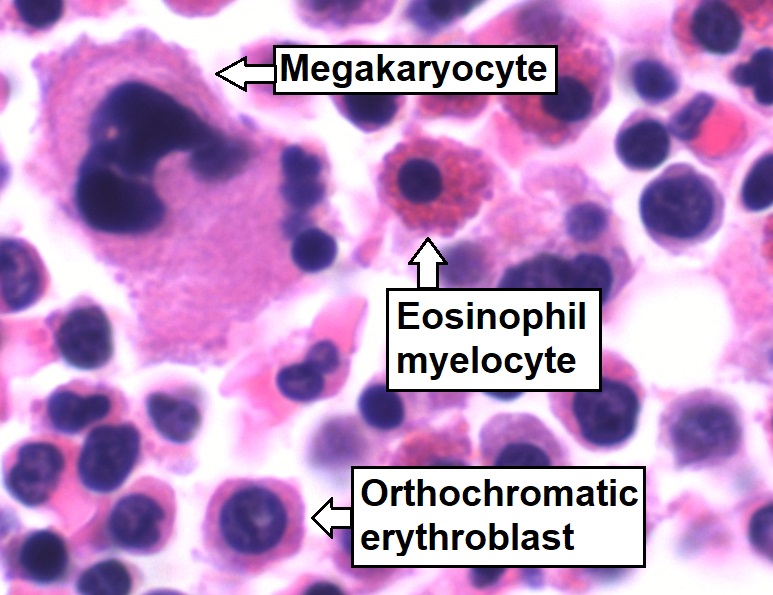
Erythrocytes, or red blood cells (RBCs), are the most abundant cells in the blood. While erythrocytes in the fetus are initially produced in the yolk sac Yolk Sac The first of four extra-embryonic membranes to form during embryogenesis. In reptiles and birds, it arises from endoderm and mesoderm to incorporate the egg yolk into the digestive tract for nourishing the embryo. In placental mammals, its nutritional function is vestigial; however, it is the source of intestinal mucosa; blood cells; and germ cells. It is sometimes called the vitelline sac, which should not be confused with the vitelline membrane of the egg. Embryoblast and Trophoblast Development then the liver Liver The liver is the largest gland in the human body. The liver is found in the superior right quadrant of the abdomen and weighs approximately 1.5 kilograms. Its main functions are detoxification, metabolism, nutrient storage (e.g., iron and vitamins), synthesis of coagulation factors, formation of bile, filtration, and storage of blood. Liver: Anatomy, the bone marrow Bone marrow The soft tissue filling the cavities of bones. Bone marrow exists in two types, yellow and red. Yellow marrow is found in the large cavities of large bones and consists mostly of fat cells and a few primitive blood cells. Red marrow is a hematopoietic tissue and is the site of production of erythrocytes and granular leukocytes. Bone marrow is made up of a framework of connective tissue containing branching fibers with the frame being filled with marrow cells. Bone Marrow: Composition and Hematopoiesis eventually becomes the main site of production. Erythropoiesis starts with hematopoietic stem cells Hematopoietic stem cells Progenitor cells from which all blood cells derived. They are found primarily in the bone marrow and also in small numbers in the peripheral blood. Bone Marrow: Composition and Hematopoiesis, which develop into lineage-committed progenitors and differentiate into mature RBCs. The process occurs in stages, and extrusion of the nuclei and organelles Organelles A cell is a complex unit that performs several complex functions. An organelle is a specialized subunit within a cell that fulfills a specific role or function. Organelles are enclosed within their own lipid bilayers or are unbound by membranes. The Cell: Organelles occurs prior to maturation. Thus, mature RBCs lack nuclei and have a biconcave shape. RBCs carry hemoglobin, and the shape allows efficient oxygen transport. Billions of RBCs are produced daily, as the life span is 120 days. Senescent or deformed RBCs are removed by macrophages Macrophages The relatively long-lived phagocytic cell of mammalian tissues that are derived from blood monocytes. Main types are peritoneal macrophages; alveolar macrophages; histiocytes; kupffer cells of the liver; and osteoclasts. They may further differentiate within chronic inflammatory lesions to epithelioid cells or may fuse to form foreign body giant cells or langhans giant cells. Innate Immunity: Phagocytes and Antigen Presentation in the spleen Spleen The spleen is the largest lymphoid organ in the body, located in the LUQ of the abdomen, superior to the left kidney and posterior to the stomach at the level of the 9th-11th ribs just below the diaphragm. The spleen is highly vascular and acts as an important blood filter, cleansing the blood of pathogens and damaged erythrocytes. Spleen: Anatomy, liver Liver The liver is the largest gland in the human body. The liver is found in the superior right quadrant of the abdomen and weighs approximately 1.5 kilograms. Its main functions are detoxification, metabolism, nutrient storage (e.g., iron and vitamins), synthesis of coagulation factors, formation of bile, filtration, and storage of blood. Liver: Anatomy, and bone marrow Bone marrow The soft tissue filling the cavities of bones. Bone marrow exists in two types, yellow and red. Yellow marrow is found in the large cavities of large bones and consists mostly of fat cells and a few primitive blood cells. Red marrow is a hematopoietic tissue and is the site of production of erythrocytes and granular leukocytes. Bone marrow is made up of a framework of connective tissue containing branching fibers with the frame being filled with marrow cells. Bone Marrow: Composition and Hematopoiesis.

Last updated: May 17, 2024

Editorial responsibility: Stanley Oiseth, Lindsay Jones, Evelin Maza

Advertisement

Advertisement

Advertisement

Advertisement

Advertisement

Advertisement

Advertisement

Advertisement

Overview

Definition and description

Erythrocytes, also called red blood cells (RBCs), are terminally differentiated structures lacking nuclei but filled with oxygen-carrying hemoglobin. Erythrocytes are the most abundant cells in the blood.
  • Diameter: 7.5 μm
  • Biconcave shape:
    • Gives RBCs high surface-to-volume ratio 
    • Sets up most hemoglobin within a short distance from the cell surface, facilitating efficient oxygen transport 
  • Life span: 120 days

Structure

  • Flexible biconcave structure: 
    • Easily bends, permitting passage through small capillaries Capillaries Capillaries are the primary structures in the circulatory system that allow the exchange of gas, nutrients, and other materials between the blood and the extracellular fluid (ECF). Capillaries are the smallest of the blood vessels. Because a capillary diameter is so small, only 1 RBC may pass through at a time. Capillaries: Histology
    • Membrane proteins Proteins Linear polypeptides that are synthesized on ribosomes and may be further modified, crosslinked, cleaved, or assembled into complex proteins with several subunits. The specific sequence of amino acids determines the shape the polypeptide will take, during protein folding, and the function of the protein. Energy Homeostasis, including spectrin and ankyrin, stabilize the membrane to keep the shape and elasticity Elasticity Resistance and recovery from distortion of shape. Skeletal Muscle Contraction of RBCs.
  • Components:
    • No nuclei (without nuclei, defective proteins Proteins Linear polypeptides that are synthesized on ribosomes and may be further modified, crosslinked, cleaved, or assembled into complex proteins with several subunits. The specific sequence of amino acids determines the shape the polypeptide will take, during protein folding, and the function of the protein. Energy Homeostasis cannot be replaced)
    • Without most organelles Organelles A cell is a complex unit that performs several complex functions. An organelle is a specialized subunit within a cell that fulfills a specific role or function. Organelles are enclosed within their own lipid bilayers or are unbound by membranes. The Cell: Organelles 
    • During erythropoiesis, which takes place in the red bone marrow Red bone marrow Bone Marrow: Composition and Hematopoiesis, precursors lose nuclei and organelles Organelles A cell is a complex unit that performs several complex functions. An organelle is a specialized subunit within a cell that fulfills a specific role or function. Organelles are enclosed within their own lipid bilayers or are unbound by membranes. The Cell: Organelles prior to release Release Release of a virus from the host cell following virus assembly and maturation. Egress can occur by host cell lysis, exocytosis, or budding through the plasma membrane. Virology into circulation Circulation The movement of the blood as it is pumped through the cardiovascular system. ABCDE Assessment.
  • Senescent or deformed RBCs are removed by macrophages Macrophages The relatively long-lived phagocytic cell of mammalian tissues that are derived from blood monocytes. Main types are peritoneal macrophages; alveolar macrophages; histiocytes; kupffer cells of the liver; and osteoclasts. They may further differentiate within chronic inflammatory lesions to epithelioid cells or may fuse to form foreign body giant cells or langhans giant cells. Innate Immunity: Phagocytes and Antigen Presentation of the liver Liver The liver is the largest gland in the human body. The liver is found in the superior right quadrant of the abdomen and weighs approximately 1.5 kilograms. Its main functions are detoxification, metabolism, nutrient storage (e.g., iron and vitamins), synthesis of coagulation factors, formation of bile, filtration, and storage of blood. Liver: Anatomy, spleen Spleen The spleen is the largest lymphoid organ in the body, located in the LUQ of the abdomen, superior to the left kidney and posterior to the stomach at the level of the 9th-11th ribs just below the diaphragm. The spleen is highly vascular and acts as an important blood filter, cleansing the blood of pathogens and damaged erythrocytes. Spleen: Anatomy, and bone marrow Bone marrow The soft tissue filling the cavities of bones. Bone marrow exists in two types, yellow and red. Yellow marrow is found in the large cavities of large bones and consists mostly of fat cells and a few primitive blood cells. Red marrow is a hematopoietic tissue and is the site of production of erythrocytes and granular leukocytes. Bone marrow is made up of a framework of connective tissue containing branching fibers with the frame being filled with marrow cells. Bone Marrow: Composition and Hematopoiesis.
  • Processing of components after degradation:
    • Globin (from hemoglobin): broken into amino acids Amino acids Organic compounds that generally contain an amino (-NH2) and a carboxyl (-COOH) group. Twenty alpha-amino acids are the subunits which are polymerized to form proteins. Basics of Amino Acids and reused by the bone marrow Bone marrow The soft tissue filling the cavities of bones. Bone marrow exists in two types, yellow and red. Yellow marrow is found in the large cavities of large bones and consists mostly of fat cells and a few primitive blood cells. Red marrow is a hematopoietic tissue and is the site of production of erythrocytes and granular leukocytes. Bone marrow is made up of a framework of connective tissue containing branching fibers with the frame being filled with marrow cells. Bone Marrow: Composition and Hematopoiesis for new RBCs
    • Iron Iron A metallic element with atomic symbol fe, atomic number 26, and atomic weight 55. 85. It is an essential constituent of hemoglobins; cytochromes; and iron-binding proteins. It plays a role in cellular redox reactions and in the transport of oxygen. Trace Elements: stored in liver Liver The liver is the largest gland in the human body. The liver is found in the superior right quadrant of the abdomen and weighs approximately 1.5 kilograms. Its main functions are detoxification, metabolism, nutrient storage (e.g., iron and vitamins), synthesis of coagulation factors, formation of bile, filtration, and storage of blood. Liver: Anatomy or spleen Spleen The spleen is the largest lymphoid organ in the body, located in the LUQ of the abdomen, superior to the left kidney and posterior to the stomach at the level of the 9th-11th ribs just below the diaphragm. The spleen is highly vascular and acts as an important blood filter, cleansing the blood of pathogens and damaged erythrocytes. Spleen: Anatomy or reused for new RBCs in the bone marrow Bone marrow The soft tissue filling the cavities of bones. Bone marrow exists in two types, yellow and red. Yellow marrow is found in the large cavities of large bones and consists mostly of fat cells and a few primitive blood cells. Red marrow is a hematopoietic tissue and is the site of production of erythrocytes and granular leukocytes. Bone marrow is made up of a framework of connective tissue containing branching fibers with the frame being filled with marrow cells. Bone Marrow: Composition and Hematopoiesis
    • Nonheme iron Iron A metallic element with atomic symbol fe, atomic number 26, and atomic weight 55. 85. It is an essential constituent of hemoglobins; cytochromes; and iron-binding proteins. It plays a role in cellular redox reactions and in the transport of oxygen. Trace Elements: become bilirubin Bilirubin A bile pigment that is a degradation product of heme. Heme Metabolism, biliverdin Biliverdin 1, 3, 6, 7-tetramethyl-4, 5-dicarboxyethyl-2, 8-divinylbilenone. Biosynthesized from hemoglobin as a precursor of bilirubin. Heme Metabolism
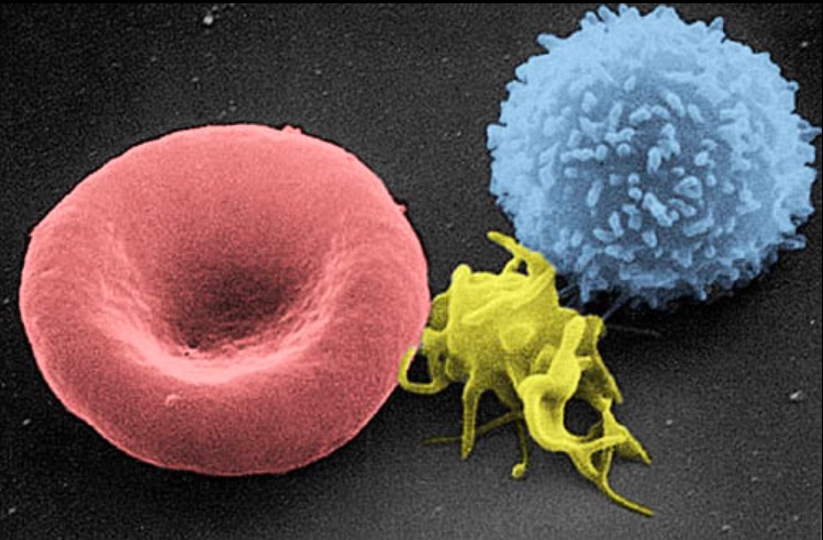
Electron micrograph of blood cells

Scanning electron micrograph of a blood cell:
Left to right: human RBC, thrombocyte (platelet), and leukocyte
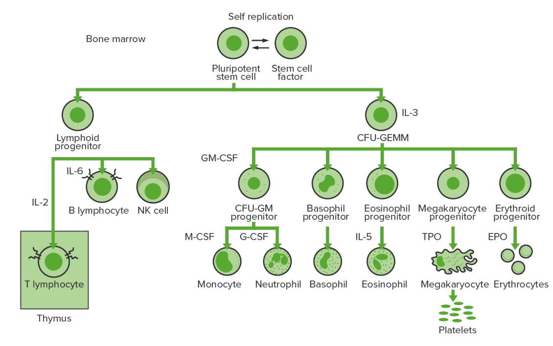
Image: “Electron micrograph of blood cells” by Electron Microscopy Facility at The National Cancer Institute at Frederick. License: Public Domain

Erythropoiesis

RBC production

  • Hematopoiesis Hematopoiesis The development and formation of various types of blood cells. Hematopoiesis can take place in the bone marrow (medullary) or outside the bone marrow (extramedullary hematopoiesis). Bone Marrow: Composition and Hematopoiesis:
    • 1st to 2nd month in utero: mesoderm Mesoderm The middle germ layer of an embryo derived from three paired mesenchymal aggregates along the neural tube. Gastrulation and Neurulation of the yolk sac Yolk Sac The first of four extra-embryonic membranes to form during embryogenesis. In reptiles and birds, it arises from endoderm and mesoderm to incorporate the egg yolk into the digestive tract for nourishing the embryo. In placental mammals, its nutritional function is vestigial; however, it is the source of intestinal mucosa; blood cells; and germ cells. It is sometimes called the vitelline sac, which should not be confused with the vitelline membrane of the egg. Embryoblast and Trophoblast Development
    • By the 2nd month, hematopoiesis Hematopoiesis The development and formation of various types of blood cells. Hematopoiesis can take place in the bone marrow (medullary) or outside the bone marrow (extramedullary hematopoiesis). Bone Marrow: Composition and Hematopoiesis moves to the liver Liver The liver is the largest gland in the human body. The liver is found in the superior right quadrant of the abdomen and weighs approximately 1.5 kilograms. Its main functions are detoxification, metabolism, nutrient storage (e.g., iron and vitamins), synthesis of coagulation factors, formation of bile, filtration, and storage of blood. Liver: Anatomy (and spleen Spleen The spleen is the largest lymphoid organ in the body, located in the LUQ of the abdomen, superior to the left kidney and posterior to the stomach at the level of the 9th-11th ribs just below the diaphragm. The spleen is highly vascular and acts as an important blood filter, cleansing the blood of pathogens and damaged erythrocytes. Spleen: Anatomy).
    • By the 5th month, hematopoiesis Hematopoiesis The development and formation of various types of blood cells. Hematopoiesis can take place in the bone marrow (medullary) or outside the bone marrow (extramedullary hematopoiesis). Bone Marrow: Composition and Hematopoiesis occurs in the bone marrow Bone marrow The soft tissue filling the cavities of bones. Bone marrow exists in two types, yellow and red. Yellow marrow is found in the large cavities of large bones and consists mostly of fat cells and a few primitive blood cells. Red marrow is a hematopoietic tissue and is the site of production of erythrocytes and granular leukocytes. Bone marrow is made up of a framework of connective tissue containing branching fibers with the frame being filled with marrow cells. Bone Marrow: Composition and Hematopoiesis and the bone marrow Bone marrow The soft tissue filling the cavities of bones. Bone marrow exists in two types, yellow and red. Yellow marrow is found in the large cavities of large bones and consists mostly of fat cells and a few primitive blood cells. Red marrow is a hematopoietic tissue and is the site of production of erythrocytes and granular leukocytes. Bone marrow is made up of a framework of connective tissue containing branching fibers with the frame being filled with marrow cells. Bone Marrow: Composition and Hematopoiesis becomes the predominant source of blood cells.
  • As with other blood cells, erythropoiesis starts with multipotent hematopoietic stem cells Hematopoietic stem cells Progenitor cells from which all blood cells derived. They are found primarily in the bone marrow and also in small numbers in the peripheral blood. Bone Marrow: Composition and Hematopoiesis (HSCs).
  • HSCs → multipotent progenitor cells Multipotent progenitor cells Specialized stem cells that are committed to give rise to cells that have a particular function; examples are myoblasts; myeloid progenitor cells; and skin stem cells. Bone Marrow: Composition and Hematopoiesis (MPPs) → common myeloid progenitor → burst-forming unit erythroid (BFU-E) → erythrocyte maturation 
Bone marrow hematopoiesis

Bone-marrow hematopoiesis: proliferation and differentiation of the formed elements of blood.
CFU-GEMM: colony-forming unit–granulocyte, erythrocyte, monocyte, megakaryocyte
CFU-GM: colony-forming unit–granulocyte-macrophage
GM-CSF: granulocyte-macrophage colony-stimulating factor
M-CSF: macrophage colony-stimulating factor
G-CSF: granulocyte colony-stimulating factor
NK: natural killer
TPO: thrombopoietin
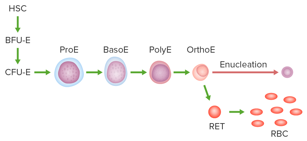
Image by Lecturio. License: CC BY-NC-SA 4.0

Stages of erythrocyte differentiation

  1. Proerythroblast:
    • Large, central, pale-staining nucleus Nucleus Within a eukaryotic cell, a membrane-limited body which contains chromosomes and one or more nucleoli (cell nucleolus). The nuclear membrane consists of a double unit-type membrane which is perforated by a number of pores; the outermost membrane is continuous with the endoplasmic reticulum. A cell may contain more than one nucleus. The Cell: Organelles with 1–2 large nucleoli
    • Polyribosomes (which provide mild basophilia) in the cytoplasm synthesize hemoglobin.
    • 14–20 μm in diameter
  2. Basophilic erythroblast: 
    • Deep blue (more intense basophilia), smaller cell than proerythroblast
    • Smaller nuclei and patchy chromatin Chromatin The material of chromosomes. It is a complex of dna; histones; and nonhistone proteins found within the nucleus of a cell. DNA Types and Structure
    • 13–16 μm in diameter
  3. Polychromatophilic erythroblast:
    • Mix of staining affinities: basophilic (from polyribosomes) and acidophilic (from increasing hemoglobin)
    • Nuclei with checkerboard chromatin Chromatin The material of chromosomes. It is a complex of dna; histones; and nonhistone proteins found within the nucleus of a cell. DNA Types and Structure
    • 12–15 μm in diameter
  4.  Orthochromatic erythroblast (normoblast): 
    • Acidophilic cytoplasm, trace basophilia
    • Small eccentric nuclei with condensed chromatin Chromatin The material of chromosomes. It is a complex of dna; histones; and nonhistone proteins found within the nucleus of a cell. DNA Types and Structure → ends with extrusion of degenerated or pyknotic nuclei
    • 8–10 μm in diameter
  5. Reticulocyte:
    • Almost similar to mature RBC
    • Difficult to identify without supravital stain
    • If stained with brilliant cresyl blue, residual polyribosomes create a blue-staining cytoplasmic precipitate. 
    • Maturation to erythrocyte occurs in 24–48 hours.
    • Enzymatic digestion Digestion Digestion refers to the process of the mechanical and chemical breakdown of food into smaller particles, which can then be absorbed and utilized by the body. Digestion and Absorption or expulsion of organelles Organelles A cell is a complex unit that performs several complex functions. An organelle is a specialized subunit within a cell that fulfills a specific role or function. Organelles are enclosed within their own lipid bilayers or are unbound by membranes. The Cell: Organelles 
  6. Mature RBC: 
    • Without nuclei
    • With biconcave shape

Regulation

Table: Regulation of erythropoiesis
Cytokines Cytokines Non-antibody proteins secreted by inflammatory leukocytes and some non-leukocytic cells, that act as intercellular mediators. They differ from classical hormones in that they are produced by a number of tissue or cell types rather than by specialized glands. They generally act locally in a paracrine or autocrine rather than endocrine manner. Adaptive Immune Response and growth factors Activities Source
SCF Stimulates all hematopoietic progenitor cells Bone marrow Bone marrow The soft tissue filling the cavities of bones. Bone marrow exists in two types, yellow and red. Yellow marrow is found in the large cavities of large bones and consists mostly of fat cells and a few primitive blood cells. Red marrow is a hematopoietic tissue and is the site of production of erythrocytes and granular leukocytes. Bone marrow is made up of a framework of connective tissue containing branching fibers with the frame being filled with marrow cells. Bone Marrow: Composition and Hematopoiesis stromal cells
GM-CSF GM-CSF An acidic glycoprotein of mw 23 kda with internal disulfide bonds. The protein is produced in response to a number of inflammatory mediators by mesenchymal cells present in the hemopoietic environment and at peripheral sites of inflammation. GM-CSF is able to stimulate the production of neutrophilic granulocytes, macrophages, and mixed granulocyte-macrophage colonies from bone marrow cells and can stimulate the formation of eosinophil colonies from fetal liver progenitor cells. GM-CSF can also stimulate some functional activities in mature granulocytes and macrophages. White Myeloid Cells: Histology Stimulates myeloid progenitor cells Myeloid progenitor cells Stem cells derived from hematopoietic stem cells. Derived from these myeloid progenitor cells are the megakaryocytes; erythroid cells; myeloid cells; and some dendritic cells. Acute Myeloid Leukemia Endothelial cells, T cells T cells Lymphocytes responsible for cell-mediated immunity. Two types have been identified – cytotoxic (t-lymphocytes, cytotoxic) and helper T-lymphocytes (t-lymphocytes, helper-inducer). They are formed when lymphocytes circulate through the thymus gland and differentiate to thymocytes. When exposed to an antigen, they divide rapidly and produce large numbers of new T cells sensitized to that antigen. T cells: Types and Functions
EPO Stimulates erythropoiesis, including differentiation Kidney, liver Liver The liver is the largest gland in the human body. The liver is found in the superior right quadrant of the abdomen and weighs approximately 1.5 kilograms. Its main functions are detoxification, metabolism, nutrient storage (e.g., iron and vitamins), synthesis of coagulation factors, formation of bile, filtration, and storage of blood. Liver: Anatomy
IL-3 Mitogen for all granulocyte and megakaryocyte Megakaryocyte Very large bone marrow cells which release mature blood platelets. Platelets: Histology/erythrocyte progenitor cells T helper cells
EPO: erythropoietin
GM-CSF: granulocyte macrophage colony-stimulating factor
SCF: stem cell factor

Other factors:

  • Iron Iron A metallic element with atomic symbol fe, atomic number 26, and atomic weight 55. 85. It is an essential constituent of hemoglobins; cytochromes; and iron-binding proteins. It plays a role in cellular redox reactions and in the transport of oxygen. Trace Elements
  • Vitamin B12
  • Intrinsic factor Intrinsic factor A glycoprotein secreted by the cells of the gastric glands that is required for the absorption of vitamin B 12 (cyanocobalamin). Deficiency of intrinsic factor leads to vitamin B12 deficiency and anemia, pernicious. Gastritis
  • Folic acid
  • Oxygen level

Clinical Relevance

  • Anemia Anemia Anemia is a condition in which individuals have low Hb levels, which can arise from various causes. Anemia is accompanied by a reduced number of RBCs and may manifest with fatigue, shortness of breath, pallor, and weakness. Subtypes are classified by the size of RBCs, chronicity, and etiology. Anemia: Overview and Types: decrease in total number of RBCs, hemoglobin, or circulating RBC mass Mass Three-dimensional lesion that occupies a space within the breast Imaging of the Breast. Anemia Anemia Anemia is a condition in which individuals have low Hb levels, which can arise from various causes. Anemia is accompanied by a reduced number of RBCs and may manifest with fatigue, shortness of breath, pallor, and weakness. Subtypes are classified by the size of RBCs, chronicity, and etiology. Anemia: Overview and Types is usually reflected in decreased hemoglobin and hematocrit Hematocrit The volume of packed red blood cells in a blood specimen. The volume is measured by centrifugation in a tube with graduated markings, or with automated blood cell counters. It is an indicator of erythrocyte status in disease. For example, anemia shows a low value; polycythemia, a high value. Neonatal Polycythemia, which can arise from reduced hematopoiesis Hematopoiesis The development and formation of various types of blood cells. Hematopoiesis can take place in the bone marrow (medullary) or outside the bone marrow (extramedullary hematopoiesis). Bone Marrow: Composition and Hematopoiesis, hemolysis, or blood loss.
  • Hemolytic anemia Hemolytic Anemia Hemolytic anemia (HA) is the term given to a large group of anemias that are caused by the premature destruction/hemolysis of circulating red blood cells (RBCs). Hemolysis can occur within (intravascular hemolysis) or outside the blood vessels (extravascular hemolysis). Hemolytic Anemia: anemia Anemia Anemia is a condition in which individuals have low Hb levels, which can arise from various causes. Anemia is accompanied by a reduced number of RBCs and may manifest with fatigue, shortness of breath, pallor, and weakness. Subtypes are classified by the size of RBCs, chronicity, and etiology. Anemia: Overview and Types due to destruction or premature Premature Childbirth before 37 weeks of pregnancy (259 days from the first day of the mother’s last menstrual period, or 245 days after fertilization). Necrotizing Enterocolitis clearance of RBCs. Hemolytic anemia Hemolytic Anemia Hemolytic anemia (HA) is the term given to a large group of anemias that are caused by the premature destruction/hemolysis of circulating red blood cells (RBCs). Hemolysis can occur within (intravascular hemolysis) or outside the blood vessels (extravascular hemolysis). Hemolytic Anemia can be characterized on the basis of increased clearance by the spleen Spleen The spleen is the largest lymphoid organ in the body, located in the LUQ of the abdomen, superior to the left kidney and posterior to the stomach at the level of the 9th-11th ribs just below the diaphragm. The spleen is highly vascular and acts as an important blood filter, cleansing the blood of pathogens and damaged erythrocytes. Spleen: Anatomy ( extravascular hemolysis Extravascular hemolysis Hemolytic Anemia) or damage caused by a narrowed vascular lumen ( intravascular hemolysis Intravascular hemolysis Hemolytic Anemia). Splenic clearance ( extravascular hemolysis Extravascular hemolysis Hemolytic Anemia) can be due to intrinsic abnormalities of the RBC (membrane, enzyme, hemoglobin) or extrinsic coating by the immune system Immune system The body’s defense mechanism against foreign organisms or substances and deviant native cells. It includes the humoral immune response and the cell-mediated response and consists of a complex of interrelated cellular, molecular, and genetic components. Primary Lymphatic Organs (e.g. ABO incompatibility ABO incompatibility Hemolytic Disease of the Fetus and Newborn).
  • Thalassemia Thalassemia Thalassemia is a hereditary cause of microcytic hypochromic anemia and results from a deficiency in either the α or β globin chains, resulting in hemoglobinopathy. The presentation of thalassemia depends on the number of defective chains present and can range from being asymptomatic to rendering the more severely affected patients to be transfusion dependent. Thalassemia: hereditary cause of microcytic, hypochromic anemia Anemia Anemia is a condition in which individuals have low Hb levels, which can arise from various causes. Anemia is accompanied by a reduced number of RBCs and may manifest with fatigue, shortness of breath, pallor, and weakness. Subtypes are classified by the size of RBCs, chronicity, and etiology. Anemia: Overview and Types. Thalassemia Thalassemia Thalassemia is a hereditary cause of microcytic hypochromic anemia and results from a deficiency in either the α or β globin chains, resulting in hemoglobinopathy. The presentation of thalassemia depends on the number of defective chains present and can range from being asymptomatic to rendering the more severely affected patients to be transfusion dependent. Thalassemia is a deficiency in either the alpha (⍺) or beta (𝛽) globin chain that results in a hemoglobinopathy. The presentation of thalassemia Thalassemia Thalassemia is a hereditary cause of microcytic hypochromic anemia and results from a deficiency in either the α or β globin chains, resulting in hemoglobinopathy. The presentation of thalassemia depends on the number of defective chains present and can range from being asymptomatic to rendering the more severely affected patients to be transfusion dependent. Thalassemia depends on the number of defective chains present. The consequent hemolytic anemia Hemolytic Anemia Hemolytic anemia (HA) is the term given to a large group of anemias that are caused by the premature destruction/hemolysis of circulating red blood cells (RBCs). Hemolysis can occur within (intravascular hemolysis) or outside the blood vessels (extravascular hemolysis). Hemolytic Anemia results in severe systemic symptoms, rendering more severely affected patients Patients Individuals participating in the health care system for the purpose of receiving therapeutic, diagnostic, or preventive procedures. Clinician–Patient Relationship transfusion-dependent.
  • Megaloblastic anemia Megaloblastic anemia Megaloblastic anemia is a subset of macrocytic anemias that arises because of impaired nucleic acid synthesis in erythroid precursors. This impairment leads to ineffective RBC production and intramedullary hemolysis that is characterized by large cells with arrested nuclear maturation. The most common causes are vitamin B12 and folic acid deficiencies. Megaloblastic Anemia: arises due to impaired nucleic acid synthesis Synthesis Polymerase Chain Reaction (PCR) in erythroid precursors. This impairment leads to ineffective RBC production and intramedullary hemolysis. Megaloblastic anemia Megaloblastic anemia Megaloblastic anemia is a subset of macrocytic anemias that arises because of impaired nucleic acid synthesis in erythroid precursors. This impairment leads to ineffective RBC production and intramedullary hemolysis that is characterized by large cells with arrested nuclear maturation. The most common causes are vitamin B12 and folic acid deficiencies. Megaloblastic Anemia is characterized by large cells with arrested nuclear maturation. The most common causes are vitamin B12 and folic acid deficiencies, which can be due to low dietary intake, underlying malabsorptive conditions and medications. Clinical presentation includes anemic and GI symptoms with neurologic manifestations more commonly seen in vitamin B12 deficiency. 
  • Aplastic anemia Aplastic Anemia Aplastic anemia (AA) is a rare, life-threatening condition characterized by pancytopenia and hypocellularity of the bone marrow (in the absence of any abnormal cells) reflecting damage to hematopoietic stem cells. Aplastic anemia can be acquired or inherited, however, most cases of AA are acquired and caused by autoimmune damage to hematopoietic stem cells. Aplastic Anemia ( AA AA Amyloidosis): rare life-threatening condition characterized by pancytopenia Pancytopenia Deficiency of all three cell elements of the blood, erythrocytes, leukocytes and platelets. Aplastic Anemia and hypocellularity of the bone marrow Bone marrow The soft tissue filling the cavities of bones. Bone marrow exists in two types, yellow and red. Yellow marrow is found in the large cavities of large bones and consists mostly of fat cells and a few primitive blood cells. Red marrow is a hematopoietic tissue and is the site of production of erythrocytes and granular leukocytes. Bone marrow is made up of a framework of connective tissue containing branching fibers with the frame being filled with marrow cells. Bone Marrow: Composition and Hematopoiesis, reflecting damage to the hematopoietic stem cells Hematopoietic stem cells Progenitor cells from which all blood cells derived. They are found primarily in the bone marrow and also in small numbers in the peripheral blood. Bone Marrow: Composition and Hematopoiesis (HSCs). Most cases of AA AA Amyloidosis are acquired and caused by autoimmune damage to HSCs. Known causes of acquired AAs include medications and chemicals, high doses of whole-body radiation Radiation Emission or propagation of acoustic waves (sound), electromagnetic energy waves (such as light; radio waves; gamma rays; or x-rays), or a stream of subatomic particles (such as electrons; neutrons; protons; or alpha particles). Osteosarcoma, viral infections Infections Invasion of the host organism by microorganisms or their toxins or by parasites that can cause pathological conditions or diseases. Chronic Granulomatous Disease, immune diseases, and pregnancy Pregnancy The status during which female mammals carry their developing young (embryos or fetuses) in utero before birth, beginning from fertilization to birth. Pregnancy: Diagnosis, Physiology, and Care. Inherited or constitutional syndromes associated with AA AA Amyloidosis include Fanconi anemia Fanconi anemia Congenital disorder affecting all bone marrow elements, resulting in anemia; leukopenia; and thrombopenia, and associated with cardiac, renal, and limb malformations as well as dermal pigmentary changes. Spontaneous chromosome breakage is a feature of this disease along with predisposition to leukemia. There are at least 7 complementation groups in Fanconi anemia: fanca, fancb, fancc, fancd1, fancd2, fance, fancf, fancg, and fancl. Aplastic Anemia, dyskeratosis Dyskeratosis Dermatomyositis congenita, and Down syndrome Down syndrome Down syndrome, or trisomy 21, is the most common chromosomal aberration and the most frequent genetic cause of developmental delay. Both boys and girls are affected and have characteristic craniofacial and musculoskeletal features, as well as multiple medical anomalies involving the cardiac, gastrointestinal, ocular, and auditory systems. Down syndrome (Trisomy 21).

References

  1. Hattangadi, S. M., et al. (2011). From stem cell to red cell: regulation of erythropoiesis at multiple levels by multiple proteins, RNAs, and chromatin modifications. Blood 118:6258–6268. https://doi.org/10.1182/blood-2011-07-356006 
  2. Mohandas, N. (2021). Structure and composition of the erythrocyte. In Kaushansky, K., et al. (Eds.), Williams Hematology, 10th ed.
  3. Mescher A.L. (Ed.). (2021). Blood. In Mescher A.L. (Ed.), Junqueira’s Basic Histology Text and Atlas (16th ed.) https://accessmedicine.mhmedical.com/content.aspx?bookid=3047&sectionid=255121436
  4. Paulsen D.F. (Ed.). (2010). Hematopoiesis. Chapter 13 of Histology & Cell Biology: Examination & Board Review, 5th ed. https://accessmedicine.mhmedical.com/content.aspx?bookid=563&sectionid=42045308
  5. Singh, V.K., et al. (2014). Manufacturing blood ex vivo: a futuristic approach to deal with the supply and safety concerns. Frontiers in Cell and Developmental Biology. https://www.frontiersin.org/article/10.3389/fcell.2014.00026 
  6. Sposi, N. (2015). Interaction between erythropoiesis and iron metabolism in human β-thalassemia—recent advances and new therapeutic approaches. In Munshi, A., Ed., Inherited Hemoglobin Disorders. https://doi.org/10.5772/61716

Create your free account or log in to continue reading!

Sign up now and get free access to Lecturio with concept pages, medical videos, and questions for your medical education.

User Reviews

Unwrap New Skills This Holiday 🎄 Save 30% on all plans now!

Details