Advertisement

Advertisement

Advertisement

Advertisement

Dissection of the Carotid and Vertebral Arteries

Carotid and vertebral artery Vertebral artery The first branch of the subclavian artery with distribution to muscles of the neck; vertebrae; spinal cord; cerebellum; and interior of the cerebrum. Lateral Medullary Syndrome (Wallenberg Syndrome) dissections occur when the integrity of the arterial wall structure fails, usually abruptly, resulting in intramural hematoma Hematoma A collection of blood outside the blood vessels. Hematoma can be localized in an organ, space, or tissue. Intussusception formation and a false lumen False lumen Aortic Dissection between the tunica media Tunica media The middle layer of blood vessel walls, composed principally of thin, cylindrical, smooth muscle cells and elastic tissue. It accounts for the bulk of the wall of most arteries. The smooth muscle cells are arranged in circular layers around the vessel, and the thickness of the coat varies with the size of the vessel. Arteries: Histology and the intimal or adventitial layers. This may result in aneurysm Aneurysm An aneurysm is a bulging, weakened area of a blood vessel that causes an abnormal widening of its diameter > 1.5 times the size of the native vessel. Aneurysms occur more often in arteries than in veins and are at risk of dissection and rupture, which can be life-threatening. Thoracic Aortic Aneurysms, stenosis Stenosis Hypoplastic Left Heart Syndrome (HLHS), or occlusion. Patients Patients Individuals participating in the health care system for the purpose of receiving therapeutic, diagnostic, or preventive procedures. Clinician–Patient Relationship typically present with unilateral head or neck Neck The part of a human or animal body connecting the head to the rest of the body. Peritonsillar Abscess pain Pain An unpleasant sensation induced by noxious stimuli which are detected by nerve endings of nociceptive neurons. Pain: Types and Pathways and/or stroke-like symptoms. Minor trauma or neck Neck The part of a human or animal body connecting the head to the rest of the body. Peritonsillar Abscess manipulation are common preceding events. Dissections require imaging to confirm and are treated with medical and sometimes surgical management. Complications can include cerebrovascular stroke and, in severe cases, death.

Last updated: Jan 16, 2024

Editorial responsibility: Stanley Oiseth, Lindsay Jones, Evelin Maza

Advertisement

Advertisement

Advertisement

Advertisement

Advertisement

Advertisement

Overview

Definition

Arterial dissection is a violation of the structural integrity of the arterial wall that results in blood accumulating between the layers.

Epidemiology

  • Mean age at onset: 44–46 years.
  • Common cause of stroke in young patients Patients Individuals participating in the health care system for the purpose of receiving therapeutic, diagnostic, or preventive procedures. Clinician–Patient Relationship
  • No clear sex Sex The totality of characteristics of reproductive structure, functions, phenotype, and genotype, differentiating the male from the female organism. Gender Dysphoria or ethnic predilection
  • Carotid artery dissection is more common overall than vertebral artery Vertebral artery The first branch of the subclavian artery with distribution to muscles of the neck; vertebrae; spinal cord; cerebellum; and interior of the cerebrum. Lateral Medullary Syndrome (Wallenberg Syndrome) dissection.
  • Combined annual incidence Incidence The number of new cases of a given disease during a given period in a specified population. It also is used for the rate at which new events occur in a defined population. It is differentiated from prevalence, which refers to all cases in the population at a given time. Measures of Disease Frequency: 2.6 per 100,000

Etiology

  • Spontaneous in majority of cases
  • Blunt or penetrating trauma:
    • Falls
    • Automobile accidents
    • Minor sport-related injuries
  • Chiropractic manipulation: no definitive evidence
  • Risk factors:
    • Hypertension Hypertension Hypertension, or high blood pressure, is a common disease that manifests as elevated systemic arterial pressures. Hypertension is most often asymptomatic and is found incidentally as part of a routine physical examination or during triage for an unrelated medical encounter. Hypertension
    • Fibromuscular dysplasia Fibromuscular dysplasia Fibromuscular dysplasia (FMD) is a nonatherosclerotic, noninflammatory, medium-sized angiopathy due to fibroplasia of the vessel wall. The condition leads to complications related to arterial stenosis, aneurysm, or dissection. Fibromuscular Dysplasia
    • Connective tissue Connective tissue Connective tissues originate from embryonic mesenchyme and are present throughout the body except inside the brain and spinal cord. The main function of connective tissues is to provide structural support to organs. Connective tissues consist of cells and an extracellular matrix. Connective Tissue: Histology disorders:
      • Marfan syndrome Marfan syndrome Marfan syndrome is a genetic condition with autosomal dominant inheritance. Marfan syndrome affects the elasticity of connective tissues throughout the body, most notably in the cardiovascular, ocular, and musculoskeletal systems. Marfan Syndrome
      • Ehlers–Danlos syndrome
    • Oral contraceptives
    • Smoking Smoking Willful or deliberate act of inhaling and exhaling smoke from burning substances or agents held by hand. Interstitial Lung Diseases
    • Infection
    • Migraine Migraine Migraine headache is a primary headache disorder and is among the most prevalent disorders in the world. Migraine is characterized by episodic, moderate to severe headaches that may be associated with increased sensitivity to light and sound, as well as nausea and/or vomiting. Migraine Headache
    • Pregnancy Pregnancy The status during which female mammals carry their developing young (embryos or fetuses) in utero before birth, beginning from fertilization to birth. Pregnancy: Diagnosis, Physiology, and Care

Pathophysiology and Clinical Presentation

Pathophysiology

Dissection formation:

Dissection of the carotid and vertebral arteries

Process of dissection formation

Image by Lecturio.

Most common locations:

  • Extracranial dissections are more common than intracranial.
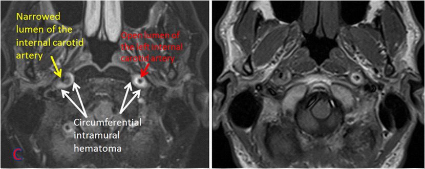
  • Carotid: internal carotid artery Internal carotid artery Branch of the common carotid artery which supplies the anterior part of the brain, the eye and its appendages, the forehead and nose. Carotid Arterial System: Anatomy 2 cm above carotid bifurcation
  • Vertebral:
    • Cervical transverse processes C2–C6
    • Segment between C2 and foramen magnum
    • Multiple synchronous dissection occurs in 13%–22% of cases.

Pathophysiologic effects:

  • Subintimal dissection usually results in vessel stenosis Stenosis Hypoplastic Left Heart Syndrome (HLHS) or occlusion.
  • Ischemic stroke Ischemic Stroke An ischemic stroke (also known as cerebrovascular accident) is an acute neurologic injury that occurs as a result of brain ischemia; this condition may be due to cerebral blood vessel occlusion by thrombosis or embolism, or rarely due to systemic hypoperfusion. Ischemic Stroke may result from hypoperfusion or a thromboembolic event (more common).
  • Subadventitial dissections result in aneurysm Aneurysm An aneurysm is a bulging, weakened area of a blood vessel that causes an abnormal widening of its diameter > 1.5 times the size of the native vessel. Aneurysms occur more often in arteries than in veins and are at risk of dissection and rupture, which can be life-threatening. Thoracic Aortic Aneurysms formation.
  • Symptoms may arise from an aneurysm Aneurysm An aneurysm is a bulging, weakened area of a blood vessel that causes an abnormal widening of its diameter > 1.5 times the size of the native vessel. Aneurysms occur more often in arteries than in veins and are at risk of dissection and rupture, which can be life-threatening. Thoracic Aortic Aneurysms compressing adjacent structures and distention of a vessel wall.
  • Rarely, dissection of thin intracranial arteries Arteries Arteries are tubular collections of cells that transport oxygenated blood and nutrients from the heart to the tissues of the body. The blood passes through the arteries in order of decreasing luminal diameter, starting in the largest artery (the aorta) and ending in the small arterioles. Arteries are classified into 3 types: large elastic arteries, medium muscular arteries, and small arteries and arterioles. Arteries: Histology may result in vessel rupture and cerebral hemorrhage.
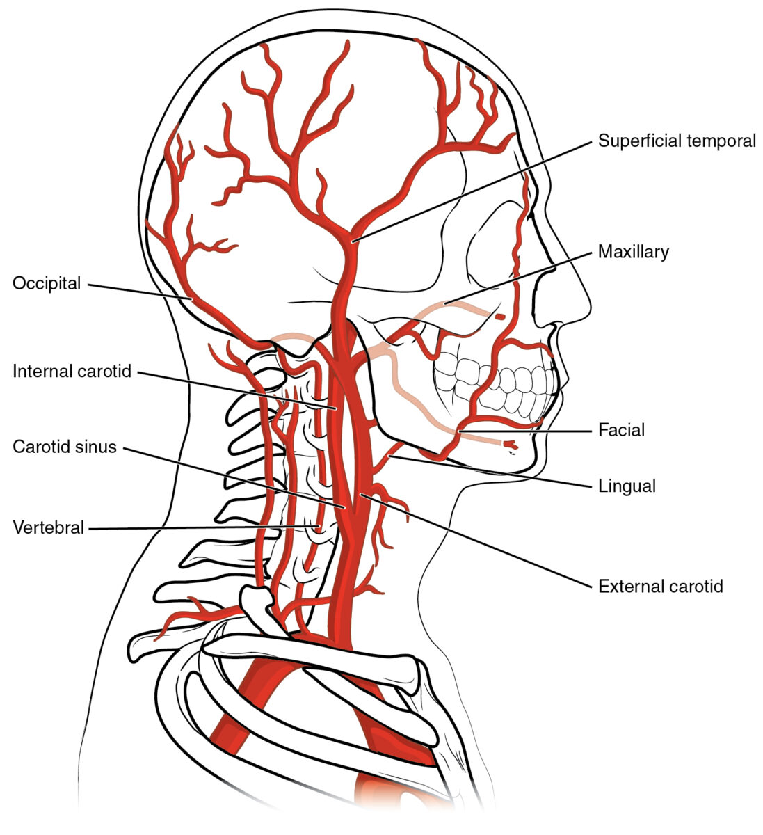
Anatomy of vertebral and carotid arteries

Anatomy of vertebral and carotid arteries

Image: “2122 Common Carotid Artery” by OpenStax College. License: CC BY 3.0

Clinical presentation

Local symptoms:

Ischemic symptoms:

  • Strokes or transient ischemic attacks (TIAs)
  • Carotid artery (anterior circulation Circulation The movement of the blood as it is pumped through the cardiovascular system. ABCDE Assessment stroke symptoms):
    • Contralateral paresis Paresis A general term referring to a mild to moderate degree of muscular weakness, occasionally used as a synonym for paralysis (severe or complete loss of motor function). In the older literature, paresis often referred specifically to paretic neurosyphilis. ‘general paresis’ and ‘general paralysis’ may still carry that connotation. Bilateral lower extremity paresis is referred to as paraparesis. Spinal Disk Herniation
    • Monocular blindness Blindness The inability to see or the loss or absence of perception of visual stimuli. This condition may be the result of eye diseases; optic nerve diseases; optic chiasm diseases; or brain diseases affecting the visual pathways or occipital lobe. Retinopathy of Prematurity
    • Upper-extremity paresis Paresis A general term referring to a mild to moderate degree of muscular weakness, occasionally used as a synonym for paralysis (severe or complete loss of motor function). In the older literature, paresis often referred specifically to paretic neurosyphilis. ‘general paresis’ and ‘general paralysis’ may still carry that connotation. Bilateral lower extremity paresis is referred to as paraparesis. Spinal Disk Herniation
    • Aphasia Aphasia A cognitive disorder marked by an impaired ability to comprehend or express language in its written or spoken form. This condition is caused by diseases which affect the language areas of the dominant hemisphere. Clinical features are used to classify the various subtypes of this condition. General categories include receptive, expressive, and mixed forms of aphasia. Ischemic Stroke
    • Ipsilateral facial weakness
  • Vertebral artery Vertebral artery The first branch of the subclavian artery with distribution to muscles of the neck; vertebrae; spinal cord; cerebellum; and interior of the cerebrum. Lateral Medullary Syndrome (Wallenberg Syndrome) ( vertebrobasilar ischemia Vertebrobasilar ischemia Localized or diffuse reduction in blood flow through the vertebrobasilar arterial system, which supplies the brain stem; cerebellum; occipital lobe; medial temporal lobe; and thalamus. Characteristic clinical features include syncope; lightheadedness; visual disturbances; and vertigo. Brain stem infarctions or other brain infarction may be associated. Subclavian Steal Syndrome):
    • Ipsilateral facial pain Facial pain Pain in the facial region including orofacial pain and craniofacial pain. Associated conditions include local inflammatory and neoplastic disorders and neuralgic syndromes involving the trigeminal, facial, and glossopharyngeal nerves. Conditions which feature recurrent or persistent facial pain as the primary manifestation of disease are referred to as facial pain syndromes. Trigeminal Neuralgia and numbness ( dysesthesia Dysesthesia Complex Regional Pain Syndrome (CRPS))
    • Loss of pain Pain An unpleasant sensation induced by noxious stimuli which are detected by nerve endings of nociceptive neurons. Pain: Types and Pathways and temperature in the ipsilateral face and contralateral trunk/limbs
    • Lateral medullary infarct Infarct Area of necrotic cells in an organ, arising mainly from hypoxia and ischemia Ischemic Cell Damage (Wallenberg) syndrome:
      • Loss of taste
      • Dysphagia Dysphagia Dysphagia is the subjective sensation of difficulty swallowing. Symptoms can range from a complete inability to swallow, to the sensation of solids or liquids becoming “stuck.” Dysphagia is classified as either oropharyngeal or esophageal, with esophageal dysphagia having 2 sub-types: functional and mechanical. Dysphagia
      • Dysarthria Dysarthria Disorders of speech articulation caused by imperfect coordination of pharynx, larynx, tongue, or face muscles. This may result from cranial nerve diseases; neuromuscular diseases; cerebellar diseases; basal ganglia diseases; brain stem diseases; or diseases of the corticobulbar tracts. The cortical language centers are intact in this condition. Wilson Disease
      • Hoarseness Hoarseness An unnaturally deep or rough quality of voice. Parapharyngeal Abscess (ipsilateral vocal cord paralysis)
      • Vertigo Vertigo Vertigo is defined as the perceived sensation of rotational motion while remaining still. A very common complaint in primary care and the ER, vertigo is more frequently experienced by women and its prevalence increases with age. Vertigo is classified into peripheral or central based on its etiology. Vertigo
      • Lack of automatic respiration Respiration The act of breathing with the lungs, consisting of inhalation, or the taking into the lungs of the ambient air, and of exhalation, or the expelling of the modified air which contains more carbon dioxide than the air taken in. Nose Anatomy (External & Internal) during sleep Sleep A readily reversible suspension of sensorimotor interaction with the environment, usually associated with recumbency and immobility. Physiology of Sleep
      • Hiccups
      • Ipsilateral Horner syndrome Horner syndrome Horner syndrome is a condition resulting from an interruption of the sympathetic innervation of the eyes. The syndrome is usually idiopathic but can be directly caused by head and neck trauma, cerebrovascular disease, or a tumor of the CNS. Horner Syndrome
      • Ipsilateral limb ataxia Ataxia Impairment of the ability to perform smoothly coordinated voluntary movements. This condition may affect the limbs, trunk, eyes, pharynx, larynx, and other structures. Ataxia may result from impaired sensory or motor function. Sensory ataxia may result from posterior column injury or peripheral nerve diseases. Motor ataxia may be associated with cerebellar diseases; cerebral cortex diseases; thalamic diseases; basal ganglia diseases; injury to the red nucleus; and other conditions. Ataxia-telangiectasia
    • Cervical cord ischemia Ischemia A hypoperfusion of the blood through an organ or tissue caused by a pathologic constriction or obstruction of its blood vessels, or an absence of blood circulation. Ischemic Cell Damage

Subarachnoid hemorrhage Subarachnoid Hemorrhage Subarachnoid hemorrhage (SAH) is a type of cerebrovascular accident (stroke) resulting from intracranial hemorrhage into the subarachnoid space between the arachnoid and the pia mater layers of the meninges surrounding the brain. Most SAHs originate from a saccular aneurysm in the circle of Willis but may also occur as a result of trauma, uncontrolled hypertension, vasculitis, anticoagulant use, or stimulant use. Subarachnoid Hemorrhage:
  • Rare
  • Can result from intracranial artery dissection

Diagnosis

History

  • Acute or subacute headache Headache The symptom of pain in the cranial region. It may be an isolated benign occurrence or manifestation of a wide variety of headache disorders. Brain Abscess or neck Neck The part of a human or animal body connecting the head to the rest of the body. Peritonsillar Abscess pain Pain An unpleasant sensation induced by noxious stimuli which are detected by nerve endings of nociceptive neurons. Pain: Types and Pathways
  • Neurologic/stroke symptoms
  • Recent trauma (even minor) or sports activities
  • Intense sneezing Sneezing The sudden, forceful, involuntary expulsion of air from the nose and mouth caused by irritation to the mucous membranes of the upper respiratory tract. Rhinovirus/coughing
  • Horner syndrome Horner syndrome Horner syndrome is a condition resulting from an interruption of the sympathetic innervation of the eyes. The syndrome is usually idiopathic but can be directly caused by head and neck trauma, cerebrovascular disease, or a tumor of the CNS. Horner Syndrome
  • History of migraines
  • History of connective tissue Connective tissue Connective tissues originate from embryonic mesenchyme and are present throughout the body except inside the brain and spinal cord. The main function of connective tissues is to provide structural support to organs. Connective tissues consist of cells and an extracellular matrix. Connective Tissue: Histology disorders

Physical exam

  • Focal neurologic signs
  • Slurred speech Slurred Speech Cerebellar Disorders
  • Ataxia Ataxia Impairment of the ability to perform smoothly coordinated voluntary movements. This condition may affect the limbs, trunk, eyes, pharynx, larynx, and other structures. Ataxia may result from impaired sensory or motor function. Sensory ataxia may result from posterior column injury or peripheral nerve diseases. Motor ataxia may be associated with cerebellar diseases; cerebral cortex diseases; thalamic diseases; basal ganglia diseases; injury to the red nucleus; and other conditions. Ataxia-telangiectasia
  • Carotid bruit Carotid bruit Carotid Artery Stenosis on auscultation

Imaging

  • CT angiography Angiography Radiography of blood vessels after injection of a contrast medium. Cardiac Surgery ( CTA CTA A non-invasive method that uses a ct scanner for capturing images of blood vessels and tissues. A contrast material is injected, which helps produce detailed images that aid in diagnosing vascular diseases. Pulmonary Function Tests) or MRA MRA Imaging of the Heart and Great Vessels is the preferred test:
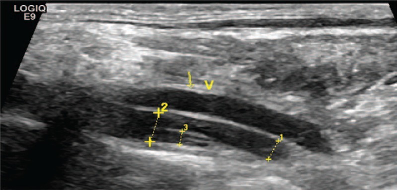
  • Arterial duplex and transcranial Doppler Doppler Ultrasonography applying the doppler effect, with frequency-shifted ultrasound reflections produced by moving targets (usually red blood cells) in the bloodstream along the ultrasound axis in direct proportion to the velocity of movement of the targets, to determine both direction and velocity of blood flow. Ultrasound (Sonography):
    • Can be used as a screening Screening Preoperative Care test or to monitor therapy
    • Relatively low sensitivity
    • Suboptimal yield for identifying dissection near the skull Skull The skull (cranium) is the skeletal structure of the head supporting the face and forming a protective cavity for the brain. The skull consists of 22 bones divided into the viscerocranium (facial skeleton) and the neurocranium. Skull: Anatomy base and vertebral artery Vertebral artery The first branch of the subclavian artery with distribution to muscles of the neck; vertebrae; spinal cord; cerebellum; and interior of the cerebrum. Lateral Medullary Syndrome (Wallenberg Syndrome) dissection within the transverse foramina
  • Conventional angiography Angiography Radiography of blood vessels after injection of a contrast medium. Cardiac Surgery:
    • Invasive
    • Used when clinical presentation is suggestive of dissection despite negative studies.

Management

Local nonischemic symptoms

  • Do not require specific treatment
  • Antiplatelet therapy is administered for stroke prevention.

TIA TIA Transient ischemic attack (TIA) is a temporary episode of neurologic dysfunction caused by ischemia without infarction that resolves completely when blood supply is restored. Transient ischemic attack is a neurologic emergency that warrants urgent medical attention. Transient Ischemic Attack (TIA) or stroke

  • Treatment should follow the same protocol as any ischemic stroke Ischemic Stroke An ischemic stroke (also known as cerebrovascular accident) is an acute neurologic injury that occurs as a result of brain ischemia; this condition may be due to cerebral blood vessel occlusion by thrombosis or embolism, or rarely due to systemic hypoperfusion. Ischemic Stroke.
  • Thrombolytic treatment started only after intracranial hemorrhage Intracranial hemorrhage Subarachnoid hemorrhage (SAH) is a type of cerebrovascular accident (stroke) resulting from intracranial hemorrhage into the subarachnoid space between the arachnoid and the pia mater layers of the meninges surrounding the brain. Most sahs originate from a saccular aneurysm in the circle of willis but may also occur as a result of trauma, uncontrolled hypertension, vasculitis, anticoagulant use, or stimulant use. Subarachnoid Hemorrhage is ruled out using noncontrast head CT.
  • Acute phase Acute phase Short Bowel Syndrome:
    • IV thrombolysis with alteplase Alteplase Thrombolytics is recommended for eligible patients Patients Individuals participating in the health care system for the purpose of receiving therapeutic, diagnostic, or preventive procedures. Clinician–Patient Relationship.
    • Time window: 3–4.5 hours after symptom onset
    • Proximal intracranial artery occlusion may also benefit from mechanical thrombectomy Thrombectomy Surgical removal of an obstructing clot or foreign material from a blood vessel at the point of its formation. Removal of a clot arising from a distant site is called embolectomy. Vascular Surgery up to 24 hours after presentation.
    • Emergency stenting can be performed in experienced centers.
  • Beyond the acute period:
    • Anticoagulation Anticoagulation Pulmonary Hypertension Drugs or antiplatelet drugs are recommended.
    • Antiplatelet drugs, not anticoagulation Anticoagulation Pulmonary Hypertension Drugs, should be used for intracranial dissections to avoid the risk of subarachnoid hemorrhage Subarachnoid Hemorrhage Subarachnoid hemorrhage (SAH) is a type of cerebrovascular accident (stroke) resulting from intracranial hemorrhage into the subarachnoid space between the arachnoid and the pia mater layers of the meninges surrounding the brain. Most SAHs originate from a saccular aneurysm in the circle of Willis but may also occur as a result of trauma, uncontrolled hypertension, vasculitis, anticoagulant use, or stimulant use. Subarachnoid Hemorrhage.
    • Should be delayed until 24 hours after IV thrombolytic therapy
    • Can be started immediately if not treated with IV thrombolytic therapy
  • Endovascular surgical repair: indicated only for ischemia Ischemia A hypoperfusion of the blood through an organ or tissue caused by a pathologic constriction or obstruction of its blood vessels, or an absence of blood circulation. Ischemic Cell Damage that recurs despite medical management

Subarachnoid hemorrhage Subarachnoid Hemorrhage Subarachnoid hemorrhage (SAH) is a type of cerebrovascular accident (stroke) resulting from intracranial hemorrhage into the subarachnoid space between the arachnoid and the pia mater layers of the meninges surrounding the brain. Most SAHs originate from a saccular aneurysm in the circle of Willis but may also occur as a result of trauma, uncontrolled hypertension, vasculitis, anticoagulant use, or stimulant use. Subarachnoid Hemorrhage

  • Neurosurgical intervention
  • Available methods include:
    • Proximal occlusion
    • Wrapping of pseudoaneurysm Pseudoaneurysm Not an aneurysm but a well-defined collection of blood and connective tissue outside the wall of a blood vessel or the heart. It is the containment of a ruptured blood vessel or heart, such as sealing a rupture of the left ventricle. False aneurysm is formed by organized thrombus and hematoma in surrounding tissue. Thoracic Aortic Aneurysms
    • Bypass
    • Embolization Embolization A method of hemostasis utilizing various agents such as gelfoam, silastic, metal, glass, or plastic pellets, autologous clot, fat, and muscle as emboli. It has been used in the treatment of spinal cord and intracranial arteriovenous malformations, renal arteriovenous fistulas, gastrointestinal bleeding, epistaxis, hypersplenism, certain highly vascular tumors, traumatic rupture of blood vessels, and control of operative hemorrhage. Gastrointestinal Bleeding
    • Stenting

Differential Diagnosis

  • Ruptured cerebral aneurysm Cerebral aneurysm Brain aneurysms, also known as intracranial or cerebral aneurysms, are dilations of the arteries along points of weakness in the brain. The majority of the aneurysms are berry (saccular) in nature and located within the anterior circulation of the circle of Willis. Brain Aneurysms: Subarachnoid hemorrhage Subarachnoid Hemorrhage Subarachnoid hemorrhage (SAH) is a type of cerebrovascular accident (stroke) resulting from intracranial hemorrhage into the subarachnoid space between the arachnoid and the pia mater layers of the meninges surrounding the brain. Most SAHs originate from a saccular aneurysm in the circle of Willis but may also occur as a result of trauma, uncontrolled hypertension, vasculitis, anticoagulant use, or stimulant use. Subarachnoid Hemorrhage results in a sudden, violent “ thunderclap headache Thunderclap Headache Subarachnoid Hemorrhage.” Diagnosis of an acute hemorrhage can be established with a noncontrast head CT. Treatment of the aneurysm Aneurysm An aneurysm is a bulging, weakened area of a blood vessel that causes an abnormal widening of its diameter > 1.5 times the size of the native vessel. Aneurysms occur more often in arteries than in veins and are at risk of dissection and rupture, which can be life-threatening. Thoracic Aortic Aneurysms involves neurosurgical vascular interventions such as clipping and embolization Embolization A method of hemostasis utilizing various agents such as gelfoam, silastic, metal, glass, or plastic pellets, autologous clot, fat, and muscle as emboli. It has been used in the treatment of spinal cord and intracranial arteriovenous malformations, renal arteriovenous fistulas, gastrointestinal bleeding, epistaxis, hypersplenism, certain highly vascular tumors, traumatic rupture of blood vessels, and control of operative hemorrhage. Gastrointestinal Bleeding.
  • Carotid stenosis Stenosis Hypoplastic Left Heart Syndrome (HLHS): chronic atherosclerotic disease resulting in narrowing of the common and internal carotid arteries Carotid Arteries Either of the two principal arteries on both sides of the neck that supply blood to the head and neck; each divides into two branches, the internal carotid artery and the external carotid artery. Carotid Arterial System: Anatomy: Acute presentation involves ischemic stroke Ischemic Stroke An ischemic stroke (also known as cerebrovascular accident) is an acute neurologic injury that occurs as a result of brain ischemia; this condition may be due to cerebral blood vessel occlusion by thrombosis or embolism, or rarely due to systemic hypoperfusion. Ischemic Stroke or TIA TIA Transient ischemic attack (TIA) is a temporary episode of neurologic dysfunction caused by ischemia without infarction that resolves completely when blood supply is restored. Transient ischemic attack is a neurologic emergency that warrants urgent medical attention. Transient Ischemic Attack (TIA) with anterior distribution similar to carotid artery dissection. Diagnosis is established with imaging, and acute treatment is focused on stroke management. Long-term management involves antiplatelet therapy and surgical carotid endarterectomy Endarterectomy Surgical excision, performed under general anesthesia, of the atheromatous tunica intima of an artery. When reconstruction of an artery is performed as an endovascular procedure through a catheter, it is called atherectomy. Intestinal Ischemia.
  • Vertebrobasilar insufficiency: stenosis Stenosis Hypoplastic Left Heart Syndrome (HLHS) of vertebral and basilar arteries Arteries Arteries are tubular collections of cells that transport oxygenated blood and nutrients from the heart to the tissues of the body. The blood passes through the arteries in order of decreasing luminal diameter, starting in the largest artery (the aorta) and ending in the small arterioles. Arteries are classified into 3 types: large elastic arteries, medium muscular arteries, and small arteries and arterioles. Arteries: Histology secondary to atherosclerosis Atherosclerosis Atherosclerosis is a common form of arterial disease in which lipid deposition forms a plaque in the blood vessel walls. Atherosclerosis is an incurable disease, for which there are clearly defined risk factors that often can be reduced through a change in lifestyle and behavior of the patient. Atherosclerosis: Vertebrobasilar insufficiency results in compromised circulation Circulation The movement of the blood as it is pumped through the cardiovascular system. ABCDE Assessment to the posterior brain Brain The part of central nervous system that is contained within the skull (cranium). Arising from the neural tube, the embryonic brain is comprised of three major parts including prosencephalon (the forebrain); mesencephalon (the midbrain); and rhombencephalon (the hindbrain). The developed brain consists of cerebrum; cerebellum; and other structures in the brain stem. Nervous System: Anatomy, Structure, and Classification. Symptoms include dizziness Dizziness An imprecise term which may refer to a sense of spatial disorientation, motion of the environment, or lightheadedness. Lateral Medullary Syndrome (Wallenberg Syndrome)/ vertigo Vertigo Vertigo is defined as the perceived sensation of rotational motion while remaining still. A very common complaint in primary care and the ER, vertigo is more frequently experienced by women and its prevalence increases with age. Vertigo is classified into peripheral or central based on its etiology. Vertigo, numbness, slurred speech Slurred Speech Cerebellar Disorders, weakness, confusion, and loss of coordination Coordination Cerebellar Disorders. Diagnosis can be made with MRA MRA Imaging of the Heart and Great Vessels or CTA CTA A non-invasive method that uses a ct scanner for capturing images of blood vessels and tissues. A contrast material is injected, which helps produce detailed images that aid in diagnosing vascular diseases. Pulmonary Function Tests, and treatment is focused on control of hypertension Hypertension Hypertension, or high blood pressure, is a common disease that manifests as elevated systemic arterial pressures. Hypertension is most often asymptomatic and is found incidentally as part of a routine physical examination or during triage for an unrelated medical encounter. Hypertension and cholesterol Cholesterol The principal sterol of all higher animals, distributed in body tissues, especially the brain and spinal cord, and in animal fats and oils. Cholesterol Metabolism and lifestyle modifications.
  • Migraine Migraine Migraine headache is a primary headache disorder and is among the most prevalent disorders in the world. Migraine is characterized by episodic, moderate to severe headaches that may be associated with increased sensitivity to light and sound, as well as nausea and/or vomiting. Migraine Headache: a recurrent headache Headache The symptom of pain in the cranial region. It may be an isolated benign occurrence or manifestation of a wide variety of headache disorders. Brain Abscess of varying intensity and characteristics: Migraines can sometimes be accompanied by vision Vision Ophthalmic Exam changes, focal neurologic symptoms, photophobia Photophobia Abnormal sensitivity to light. This may occur as a manifestation of eye diseases; migraine; subarachnoid hemorrhage; meningitis; and other disorders. Photophobia may also occur in association with depression and other mental disorders. Migraine Headache and phonophobia Phonophobia Specific Phobias. Diagnosis is clinical, and management can include a variety of therapies, such as NSAIDs NSAIDS Primary vs Secondary Headaches, triptans Triptans Triptans and ergot alkaloids are agents used mainly for the management of acute migraines. The therapeutic effect is induced by binding to serotonin receptors, which causes reduced vasoactive neuropeptide release, pain conduction, and intracranial vasoconstriction. Triptans and Ergot Alkaloids, compazine, caffeine Caffeine A methylxanthine naturally occurring in some beverages and also used as a pharmacological agent. Caffeine’s most notable pharmacological effect is as a central nervous system stimulant, increasing alertness and producing agitation. Several cellular actions of caffeine have been observed, but it is not entirely clear how each contributes to its pharmacological profile. Among the most important are inhibition of cyclic nucleotide phosphodiesterases, antagonism of adenosine receptors, and modulation of intracellular calcium handling. Stimulants, and oxygen.
  • Horner syndrome Horner syndrome Horner syndrome is a condition resulting from an interruption of the sympathetic innervation of the eyes. The syndrome is usually idiopathic but can be directly caused by head and neck trauma, cerebrovascular disease, or a tumor of the CNS. Horner Syndrome: disorder characterized by a constricted pupil Pupil The pupil is the space within the eye that permits light to project onto the retina. Anatomically located in front of the lens, the pupil’s size is controlled by the surrounding iris. The pupil provides insight into the function of the central and autonomic nervous systems. Pupil: Physiology and Abnormalities ( miosis Miosis Pupil: Physiology and Abnormalities), drooping of the upper eyelid ( ptosis Ptosis Cranial Nerve Palsies), absence of sweating of the face (anhidrosis), and sinking of the eyeball into the bony cavity ( enophthalmos Enophthalmos Recession of the eyeball into the orbit. Marfan Syndrome): Horner syndrome Horner syndrome Horner syndrome is a condition resulting from an interruption of the sympathetic innervation of the eyes. The syndrome is usually idiopathic but can be directly caused by head and neck trauma, cerebrovascular disease, or a tumor of the CNS. Horner Syndrome is most commonly caused by apical lung cancer Lung cancer Lung cancer is the malignant transformation of lung tissue and the leading cause of cancer-related deaths. The majority of cases are associated with long-term smoking. The disease is generally classified histologically as either small cell lung cancer or non-small cell lung cancer. Symptoms include cough, dyspnea, weight loss, and chest discomfort. Lung Cancer compressing the sympathetic plexus. Internal carotid artery dissection Internal Carotid Artery Dissection The splitting of the vessel wall in one or both (left and right) internal carotid arteries. Interstitial hemorrhage into the media of the vessel wall can lead to occlusion of the internal carotid artery and aneurysm formation. Cranial Nerve Palsies can cause partial Horner syndrome Horner syndrome Horner syndrome is a condition resulting from an interruption of the sympathetic innervation of the eyes. The syndrome is usually idiopathic but can be directly caused by head and neck trauma, cerebrovascular disease, or a tumor of the CNS. Horner Syndrome, which is characterized by the absence of anhidrosis.

References

  1. Blum, C. A., Yaghi, S. (2015). Cervical artery dissection: a review of the epidemiology, pathophysiology, treatment, and outcome. Archives of Neuroscience 2(4):e26670. https://doi.org/10.5812/archneurosci.26670
  2. Arnold, M., Bousser, M. (2005). Carotid and vertebral artery dissection. Practical Neurology 5(2):100–109. https://www.researchgate.net/publication/238331335_Carotid_and_Vertebral_Artery_Dissection
  3. Baumgartner, R.W., et al. (2001). Carotid dissection with and without ischemic events: local symptoms and cerebral artery findings. Neurology 57(5):827–832. https://doi.org/10.1212/wnl.57.5.827
  4. Powers, et al. (2019). Guidelines for the early management of patients with acute ischemic stroke: 2019 update to the 2018 guidelines for the early management of acute ischemic stroke: a guideline for healthcare professionals from the American Heart Association/American Stroke Association. Stroke 50(12):e344–e418. https://doi.org/10.1161/str.0000000000000211

Create your free account or log in to continue reading!

Sign up now and get free access to Lecturio with concept pages, medical videos, and questions for your medical education.

User Reviews

Unwrap New Skills This Holiday 🎄 Save 30% on all plans now!

Details