Advertisement

Advertisement

Advertisement

Advertisement

Heart Failure

Heart failure (HF) is the complex condition resulting from a structural and/or functional inability of the heart to supply the normal cardiac output to meet metabolic needs. In HF, impaired ventricular filling or ejection of blood leads to fatigue, dyspnea, and fluid retention or edema. Echocardiography can confirm the diagnosis, providing information about the ejection fraction (EF) and the adequacy of ventricular filling and relaxation. Treatment is directed at the removal of excess fluid and decreasing oxygen demand of the heart. Prognosis depends on the underlying cause, compliance with medical therapy, and presence of comorbidities.

Last updated: May 16, 2024
Editorial responsibility: Stanley Oiseth, Lindsay Jones, Evelin Maza

Advertisement

Advertisement

Advertisement

Advertisement

Advertisement

Advertisement

Epidemiology

  • Prevalence Prevalence The total number of cases of a given disease in a specified population at a designated time. It is differentiated from incidence, which refers to the number of new cases in the population at a given time. Measures of Disease Frequency in the United States: 1%–2%
  • More common in men
  • Prevalence Prevalence The total number of cases of a given disease in a specified population at a designated time. It is differentiated from incidence, which refers to the number of new cases in the population at a given time. Measures of Disease Frequency in African Americans is 25% higher than in Whites.
  • Incidence Incidence The number of new cases of a given disease during a given period in a specified population. It also is used for the rate at which new events occur in a defined population. It is differentiated from prevalence, which refers to all cases in the population at a given time. Measures of Disease Frequency and prevalence Prevalence The total number of cases of a given disease in a specified population at a designated time. It is differentiated from incidence, which refers to the number of new cases in the population at a given time. Measures of Disease Frequency increase steeply with age.
  • Most common cause of hospitalization Hospitalization The confinement of a patient in a hospital. Delirium in patients Patients Individuals participating in the health care system for the purpose of receiving therapeutic, diagnostic, or preventive procedures. Clinician–Patient Relationship > 60 years of age

Etiology

Underlying conditions include:

  • Hypertension Hypertension Hypertension, or high blood pressure, is a common disease that manifests as elevated systemic arterial pressures. Hypertension is most often asymptomatic and is found incidentally as part of a routine physical examination or during triage for an unrelated medical encounter. Hypertension
  • Coronary artery Coronary Artery Truncus Arteriosus disease 
  • Diabetes Diabetes Diabetes mellitus (DM) is a metabolic disease characterized by hyperglycemia and dysfunction of the regulation of glucose metabolism by insulin. Type 1 DM is diagnosed mostly in children and young adults as the result of autoimmune destruction of β cells in the pancreas and the resulting lack of insulin. Type 2 DM has a significant association with obesity and is characterized by insulin resistance. Diabetes Mellitus mellitus
  • Myocardial infarction Myocardial infarction MI is ischemia and death of an area of myocardial tissue due to insufficient blood flow and oxygenation, usually from thrombus formation on a ruptured atherosclerotic plaque in the epicardial arteries. Clinical presentation is most commonly with chest pain, but women and patients with diabetes may have atypical symptoms. Myocardial Infarction ( MI MI MI is ischemia and death of an area of myocardial tissue due to insufficient blood flow and oxygenation, usually from thrombus formation on a ruptured atherosclerotic plaque in the epicardial arteries. Clinical presentation is most commonly with chest pain, but women and patients with diabetes may have atypical symptoms. Myocardial Infarction)
  • Cardiomyopathy Cardiomyopathy Cardiomyopathy refers to a group of myocardial diseases associated with structural changes of the heart muscles (myocardium) and impaired systolic and/or diastolic function in the absence of other heart disorders (coronary artery disease, hypertension, valvular disease, and congenital heart disease). Cardiomyopathy: Overview and Types (includes postpartum and stress-related types)
  • Left ventricular hypertrophy Ventricular Hypertrophy Tetralogy of Fallot
  • Pulmonary hypertension Pulmonary Hypertension Pulmonary hypertension (PH) or pulmonary arterial hypertension (PAH) is characterized by elevated pulmonary arterial pressure, which can lead to chronic progressive right heart failure. Pulmonary hypertension is grouped into 5 categories based on etiology, which include primary PAH, and PH due to cardiac disease, lung or hypoxic disease, chronic thromboembolic disease, and multifactorial or unclear etiologies. Pulmonary Hypertension
  • Cor pulmonale Cor Pulmonale Cor pulmonale is right ventricular (RV) dysfunction caused by lung disease that results in pulmonary artery hypertension. The most common cause of cor pulmonale is chronic obstructive pulmonary disease. Dyspnea is the usual presenting symptom. Cor Pulmonale (e.g., chronic obstructive pulmonary disease Pulmonary disease Diseases involving the respiratory system. Blastomyces/Blastomycosis)
  • Valvular heart diseases (e.g., aortic stenosis Stenosis Hypoplastic Left Heart Syndrome (HLHS))
  • Renal failure Renal failure Conditions in which the kidneys perform below the normal level in the ability to remove wastes, concentrate urine, and maintain electrolyte balance; blood pressure; and calcium metabolism. Renal insufficiency can be classified by the degree of kidney damage (as measured by the level of proteinuria) and reduction in glomerular filtration rate. Crush Syndrome
  • Pericardial diseases (e.g., constrictive pericarditis Constrictive pericarditis Inflammation of the pericardium that is characterized by the fibrous scarring and adhesion of both serous layers, the visceral pericardium and the parietal pericardium leading to the loss of pericardial cavity. The thickened pericardium severely restricts cardiac filling. Clinical signs include fatigue, muscle wasting, and weight loss. Pericarditis)
  • Myocarditis Myocarditis Myocarditis is an inflammatory disease of the myocardium, which may occur alone or in association with a systemic process. There are numerous etiologies of myocarditis, but all lead to inflammation and myocyte injury, most often leading to signs and symptoms of heart failure. Myocarditis
  • Hemochromatosis Hemochromatosis A disorder of iron metabolism characterized by a triad of hemosiderosis; liver cirrhosis; and diabetes mellitus. It is caused by massive iron deposits in parenchymal cells that may develop after a prolonged increase of iron absorption. Hereditary Hemochromatosis
  • Amyloidosis Amyloidosis Amyloidosis is a disease caused by abnormal extracellular tissue deposition of fibrils composed of various misfolded low-molecular-weight protein subunits. These proteins are frequently byproducts of other pathological processes (e.g., multiple myeloma). Amyloidosis
  • Arrhythmias (e.g., bradyarrhythmias Bradyarrhythmias Bradyarrhythmia is a rhythm in which the heart rate is less than 60/min. Bradyarrhythmia can be physiologic, without symptoms or hemodynamic change. Pathologic bradyarrhythmia results in reduced cardiac output and hemodynamic instability causing syncope, dizziness, or dyspnea. Bradyarrhythmias, tachyarrhythmias)
  • Substance use disorder
  • Chemotherapy Chemotherapy Osteosarcoma or cardiotoxic drugs

Risk factors include:

  • Substance use disorder (e.g., ethanol Ethanol A clear, colorless liquid rapidly absorbed from the gastrointestinal tract and distributed throughout the body. It has bactericidal activity and is used often as a topical disinfectant. It is widely used as a solvent and preservative in pharmaceutical preparations as well as serving as the primary ingredient in alcoholic beverages. Ethanol Metabolism, cocaine Cocaine An alkaloid ester extracted from the leaves of plants including coca. It is a local anesthetic and vasoconstrictor and is clinically used for that purpose, particularly in the eye, ear, nose, and throat. It also has powerful central nervous system effects similar to the amphetamines and is a drug of abuse. Cocaine, like amphetamines, acts by multiple mechanisms on brain catecholaminergic neurons; the mechanism of its reinforcing effects is thought to involve inhibition of dopamine uptake. Local Anesthetics)
  • Smoking Smoking Willful or deliberate act of inhaling and exhaling smoke from burning substances or agents held by hand. Interstitial Lung Diseases
  • Obesity Obesity Obesity is a condition associated with excess body weight, specifically with the deposition of excessive adipose tissue. Obesity is considered a global epidemic. Major influences come from the western diet and sedentary lifestyles, but the exact mechanisms likely include a mixture of genetic and environmental factors. Obesity
  • Sedentary lifestyle

Pathophysiology

  • Heart failure Heart Failure A heterogeneous condition in which the heart is unable to pump out sufficient blood to meet the metabolic need of the body. Heart failure can be caused by structural defects, functional abnormalities (ventricular dysfunction), or a sudden overload beyond its capacity. Chronic heart failure is more common than acute heart failure which results from sudden insult to cardiac function, such as myocardial infarction. Total Anomalous Pulmonary Venous Return (TAPVR) (HF) can be due to systolic and/or diastolic dysfunction Diastolic dysfunction Restrictive Cardiomyopathy.
    • HF with reduced EF EF Cardiac Cycle (HFrEF), or systolic heart failure Heart Failure A heterogeneous condition in which the heart is unable to pump out sufficient blood to meet the metabolic need of the body. Heart failure can be caused by structural defects, functional abnormalities (ventricular dysfunction), or a sudden overload beyond its capacity. Chronic heart failure is more common than acute heart failure which results from sudden insult to cardiac function, such as myocardial infarction. Total Anomalous Pulmonary Venous Return (TAPVR):
      • Impaired myocyte contractility and dilated heart → reduced stroke volume Stroke volume The amount of blood pumped out of the heart per beat, not to be confused with cardiac output (volume/time). It is calculated as the difference between the end-diastolic volume and the end-systolic volume. Cardiac Cycle and EF EF Cardiac Cycle 
      • Left ventricular ejection fraction Ejection fraction Cardiac Cycle (LVEF) ≤ 40%
    • HF with preserved EF EF Cardiac Cycle (HFpEF), or diastolic heart failure Heart Failure A heterogeneous condition in which the heart is unable to pump out sufficient blood to meet the metabolic need of the body. Heart failure can be caused by structural defects, functional abnormalities (ventricular dysfunction), or a sudden overload beyond its capacity. Chronic heart failure is more common than acute heart failure which results from sudden insult to cardiac function, such as myocardial infarction. Total Anomalous Pulmonary Venous Return (TAPVR):
      • Inadequate relaxation and filling of the ventricle but normal myocyte contractility → preserved EF EF Cardiac Cycle despite low stroke volume Stroke volume The amount of blood pumped out of the heart per beat, not to be confused with cardiac output (volume/time). It is calculated as the difference between the end-diastolic volume and the end-systolic volume. Cardiac Cycle
      • LVEF ≥ 50%
    • Heart failure Heart Failure A heterogeneous condition in which the heart is unable to pump out sufficient blood to meet the metabolic need of the body. Heart failure can be caused by structural defects, functional abnormalities (ventricular dysfunction), or a sudden overload beyond its capacity. Chronic heart failure is more common than acute heart failure which results from sudden insult to cardiac function, such as myocardial infarction. Total Anomalous Pulmonary Venous Return (TAPVR) with midrange EF EF Cardiac Cycle (HFmrEF):
      • LVEF between 40% and 49%
    • HF with improved EF EF Cardiac Cycle (HFiEF): previous LVEF ≤ 40% but with better EF EF Cardiac Cycle (> 40%) on follow-up measurement
  • HF can also categorized by the area affected:
    • Left-sided heart failure Heart Failure A heterogeneous condition in which the heart is unable to pump out sufficient blood to meet the metabolic need of the body. Heart failure can be caused by structural defects, functional abnormalities (ventricular dysfunction), or a sudden overload beyond its capacity. Chronic heart failure is more common than acute heart failure which results from sudden insult to cardiac function, such as myocardial infarction. Total Anomalous Pulmonary Venous Return (TAPVR): results from abnormalities on the left side of the heart (left ventricle, aortic valve Aortic valve The valve between the left ventricle and the ascending aorta which prevents backflow into the left ventricle. Heart: Anatomy, mitral valve Mitral valve The valve between the left atrium and left ventricle of the heart. Heart: Anatomy)
    • Right-sided heart failure Right-Sided Heart Failure Ebstein’s Anomaly: results from abnormalities on the right side of the heart (right ventricle, tricuspid valve Tricuspid valve The valve consisting of three cusps situated between the right atrium and right ventricle of the heart. Heart: Anatomy, pulmonic valve)
    • Left- and right-sided HF can occur at the same time.
  • Compensatory mechanisms induced by decreased cardiac output Cardiac output The volume of blood passing through the heart per unit of time. It is usually expressed as liters (volume) per minute so as not to be confused with stroke volume (volume per beat). Cardiac Mechanics:
    • Activation of the sympathetic nervous system Nervous system The nervous system is a small and complex system that consists of an intricate network of neural cells (or neurons) and even more glial cells (for support and insulation). It is divided according to its anatomical components as well as its functional characteristics. The brain and spinal cord are referred to as the central nervous system, and the branches of nerves from these structures are referred to as the peripheral nervous system. Nervous System: Anatomy, Structure, and Classification
    • Activation of the renin-angiotensin-aldosterone system Renin-angiotensin-aldosterone system A blood pressure regulating system of interacting components that include renin; angiotensinogen; angiotensin converting enzyme; angiotensin i; angiotensin ii; and angiotensinase. Renin, an enzyme produced in the kidney, acts on angiotensinogen, an alpha-2 globulin produced by the liver, forming angiotensin I. Angiotensin-converting enzyme, contained in the lung, acts on angiotensin I in the plasma converting it to angiotensin II, an extremely powerful vasoconstrictor. Angiotensin II causes contraction of the arteriolar and renal vascular smooth muscle, leading to retention of salt and water in the kidney and increased arterial blood pressure. In addition, angiotensin II stimulates the release of aldosterone from the adrenal cortex, which in turn also increases salt and water retention in the kidney. Angiotensin-converting enzyme also breaks down bradykinin, a powerful vasodilator and component of the kallikrein-kinin system. Adrenal Hormones
    • Antidiuretic hormone Antidiuretic hormone Antidiuretic hormones released by the neurohypophysis of all vertebrates (structure varies with species) to regulate water balance and osmolarity. In general, vasopressin is a nonapeptide consisting of a six-amino-acid ring with a cysteine 1 to cysteine 6 disulfide bridge or an octapeptide containing a cystine. All mammals have arginine vasopressin except the pig with a lysine at position 8. Vasopressin, a vasoconstrictor, acts on the kidney collecting ducts to increase water reabsorption, increase blood volume and blood pressure. Hypernatremia release
    • Atrial natriuretic peptide Atrial natriuretic peptide A potent natriuretic and vasodilatory peptide or mixture of different-sized low molecular weight peptides derived from a common precursor and secreted mainly by the heart atrium. All these peptides share a sequence of about 20 amino acids. Renal Sodium and Water Regulation/ brain Brain The part of central nervous system that is contained within the skull (cranium). Arising from the neural tube, the embryonic brain is comprised of three major parts including prosencephalon (the forebrain); mesencephalon (the midbrain); and rhombencephalon (the hindbrain). The developed brain consists of cerebrum; cerebellum; and other structures in the brain stem. Nervous System: Anatomy, Structure, and Classification natriuretic peptide ( BNP BNP A peptide that is secreted by the brain and the heart atria, stored mainly in cardiac ventricular myocardium. It can cause natriuresis; diuresis; vasodilation; and inhibits secretion of renin and aldosterone. It improves heart function. It contains 32 amino acids. Renal Sodium and Water Regulation) release
    • Cardiac remodeling/ hypertrophy Hypertrophy General increase in bulk of a part or organ due to cell enlargement and accumulation of fluids and secretions, not due to tumor formation, nor to an increase in the number of cells (hyperplasia). Cellular Adaptation
    • Effects:
      • ↑ Left ventricular and atrial pressures Atrial pressures The pressure within the cardiac atrium. It can be measured directly by using a pressure catheter. It can be also estimated using various imaging techniques or other pressure readings such as pulmonary capillary wedge pressure (an estimate of left atrial pressure) and central venous pressure (an estimate of right atrial pressure). Cardiac Cycle → ↑ pulmonary capillary pressure → pulmonary edema Pulmonary edema Pulmonary edema is a condition caused by excess fluid within the lung parenchyma and alveoli as a consequence of a disease process. Based on etiology, pulmonary edema is classified as cardiogenic or noncardiogenic. Patients may present with progressive dyspnea, orthopnea, cough, or respiratory failure. Pulmonary Edema ( dyspnea Dyspnea Dyspnea is the subjective sensation of breathing discomfort. Dyspnea is a normal manifestation of heavy physical or psychological exertion, but also may be caused by underlying conditions (both pulmonary and extrapulmonary). Dyspnea)
      • Impaired left ventricular filling Ventricular filling Cardiac Cycle → promotes sodium Sodium A member of the alkali group of metals. It has the atomic symbol na, atomic number 11, and atomic weight 23. Hyponatremia and water retention → ↑ volume → extremity/ peripheral edema Peripheral edema Peripheral edema is the swelling of the lower extremities, namely, legs, feet, and ankles. Edema 
      • Chronic passive hepatic congestion → liver Liver The liver is the largest gland in the human body. The liver is found in the superior right quadrant of the abdomen and weighs approximately 1.5 kilograms. Its main functions are detoxification, metabolism, nutrient storage (e.g., iron and vitamins), synthesis of coagulation factors, formation of bile, filtration, and storage of blood. Liver: Anatomy injury (congestive hepatopathy)
      • Inadequate blood pressure and ↓ renal perfusion → acute kidney injury Acute Kidney Injury Acute kidney injury refers to sudden and often reversible loss of renal function, which develops over days or weeks. Azotemia refers to elevated levels of nitrogen-containing substances in the blood that accompany AKI, which include BUN and creatinine. Acute Kidney Injury to progressive chronic disease (cardiorenal syndrome)
      • Inability to supply sufficient blood to other areas → confusion, fatigue Fatigue The state of weariness following a period of exertion, mental or physical, characterized by a decreased capacity for work and reduced efficiency to respond to stimuli. Fibromyalgia, diaphoresis
  • Initially, these adaptations can increase cardiac output Cardiac output The volume of blood passing through the heart per unit of time. It is usually expressed as liters (volume) per minute so as not to be confused with stroke volume (volume per beat). Cardiac Mechanics and prevent the development of symptoms (compensated HF).
  • Over time, hypertrophy Hypertrophy General increase in bulk of a part or organ due to cell enlargement and accumulation of fluids and secretions, not due to tumor formation, nor to an increase in the number of cells (hyperplasia). Cellular Adaptation decreases lumen size and fails to increase contractility → exacerbation of HF due to increased oxygen demand and decreased contractility → decompensated HF
  • Acute decompensated HF is usually due to myocardial ischemia Myocardial ischemia A disorder of cardiac function caused by insufficient blood flow to the muscle tissue of the heart. The decreased blood flow may be due to narrowing of the coronary arteries (coronary artery disease), to obstruction by a thrombus (coronary thrombosis), or less commonly, to diffuse narrowing of arterioles and other small vessels within the heart. Coronary Heart Disease (e.g., MI MI MI is ischemia and death of an area of myocardial tissue due to insufficient blood flow and oxygenation, usually from thrombus formation on a ruptured atherosclerotic plaque in the epicardial arteries. Clinical presentation is most commonly with chest pain, but women and patients with diabetes may have atypical symptoms. Myocardial Infarction).

Clinical Presentation

Symptoms of HF

  • Chest pain Pain An unpleasant sensation induced by noxious stimuli which are detected by nerve endings of nociceptive neurons. Pain: Types and Pathways
  • Dyspnea Dyspnea Dyspnea is the subjective sensation of breathing discomfort. Dyspnea is a normal manifestation of heavy physical or psychological exertion, but also may be caused by underlying conditions (both pulmonary and extrapulmonary). Dyspnea (exertional in early HF)
  • Orthopnea Orthopnea Pulmonary Edema
  • Paroxysmal nocturnal dyspnea Paroxysmal nocturnal dyspnea A disorder characterized by sudden attacks of respiratory distress in at rest patients with heart failure and pulmonary edema. It usually occurs at night after several hours of sleep in a reclining position. Patients awaken with a feeling of suffocation, coughing, a cold sweat, and tachycardia. When there is significant wheezing, it is called cardiac asthma. Pulmonary Edema
  • Hypotension Hypotension Hypotension is defined as low blood pressure, specifically < 90/60 mm Hg, and is most commonly a physiologic response. Hypotension may be mild, serious, or life threatening, depending on the cause. Hypotension 
  • Syncope Syncope Syncope is a short-term loss of consciousness and loss of postural stability followed by spontaneous return of consciousness to the previous neurologic baseline without the need for resuscitation. The condition is caused by transient interruption of cerebral blood flow that may be benign or related to a underlying life-threatening condition. Syncope
  • Fatigue Fatigue The state of weariness following a period of exertion, mental or physical, characterized by a decreased capacity for work and reduced efficiency to respond to stimuli. Fibromyalgia
  • Confusion
  • Palpitations Palpitations Ebstein’s Anomaly
  • Dizziness Dizziness An imprecise term which may refer to a sense of spatial disorientation, motion of the environment, or lightheadedness. Lateral Medullary Syndrome (Wallenberg Syndrome)
  • Cough
  • Exercise intolerance
  • Unintentional weight loss Weight loss Decrease in existing body weight. Bariatric Surgery (cardiac cachexia)

Signs of HF

  • Vital sign findings can include:
    • Tachycardia Tachycardia Abnormally rapid heartbeat, usually with a heart rate above 100 beats per minute for adults. Tachycardia accompanied by disturbance in the cardiac depolarization (cardiac arrhythmia) is called tachyarrhythmia. Sepsis in Children
    • Narrow pulse pressure
    • Hypotension Hypotension Hypotension is defined as low blood pressure, specifically < 90/60 mm Hg, and is most commonly a physiologic response. Hypotension may be mild, serious, or life threatening, depending on the cause. Hypotension seen in severe HF
    • Pulsus alternans (alternating strong and weak pulses): incomplete recovery of the ventricular contraction every other cardiac cycle Cardiac cycle The cardiac cycle describes a complete contraction and relaxation of all 4 chambers of the heart during a standard heartbeat. The cardiac cycle includes 7 phases, which together describe the cycle of ventricular filling, isovolumetric contraction, ventricular ejection, and isovolumetric relaxation. Cardiac Cycle → alternating stroke volume Stroke volume The amount of blood pumped out of the heart per beat, not to be confused with cardiac output (volume/time). It is calculated as the difference between the end-diastolic volume and the end-systolic volume. Cardiac Cycle
  • Appearance:
    • Cyanosis Cyanosis A bluish or purplish discoloration of the skin and mucous membranes due to an increase in the amount of deoxygenated hemoglobin in the blood or a structural defect in the hemoglobin molecule. Pulmonary Examination (from poor perfusion)
    • Diaphoresis
  • Chest and cardiovascular examination Cardiovascular examination Examination of the cardiovascular system (CVS) is a critical component of a thorough physical examination. As with all components of a complete physical examination, the CVS examination consists of inspection, palpation, and auscultation. The evaluation of the CVS focuses on the heart, but also includes an assessment of the arterial system throughout the body. Cardiovascular Examination:
  • Other physical findings:
    • Hepatosplenomegaly Hepatosplenomegaly Cytomegalovirus (which can cause tenderness)
    • Hepatojugular reflux (↑ jugular venous pressure Jugular Venous Pressure Portal Hypertension with RUQ compression Compression Blunt Chest Trauma)
    • Ascites Ascites Ascites is the pathologic accumulation of fluid within the peritoneal cavity that occurs due to an osmotic and/or hydrostatic pressure imbalance secondary to portal hypertension (cirrhosis, heart failure) or non-portal hypertension (hypoalbuminemia, malignancy, infection). Ascites
    • Symmetric and/or pitting lower-extremity edema Edema Edema is a condition in which excess serous fluid accumulates in the body cavity or interstitial space of connective tissues. Edema is a symptom observed in several medical conditions. It can be categorized into 2 types, namely, peripheral (in the extremities) and internal (in an organ or body cavity). Edema:
      • Consistent with volume overload
      • Over a long time, skin Skin The skin, also referred to as the integumentary system, is the largest organ of the body. The skin is primarily composed of the epidermis (outer layer) and dermis (deep layer). The epidermis is primarily composed of keratinocytes that undergo rapid turnover, while the dermis contains dense layers of connective tissue. Skin: Structure and Functions induration Induration Dermatologic Examination occurs.
Signs of chf

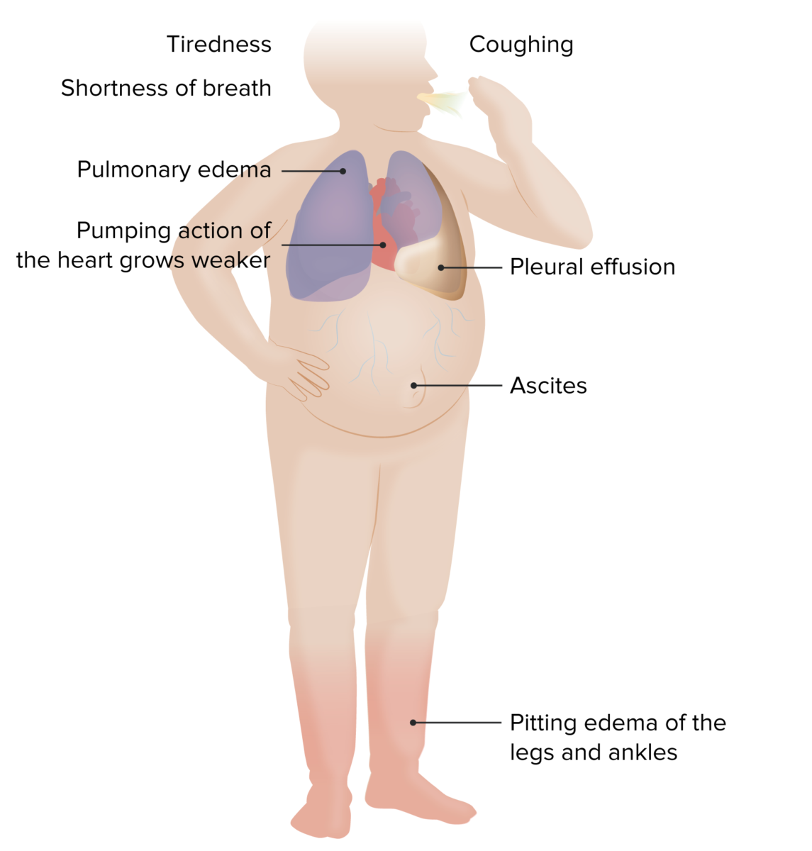
Symptoms/signs of HF

Image by Lecturio.

New York Heart Association (NYHA) functional classification

  • Class I: no limitations Limitations Conflict of Interest of physical activity
  • Class II: comfortable at rest but slight limitation during moderate/prolonged physical activity
  • Class III: comfortable only at rest. Daily activities such as dressing result in marked limitations Limitations Conflict of Interest of physical activity.
  • Class IV: symptomatic even at rest

American Heart Association classification

  • Stage A (at risk for HF): high risk for heart failure Heart Failure A heterogeneous condition in which the heart is unable to pump out sufficient blood to meet the metabolic need of the body. Heart failure can be caused by structural defects, functional abnormalities (ventricular dysfunction), or a sudden overload beyond its capacity. Chronic heart failure is more common than acute heart failure which results from sudden insult to cardiac function, such as myocardial infarction. Total Anomalous Pulmonary Venous Return (TAPVR) but without structural heart disease, objective evidence of cardiovascular disease, or symptoms of heart failure Heart Failure A heterogeneous condition in which the heart is unable to pump out sufficient blood to meet the metabolic need of the body. Heart failure can be caused by structural defects, functional abnormalities (ventricular dysfunction), or a sudden overload beyond its capacity. Chronic heart failure is more common than acute heart failure which results from sudden insult to cardiac function, such as myocardial infarction. Total Anomalous Pulmonary Venous Return (TAPVR)
  • Stage B (pre-HF): structural heart disease/objective evidence of minimal cardiovascular disease but without signs or symptoms of heart failure Heart Failure A heterogeneous condition in which the heart is unable to pump out sufficient blood to meet the metabolic need of the body. Heart failure can be caused by structural defects, functional abnormalities (ventricular dysfunction), or a sudden overload beyond its capacity. Chronic heart failure is more common than acute heart failure which results from sudden insult to cardiac function, such as myocardial infarction. Total Anomalous Pulmonary Venous Return (TAPVR)
  • Stage C (symptomatic HF): structural heart disease/objective evidence of moderate to severe cardiovascular disease with prior or current symptoms of heart failure Heart Failure A heterogeneous condition in which the heart is unable to pump out sufficient blood to meet the metabolic need of the body. Heart failure can be caused by structural defects, functional abnormalities (ventricular dysfunction), or a sudden overload beyond its capacity. Chronic heart failure is more common than acute heart failure which results from sudden insult to cardiac function, such as myocardial infarction. Total Anomalous Pulmonary Venous Return (TAPVR)
  • Stage D (advanced HF): refractory heart failure Heart Failure A heterogeneous condition in which the heart is unable to pump out sufficient blood to meet the metabolic need of the body. Heart failure can be caused by structural defects, functional abnormalities (ventricular dysfunction), or a sudden overload beyond its capacity. Chronic heart failure is more common than acute heart failure which results from sudden insult to cardiac function, such as myocardial infarction. Total Anomalous Pulmonary Venous Return (TAPVR)/objective evidence of severe cardiovascular disease requiring specialized interventions

Diagnosis

Initial approach

  • Diagnosis is often made based on:
    • Clinical evaluation:
      • Signs and symptoms of congestion
      • Evidence of fluid retention and/or hypoperfusion
      • Impairment of functional capacity
      • Precipitating factors cause exacerbation owing to ↑ cardiac workload.
      • Other factors: family history Family History Adult Health Maintenance of cardiac disease
    • Evidence of cardiac dysfunction on echocardiography Echocardiography Ultrasonic recording of the size, motion, and composition of the heart and surrounding tissues. The standard approach is transthoracic. Tricuspid Valve Atresia (TVA) (or cardiac catheterization Cardiac Catheterization Procedures in which placement of cardiac catheters is performed for therapeutic or diagnostic procedures. Cardiac Surgery)
  • Initial evaluation of suspected HF involves laboratory tests and imaging studies.
  • Laboratory tests evaluate risk factors, preexisting conditions and reversible causes of heart dysfunction.
    • CBC:
      • Anemia Anemia Anemia is a condition in which individuals have low Hb levels, which can arise from various causes. Anemia is accompanied by a reduced number of RBCs and may manifest with fatigue, shortness of breath, pallor, and weakness. Subtypes are classified by the size of RBCs, chronicity, and etiology. Anemia: Overview and Types can cause high-output cardiac failure.
      • Hemoglobin < 8 mg/dL is associated with impaired delivery of oxygen.
    • Fasting glucose Glucose A primary source of energy for living organisms. It is naturally occurring and is found in fruits and other parts of plants in its free state. It is used therapeutically in fluid and nutrient replacement. Lactose Intolerance and hemoglobin A1c: Diabetes Diabetes Diabetes mellitus (DM) is a metabolic disease characterized by hyperglycemia and dysfunction of the regulation of glucose metabolism by insulin. Type 1 DM is diagnosed mostly in children and young adults as the result of autoimmune destruction of β cells in the pancreas and the resulting lack of insulin. Type 2 DM has a significant association with obesity and is characterized by insulin resistance. Diabetes Mellitus mellitus is a common comorbidity.
    • BUN and creatinine:
      • BUN/creatinine: > 20 = prerenal failure → worse prognosis Prognosis A prediction of the probable outcome of a disease based on a individual’s condition and the usual course of the disease as seen in similar situations. Non-Hodgkin Lymphomas
      • Increased BUN → increased mortality Mortality All deaths reported in a given population. Measures of Health Status
      • Increase of ≥ 0.3 mg/dL in creatinine → increased mortality Mortality All deaths reported in a given population. Measures of Health Status[27]
    • Electrolytes Electrolytes Electrolytes are mineral salts that dissolve in water and dissociate into charged particles called ions, which can be either be positively (cations) or negatively (anions) charged. Electrolytes are distributed in the extracellular and intracellular compartments in different concentrations. Electrolytes are essential for various basic life-sustaining functions. Electrolytes:
      • Hyponatremia Hyponatremia Hyponatremia is defined as a decreased serum sodium (sNa+) concentration less than 135 mmol/L. Serum sodium is the greatest contributor to plasma osmolality, which is very tightly controlled via antidiuretic hormone (ADH) release from the hypothalamus and by the thirst mechanism. Hyponatremia → worse prognosis Prognosis A prediction of the probable outcome of a disease based on a individual’s condition and the usual course of the disease as seen in similar situations. Non-Hodgkin Lymphomas
      • Sodium Sodium A member of the alkali group of metals. It has the atomic symbol na, atomic number 11, and atomic weight 23. Hyponatremia < 137 mEq/L → decreased survival
    • Albumin Albumin Serum albumin from humans. It is an essential carrier of both endogenous substances, such as fatty acids and bilirubin, and of xenobiotics in the blood. Liver Function Tests: hypoalbuminemia Hypoalbuminemia A condition in which albumin level in blood (serum albumin) is below the normal range. Hypoalbuminemia may be due to decreased hepatic albumin synthesis, increased albumin catabolism, altered albumin distribution, or albumin loss through the urine (albuminuria). Nephrotic Syndrome in Children → worse prognosis Prognosis A prediction of the probable outcome of a disease based on a individual’s condition and the usual course of the disease as seen in similar situations. Non-Hodgkin Lymphomas
    • Bilirubin Bilirubin A bile pigment that is a degradation product of heme. Heme Metabolism:
      • Elevated total bilirubin Bilirubin A bile pigment that is a degradation product of heme. Heme Metabolism → worse prognosis Prognosis A prediction of the probable outcome of a disease based on a individual’s condition and the usual course of the disease as seen in similar situations. Non-Hodgkin Lymphomas
      • Results from congestive hepatopathy
    • Serum aminotransferase levels: reflect liver Liver The liver is the largest gland in the human body. The liver is found in the superior right quadrant of the abdomen and weighs approximately 1.5 kilograms. Its main functions are detoxification, metabolism, nutrient storage (e.g., iron and vitamins), synthesis of coagulation factors, formation of bile, filtration, and storage of blood. Liver: Anatomy function
    • Thyroid function tests Thyroid Function Tests Blood tests used to evaluate the functioning of the thyroid gland. Ion Channel Myopathy: Hyperthyroidism Hyperthyroidism Hypersecretion of thyroid hormones from the thyroid gland. Elevated levels of thyroid hormones increase basal metabolic rate. Thyrotoxicosis and Hyperthyroidism and hypothyroidism Hypothyroidism Hypothyroidism is a condition characterized by a deficiency of thyroid hormones. Iodine deficiency is the most common cause worldwide, but Hashimoto’s disease (autoimmune thyroiditis) is the leading cause in non-iodine-deficient regions. Hypothyroidism can lead to severe heart failure Heart Failure A heterogeneous condition in which the heart is unable to pump out sufficient blood to meet the metabolic need of the body. Heart failure can be caused by structural defects, functional abnormalities (ventricular dysfunction), or a sudden overload beyond its capacity. Chronic heart failure is more common than acute heart failure which results from sudden insult to cardiac function, such as myocardial infarction. Total Anomalous Pulmonary Venous Return (TAPVR) or contribute to the severity of existing heart failure Heart Failure A heterogeneous condition in which the heart is unable to pump out sufficient blood to meet the metabolic need of the body. Heart failure can be caused by structural defects, functional abnormalities (ventricular dysfunction), or a sudden overload beyond its capacity. Chronic heart failure is more common than acute heart failure which results from sudden insult to cardiac function, such as myocardial infarction. Total Anomalous Pulmonary Venous Return (TAPVR).
    • Lipid panel: ↑ Lipids Lipids Lipids are a diverse group of hydrophobic organic molecules, which include fats, oils, sterols, and waxes. Fatty Acids and Lipids → ↑ risk for atherosclerotic cardiovascular disease
    • BNP BNP A peptide that is secreted by the brain and the heart atria, stored mainly in cardiac ventricular myocardium. It can cause natriuresis; diuresis; vasodilation; and inhibits secretion of renin and aldosterone. It improves heart function. It contains 32 amino acids. Renal Sodium and Water Regulation/N-terminal pro-BNP (NT-proBNP) levels: high sensitivity and predictive value
    • Troponin: Obtained in the emergency setting during acute decompensated HF to determine the presence of acute coronary syndrome
  • Cardiopulmonary tests:
    • ECG ECG An electrocardiogram (ECG) is a graphic representation of the electrical activity of the heart plotted against time. Adhesive electrodes are affixed to the skin surface allowing measurement of cardiac impulses from many angles. The ECG provides 3-dimensional information about the conduction system of the heart, the myocardium, and other cardiac structures. Electrocardiogram (ECG):
      • Detects abnormalities in cardiac rhythm and conduction
      • Can show increased QRS voltage in leads I and aVL, Q waves, and ST- and T-wave abnormalities in patients Patients Individuals participating in the health care system for the purpose of receiving therapeutic, diagnostic, or preventive procedures. Clinician–Patient Relationship with prior MI MI MI is ischemia and death of an area of myocardial tissue due to insufficient blood flow and oxygenation, usually from thrombus formation on a ruptured atherosclerotic plaque in the epicardial arteries. Clinical presentation is most commonly with chest pain, but women and patients with diabetes may have atypical symptoms. Myocardial Infarction or with cardiomyopathy Cardiomyopathy Cardiomyopathy refers to a group of myocardial diseases associated with structural changes of the heart muscles (myocardium) and impaired systolic and/or diastolic function in the absence of other heart disorders (coronary artery disease, hypertension, valvular disease, and congenital heart disease). Cardiomyopathy: Overview and Types
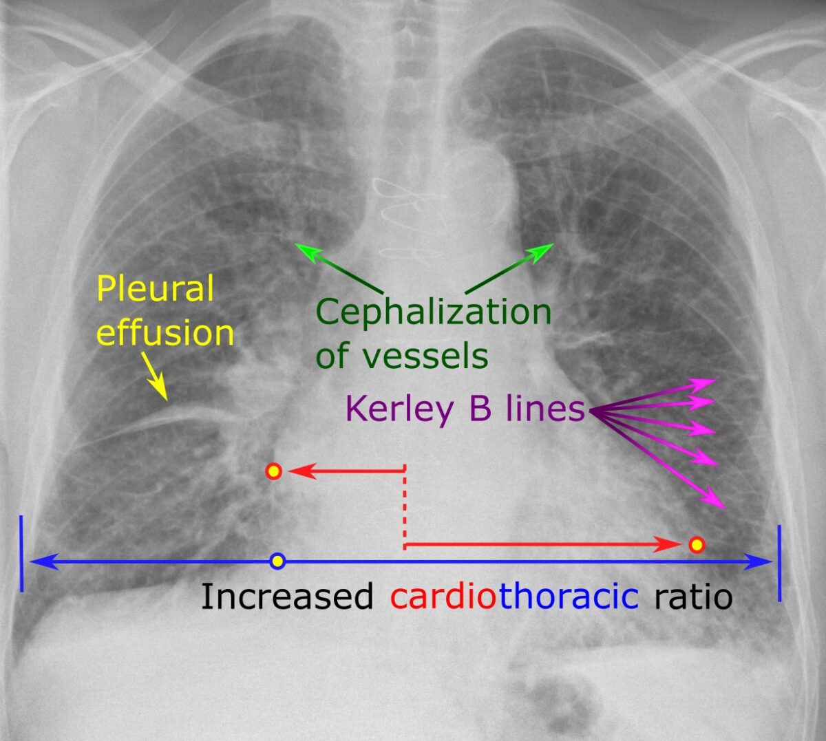
    • Chest X-ray X-ray Penetrating electromagnetic radiation emitted when the inner orbital electrons of an atom are excited and release radiant energy. X-ray wavelengths range from 1 pm to 10 nm. Hard x-rays are the higher energy, shorter wavelength x-rays. Soft x-rays or grenz rays are less energetic and longer in wavelength. The short wavelength end of the x-ray spectrum overlaps the gamma rays wavelength range. The distinction between gamma rays and x-rays is based on their radiation source. Pulmonary Function Tests: Can show cardiomegaly Cardiomegaly Enlargement of the heart, usually indicated by a cardiothoracic ratio above 0. 50. Heart enlargement may involve the right, the left, or both heart ventricles or heart atria. Cardiomegaly is a nonspecific symptom seen in patients with chronic systolic heart failure (heart failure) or several forms of cardiomyopathies. Ebstein’s Anomaly (ratio of cardiac size to thoracic size > 0.5), pulmonary edema Pulmonary edema Pulmonary edema is a condition caused by excess fluid within the lung parenchyma and alveoli as a consequence of a disease process. Based on etiology, pulmonary edema is classified as cardiogenic or noncardiogenic. Patients may present with progressive dyspnea, orthopnea, cough, or respiratory failure. Pulmonary Edema, pleural effusion Pleural Effusion Pleural effusion refers to the accumulation of fluid between the layers of the parietal and visceral pleura. Common causes of this condition include infection, malignancy, autoimmune disorders, or volume overload. Clinical manifestations include chest pain, cough, and dyspnea. Pleural Effusion, pulmonary vascular congestion, Kerley B lines Kerley B Lines Pulmonary Edema
    • Transthoracic echocardiogram Transthoracic echocardiogram Endocarditis ( TTE TTE Imaging of the Heart and Great Vessels) with Doppler Doppler Ultrasonography applying the doppler effect, with frequency-shifted ultrasound reflections produced by moving targets (usually red blood cells) in the bloodstream along the ultrasound axis in direct proportion to the velocity of movement of the targets, to determine both direction and velocity of blood flow. Ultrasound (Sonography) imaging:
      • Measures ejection fraction Ejection fraction Cardiac Cycle and assesses cardiac anatomy
      • Detects abnormalities in the valves, myocardium Myocardium The muscle tissue of the heart. It is composed of striated, involuntary muscle cells connected to form the contractile pump to generate blood flow. Heart: Anatomy, and pericardium Pericardium A conical fibroserous sac surrounding the heart and the roots of the great vessels (aorta; venae cavae; pulmonary artery). Pericardium consists of two sacs: the outer fibrous pericardium and the inner serous pericardium. The latter consists of an outer parietal layer facing the fibrous pericardium, and an inner visceral layer (epicardium) resting next to the heart, and a pericardial cavity between these two layers. Heart: Anatomy
    • Cardiac magnetic resonance Cardiac magnetic resonance Aortic Regurgitation imaging ( CMR CMR Imaging of the Heart and Great Vessels):
      • Characterizes the myocardium Myocardium The muscle tissue of the heart. It is composed of striated, involuntary muscle cells connected to form the contractile pump to generate blood flow. Heart: Anatomy
      • Detects damaged myocardium Myocardium The muscle tissue of the heart. It is composed of striated, involuntary muscle cells connected to form the contractile pump to generate blood flow. Heart: Anatomy, fibrosis Fibrosis Any pathological condition where fibrous connective tissue invades any organ, usually as a consequence of inflammation or other injury. Bronchiolitis Obliterans, and MI MI MI is ischemia and death of an area of myocardial tissue due to insufficient blood flow and oxygenation, usually from thrombus formation on a ruptured atherosclerotic plaque in the epicardial arteries. Clinical presentation is most commonly with chest pain, but women and patients with diabetes may have atypical symptoms. Myocardial Infarction (acute and chronic)
    • Multigated acquisition scan (radionuclide ventriculography):
      • Most accurate way to assess EF EF Cardiac Cycle, but it is rarely done
      • Usual indication: to obtain baseline assessment of ventricular function and to monitor cardiotoxicity of anthracyclines Anthracyclines Organic compounds that have a tetrahydronaphthacenedione ring structure attached by a glycosidic linkage to the amino sugar daunosamine. Antitumor Antibiotics ( chemotherapy Chemotherapy Osteosarcoma)
    • Cardiopulmonary exercise testing (CPET):
      • Can detect ischemic heart disease Ischemic heart disease Coronary heart disease (CHD), or ischemic heart disease, describes a situation in which an inadequate supply of blood to the myocardium exists due to a stenosis of the coronary arteries, typically from atherosclerosis. Coronary Heart Disease
      • Provides information regarding exercise capacity, thus helping to determine prognosis Prognosis A prediction of the probable outcome of a disease based on a individual’s condition and the usual course of the disease as seen in similar situations. Non-Hodgkin Lymphomas
      • Also helps assess appropriateness of advanced treatment such as transplantation
Table: Common diagnostic findings in HF
Test Findings
BNP BNP A peptide that is secreted by the brain and the heart atria, stored mainly in cardiac ventricular myocardium. It can cause natriuresis; diuresis; vasodilation; and inhibits secretion of renin and aldosterone. It improves heart function. It contains 32 amino acids. Renal Sodium and Water Regulation/pro-BNP
  • BNP BNP A peptide that is secreted by the brain and the heart atria, stored mainly in cardiac ventricular myocardium. It can cause natriuresis; diuresis; vasodilation; and inhibits secretion of renin and aldosterone. It improves heart function. It contains 32 amino acids. Renal Sodium and Water Regulation > 400 pg/mL
  • Pro-BNP
    • > 450 pg/mL (< 50 years of age)
    • > 900 pg/mL (50 to 75 years of age)
    • > 1800 pg/mL (> 75 years of age)
Chest X-ray X-ray Penetrating electromagnetic radiation emitted when the inner orbital electrons of an atom are excited and release radiant energy. X-ray wavelengths range from 1 pm to 10 nm. Hard x-rays are the higher energy, shorter wavelength x-rays. Soft x-rays or grenz rays are less energetic and longer in wavelength. The short wavelength end of the x-ray spectrum overlaps the gamma rays wavelength range. The distinction between gamma rays and x-rays is based on their radiation source. Pulmonary Function Tests
  • Pulmonary edema Pulmonary edema Pulmonary edema is a condition caused by excess fluid within the lung parenchyma and alveoli as a consequence of a disease process. Based on etiology, pulmonary edema is classified as cardiogenic or noncardiogenic. Patients may present with progressive dyspnea, orthopnea, cough, or respiratory failure. Pulmonary Edema
  • Cardiac/thoracic width > 0.5
ECG ECG An electrocardiogram (ECG) is a graphic representation of the electrical activity of the heart plotted against time. Adhesive electrodes are affixed to the skin surface allowing measurement of cardiac impulses from many angles. The ECG provides 3-dimensional information about the conduction system of the heart, the myocardium, and other cardiac structures. Electrocardiogram (ECG)
  • Left-axis deviation
  • Increased QRS duration
Echocardiography Echocardiography Ultrasonic recording of the size, motion, and composition of the heart and surrounding tissues. The standard approach is transthoracic. Tricuspid Valve Atresia (TVA)
  • Reduced/preserved EF EF Cardiac Cycle (normal > 55%)
  • Ventricular size and wall-motion abnormalities dependent on etiology
Congestive heart failure radiograph

Chest radiograph showing characteristic findings of HF

Image: “Chest radiograph with signs of congestive heart failure – annotated” by Mikael Häggström – Own work. License: CC0 1.0

Mnemonic

A good mnemonic to remember these findings on CXR CXR X-ray visualization of the chest and organs of the thoracic cavity. It is not restricted to visualization of the lungs. Pulmonary Function Tests is ABCDE:
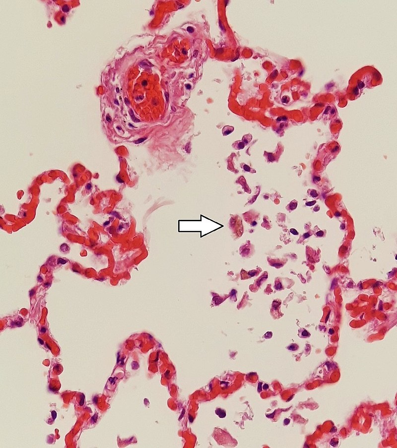
  • A: alveolar edema Edema Edema is a condition in which excess serous fluid accumulates in the body cavity or interstitial space of connective tissues. Edema is a symptom observed in several medical conditions. It can be categorized into 2 types, namely, peripheral (in the extremities) and internal (in an organ or body cavity). Edema (batwing)
  • B: Kerley B lines Kerley B Lines Pulmonary Edema
  • C: cardiomegaly Cardiomegaly Enlargement of the heart, usually indicated by a cardiothoracic ratio above 0. 50. Heart enlargement may involve the right, the left, or both heart ventricles or heart atria. Cardiomegaly is a nonspecific symptom seen in patients with chronic systolic heart failure (heart failure) or several forms of cardiomyopathies. Ebstein’s Anomaly
  • D: dilated upper lobe vessels
  • E: pleural effusion Pleural Effusion Pleural effusion refers to the accumulation of fluid between the layers of the parietal and visceral pleura. Common causes of this condition include infection, malignancy, autoimmune disorders, or volume overload. Clinical manifestations include chest pain, cough, and dyspnea. Pleural Effusion

Further testing

  • When clinical evaluation and initial tests confirm HF, additional tests to determine the etiology and risk stratification by classifying the stage of HF are performed.
  • Workup for coronary artery Coronary Artery Truncus Arteriosus disease (especially for unexplained HF):
    • Cardiac stress test: can assess functional impairment due to coronary heart disease Coronary heart disease Coronary heart disease (CHD), or ischemic heart disease, describes a situation in which an inadequate supply of blood to the myocardium exists due to a stenosis of the coronary arteries, typically from atherosclerosis. Coronary Heart Disease
      • Exercise ECG ECG An electrocardiogram (ECG) is a graphic representation of the electrical activity of the heart plotted against time. Adhesive electrodes are affixed to the skin surface allowing measurement of cardiac impulses from many angles. The ECG provides 3-dimensional information about the conduction system of the heart, the myocardium, and other cardiac structures. Electrocardiogram (ECG) testing
      • Radionuclide myocardial perfusion imaging Myocardial perfusion imaging The creation and display of functional images showing where the blood is flowing into the myocardium by following over time the distribution of tracers injected into the bloodstream. Nuclear Imaging (rMPI)
      • Stress echocardiography Echocardiography Ultrasonic recording of the size, motion, and composition of the heart and surrounding tissues. The standard approach is transthoracic. Tricuspid Valve Atresia (TVA)
    • Coronary angiography Angiography Radiography of blood vessels after injection of a contrast medium. Cardiac Surgery
  • Endomyocardial biopsy Endomyocardial biopsy Myocarditis:
    • Not a routine test 
    • An invasive procedure whose risks include cardiac tamponade Tamponade Pericardial effusion, usually of rapid onset, exceeding ventricular filling pressures and causing collapse of the heart with a markedly reduced cardiac output. Pericarditis, perforation Perforation A pathological hole in an organ, blood vessel or other soft part of the body, occurring in the absence of external force. Esophagitis, and thrombus formation
    • Performed when a diagnosis (e.g., amyloidosis Amyloidosis Amyloidosis is a disease caused by abnormal extracellular tissue deposition of fibrils composed of various misfolded low-molecular-weight protein subunits. These proteins are frequently byproducts of other pathological processes (e.g., multiple myeloma). Amyloidosis) is expected to influence management and the information provided by the test outweighs its risks
  • Right heart ( pulmonary artery Pulmonary artery The short wide vessel arising from the conus arteriosus of the right ventricle and conveying unaerated blood to the lungs. Lungs: Anatomy) catheterization:
  • As clinically indicated, specific workup is done for nonischemic conditions.
  • Additional workup is performed if there are complications such as congestive hepatopathy.
    • Rarely, invasive tests are needed.
    • In individuals with equivocal cases (especially in those with underlying liver Liver The liver is the largest gland in the human body. The liver is found in the superior right quadrant of the abdomen and weighs approximately 1.5 kilograms. Its main functions are detoxification, metabolism, nutrient storage (e.g., iron and vitamins), synthesis of coagulation factors, formation of bile, filtration, and storage of blood. Liver: Anatomy disease), percutaneous liver Liver The liver is the largest gland in the human body. The liver is found in the superior right quadrant of the abdomen and weighs approximately 1.5 kilograms. Its main functions are detoxification, metabolism, nutrient storage (e.g., iron and vitamins), synthesis of coagulation factors, formation of bile, filtration, and storage of blood. Liver: Anatomy biopsy Biopsy Removal and pathologic examination of specimens from the living body. Ewing Sarcoma is done.
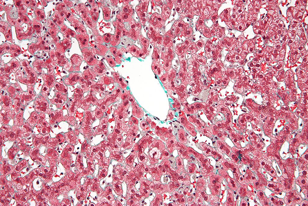
    • “Nutmeg liver Liver The liver is the largest gland in the human body. The liver is found in the superior right quadrant of the abdomen and weighs approximately 1.5 kilograms. Its main functions are detoxification, metabolism, nutrient storage (e.g., iron and vitamins), synthesis of coagulation factors, formation of bile, filtration, and storage of blood. Liver: Anatomy”: Gross appearance of red central areas (representing sinusoidal congestion) alternates with yellow areas (normal or fatty liver Liver The liver is the largest gland in the human body. The liver is found in the superior right quadrant of the abdomen and weighs approximately 1.5 kilograms. Its main functions are detoxification, metabolism, nutrient storage (e.g., iron and vitamins), synthesis of coagulation factors, formation of bile, filtration, and storage of blood. Liver: Anatomy tissue).
    • Histologically, liver Liver The liver is the largest gland in the human body. The liver is found in the superior right quadrant of the abdomen and weighs approximately 1.5 kilograms. Its main functions are detoxification, metabolism, nutrient storage (e.g., iron and vitamins), synthesis of coagulation factors, formation of bile, filtration, and storage of blood. Liver: Anatomy changes are seen with hemosiderin-laden macrophages Macrophages The relatively long-lived phagocytic cell of mammalian tissues that are derived from blood monocytes. Main types are peritoneal macrophages; alveolar macrophages; histiocytes; kupffer cells of the liver; and osteoclasts. They may further differentiate within chronic inflammatory lesions to epithelioid cells or may fuse to form foreign body giant cells or langhans giant cells. Innate Immunity: Phagocytes and Antigen Presentation.

Management

Management of acute decompensated HF

Acute decompensated HF causes respiratory distress, generally due to accumulation of fluid in the lungs Lungs Lungs are the main organs of the respiratory system. Lungs are paired viscera located in the thoracic cavity and are composed of spongy tissue. The primary function of the lungs is to oxygenate blood and eliminate CO2. Lungs: Anatomy ( pulmonary edema Pulmonary edema Pulmonary edema is a condition caused by excess fluid within the lung parenchyma and alveoli as a consequence of a disease process. Based on etiology, pulmonary edema is classified as cardiogenic or noncardiogenic. Patients may present with progressive dyspnea, orthopnea, cough, or respiratory failure. Pulmonary Edema).

Initial treatment measures

  • Assess airway Airway ABCDE Assessment and oxygenation:
  • Continuous cardiac telemetry Telemetry Transmission of the readings of instruments to a remote location by means of wires, radio waves, or other means. Crush Syndrome
  • Vital signs: check for hypertension Hypertension Hypertension, or high blood pressure, is a common disease that manifests as elevated systemic arterial pressures. Hypertension is most often asymptomatic and is found incidentally as part of a routine physical examination or during triage for an unrelated medical encounter. Hypertension or hypotension Hypotension Hypotension is defined as low blood pressure, specifically < 90/60 mm Hg, and is most commonly a physiologic response. Hypotension may be mild, serious, or life threatening, depending on the cause. Hypotension
  • Seated posture or upright position
  • IV diuretic therapy:
    • Relieves pulmonary edema Pulmonary edema Pulmonary edema is a condition caused by excess fluid within the lung parenchyma and alveoli as a consequence of a disease process. Based on etiology, pulmonary edema is classified as cardiogenic or noncardiogenic. Patients may present with progressive dyspnea, orthopnea, cough, or respiratory failure. Pulmonary Edema/acute volume overload
    • Options:
      • Furosemide Furosemide A benzoic-sulfonamide-furan. It is a diuretic with fast onset and short duration that is used for edema and chronic renal insufficiency. Loop Diuretics
      • Bumetanide Bumetanide A sulfamyl diuretic. Loop Diuretics
      • Torsemide Torsemide A pyridine and sulfonamide derivative that acts as a sodium-potassium chloride symporter inhibitor. It is used for the treatment of edema associated with congestive heart failure; chronic renal insufficiency; and liver diseases. It is also used for the management of hypertension. Loop Diuretics
  • For severe hypertension Severe hypertension A confirmed blood pressure ≥ 180 mm Hg systolic and/or ≥ 120 mm Hg diastolic. Uncontrolled Hypertension, administer vasodilator therapy.
    • Nitroprusside Nitroprusside A powerful vasodilator used in emergencies to lower blood pressure or to improve cardiac function. It is also an indicator for free sulfhydryl groups in proteins. Nitrates:
    • Nitroglycerin Nitroglycerin A volatile vasodilator which relieves angina pectoris by stimulating guanylate cyclase and lowering cytosolic calcium. It is also sometimes used for tocolysis and explosives. Nitrates:
      • Reduces venous tone
      • Can be given to those who have not responded fully to diuretic therapy
  • For hypotension Hypotension Hypotension is defined as low blood pressure, specifically < 90/60 mm Hg, and is most commonly a physiologic response. Hypotension may be mild, serious, or life threatening, depending on the cause. Hypotension/ cardiogenic shock Cardiogenic shock Shock resulting from diminution of cardiac output in heart disease. Types of Shock:
    • Inotropic agents: given in HFrEF (LV systolic dysfunction Systolic dysfunction Dilated Cardiomyopathy)
      • Dobutamine Dobutamine A catecholamine derivative with specificity for beta-1 adrenergic receptors. Sympathomimetic Drugs 
      • Milrinone Milrinone A positive inotropic cardiotonic agent with vasodilator properties. It inhibits camp phosphodiesterase type 3 activity in myocardium and vascular smooth muscle. Milrinone is a derivative of amrinone and has 20-30 times the inotropic potency of amrinone. Phosphodiesterase Inhibitors 
      • Low-dose dopamine Dopamine One of the catecholamine neurotransmitters in the brain. It is derived from tyrosine and is the precursor to norepinephrine and epinephrine. Dopamine is a major transmitter in the extrapyramidal system of the brain, and important in regulating movement. Receptors and Neurotransmitters of the CNS 
    • Vasopressors Vasopressors Sepsis in Children: option in HFpEF and LV outflow tract obstruction
      • Norepinephrine Norepinephrine Precursor of epinephrine that is secreted by the adrenal medulla and is a widespread central and autonomic neurotransmitter. Norepinephrine is the principal transmitter of most postganglionic sympathetic fibers, and of the diffuse projection system in the brain that arises from the locus ceruleus. Receptors and Neurotransmitters of the CNS 
      • High-dose dopamine Dopamine One of the catecholamine neurotransmitters in the brain. It is derived from tyrosine and is the precursor to norepinephrine and epinephrine. Dopamine is a major transmitter in the extrapyramidal system of the brain, and important in regulating movement. Receptors and Neurotransmitters of the CNS 
      • Vasopressin

Mnemonic

To recall the management of acute heart failure Heart Failure A heterogeneous condition in which the heart is unable to pump out sufficient blood to meet the metabolic need of the body. Heart failure can be caused by structural defects, functional abnormalities (ventricular dysfunction), or a sudden overload beyond its capacity. Chronic heart failure is more common than acute heart failure which results from sudden insult to cardiac function, such as myocardial infarction. Total Anomalous Pulmonary Venous Return (TAPVR) or exacerbation of HF, remember LMNOP:

  • Lasix/Loop diuretic
  • Morphine (for ACS)
  • Nitroglycerin
  • Oxygen
  • Position or Prop up the patient

Long-term management of HF

Lifestyle modifications decrease morbidity Morbidity The proportion of patients with a particular disease during a given year per given unit of population. Measures of Health Status and mortality Mortality All deaths reported in a given population. Measures of Health Status in heart failure Heart Failure A heterogeneous condition in which the heart is unable to pump out sufficient blood to meet the metabolic need of the body. Heart failure can be caused by structural defects, functional abnormalities (ventricular dysfunction), or a sudden overload beyond its capacity. Chronic heart failure is more common than acute heart failure which results from sudden insult to cardiac function, such as myocardial infarction. Total Anomalous Pulmonary Venous Return (TAPVR).

  • Weight loss Weight loss Decrease in existing body weight. Bariatric Surgery
  • Exercise (contraindicated if decompensating)
  • Restriction of sodium Sodium A member of the alkali group of metals. It has the atomic symbol na, atomic number 11, and atomic weight 23. Hyponatremia to 3 g/day
  • Restriction of fluids to 1.5–2 L/day in volume overload/ edema Edema Edema is a condition in which excess serous fluid accumulates in the body cavity or interstitial space of connective tissues. Edema is a symptom observed in several medical conditions. It can be categorized into 2 types, namely, peripheral (in the extremities) and internal (in an organ or body cavity). Edema
  • Weight monitoring (if > 5 lb weight gain, consult a physician)
  • Cessation of smoking Smoking Willful or deliberate act of inhaling and exhaling smoke from burning substances or agents held by hand. Interstitial Lung Diseases and alcohol consumption
  • Avoidance of illicit drug use
  • Avoid drugs that worsen HF or trigger Trigger The type of signal that initiates the inspiratory phase by the ventilator Invasive Mechanical Ventilation exacerbation, such as:
    • NSAIDs NSAIDS Primary vs Secondary Headaches
    • Thiazolidinediones Thiazolidinediones Thiazoles with two keto oxygens. Members are insulin-sensitizing agents which overcome insulin resistance by activation of the peroxisome proliferator activated receptor gamma (ppar-gamma). Non-insulinotropic Diabetes Drugs
    • Nondihydropyridine calcium Calcium A basic element found in nearly all tissues. It is a member of the alkaline earth family of metals with the atomic symbol ca, atomic number 20, and atomic weight 40. Calcium is the most abundant mineral in the body and combines with phosphorus to form calcium phosphate in the bones and teeth. It is essential for the normal functioning of nerves and muscles and plays a role in blood coagulation (as factor IV) and in many enzymatic processes. Electrolytes channel blockers
    • Trimethoprim Trimethoprim The sulfonamides are a class of antimicrobial drugs inhibiting folic acid synthesize in pathogens. The prototypical drug in the class is sulfamethoxazole. Although not technically sulfonamides, trimethoprim, dapsone, and pyrimethamine are also important antimicrobial agents inhibiting folic acid synthesis. The agents are often combined with sulfonamides, resulting in a synergistic effect. Sulfonamides and Trimethoprim sulfamethoxazole Sulfamethoxazole A bacteriostatic antibacterial agent that interferes with folic acid synthesis in susceptible bacteria. Its broad spectrum of activity has been limited by the development of resistance. Sulfonamides and Trimethoprim (↑ potassium Potassium An element in the alkali group of metals with an atomic symbol k, atomic number 19, and atomic weight 39. 10. It is the chief cation in the intracellular fluid of muscle and other cells. Potassium ion is a strong electrolyte that plays a significant role in the regulation of fluid volume and maintenance of the water-electrolyte balance. Hyperkalemia when taken with drugs for HF)
    • Phosphodiesterase-3 inhibitors Phosphodiesterase-3 Inhibitors Compounds that specifically inhibit phosphodiesterase 3. Phosphodiesterase Inhibitors
    • Antidepressants, especially tricyclic antidepressants Tricyclic antidepressants Tricyclic antidepressants (TCAs) are a class of medications used in the management of mood disorders, primarily depression. These agents, named after their 3-ring chemical structure, act via reuptake inhibition of neurotransmitters (particularly norepinephrine and serotonin) in the brain. Tricyclic Antidepressants
    • Antiarrhythmics such as class 1C and dronedarone Dronedarone A non-iodinated derivative of amiodarone that is used for the treatment of arrhythmia. Class 3 Antiarrhythmic Drugs (Potassium Channel Blockers) ( negative inotropic effect Negative Inotropic Effect Class 4 Antiarrhythmic Drugs (Calcium Channel Blockers))
    • Dipeptidyl peptidase-4 Dipeptidyl peptidase-4 A serine protease that catalyses the release of an n-terminal dipeptide. Several biologically-active peptides have been identified as dipeptidyl peptidase 4 substrates including incretins; neuropeptides; and chemokines. The protein is also found bound to adenosine deaminase on the t-cell surface and is believed to play a role in t-cell activation. Angioedema inhibitors (alogliptin, saxagliptin)
    • Cardiotoxic chemotherapy Chemotherapy Osteosarcoma
    • Tumor Tumor Inflammation necrosis Necrosis The death of cells in an organ or tissue due to disease, injury or failure of the blood supply. Ischemic Cell Damage factor–alpha inhibitors 
  • Pneumococcal polysaccharide (PPSV23) vaccine Vaccine Suspensions of killed or attenuated microorganisms (bacteria, viruses, fungi, protozoa), antigenic proteins, synthetic constructs, or other bio-molecular derivatives, administered for the prevention, amelioration, or treatment of infectious and other diseases. Vaccination (individuals with HF are more susceptible to pneumococcal pneumonia Pneumonia Pneumonia or pulmonary inflammation is an acute or chronic inflammation of lung tissue. Causes include infection with bacteria, viruses, or fungi. In more rare cases, pneumonia can also be caused through toxic triggers through inhalation of toxic substances, immunological processes, or in the course of radiotherapy. Pneumonia)
  • Annual influenza Influenza Influenza viruses are members of the Orthomyxoviridae family and the causative organisms of influenza, a highly contagious febrile respiratory disease. There are 3 primary influenza viruses (A, B, and C) and various subtypes, which are classified based on their virulent surface antigens, hemagglutinin (HA) and neuraminidase (NA). Influenza typically presents with a fever, myalgia, headache, and symptoms of an upper respiratory infection. Influenza Viruses/Influenza vaccine Vaccine Suspensions of killed or attenuated microorganisms (bacteria, viruses, fungi, protozoa), antigenic proteins, synthetic constructs, or other bio-molecular derivatives, administered for the prevention, amelioration, or treatment of infectious and other diseases. Vaccination

Pharmacologic therapy

Overview:

  • Guideline-directed medical therapy (4 core drug classes included in the optimal treatment proven to reduce morbidity Morbidity The proportion of patients with a particular disease during a given year per given unit of population. Measures of Health Status and mortality Mortality All deaths reported in a given population. Measures of Health Status):
    • Renin Renin A highly specific (leu-leu) endopeptidase that generates angiotensin I from its precursor angiotensinogen, leading to a cascade of reactions which elevate blood pressure and increase sodium retention by the kidney in the renin-angiotensin system. Renal Sodium and Water Regulation–angiotensin system inhibitor:
      • Angiotensin-receptor– neprilysin Neprilysin Enzyme that is a major constituent of kidney brush-border membranes and is also present to a lesser degree in the brain and other tissues. It preferentially catalyzes cleavage at the amino group of hydrophobic residues of the b-chain of insulin as well as opioid peptides and other biologically active peptides. The enzyme is inhibited primarily by edta, phosphoramidon, and thiorphan and is reactivated by zinc. Neprilysin is identical to common acute lymphoblastic leukemia antigen (CALLA antigen), an important marker in the diagnosis of human acute lymphocytic leukemia. There is no relationship with calla plant. Renin-Angiotensin-Aldosterone System Inhibitors inhibitor ( ARNi ARNi Renin-Angiotensin-Aldosterone System Inhibitors), OR
      • Angiotensin-converting–enzyme inhibitor ( ACEi ACEi A class of drugs whose main indications are the treatment of hypertension and heart failure. They exert their hemodynamic effect mainly by inhibiting the renin-angiotensin system. They also modulate sympathetic nervous system activity and increase prostaglandin synthesis. They cause mainly vasodilation and mild natriuresis without affecting heart rate and contractility. Renin-Angiotensin-Aldosterone System Inhibitors), OR 
      • Angiotensin receptor Receptor Receptors are proteins located either on the surface of or within a cell that can bind to signaling molecules known as ligands (e.g., hormones) and cause some type of response within the cell. Receptors blocker (ARB))
    • Beta blocker
    • Mineralocorticoid receptor Receptor Receptors are proteins located either on the surface of or within a cell that can bind to signaling molecules known as ligands (e.g., hormones) and cause some type of response within the cell. Receptors antagonist ( MRA MRA Imaging of the Heart and Great Vessels)
    • Sodium Sodium A member of the alkali group of metals. It has the atomic symbol na, atomic number 11, and atomic weight 23. Hyponatremia glucose Glucose A primary source of energy for living organisms. It is naturally occurring and is found in fruits and other parts of plants in its free state. It is used therapeutically in fluid and nutrient replacement. Lactose Intolerance cotransporter 2 inhibitor (SGLT2i)
  • In HFrEF:
    • Depending on the stage of HFrEF, general initial long-term therapy includes:
      • Renin Renin A highly specific (leu-leu) endopeptidase that generates angiotensin I from its precursor angiotensinogen, leading to a cascade of reactions which elevate blood pressure and increase sodium retention by the kidney in the renin-angiotensin system. Renal Sodium and Water Regulation–angiotensin system inhibitor
      • Beta blocker
      • Diuretic as needed for volume overload 
    • Secondary long-term agents (added to initial therapy for specific criteria):
      • MRA MRA Imaging of the Heart and Great Vessels
      • SGLT2i
      • Ivabradine Ivabradine A benzazepine derivative and selective hyperpolarization-activated cyclic nucleotide-gated channels inhibitor that lowers the heart rate. It is used in the treatment of chronic stable angina in patients unable to take beta-adrenergic blockers, and in the treatment of heart failure. Heart Failure and Angina Medication
      • Hydralazine Hydralazine A direct-acting vasodilator that is used as an antihypertensive agent. Heart Failure and Angina Medication plus nitrate
      • Digoxin Digoxin A cardiotonic glycoside obtained mainly from digitalis lanata; it consists of three sugars and the aglycone digoxigenin. Digoxin has positive inotropic and negative chronotropic activity. It is used to control ventricular rate in atrial fibrillation and in the management of congestive heart failure with atrial fibrillation. Its use in congestive heart failure and sinus rhythm is less certain. The margin between toxic and therapeutic doses is small. Cardiac Glycosides
      • Vericiguat
  • In HFpEF:

Drug classes in detail:

  • ARNi ARNi Renin-Angiotensin-Aldosterone System Inhibitors (sacubitril– valsartan Valsartan A tetrazole derivative and angiotensin II type 1 receptor blocker that is used to treat hypertension. Hypertension Drugs):
    • Role of neprilysin Neprilysin Enzyme that is a major constituent of kidney brush-border membranes and is also present to a lesser degree in the brain and other tissues. It preferentially catalyzes cleavage at the amino group of hydrophobic residues of the b-chain of insulin as well as opioid peptides and other biologically active peptides. The enzyme is inhibited primarily by edta, phosphoramidon, and thiorphan and is reactivated by zinc. Neprilysin is identical to common acute lymphoblastic leukemia antigen (CALLA antigen), an important marker in the diagnosis of human acute lymphocytic leukemia. There is no relationship with calla plant. Renin-Angiotensin-Aldosterone System Inhibitors:
      • Degrades BNP BNP A peptide that is secreted by the brain and the heart atria, stored mainly in cardiac ventricular myocardium. It can cause natriuresis; diuresis; vasodilation; and inhibits secretion of renin and aldosterone. It improves heart function. It contains 32 amino acids. Renal Sodium and Water Regulation; so if neprilysin Neprilysin Enzyme that is a major constituent of kidney brush-border membranes and is also present to a lesser degree in the brain and other tissues. It preferentially catalyzes cleavage at the amino group of hydrophobic residues of the b-chain of insulin as well as opioid peptides and other biologically active peptides. The enzyme is inhibited primarily by edta, phosphoramidon, and thiorphan and is reactivated by zinc. Neprilysin is identical to common acute lymphoblastic leukemia antigen (CALLA antigen), an important marker in the diagnosis of human acute lymphocytic leukemia. There is no relationship with calla plant. Renin-Angiotensin-Aldosterone System Inhibitors is inhibited, BNP BNP A peptide that is secreted by the brain and the heart atria, stored mainly in cardiac ventricular myocardium. It can cause natriuresis; diuresis; vasodilation; and inhibits secretion of renin and aldosterone. It improves heart function. It contains 32 amino acids. Renal Sodium and Water Regulation increases.
      • NT-proBNP, however, is not degraded by neprilysin Neprilysin Enzyme that is a major constituent of kidney brush-border membranes and is also present to a lesser degree in the brain and other tissues. It preferentially catalyzes cleavage at the amino group of hydrophobic residues of the b-chain of insulin as well as opioid peptides and other biologically active peptides. The enzyme is inhibited primarily by edta, phosphoramidon, and thiorphan and is reactivated by zinc. Neprilysin is identical to common acute lymphoblastic leukemia antigen (CALLA antigen), an important marker in the diagnosis of human acute lymphocytic leukemia. There is no relationship with calla plant. Renin-Angiotensin-Aldosterone System Inhibitors; so NT-proBNP can be used for HF monitoring while on ARNi ARNi Renin-Angiotensin-Aldosterone System Inhibitors.
    • Drug components and mechanism:
      • Sacubitril: inhibits neprilysin Neprilysin Enzyme that is a major constituent of kidney brush-border membranes and is also present to a lesser degree in the brain and other tissues. It preferentially catalyzes cleavage at the amino group of hydrophobic residues of the b-chain of insulin as well as opioid peptides and other biologically active peptides. The enzyme is inhibited primarily by edta, phosphoramidon, and thiorphan and is reactivated by zinc. Neprilysin is identical to common acute lymphoblastic leukemia antigen (CALLA antigen), an important marker in the diagnosis of human acute lymphocytic leukemia. There is no relationship with calla plant. Renin-Angiotensin-Aldosterone System Inhibitors → ↑ levels of peptides (including natriuretic peptides Natriuretic peptides Peptides that regulate the water-electrolyte balance in the body, also known as natriuretic peptide hormones. Several have been sequenced (atrial natriuretic factor; brain natriuretic peptide; c-type natriuretic peptide). Arterial Pressure Regulation, bradykinin Bradykinin A nonapeptide messenger that is enzymatically produced from kallidin in the blood where it is a potent but short-lived agent of arteriolar dilation and increased capillary permeability. Bradykinin is also released from mast cells during asthma attacks, from gut walls as a gastrointestinal vasodilator, from damaged tissues as a pain signal, and may be a neurotransmitter. Hereditary Angioedema (C1 Esterase Inhibitor Deficiency), and adrenomedullin); causes vasodilation Vasodilation The physiological widening of blood vessels by relaxing the underlying vascular smooth muscle. Pulmonary Hypertension Drugs and natriuresis 
      • Valsartan Valsartan A tetrazole derivative and angiotensin II type 1 receptor blocker that is used to treat hypertension. Hypertension Drugs: an ARB
    • Benefits: improved outcomes (↓ mortality Mortality All deaths reported in a given population. Measures of Health Status and rehospitalization) in NYHA class II–III HF as compared with enalapril Enalapril An angiotensin-converting enzyme inhibitor that is used to treat hypertension and heart failure. Hypertension Drugs
  • Other renin Renin A highly specific (leu-leu) endopeptidase that generates angiotensin I from its precursor angiotensinogen, leading to a cascade of reactions which elevate blood pressure and increase sodium retention by the kidney in the renin-angiotensin system. Renal Sodium and Water Regulation–angiotensin system inhibitors (given when ARNi ARNi Renin-Angiotensin-Aldosterone System Inhibitors is not feasible):
    • ACE inhibitors ACE inhibitors Truncus Arteriosus:
      • Mechanism: prevents formation of angiotensin II Angiotensin II An octapeptide that is a potent but labile vasoconstrictor. It is produced from angiotensin I after the removal of two amino acids at the c-terminal by angiotensin converting enzyme. The amino acid in position 5 varies in different species. To block vasoconstriction and hypertension effect of angiotensin II, patients are often treated with ace inhibitors or with angiotensin II type 1 receptor blockers. Renal Sodium and Water Regulation (a vasoconstrictor) from angiotensin I
      • Effects: ↓ cardiac remodeling  and ↓ adverse effects of angiotensin on the heart, kidneys Kidneys The kidneys are a pair of bean-shaped organs located retroperitoneally against the posterior wall of the abdomen on either side of the spine. As part of the urinary tract, the kidneys are responsible for blood filtration and excretion of water-soluble waste in the urine. Kidneys: Anatomy, and blood vessels → ↓ LV afterload Afterload Afterload is the resistance in the aorta that prevents blood from leaving the heart. Afterload represents the pressure the LV needs to overcome to eject blood into the aorta. Cardiac Mechanics with improved vascular tone and endothelial function 
      • Benefits: decreased mortality Mortality All deaths reported in a given population. Measures of Health Status and rehospitalization in HFrEF
      • Cannot be used with ARNi ARNi Renin-Angiotensin-Aldosterone System Inhibitors
      • Contraindicated in pregnancy Pregnancy The status during which female mammals carry their developing young (embryos or fetuses) in utero before birth, beginning from fertilization to birth. Pregnancy: Diagnosis, Physiology, and Care and in individuals with history of angioedema Angioedema Angioedema is a localized, self-limited (but potentially life-threatening), nonpitting, asymmetrical edema occurring in the deep layers of the skin and mucosal tissue. The common underlying pathophysiology involves inflammatory mediators triggering significant vasodilation and increased capillary permeability. Angioedema
      • Monitor electrolytes Electrolytes Electrolytes are mineral salts that dissolve in water and dissociate into charged particles called ions, which can be either be positively (cations) or negatively (anions) charged. Electrolytes are distributed in the extracellular and intracellular compartments in different concentrations. Electrolytes are essential for various basic life-sustaining functions. Electrolytes (especially potassium Potassium An element in the alkali group of metals with an atomic symbol k, atomic number 19, and atomic weight 39. 10. It is the chief cation in the intracellular fluid of muscle and other cells. Potassium ion is a strong electrolyte that plays a significant role in the regulation of fluid volume and maintenance of the water-electrolyte balance. Hyperkalemia) and renal function
      • Examples: enalapril Enalapril An angiotensin-converting enzyme inhibitor that is used to treat hypertension and heart failure. Hypertension Drugs, lisinopril Lisinopril One of the angiotensin-converting enzyme inhibitors (ACE inhibitors), orally active, that has been used in the treatment of hypertension and congestive heart failure. Renin-Angiotensin-Aldosterone System Inhibitors, ramipril Ramipril A long-acting angiotensin-converting enzyme inhibitor. It is a prodrug that is transformed in the liver to its active metabolite ramiprilat. Hypertension Drugs
    • ARBs ARBs Agents that antagonize angiotensin receptors. Many drugs in this class specifically target the angiotensin type 1 receptor. Heart Failure and Angina Medication:
      • Mechanism: blocks angiotensin II Angiotensin II An octapeptide that is a potent but labile vasoconstrictor. It is produced from angiotensin I after the removal of two amino acids at the c-terminal by angiotensin converting enzyme. The amino acid in position 5 varies in different species. To block vasoconstriction and hypertension effect of angiotensin II, patients are often treated with ace inhibitors or with angiotensin II type 1 receptor blockers. Renal Sodium and Water Regulation receptors Receptors Receptors are proteins located either on the surface of or within a cell that can bind to signaling molecules known as ligands (e.g., hormones) and cause some type of response within the cell. Receptors (displaces angiotensin II Angiotensin II An octapeptide that is a potent but labile vasoconstrictor. It is produced from angiotensin I after the removal of two amino acids at the c-terminal by angiotensin converting enzyme. The amino acid in position 5 varies in different species. To block vasoconstriction and hypertension effect of angiotensin II, patients are often treated with ace inhibitors or with angiotensin II type 1 receptor blockers. Renal Sodium and Water Regulation from the receptor Receptor Receptors are proteins located either on the surface of or within a cell that can bind to signaling molecules known as ligands (e.g., hormones) and cause some type of response within the cell. Receptors)
      • Effects: ↓ adverse effects of angiotensin II Angiotensin II An octapeptide that is a potent but labile vasoconstrictor. It is produced from angiotensin I after the removal of two amino acids at the c-terminal by angiotensin converting enzyme. The amino acid in position 5 varies in different species. To block vasoconstriction and hypertension effect of angiotensin II, patients are often treated with ace inhibitors or with angiotensin II type 1 receptor blockers. Renal Sodium and Water Regulation
      • Benefits: decreased mortality Mortality All deaths reported in a given population. Measures of Health Status and rehospitalization in HFrEF
      • For individuals with contraindication or intolerance to ACEis ACEIs A class of drugs whose main indications are the treatment of hypertension and heart failure. They exert their hemodynamic effect mainly by inhibiting the renin-angiotensin system. They also modulate sympathetic nervous system activity and increase prostaglandin synthesis. They cause mainly vasodilation and mild natriuresis without affecting heart rate and contractility. Heart Failure and Angina Medication (e.g., cough which is due to kinin accumulation from ACE inhibition)
      • Avoid combination with ACEis ACEIs A class of drugs whose main indications are the treatment of hypertension and heart failure. They exert their hemodynamic effect mainly by inhibiting the renin-angiotensin system. They also modulate sympathetic nervous system activity and increase prostaglandin synthesis. They cause mainly vasodilation and mild natriuresis without affecting heart rate and contractility. Heart Failure and Angina Medication.
      • Monitor electrolytes Electrolytes Electrolytes are mineral salts that dissolve in water and dissociate into charged particles called ions, which can be either be positively (cations) or negatively (anions) charged. Electrolytes are distributed in the extracellular and intracellular compartments in different concentrations. Electrolytes are essential for various basic life-sustaining functions. Electrolytes (especially potassium Potassium An element in the alkali group of metals with an atomic symbol k, atomic number 19, and atomic weight 39. 10. It is the chief cation in the intracellular fluid of muscle and other cells. Potassium ion is a strong electrolyte that plays a significant role in the regulation of fluid volume and maintenance of the water-electrolyte balance. Hyperkalemia) and renal function
      • Examples: valsartan Valsartan A tetrazole derivative and angiotensin II type 1 receptor blocker that is used to treat hypertension. Hypertension Drugs, losartan Losartan An antagonist of angiotensin type 1 receptor with antihypertensive activity due to the reduced pressor effect of angiotensin II. Hypertension Drugs, candesartan
  • Beta blockers:
    • Mechanism: ↓ catecholamine effects on the myocardium Myocardium The muscle tissue of the heart. It is composed of striated, involuntary muscle cells connected to form the contractile pump to generate blood flow. Heart: Anatomy, ↓ vasoconstrictors
    • Effects: ↓ heart rate Heart rate The number of times the heart ventricles contract per unit of time, usually per minute. Cardiac Physiology, ↓ myocardial demand, ↓ premature Premature Childbirth before 37 weeks of pregnancy (259 days from the first day of the mother’s last menstrual period, or 245 days after fertilization). Necrotizing Enterocolitis ventricular beats
    • Benefits: associated with decreased mortality Mortality All deaths reported in a given population. Measures of Health Status and incidence Incidence The number of new cases of a given disease during a given period in a specified population. It also is used for the rate at which new events occur in a defined population. It is differentiated from prevalence, which refers to all cases in the population at a given time. Measures of Disease Frequency of sudden cardiac death Sudden cardiac death Cardiac arrest is the sudden, complete cessation of cardiac output with hemodynamic collapse. Patients present as pulseless, unresponsive, and apneic. Rhythms associated with cardiac arrest are ventricular fibrillation/tachycardia, asystole, or pulseless electrical activity. Cardiac Arrest
    • Use after stabilizing with ARNi ARNi Renin-Angiotensin-Aldosterone System Inhibitors/ ACEi ACEi A class of drugs whose main indications are the treatment of hypertension and heart failure. They exert their hemodynamic effect mainly by inhibiting the renin-angiotensin system. They also modulate sympathetic nervous system activity and increase prostaglandin synthesis. They cause mainly vasodilation and mild natriuresis without affecting heart rate and contractility. Renin-Angiotensin-Aldosterone System Inhibitors/ARB
    • Contraindicated in acute decompensated HF owing to negative inotropic/chronotropic effects
    • Established beta blockers with benefits in HF:
      • Carvedilol Carvedilol A carbazole and propanol derivative that acts as a non-cardioselective beta blocker and vasodilator. It has blocking activity for alpha 1 adrenergic receptors and, at higher doses, may function as a blocker of calcium channels; it also has antioxidant properties. Carvedilol is used in the treatment of hypertension; angina pectoris; and heart failure. It can also reduce the risk of death following myocardial infarction. Class 2 Antiarrhythmic Drugs (Beta Blockers) (both immediate-release and extended-release formulations)
      • Extended-release metoprolol Metoprolol A selective adrenergic beta-1 blocking agent that is commonly used to treat angina pectoris; hypertension; and cardiac arrhythmias. Antiadrenergic Drugs succinate
      • Bisoprolol Bisoprolol A cardioselective beta-1 adrenergic blocker. It is effective in the management of hypertension and angina pectoris. Class 2 Antiarrhythmic Drugs (Beta Blockers) 
  • Diuretics Diuretics Agents that promote the excretion of urine through their effects on kidney function. Heart Failure and Angina Medication:
    • Mechanism:
      • Loop diuretic: ↓ reabsorption of sodium Sodium A member of the alkali group of metals. It has the atomic symbol na, atomic number 11, and atomic weight 23. Hyponatremia in the thick ascending limb Thick ascending limb Renal Sodium and Water Regulation of the loop of Henle Loop of Henle The U-shaped portion of the renal tubule in the kidney medulla, consisting of a descending limb and an ascending limb. It is situated between the proximal kidney tubule and the distal kidney tubule. Tubular System
      • Thiazide Thiazide Heterocyclic compounds with sulfur and nitrogen in the ring. This term commonly refers to the benzothiadiazines that inhibit sodium-potassium-chloride symporters and are used as diuretics. Hyponatremia: inhibits reabsorption of sodium Sodium A member of the alkali group of metals. It has the atomic symbol na, atomic number 11, and atomic weight 23. Hyponatremia in the distal tubules
    • Effects: ↑ excretion of sodium Sodium A member of the alkali group of metals. It has the atomic symbol na, atomic number 11, and atomic weight 23. Hyponatremia and water → symptomatic relief from volume overload
    • No improvement in prognosis Prognosis A prediction of the probable outcome of a disease based on a individual’s condition and the usual course of the disease as seen in similar situations. Non-Hodgkin Lymphomas/does not decrease mortality Mortality All deaths reported in a given population. Measures of Health Status
    • Examples:
      • Loop diuretics Diuretics Agents that promote the excretion of urine through their effects on kidney function. Heart Failure and Angina Medication: furosemide Furosemide A benzoic-sulfonamide-furan. It is a diuretic with fast onset and short duration that is used for edema and chronic renal insufficiency. Loop Diuretics, torsemide Torsemide A pyridine and sulfonamide derivative that acts as a sodium-potassium chloride symporter inhibitor. It is used for the treatment of edema associated with congestive heart failure; chronic renal insufficiency; and liver diseases. It is also used for the management of hypertension. Loop Diuretics, bumetanide Bumetanide A sulfamyl diuretic. Loop Diuretics
      • Thiazides: metolazone Metolazone A quinazoline-sulfonamide derived diuretic that functions by inhibiting sodium chloride symporters. Hypertension Drugs, chlorothiazide Chlorothiazide A thiazide diuretic with actions and uses similar to those of hydrochlorothiazide. Hypertension Drugs, indapamide Indapamide A benzamide-sulfonamide-indole derived diuretic that functions by inhibiting sodium chloride symporters. Hypertension Drugs, chlorthalidone Chlorthalidone A benzenesulfonamide-phthalimidine that tautomerizes to a benzophenones form. It is considered a thiazide-like diuretic. Thiazide Diuretics, hydrochlorothiazide Hydrochlorothiazide A thiazide diuretic often considered the prototypical member of this class. It reduces the reabsorption of electrolytes from the renal tubules. This results in increased excretion of water and electrolytes, including sodium, potassium, chloride, and magnesium. It is used in the treatment of several disorders including edema, hypertension, diabetes insipidus, and hypoparathyroidism. Thiazide Diuretics 
  • MRA MRA Imaging of the Heart and Great Vessels:
    • Mechanism: blocks adverse effects of aldosterone Aldosterone A hormone secreted by the adrenal cortex that regulates electrolyte and water balance by increasing the renal retention of sodium and the excretion of potassium. Hyperkalemia on the heart, kidneys Kidneys The kidneys are a pair of bean-shaped organs located retroperitoneally against the posterior wall of the abdomen on either side of the spine. As part of the urinary tract, the kidneys are responsible for blood filtration and excretion of water-soluble waste in the urine. Kidneys: Anatomy, and blood vessels
    • Effects: diuretic and blood pressure–lowering effects
    • Benefits: decreases mortality Mortality All deaths reported in a given population. Measures of Health Status and hospitalizations
    • Given in HFrEF and NYHA class II–IV symptoms and post-MI with EF EF Cardiac Cycle ≤ 40% 
    • Associated with increased potassium Potassium An element in the alkali group of metals with an atomic symbol k, atomic number 19, and atomic weight 39. 10. It is the chief cation in the intracellular fluid of muscle and other cells. Potassium ion is a strong electrolyte that plays a significant role in the regulation of fluid volume and maintenance of the water-electrolyte balance. Hyperkalemia (electrolyte and renal function monitoring needed)
    • Examples: spironolactone Spironolactone A potassium sparing diuretic that acts by antagonism of aldosterone in the distal renal tubules. It is used mainly in the treatment of refractory edema in patients with congestive heart failure, nephrotic syndrome, or hepatic cirrhosis. Its effects on the endocrine system are utilized in the treatments of hirsutism and acne but they can lead to adverse effects. Potassium-sparing Diuretics, eplerenone Eplerenone A spironolactone derivative and selective aldosterone receptor antagonist that is used in the management of hypertension and congestive heart failure, post-myocardial infarction. Potassium-sparing Diuretics
  • SGLT2i:
    • Mechanism: ↑ osmotic diuresis Osmotic diuresis Volume Depletion and Dehydration and natriuresis,  ↑ urinary glucose Glucose A primary source of energy for living organisms. It is naturally occurring and is found in fruits and other parts of plants in its free state. It is used therapeutically in fluid and nutrient replacement. Lactose Intolerance excretion 
    • Effects:
      • With natriuresis → reduced preload Preload Cardiac Mechanics
      • With improved endothelial function and vasodilation Vasodilation The physiological widening of blood vessels by relaxing the underlying vascular smooth muscle. Pulmonary Hypertension Drugs → reduced afterload Afterload Afterload is the resistance in the aorta that prevents blood from leaving the heart. Afterload represents the pressure the LV needs to overcome to eject blood into the aorta. Cardiac Mechanics
    • Benefits: decreased mortality Mortality All deaths reported in a given population. Measures of Health Status and hospitalizations in those with HF with or without diabetes Diabetes Diabetes mellitus (DM) is a metabolic disease characterized by hyperglycemia and dysfunction of the regulation of glucose metabolism by insulin. Type 1 DM is diagnosed mostly in children and young adults as the result of autoimmune destruction of β cells in the pancreas and the resulting lack of insulin. Type 2 DM has a significant association with obesity and is characterized by insulin resistance. Diabetes Mellitus
    • Examples:
  • Ivabradine Ivabradine A benzazepine derivative and selective hyperpolarization-activated cyclic nucleotide-gated channels inhibitor that lowers the heart rate. It is used in the treatment of chronic stable angina in patients unable to take beta-adrenergic blockers, and in the treatment of heart failure. Heart Failure and Angina Medication:
    • Mechanism and effect: selectively and specifically inhibits the cardiac pacemaker Pacemaker A device designed to stimulate, by electric impulses, contraction of the heart muscles. It may be temporary (external) or permanent (internal or internal-external). Bradyarrhythmias current (If) → decrease in heart rate Heart rate The number of times the heart ventricles contract per unit of time, usually per minute. Cardiac Physiology
    • Benefits: decreases mortality Mortality All deaths reported in a given population. Measures of Health Status and hospitalizations
    • Given in HFrEF in sinus rhythm Sinus rhythm A heart rate and rhythm driven by the regular firing of the SA node (60–100 beats per minute) Cardiac Physiology and resting heart rate Heart rate The number of times the heart ventricles contract per unit of time, usually per minute. Cardiac Physiology ≥ 70/min even with maximally tolerated dose of beta blocker
  • Hydralazine Hydralazine A direct-acting vasodilator that is used as an antihypertensive agent. Heart Failure and Angina Medication + nitrates Nitrates Nitrates are a class of medications that cause systemic vasodilation (veins > arteries) by smooth muscle relaxation. Nitrates are primarily indicated for the treatment of angina, where preferential venodilation causes pooling of blood, decreased preload, and ultimately decreased myocardial O2 demand. Nitrates ( isosorbide dinitrate Isosorbide dinitrate A vasodilator used in the treatment of angina pectoris. Its actions are similar to nitroglycerin but with a slower onset of action. Nitrates):
    • Mechanism: hydralazine Hydralazine A direct-acting vasodilator that is used as an antihypertensive agent. Heart Failure and Angina Medication (arterial vasodilator → ↓ afterload Afterload Afterload is the resistance in the aorta that prevents blood from leaving the heart. Afterload represents the pressure the LV needs to overcome to eject blood into the aorta. Cardiac Mechanics) + nitrate (venodilator → ↓  preload Preload Cardiac Mechanics)
    • Effects: increases cardiac output Cardiac output The volume of blood passing through the heart per unit of time. It is usually expressed as liters (volume) per minute so as not to be confused with stroke volume (volume per beat). Cardiac Mechanics and stroke volume Stroke volume The amount of blood pumped out of the heart per beat, not to be confused with cardiac output (volume/time). It is calculated as the difference between the end-diastolic volume and the end-systolic volume. Cardiac Cycle, decreases vascular resistance Resistance Physiologically, the opposition to flow of air caused by the forces of friction. As a part of pulmonary function testing, it is the ratio of driving pressure to the rate of air flow. Ventilation: Mechanics of Breathing
    • Benefits: shown to be an effective therapy (improved survival and reduced hospitalizations) in African Americans
    • Also given to individuals who are intolerant to ACEi ACEi A class of drugs whose main indications are the treatment of hypertension and heart failure. They exert their hemodynamic effect mainly by inhibiting the renin-angiotensin system. They also modulate sympathetic nervous system activity and increase prostaglandin synthesis. They cause mainly vasodilation and mild natriuresis without affecting heart rate and contractility. Renin-Angiotensin-Aldosterone System Inhibitors/ARB and with class III–IV symptoms
  • Digoxin Digoxin A cardiotonic glycoside obtained mainly from digitalis lanata; it consists of three sugars and the aglycone digoxigenin. Digoxin has positive inotropic and negative chronotropic activity. It is used to control ventricular rate in atrial fibrillation and in the management of congestive heart failure with atrial fibrillation. Its use in congestive heart failure and sinus rhythm is less certain. The margin between toxic and therapeutic doses is small. Cardiac Glycosides:
    • Mechanism and effects: inhibits the sodium Sodium A member of the alkali group of metals. It has the atomic symbol na, atomic number 11, and atomic weight 23. Hyponatremia potassium Potassium An element in the alkali group of metals with an atomic symbol k, atomic number 19, and atomic weight 39. 10. It is the chief cation in the intracellular fluid of muscle and other cells. Potassium ion is a strong electrolyte that plays a significant role in the regulation of fluid volume and maintenance of the water-electrolyte balance. Hyperkalemia adenosine Adenosine A nucleoside that is composed of adenine and d-ribose. Adenosine or adenosine derivatives play many important biological roles in addition to being components of DNA and RNA. Adenosine itself is a neurotransmitter. Class 5 Antiarrhythmic Drugs triphosphatase (Na+/K+-ATPase) cycle in the myocardium Myocardium The muscle tissue of the heart. It is composed of striated, involuntary muscle cells connected to form the contractile pump to generate blood flow. Heart: Anatomy → ↑ calcium Calcium A basic element found in nearly all tissues. It is a member of the alkaline earth family of metals with the atomic symbol ca, atomic number 20, and atomic weight 40. Calcium is the most abundant mineral in the body and combines with phosphorus to form calcium phosphate in the bones and teeth. It is essential for the normal functioning of nerves and muscles and plays a role in blood coagulation (as factor IV) and in many enzymatic processes. Electrolytes influx → ↑ contractility
    • Benefits: ↓ HF symptoms and can help reduce hospitalizations
    • Given in those with persistent class III–IV symptoms despite optimization of initial therapy
  • Vericiguat:
    • Mechanism: guanylate cyclase Guanylate cyclase An enzyme that catalyzes the conversion of GTP to 3. Diarrheagenic E. coli stimulator (leading to production of cGMP cGMP Guanosine cyclic 3. Phosphodiesterase Inhibitors
    • Effect: smooth muscle relaxation and vasodilation Vasodilation The physiological widening of blood vessels by relaxing the underlying vascular smooth muscle. Pulmonary Hypertension Drugs
    • Given in those with persistent symptoms despite optimization of therapy 
    • Contraindicated in:
      • Pregnancy Pregnancy The status during which female mammals carry their developing young (embryos or fetuses) in utero before birth, beginning from fertilization to birth. Pregnancy: Diagnosis, Physiology, and Care
      • Those on long-acting nitrate or a phosphodiesterase-5 inhibitor (will lead to ↓↓ BP)

Invasive procedures

  • Implantable cardioverter– defibrillator Defibrillator Cardiac electrical stimulators that apply brief high-voltage electroshocks to the heart. These stimulators are used to restore normal rhythm and contractile function in hearts of patients who are experiencing ventricular fibrillation or ventricular tachycardia that is not accompanied by a palpable pulse. Some defibrillators may also be used to correct certain noncritical dysrhythmias (called synchronized defibrillation or cardioversion), using relatively low-level discharges synchronized to the patient’s ECG waveform. Cardiac Arrest (ICD):
    • Device that detects and terminates ventricular tachycardia Tachycardia Abnormally rapid heartbeat, usually with a heart rate above 100 beats per minute for adults. Tachycardia accompanied by disturbance in the cardiac depolarization (cardiac arrhythmia) is called tachyarrhythmia. Sepsis in Children and ventricular fibrillation Ventricular fibrillation Ventricular fibrillation (VF or V-fib) is a type of ventricular tachyarrhythmia (> 300/min) often preceded by ventricular tachycardia. In this arrhythmia, the ventricle beats rapidly and sporadically. The ventricular contraction is uncoordinated, leading to a decrease in cardiac output and immediate hemodynamic collapse. Ventricular Fibrillation (V-fib) (a common cause of sudden cardiac death Sudden cardiac death Cardiac arrest is the sudden, complete cessation of cardiac output with hemodynamic collapse. Patients present as pulseless, unresponsive, and apneic. Rhythms associated with cardiac arrest are ventricular fibrillation/tachycardia, asystole, or pulseless electrical activity. Cardiac Arrest)
    • Associated with decreased mortality Mortality All deaths reported in a given population. Measures of Health Status
    • Individuals considered for ICD:
      • > 1-year survival
      • > 40 days since MI MI MI is ischemia and death of an area of myocardial tissue due to insufficient blood flow and oxygenation, usually from thrombus formation on a ruptured atherosclerotic plaque in the epicardial arteries. Clinical presentation is most commonly with chest pain, but women and patients with diabetes may have atypical symptoms. Myocardial Infarction 
    • Primary prevention of arrhythmia:
      • Those with EF EF Cardiac Cycle ≤ 35% and class II–III symptoms despite optimized guideline-directed medical therapy
      • Those with EF EF Cardiac Cycle ≤ 30% and previous MI MI MI is ischemia and death of an area of myocardial tissue due to insufficient blood flow and oxygenation, usually from thrombus formation on a ruptured atherosclerotic plaque in the epicardial arteries. Clinical presentation is most commonly with chest pain, but women and patients with diabetes may have atypical symptoms. Myocardial Infarction or ischemic heart disease Ischemic heart disease Coronary heart disease (CHD), or ischemic heart disease, describes a situation in which an inadequate supply of blood to the myocardium exists due to a stenosis of the coronary arteries, typically from atherosclerosis. Coronary Heart Disease
    • Secondary prevention of arrhythmia: individuals with a history of ventricular arrhythmias or sudden cardiac arrest Cardiac arrest Cardiac arrest is the sudden, complete cessation of cardiac output with hemodynamic collapse. Patients present as pulseless, unresponsive, and apneic. Rhythms associated with cardiac arrest are ventricular fibrillation/tachycardia, asystole, or pulseless electrical activity. Cardiac Arrest
  • Cardiac resynchronization therapy (CRT):
    • Biventricular pacing device used for LV systolic function and dyssynchronous ventricular activation
    • Leads:
      • Right ventricular (RV) endocardial lead (with or without a right atrial lead) for RV pacing
      • Coronary sinus Coronary Sinus A short vein that collects about two thirds of the venous blood from the myocardium and drains into the right atrium. Coronary sinus, normally located between the left atrium and left ventricle on the posterior surface of the heart, can serve as an anatomical reference for cardiac procedures. Atrial Septal Defect (ASD) lead for LV pacing
    • Types:
      • CRT-P: with pacemaker Pacemaker A device designed to stimulate, by electric impulses, contraction of the heart muscles. It may be temporary (external) or permanent (internal or internal-external). Bradyarrhythmias
      • CRT-D: with defibrillator Defibrillator Cardiac electrical stimulators that apply brief high-voltage electroshocks to the heart. These stimulators are used to restore normal rhythm and contractile function in hearts of patients who are experiencing ventricular fibrillation or ventricular tachycardia that is not accompanied by a palpable pulse. Some defibrillators may also be used to correct certain noncritical dysrhythmias (called synchronized defibrillation or cardioversion), using relatively low-level discharges synchronized to the patient’s ECG waveform. Cardiac Arrest (used in majority of individuals with EF EF Cardiac Cycle ≤ 35%, as there is often a concomitant indication for ICD)
    • Benefits:
    • To be considered for CRT:
      • > 1-year survival
      • > 40 days after an MI MI MI is ischemia and death of an area of myocardial tissue due to insufficient blood flow and oxygenation, usually from thrombus formation on a ruptured atherosclerotic plaque in the epicardial arteries. Clinical presentation is most commonly with chest pain, but women and patients with diabetes may have atypical symptoms. Myocardial Infarction
    • Recommended in those with:
      • LVEF ≤ 35%, symptomatic (class II–III or ambulatory class IV despite optimized guideline-directed medical therapy), sinus rhythm Sinus rhythm A heart rate and rhythm driven by the regular firing of the SA node (60–100 beats per minute) Cardiac Physiology, QRS ≥ 150 msec with left bundle branch block Left bundle branch block Bundle Branch and Fascicular Blocks ( LBBB LBBB Bundle Branch and Fascicular Blocks
      • LVEF ≤ 35% with high-grade AV block AV block Atrioventricular (AV) block is a bradyarrhythmia caused by delay, or interruption, in the electrical conduction between the atria and the ventricles. Atrioventricular block occurs due to either anatomic or functional impairment, and is classified into 3 types. Atrioventricular block (AV block) (requiring a pacemaker Pacemaker A device designed to stimulate, by electric impulses, contraction of the heart muscles. It may be temporary (external) or permanent (internal or internal-external). Bradyarrhythmias)
  • Coronary revascularization Revascularization Thromboangiitis Obliterans (Buerger’s Disease) (percutaneous transluminal coronary angioplasty Coronary angioplasty A family of percutaneous techniques that are used to manage coronary occlusion, including standard balloon angioplasty (percutaneous transluminal coronary angioplasty), the placement of intracoronary stents, and atheroablative technologies (e.g., atherectomy; endarterectomy; thrombectomy; percutaneous transluminal laser angioplasty). Ptca was the dominant form of pci, before the widespread use of stenting. Cardiac Surgery or coronary artery Coronary Artery Truncus Arteriosus bypass surgery):
  • Mechanical circulatory support (MCS):
    • Indicated in stage D (class IV symptoms despite optimized therapy) or in individuals dependent on continuous inotrope therapy:
      • Short-term (acting as a bridge to transplantation)
      • Long-term or destination therapy (especially if not a candidate for surgery or transplantation): prolongs survival and improves functional status
    • Short-term MCS:
    • Durable MCS for long-term therapy:
      • Left ventricular assistive device
      • Total artificial heart
  • Cardiac transplantation:
    • Indicated in stage D (refractory to drug therapy and surgical optimization)
    • Associated with increased survival and improved functional status
Table: Therapy according to stages of HF (HFrEF)
Classification Stage A Stage B Stage C Stage D
At risk for HF Structural heart disease, no symptoms Structural heart disease, with symptoms Advanced HF
N/A NYHA class I NYHA class I–IV NYHA IV
General measures
  • Treat hypertension Hypertension Hypertension, or high blood pressure, is a common disease that manifests as elevated systemic arterial pressures. Hypertension is most often asymptomatic and is found incidentally as part of a routine physical examination or during triage for an unrelated medical encounter. Hypertension (goal < 130/80 mm Hg)
  • Treat lipid disorders Lipid disorders An abnormal amount of lipid in blood is called dyslipidemia, which includes abnormal levels of cholesterol, triglycerides, and/or lipoproteins. Dyslipidemia may be primary (familial) or secondary (acquired). Both primary and secondary causes can lead to the development of premature cardiovascular (atherosclerosis) disease. Lipid Disorders
  • Smoking Smoking Willful or deliberate act of inhaling and exhaling smoke from burning substances or agents held by hand. Interstitial Lung Diseases cessation
  • Discourage alcohol intake
  • Avoid illicit drug use
  • Regular Regular Insulin exercise (as tolerated)
Initial drug therapy As indicated by other risk factors
  • ACEi ACEi A class of drugs whose main indications are the treatment of hypertension and heart failure. They exert their hemodynamic effect mainly by inhibiting the renin-angiotensin system. They also modulate sympathetic nervous system activity and increase prostaglandin synthesis. They cause mainly vasodilation and mild natriuresis without affecting heart rate and contractility. Renin-Angiotensin-Aldosterone System Inhibitors/ARB in HFrEF
  • Beta blocker
  • Diuretic as needed
  • ARNi ARNi Renin-Angiotensin-Aldosterone System Inhibitors preferred in class II–III, or ACEi ACEi A class of drugs whose main indications are the treatment of hypertension and heart failure. They exert their hemodynamic effect mainly by inhibiting the renin-angiotensin system. They also modulate sympathetic nervous system activity and increase prostaglandin synthesis. They cause mainly vasodilation and mild natriuresis without affecting heart rate and contractility. Renin-Angiotensin-Aldosterone System Inhibitors/ARB
  • Beta blocker
  • Diuretic as needed
  • ACEi ACEi A class of drugs whose main indications are the treatment of hypertension and heart failure. They exert their hemodynamic effect mainly by inhibiting the renin-angiotensin system. They also modulate sympathetic nervous system activity and increase prostaglandin synthesis. They cause mainly vasodilation and mild natriuresis without affecting heart rate and contractility. Renin-Angiotensin-Aldosterone System Inhibitors/ARB
  • Beta blocker
  • Diuretic as needed
Secondary drug therapy (add-on) N/A N/A In class II-IV, add:
ACEi: angiotensin-converting enzyme inhibitor
ARB: angiotensin receptor blocker
ARNi: angiotensin receptor-neprilysin inhibitor
EF: ejection fraction
MRA: mineralocorticoid receptor antagonist
N/A: not applicable
SGLT2i: sodium–glucose cotransporter 2 inhibitor

Complications

Aside from cardiovascular complications such as arrhythmias and valvular dysfunction, long-term complications may include:

  • Cardiorenal syndrome:
    • Bidirectional interplay between the heart and the kidneys Kidneys The kidneys are a pair of bean-shaped organs located retroperitoneally against the posterior wall of the abdomen on either side of the spine. As part of the urinary tract, the kidneys are responsible for blood filtration and excretion of water-soluble waste in the urine. Kidneys: Anatomy leading to deterioration of renal and/or cardiac function
    • In efforts to preserve function of either the heart or the kidneys Kidneys The kidneys are a pair of bean-shaped organs located retroperitoneally against the posterior wall of the abdomen on either side of the spine. As part of the urinary tract, the kidneys are responsible for blood filtration and excretion of water-soluble waste in the urine. Kidneys: Anatomy with therapy, the other organ is compromised.
  • Congestive hepatopathy: liver Liver The liver is the largest gland in the human body. The liver is found in the superior right quadrant of the abdomen and weighs approximately 1.5 kilograms. Its main functions are detoxification, metabolism, nutrient storage (e.g., iron and vitamins), synthesis of coagulation factors, formation of bile, filtration, and storage of blood. Liver: Anatomy injury resulting from right-sided heart failure Right-Sided Heart Failure Ebstein’s Anomaly
  • Pulmonary effects (including congestion, pleural effusion Pleural Effusion Pleural effusion refers to the accumulation of fluid between the layers of the parietal and visceral pleura. Common causes of this condition include infection, malignancy, autoimmune disorders, or volume overload. Clinical manifestations include chest pain, cough, and dyspnea. Pleural Effusion): leads to loss of lung function and poor air exchange
  • Predisposition to thromboembolism Thromboembolism Obstruction of a blood vessel (embolism) by a blood clot (thrombus) in the blood stream. Systemic Lupus Erythematosus
    • Incidence Incidence The number of new cases of a given disease during a given period in a specified population. It also is used for the rate at which new events occur in a defined population. It is differentiated from prevalence, which refers to all cases in the population at a given time. Measures of Disease Frequency of strokes/thromboembolic events
    • Higher risk in HFrEF than HFpEF

Prognosis

  • Hospitalization Hospitalization The confinement of a patient in a hospital. Delirium for HF:
    • A marker for poor prognosis Prognosis A prediction of the probable outcome of a disease based on a individual’s condition and the usual course of the disease as seen in similar situations. Non-Hodgkin Lymphomas
    • Up to 22% mortality Mortality All deaths reported in a given population. Measures of Health Status rate within a year
  • Demographic factors:
    • Survival worsens with ↑ age.
    • Women, in general, have a better prognosis Prognosis A prediction of the probable outcome of a disease based on a individual’s condition and the usual course of the disease as seen in similar situations. Non-Hodgkin Lymphomas
    • Non-Hispanic African Americans have the highest death rates.
    • Individuals with lower socioeconomic status have reduced survival rate.
  • Stage of disease ( mortality Mortality All deaths reported in a given population. Measures of Health Status rate):
    • NYHA class II–III: 25% in 2 years
    • Stage D/NYHA class IV:  up to 52% in 1 year
  • Mortality Mortality All deaths reported in a given population. Measures of Health Status rate and HF etiology:
    • Inpatient mortality Mortality All deaths reported in a given population. Measures of Health Status rate approaches 40% in HF with acute MI MI MI is ischemia and death of an area of myocardial tissue due to insufficient blood flow and oxygenation, usually from thrombus formation on a ruptured atherosclerotic plaque in the epicardial arteries. Clinical presentation is most commonly with chest pain, but women and patients with diabetes may have atypical symptoms. Myocardial Infarction.
    • ↓ Survival noted with HF associated with amyloidosis Amyloidosis Amyloidosis is a disease caused by abnormal extracellular tissue deposition of fibrils composed of various misfolded low-molecular-weight protein subunits. These proteins are frequently byproducts of other pathological processes (e.g., multiple myeloma). Amyloidosis, hemochromatosis Hemochromatosis A disorder of iron metabolism characterized by a triad of hemosiderosis; liver cirrhosis; and diabetes mellitus. It is caused by massive iron deposits in parenchymal cells that may develop after a prolonged increase of iron absorption. Hereditary Hemochromatosis, doxorubicin toxicity Toxicity Dosage Calculation. and HIV HIV Anti-HIV Drugs infection. 
  • Other factors associated with poor prognosis Prognosis A prediction of the probable outcome of a disease based on a individual’s condition and the usual course of the disease as seen in similar situations. Non-Hodgkin Lymphomas:
    • Comorbidities Comorbidities The presence of co-existing or additional diseases with reference to an initial diagnosis or with reference to the index condition that is the subject of study. Comorbidity may affect the ability of affected individuals to function and also their survival; it may be used as a prognostic indicator for length of hospital stay, cost factors, and outcome or survival. St. Louis Encephalitis Virus ( DM DM Diabetes mellitus (DM) is a metabolic disease characterized by hyperglycemia and dysfunction of the regulation of glucose metabolism by insulin. Type 1 DM is diagnosed mostly in children and young adults as the result of autoimmune destruction of β cells in the pancreas and the resulting lack of insulin. Type 2 DM has a significant association with obesity and is characterized by insulin resistance. Diabetes Mellitus, hypertension Hypertension Hypertension, or high blood pressure, is a common disease that manifests as elevated systemic arterial pressures. Hypertension is most often asymptomatic and is found incidentally as part of a routine physical examination or during triage for an unrelated medical encounter. Hypertension, anemia Anemia Anemia is a condition in which individuals have low Hb levels, which can arise from various causes. Anemia is accompanied by a reduced number of RBCs and may manifest with fatigue, shortness of breath, pallor, and weakness. Subtypes are classified by the size of RBCs, chronicity, and etiology. Anemia: Overview and Types)
    • ↑ Frequency of hospitalizations and length of hospital stay
    • Elevated BNP BNP A peptide that is secreted by the brain and the heart atria, stored mainly in cardiac ventricular myocardium. It can cause natriuresis; diuresis; vasodilation; and inhibits secretion of renin and aldosterone. It improves heart function. It contains 32 amino acids. Renal Sodium and Water Regulation
    • Hyponatremia Hyponatremia Hyponatremia is defined as a decreased serum sodium (sNa+) concentration less than 135 mmol/L. Serum sodium is the greatest contributor to plasma osmolality, which is very tightly controlled via antidiuretic hormone (ADH) release from the hypothalamus and by the thirst mechanism. Hyponatremia
  • Main causes of death:

Clinical Relevance

The following conditions are risk factors and/or causes of heart failure Heart Failure A heterogeneous condition in which the heart is unable to pump out sufficient blood to meet the metabolic need of the body. Heart failure can be caused by structural defects, functional abnormalities (ventricular dysfunction), or a sudden overload beyond its capacity. Chronic heart failure is more common than acute heart failure which results from sudden insult to cardiac function, such as myocardial infarction. Total Anomalous Pulmonary Venous Return (TAPVR):

  • Hypertension Hypertension Hypertension, or high blood pressure, is a common disease that manifests as elevated systemic arterial pressures. Hypertension is most often asymptomatic and is found incidentally as part of a routine physical examination or during triage for an unrelated medical encounter. Hypertension: blood pressure that is higher than > 130 mm Hg in systole Systole Period of contraction of the heart, especially of the heart ventricles. Cardiac Cycle and > 80 mm Hg in diastole Diastole Post-systolic relaxation of the heart, especially the heart ventricles. Cardiac Cycle. It is a risk factor of secondary diseases such as stroke, HF, and cardiac insufficiency
  • Diabetes Diabetes Diabetes mellitus (DM) is a metabolic disease characterized by hyperglycemia and dysfunction of the regulation of glucose metabolism by insulin. Type 1 DM is diagnosed mostly in children and young adults as the result of autoimmune destruction of β cells in the pancreas and the resulting lack of insulin. Type 2 DM has a significant association with obesity and is characterized by insulin resistance. Diabetes Mellitus mellitus: a heterogeneous group of metabolic diseases whose basic feature is chronic hyperglycemia Hyperglycemia Abnormally high blood glucose level. Diabetes Mellitus with subsequently elevated sugar contents in the urine
  • Coronary heart disease Coronary heart disease Coronary heart disease (CHD), or ischemic heart disease, describes a situation in which an inadequate supply of blood to the myocardium exists due to a stenosis of the coronary arteries, typically from atherosclerosis. Coronary Heart Disease: the leading cause of death worldwide. It is a condition that occurs as a result of atherosclerotic changes of the coronary arteries Arteries Arteries are tubular collections of cells that transport oxygenated blood and nutrients from the heart to the tissues of the body. The blood passes through the arteries in order of decreasing luminal diameter, starting in the largest artery (the aorta) and ending in the small arterioles. Arteries are classified into 3 types: large elastic arteries, medium muscular arteries, and small arteries and arterioles. Arteries: Histology with subsequent narrowing of the vessels, preventing their dilation. 
  • Myocardial infarction Myocardial infarction MI is ischemia and death of an area of myocardial tissue due to insufficient blood flow and oxygenation, usually from thrombus formation on a ruptured atherosclerotic plaque in the epicardial arteries. Clinical presentation is most commonly with chest pain, but women and patients with diabetes may have atypical symptoms. Myocardial Infarction: ischemia Ischemia A hypoperfusion of the blood through an organ or tissue caused by a pathologic constriction or obstruction of its blood vessels, or an absence of blood circulation. Ischemic Cell Damage of the myocardial tissue due to a complete obstruction or drastic constriction of the coronary artery Coronary Artery Truncus Arteriosus. This is usually accompanied by an increase in cardiac enzymes Enzymes Enzymes are complex protein biocatalysts that accelerate chemical reactions without being consumed by them. Due to the body’s constant metabolic needs, the absence of enzymes would make life unsustainable, as reactions would occur too slowly without these molecules. Basics of Enzymes, typical ECG ECG An electrocardiogram (ECG) is a graphic representation of the electrical activity of the heart plotted against time. Adhesive electrodes are affixed to the skin surface allowing measurement of cardiac impulses from many angles. The ECG provides 3-dimensional information about the conduction system of the heart, the myocardium, and other cardiac structures. Electrocardiogram (ECG) changes (ST elevations), and chest pain Pain An unpleasant sensation induced by noxious stimuli which are detected by nerve endings of nociceptive neurons. Pain: Types and Pathways.
  • Ventricular fibrillation Ventricular fibrillation Ventricular fibrillation (VF or V-fib) is a type of ventricular tachyarrhythmia (> 300/min) often preceded by ventricular tachycardia. In this arrhythmia, the ventricle beats rapidly and sporadically. The ventricular contraction is uncoordinated, leading to a decrease in cardiac output and immediate hemodynamic collapse. Ventricular Fibrillation (V-fib): a type of ventricular tachyarrhythmia Tachyarrhythmia A tachyarrhythmia is a rapid heart rhythm, regular or irregular, with a rate > 100 beats/min. Tachyarrhythmia may or may not be accompanied by symptoms of hemodynamic change. Tachyarrhythmias often preceded by ventricular tachycardia Tachycardia Abnormally rapid heartbeat, usually with a heart rate above 100 beats per minute for adults. Tachycardia accompanied by disturbance in the cardiac depolarization (cardiac arrhythmia) is called tachyarrhythmia. Sepsis in Children. In this arrhythmia, the ventricle beats rapidly and sporadically. The heartbeat is uncoordinated, resulting in a decrease in cardiac output Cardiac output The volume of blood passing through the heart per unit of time. It is usually expressed as liters (volume) per minute so as not to be confused with stroke volume (volume per beat). Cardiac Mechanics and immediate hemodynamic collapse. 
  • Cardiomyopathy Cardiomyopathy Cardiomyopathy refers to a group of myocardial diseases associated with structural changes of the heart muscles (myocardium) and impaired systolic and/or diastolic function in the absence of other heart disorders (coronary artery disease, hypertension, valvular disease, and congenital heart disease). Cardiomyopathy: Overview and Types: a group of myocardial diseases associated with impaired systolic and diastolic function. The World Health Organization classifies 5 types based on cardiac changes: dilated, hypertrophic nonobstructive or obstructive, restrictive, arrhythmogenic right ventricular, and unclassified cardiomyopathies Cardiomyopathies A group of diseases in which the dominant feature is the involvement of the cardiac muscle itself. Cardiomyopathies are classified according to their predominant pathophysiological features (dilated cardiomyopathy; hypertrophic cardiomyopathy; restrictive cardiomyopathy) or their etiological/pathological factors (cardiomyopathy, alcoholic; endocardial fibroelastosis). Cardiomyopathy: Overview and Types
  • Myocarditis Myocarditis Myocarditis is an inflammatory disease of the myocardium, which may occur alone or in association with a systemic process. There are numerous etiologies of myocarditis, but all lead to inflammation and myocyte injury, most often leading to signs and symptoms of heart failure. Myocarditis: an inflammatory disease of the heart muscle that mostly arises due to infections Infections Invasion of the host organism by microorganisms or their toxins or by parasites that can cause pathological conditions or diseases. Chronic Granulomatous Disease with cardiotropic viruses Viruses Minute infectious agents whose genomes are composed of DNA or RNA, but not both. They are characterized by a lack of independent metabolism and the inability to replicate outside living host cells. Virology, especially infections Infections Invasion of the host organism by microorganisms or their toxins or by parasites that can cause pathological conditions or diseases. Chronic Granulomatous Disease with the coxsackievirus Coxsackievirus Coxsackievirus is a member of a family of viruses called Picornaviridae and the genus Enterovirus. Coxsackieviruses are single-stranded, positive-sense RNA viruses, and are divided into coxsackie group A and B viruses. Both groups of viruses cause upper respiratory infections, rashes, aseptic meningitis, or encephalitis. Coxsackievirus
  • Atrial fibrillation Atrial fibrillation Atrial fibrillation (AF or Afib) is a supraventricular tachyarrhythmia and the most common kind of arrhythmia. It is caused by rapid, uncontrolled atrial contractions and uncoordinated ventricular responses. Atrial Fibrillation: the most common form of supraventricular arrhythmia. Chronic AF AF Atrial fibrillation (AF or Afib) is a supraventricular tachyarrhythmia and the most common kind of arrhythmia. It is caused by rapid, uncontrolled atrial contractions and uncoordinated ventricular responses. Atrial Fibrillation increases the risk of thromboembolic events, especially for individuals in advanced age. AF AF Atrial fibrillation (AF or Afib) is a supraventricular tachyarrhythmia and the most common kind of arrhythmia. It is caused by rapid, uncontrolled atrial contractions and uncoordinated ventricular responses. Atrial Fibrillation reduces quality Quality Activities and programs intended to assure or improve the quality of care in either a defined medical setting or a program. The concept includes the assessment or evaluation of the quality of care; identification of problems or shortcomings in the delivery of care; designing activities to overcome these deficiencies; and follow-up monitoring to ensure effectiveness of corrective steps. Quality Measurement and Improvement of life and increases mortality Mortality All deaths reported in a given population. Measures of Health Status in affected individuals.
  • Amyloidosis Amyloidosis Amyloidosis is a disease caused by abnormal extracellular tissue deposition of fibrils composed of various misfolded low-molecular-weight protein subunits. These proteins are frequently byproducts of other pathological processes (e.g., multiple myeloma). Amyloidosis: a disorder caused by extracellular deposition of insoluble abnormal amyloid fibrils that alter the function of tissues. Amyloid is a protein produced in the bone marrow Bone marrow The soft tissue filling the cavities of bones. Bone marrow exists in two types, yellow and red. Yellow marrow is found in the large cavities of large bones and consists mostly of fat cells and a few primitive blood cells. Red marrow is a hematopoietic tissue and is the site of production of erythrocytes and granular leukocytes. Bone marrow is made up of a framework of connective tissue containing branching fibers with the frame being filled with marrow cells. Bone Marrow: Composition and Hematopoiesis and can be deposited in any organ.
  • Hemochromatosis Hemochromatosis A disorder of iron metabolism characterized by a triad of hemosiderosis; liver cirrhosis; and diabetes mellitus. It is caused by massive iron deposits in parenchymal cells that may develop after a prolonged increase of iron absorption. Hereditary Hemochromatosis: a genetic autosomal recessive Autosomal recessive Autosomal inheritance, both dominant and recessive, refers to the transmission of genes from the 22 autosomal chromosomes. Autosomal recessive diseases are only expressed when 2 copies of the recessive allele are inherited. Autosomal Recessive and Autosomal Dominant Inheritance disorder that occurs as a result of genetic mutations Genetic Mutations Carcinogenesis of certain genes Genes A category of nucleic acid sequences that function as units of heredity and which code for the basic instructions for the development, reproduction, and maintenance of organisms. DNA Types and Structure (e.g., HFE gene HFE gene Hereditary Hemochromatosis) involved in the metabolism of iron Iron A metallic element with atomic symbol fe, atomic number 26, and atomic weight 55. 85. It is an essential constituent of hemoglobins; cytochromes; and iron-binding proteins. It plays a role in cellular redox reactions and in the transport of oxygen. Trace Elements, resulting in increased intestinal iron absorption Iron absorption Digestion and Absorption
  • Cor pulmonale Cor Pulmonale Cor pulmonale is right ventricular (RV) dysfunction caused by lung disease that results in pulmonary artery hypertension. The most common cause of cor pulmonale is chronic obstructive pulmonary disease. Dyspnea is the usual presenting symptom. Cor Pulmonale: the abnormal enlargement of the right ventricle as a result of primary disease of the lungs Lungs Lungs are the main organs of the respiratory system. Lungs are paired viscera located in the thoracic cavity and are composed of spongy tissue. The primary function of the lungs is to oxygenate blood and eliminate CO2. Lungs: Anatomy or the pulmonary blood vessels
  • Obesity Obesity Obesity is a condition associated with excess body weight, specifically with the deposition of excessive adipose tissue. Obesity is considered a global epidemic. Major influences come from the western diet and sedentary lifestyles, but the exact mechanisms likely include a mixture of genetic and environmental factors. Obesity: a disorder involving excessive body fat deposition that increases the risk of health problems such as cardiovascular diseases, sleep apnea Sleep apnea Repeated cessation of breathing for > 10 seconds during sleep and results in sleep interruption, fatigue, and daytime sleepiness. Obstructive Sleep Apnea, and type II diabetes Diabetes Diabetes mellitus (DM) is a metabolic disease characterized by hyperglycemia and dysfunction of the regulation of glucose metabolism by insulin. Type 1 DM is diagnosed mostly in children and young adults as the result of autoimmune destruction of β cells in the pancreas and the resulting lack of insulin. Type 2 DM has a significant association with obesity and is characterized by insulin resistance. Diabetes Mellitus. It is measured using the body mass index Body mass index An indicator of body density as determined by the relationship of body weight to body height. Bmi=weight (kg)/height squared (m2). Bmi correlates with body fat (adipose tissue). Their relationship varies with age and gender. For adults, bmi falls into these categories: below 18. 5 (underweight); 18. 5-24. 9 (normal); 25. 0-29. 9 (overweight); 30. 0 and above (obese). Obesity.
  • Aortic stenosis Stenosis Hypoplastic Left Heart Syndrome (HLHS): a condition characterized by narrowing of the aortic valve Aortic valve The valve between the left ventricle and the ascending aorta which prevents backflow into the left ventricle. Heart: Anatomy, in which the left ventricle has to use more strength to maintain the cardiac output Cardiac output The volume of blood passing through the heart per unit of time. It is usually expressed as liters (volume) per minute so as not to be confused with stroke volume (volume per beat). Cardiac Mechanics against the pathologically increased pressure gradient Pressure gradient Vascular Resistance, Flow, and Mean Arterial Pressure of the valve. This results in concentric hypertrophy Hypertrophy General increase in bulk of a part or organ due to cell enlargement and accumulation of fluids and secretions, not due to tumor formation, nor to an increase in the number of cells (hyperplasia). Cellular Adaptation of the left ventricle. Long term, this creates a diastolic dysfunction Diastolic dysfunction Restrictive Cardiomyopathy.
  • Hyperthyroidism Hyperthyroidism Hypersecretion of thyroid hormones from the thyroid gland. Elevated levels of thyroid hormones increase basal metabolic rate. Thyrotoxicosis and Hyperthyroidism: Hyperthyroidism Hyperthyroidism Hypersecretion of thyroid hormones from the thyroid gland. Elevated levels of thyroid hormones increase basal metabolic rate. Thyrotoxicosis and Hyperthyroidism is caused by an excess of thyroid Thyroid The thyroid gland is one of the largest endocrine glands in the human body. The thyroid gland is a highly vascular, brownish-red gland located in the visceral compartment of the anterior region of the neck. Thyroid Gland: Anatomy hormones Hormones Hormones are messenger molecules that are synthesized in one part of the body and move through the bloodstream to exert specific regulatory effects on another part of the body. Hormones play critical roles in coordinating cellular activities throughout the body in response to the constant changes in both the internal and external environments. Hormones: Overview and Types T3 T3 A T3 thyroid hormone normally synthesized and secreted by the thyroid gland in much smaller quantities than thyroxine (T4). Most T3 is derived from peripheral monodeiodination of T4 at the 5′ position of the outer ring of the iodothyronine nucleus. The hormone finally delivered and used by the tissues is mainly t3. Thyroid Hormones and T4 T4 The major hormone derived from the thyroid gland. Thyroxine is synthesized via the iodination of tyrosines (monoiodotyrosine) and the coupling of iodotyrosines (diiodotyrosine) in the thyroglobulin. Thyroxine is released from thyroglobulin by proteolysis and secreted into the blood. Thyroxine is peripherally deiodinated to form triiodothyronine which exerts a broad spectrum of stimulatory effects on cell metabolism. Thyroid Hormones. Clinical features are mostly due to the increased body’s metabolic rate. Hyperthyroidism Hyperthyroidism Hypersecretion of thyroid hormones from the thyroid gland. Elevated levels of thyroid hormones increase basal metabolic rate. Thyrotoxicosis and Hyperthyroidism can lead to severe HF or contribute to the severity of HF due to other common causes. Therefore, thyroid Thyroid The thyroid gland is one of the largest endocrine glands in the human body. The thyroid gland is a highly vascular, brownish-red gland located in the visceral compartment of the anterior region of the neck. Thyroid Gland: Anatomy function should be assessed and corrected before classifying HF as advanced.
  • Hypothyroidism Hypothyroidism Hypothyroidism is a condition characterized by a deficiency of thyroid hormones. Iodine deficiency is the most common cause worldwide, but Hashimoto’s disease (autoimmune thyroiditis) is the leading cause in non-iodine-deficient regions. Hypothyroidism: Hypothyroidism Hypothyroidism Hypothyroidism is a condition characterized by a deficiency of thyroid hormones. Iodine deficiency is the most common cause worldwide, but Hashimoto’s disease (autoimmune thyroiditis) is the leading cause in non-iodine-deficient regions. Hypothyroidism is caused by the deficiency of the T3 T3 A T3 thyroid hormone normally synthesized and secreted by the thyroid gland in much smaller quantities than thyroxine (T4). Most T3 is derived from peripheral monodeiodination of T4 at the 5′ position of the outer ring of the iodothyronine nucleus. The hormone finally delivered and used by the tissues is mainly t3. Thyroid Hormones and T4 T4 The major hormone derived from the thyroid gland. Thyroxine is synthesized via the iodination of tyrosines (monoiodotyrosine) and the coupling of iodotyrosines (diiodotyrosine) in the thyroglobulin. Thyroxine is released from thyroglobulin by proteolysis and secreted into the blood. Thyroxine is peripherally deiodinated to form triiodothyronine which exerts a broad spectrum of stimulatory effects on cell metabolism. Thyroid Hormones hormones Hormones Hormones are messenger molecules that are synthesized in one part of the body and move through the bloodstream to exert specific regulatory effects on another part of the body. Hormones play critical roles in coordinating cellular activities throughout the body in response to the constant changes in both the internal and external environments. Hormones: Overview and Types. Clinical features are primarily due to the accumulation of matrix substances and a decreased metabolic rate. Hypothyroidism Hypothyroidism Hypothyroidism is a condition characterized by a deficiency of thyroid hormones. Iodine deficiency is the most common cause worldwide, but Hashimoto’s disease (autoimmune thyroiditis) is the leading cause in non-iodine-deficient regions. Hypothyroidism can lead to severe HF or contribute to the severity of HF due to other common causes. Therefore, thyroid Thyroid The thyroid gland is one of the largest endocrine glands in the human body. The thyroid gland is a highly vascular, brownish-red gland located in the visceral compartment of the anterior region of the neck. Thyroid Gland: Anatomy function should be assessed and corrected before classifying HF as advanced.
  • Obstructive sleep apnea Sleep apnea Repeated cessation of breathing for > 10 seconds during sleep and results in sleep interruption, fatigue, and daytime sleepiness. Obstructive Sleep Apnea: Obstructive sleep apnea Sleep apnea Repeated cessation of breathing for > 10 seconds during sleep and results in sleep interruption, fatigue, and daytime sleepiness. Obstructive Sleep Apnea is a disorder characterized by recurrent obstruction of the upper airway Airway ABCDE Assessment during sleep Sleep A readily reversible suspension of sensorimotor interaction with the environment, usually associated with recumbency and immobility. Physiology of Sleep, which causes hypoxia Hypoxia Sub-optimal oxygen levels in the ambient air of living organisms. Ischemic Cell Damage and fragmented sleep Sleep A readily reversible suspension of sensorimotor interaction with the environment, usually associated with recumbency and immobility. Physiology of Sleep. Sleep-disordered breathing Sleep-disordered breathing Bradyarrhythmias is common in patients Patients Individuals participating in the health care system for the purpose of receiving therapeutic, diagnostic, or preventive procedures. Clinician–Patient Relationship with HF and can impair systolic and diastolic cardiac function. The presence of obstructive sleep apnea Sleep apnea Repeated cessation of breathing for > 10 seconds during sleep and results in sleep interruption, fatigue, and daytime sleepiness. Obstructive Sleep Apnea in patients Patients Individuals participating in the health care system for the purpose of receiving therapeutic, diagnostic, or preventive procedures. Clinician–Patient Relationship with HF is associated with a worse prognosis Prognosis A prediction of the probable outcome of a disease based on a individual’s condition and the usual course of the disease as seen in similar situations. Non-Hodgkin Lymphomas
  • Anemia Anemia Anemia is a condition in which individuals have low Hb levels, which can arise from various causes. Anemia is accompanied by a reduced number of RBCs and may manifest with fatigue, shortness of breath, pallor, and weakness. Subtypes are classified by the size of RBCs, chronicity, and etiology. Anemia: Overview and Types: Severe anemia Anemia Anemia is a condition in which individuals have low Hb levels, which can arise from various causes. Anemia is accompanied by a reduced number of RBCs and may manifest with fatigue, shortness of breath, pallor, and weakness. Subtypes are classified by the size of RBCs, chronicity, and etiology. Anemia: Overview and Types is defined as hemoglobin < 8 mg/dL and can result in impaired oxygen delivery to tissues. This may contribute to symptoms such as dyspnea Dyspnea Dyspnea is the subjective sensation of breathing discomfort. Dyspnea is a normal manifestation of heavy physical or psychological exertion, but also may be caused by underlying conditions (both pulmonary and extrapulmonary). Dyspnea and fatigue Fatigue The state of weariness following a period of exertion, mental or physical, characterized by a decreased capacity for work and reduced efficiency to respond to stimuli. Fibromyalgia in patients Patients Individuals participating in the health care system for the purpose of receiving therapeutic, diagnostic, or preventive procedures. Clinician–Patient Relationship with suspected advanced HF. Treatment of the anemia Anemia Anemia is a condition in which individuals have low Hb levels, which can arise from various causes. Anemia is accompanied by a reduced number of RBCs and may manifest with fatigue, shortness of breath, pallor, and weakness. Subtypes are classified by the size of RBCs, chronicity, and etiology. Anemia: Overview and Types may improve HF symptoms.

References

  1. Givertz M.M., & Mehra M.R. (2022). Heart failure: pathophysiology and diagnosis. Loscalzo J, & Fauci A, & Kasper D, & Hauser S, & Longo D, & Jameson J(Eds.),Harrison’s Principles of Internal Medicine 21e. McGraw Hill. https://accessmedicine.mhmedical.com/content.aspx?bookid=3095&sectionid=265451573
  2. Heidenreich, P. A., Bozkurt, B., Aguilar, D., Allen, L. A., Byun, J. J., Colvin, M. M., Deswal, A., Drazner, M. H., Dunlay, S. M., Evers, L. R., Fang, J. C., Fedson, S. E., Fonarow, G. C., Hayek, S. S., Hernandez, A. F., Khazanie, P., Kittleson, M. M., Lee, C. S., Link, M. S., Milano, C. A., … Yancy, C. W. (2022). 2022 AHA/ACC/HFSA Guideline for the Management of Heart Failure: A Report of the American College of Cardiology/American Heart Association Joint Committee on Clinical Practice Guidelines. Circulation, 145(18), e895–e1032. https://doi.org/10.1161/CIR.0000000000001063
  3. Colucci, W., Borlaug, B. (2022) Heart Failure: Clinical manifestations and diagnosis in adults. UpToDate. Retrieved July 9, 2022, from https://www.uptodate.com/contents/heart-failure-clinical-manifestations-and-diagnosis-in-adults
  4. Colucci, W., Chen, H. (2022) Natriuretic peptide measurement in heart failure. UpToDate. Retrieved July 5, 2022, from https://www.uptodate.com/contents/natriuretic-peptide-measurement-in-heart-failure
  5. Colvin, M. (2022) 2022 AHA/ACC/HFSA Heart Failure Guideline: Key Perspectives. American College of Cardiology. Retrieved Jul 14, 2022, from https://www.acc.org/Latest-in-Cardiology/ten-points-to-remember/2022/03/29/19/53/2022-AHA-ACC-HFSA-Heart-Failure-Guideline-gl-hf
  6. Colucci, W. (2022) Determining the etiology and severity of heart failure or cardiomyopathy. UpToDate. Retrieved July 11, 2022, from https://www.uptodate.com/contents/determining-the-etiology-and-severity-of-heart-failure-or-cardiomyopathy
  7. Sterns, H., Gottlieb, S. (2020) Hyponatremia in heart failure. UpToDate. Retrieved July 9, 2022, from https://www.uptodate.com/contents/hyponatremia-in-patients-with-heart-failure
  8. Chow, S. L., Maisel, A. S., Anand, I., Bozkurt, B., de Boer, R. A., Felker, G. M., Fonarow, G. C., Greenberg, B., Januzzi, J. L., Jr, Kiernan, M. S., Liu, P. P., Wang, T. J., Yancy, C. W., Zile, M. R., & American Heart Association Clinical Pharmacology Committee of the Council on Clinical Cardiology; Council on Basic Cardiovascular Sciences; Council on Cardiovascular Disease in the Young; Council on Cardiovascular and Stroke Nursing; Council on Cardiopulmonary, Critical Care, Perioperative and Resuscitation; Council on Epidemiology and Prevention; Council on Functional Genomics and Translational Biology; and Council on Quality of Care and Outcomes Research (2017). Role of Biomarkers for the Prevention, Assessment, and Management of Heart Failure: A Scientific Statement From the American Heart Association. Circulation, 135(22), e1054–e1091. https://doi.org/10.1161/CIR.0000000000000490
  9. Garber, A., Chareonthaitawee, P., Wells Askew, J. (2020) Stress testing for the diagnosis of obstructive coronary heart disease. UpToDate. Retrieved July 10, 2022, from https://www.uptodate.com/contents/stress-testing-for-the-diagnosis-of-obstructive-coronary-heart-disease
  10. Colucci, W. (2022) Treatment of acute decompensated heart failure: Specific therapies. UpToDate. Retrieved Jul 11, 2022, from https://www.uptodate.com/contents/treatment-of-acute-decompensated-heart-failure-specific-therapies
  11. Colucci, W. (2021) Treatment of acute decompensated heart failure in coronary syndromes. UpToDate. Retrieved Jul 13, 2022, from https://www.uptodate.com/contents/treatment-of-acute-decompensated-heart-failure-in-acute-coronary-syndromes
  12. Borlaug, B., Colucci, W. (2022) Treatment and prognosis of heart failure with preserved ejection fraction. UpToDate. Retrieved Jul 14, 2022, from https://www.uptodate.com/contents/treatment-and-prognosis-of-heart-failure-with-preserved-ejection-fraction
  13. Colucci, W. (2022) Overview of the management of heart failure with reduced ejection fraction in adults. UpToDate. Retrieved Jul 14, 2022, from https://www.uptodate.com/contents/overview-of-the-management-of-heart-failure-with-reduced-ejection-fraction-in-adults
  14. Givertz, M., Colucci, W. (2020) Pharmacologic therapy of heart failure with reduced ejection fraction: Mechanisms of action. UpToDate. Retrieved Jul 15, 2022, from https://www.uptodate.com/contents/pharmacologic-therapy-of-heart-failure-with-reduced-ejection-fraction-mechanisms-of-action
  15. McDonagh, T. A., Metra, M., Adamo, M., Gardner, R. S., Baumbach, A., Böhm, M., Burri, H., Butler, J., Čelutkienė, J., Chioncel, O., Cleland, J., Coats, A., Crespo-Leiro, M. G., Farmakis, D., Gilard, M., Heymans, S., Hoes, A. W., Jaarsma, T., Jankowska, E. A., Lainscak, M., … ESC Scientific Document Group (2021). 2021 ESC Guidelines for the diagnosis and treatment of acute and chronic heart failure. European heart journal, 42(36), 3599–3726. https://doi.org/10.1093/eurheartj/ehab368
  16. National Institute for Health and Care Excellence (2018) Chronic Heart Failure in Adults: diagnosis and management. NICE. Retrieved Jul 17, 2022 from https://www.nice.org.uk/guidance/ng106
  17. Piccini, J. (2020) Implantable cardioverter-defibrillators: overview of indications, components and functions. UpToDate. Retrieved Jul 16, 2022, from https://www.uptodate.com/contents/implantable-cardioverter-defibrillators-overview-of-indications-components-and-functions
  18. Marine, J., Russo, A. (2021) Primary prevention in sudden cardiac death in patients with cardiomyopathy and heart failure with reduced LVEF. UpToDate. Retrieved Jul 16, 2022, from https://www.uptodate.com/contents/primary-prevention-of-sudden-cardiac-death-in-patients-with-cardiomyopathy-and-heart-failure-with-reduced-lvef
  19. Cantillon, D. (2019) Cardiac resynchronization therapy in heart failure: Implantation and other considerations. UpToDate. Retrieved Jul 16, 2022, from https://www.uptodate.com/contents/cardiac-resynchronization-therapy-in-heart-failure-implantation-and-other-considerations
  20. Fang, J. Aranki, S., Anyanwu, A. (2022) Treatment of ischemic cardiomyopathy. UpToDate. Retrieved Jul 16, 2022, from https://www.uptodate.com/contents/treatment-of-ischemic-cardiomyopathy
  21. Fitzsimons, M., Nicoara, A., Maisonave, Y. (2021) Short-term mechanical circulatory support: Initiation and considerations. UpToDate. Retrieved Jul 16, 2022, from https://www.uptodate.com/contents/short-term-mechanical-circulatory-support-initiation-and-management-considerations
  22. Birks, E., Mancini, D. (2022) Treatment of advanced heart failure with durable mechanical circulatory device. UpToDate. Retrieved Jul 16, 2022, from https://www.uptodate.com/contents/treatment-of-advanced-heart-failure-with-a-durable-mechanical-circulatory-support-device
  23. Colucci, W. (2021) Prognosis of heart failure. UpToDate. Retrieved Jul 16, 2022, from https://www.uptodate.com/contents/prognosis-of-heart-failure
  24. Colucci, W., Dunlay, S. (2022) Clinical manifestations and diagnosis of heart failure. UpToDate. Retrieved Jul 16, 2022, from https://www.uptodate.com/contents/clinical-manifestations-and-diagnosis-of-advanced-heart-failure
  25. Desai A.S., & Mehra M.R. (2022). Heart failure: management. Loscalzo J, & Fauci A, & Kasper D, & Hauser S, & Longo D, & Jameson J(Eds.),  Harrison’s Principles of Internal Medicine 21e. McGraw Hill. https://accessmedicine.mhmedical.com/content.aspx?bookid=3095&sectionid=265451702
  26. Dumitru, I. (2022) Heart Failure. Medscape. Retrieved Jul 16, 2022, from https://emedicine.medscape.com/article/163062-overview#a6
  27. Kiernan, M., Udelson, J., Sarnak, M. (2022) Cardiorenal syndrome. UpToDate. Retrieved Jul 17, 2022, from https://www.uptodate.com/contents/cardiorenal-syndrome-definition-prevalence-diagnosis-and-pathophysiology
  28. Friedman, L. (2021) Congestive hepatopathy. UpToDate. Retrieved Jul 16, 2022, from https://www.uptodate.com/contents/congestive-hepatopathy
  29. Apostolo, A., Giusti, G., Gargiulo, P., Bussotti, M., Agostoni, P. (2012) Lungs in Heart Failure. Pulmonary Medicine, Article ID 952741, 9 pages. https://doi.org/10.1155/2012/952741

Create your free account or log in to continue reading!

Sign up now and get free access to Lecturio with concept pages, medical videos, and questions for your medical education.

User Reviews

Unwrap New Skills This Holiday 🎄 Save 30% on all plans now!

Details