Advertisement

Advertisement

Advertisement

Advertisement

Coma

Coma is defined as a deep state of unarousable unresponsiveness, characterized by a score of 3 points on the GCS. A comatose state can be caused by a multitude of conditions, making the precise etiology and prognosis of coma difficult to determine. Diagnosis is made clinically with a thorough neurologic examination, including assessment of the brain stem to evaluate for the presence of brain death. Imaging studies and laboratory tests are dictated by the presentation and history. Definitive management depends on the underlying cause.

Last updated: Jan 17, 2024

Editorial responsibility: Stanley Oiseth, Lindsay Jones, Evelin Maza

Advertisement

Advertisement

Advertisement

Advertisement

Advertisement

Advertisement

Overview

Definition

  • A deep state of unarousable unresponsiveness
  • Lowest level of responsiveness on the GCS = 3
Table: Glasgow Coma Scale Scale Dermatologic Examination (GCS)
Description Points
Eye opening Spontaneous 4
Responds to speech 3
Responds to pain Pain An unpleasant sensation induced by noxious stimuli which are detected by nerve endings of nociceptive neurons. Pain: Types and Pathways 2
None 1
Best verbal response Oriented 5
Confused 4
Inappropriate words 3
Incomprehensible words 2
None 1
Best motor Motor Neurons which send impulses peripherally to activate muscles or secretory cells. Nervous System: Histology response Obeys commands 6
Localizes pain Pain An unpleasant sensation induced by noxious stimuli which are detected by nerve endings of nociceptive neurons. Pain: Types and Pathways 5
Withdraws from pain Pain An unpleasant sensation induced by noxious stimuli which are detected by nerve endings of nociceptive neurons. Pain: Types and Pathways 4
Decorticate posturing Decorticate Posturing Increased Intracranial Pressure (ICP) ( flexion Flexion Examination of the Upper Limbs) 3
Decerebrate posturing Decerebrate posturing A condition characterized by abnormal posturing of the limbs that is associated with injury to the brainstem. This may occur as a clinical manifestation or induced experimentally in animals. The extensor reflexes are exaggerated leading to rigid extension of the limbs accompanied by hyperreflexia and opisthotonus. This condition is usually caused by lesions which occur in the region of the brainstem that lies between the red nuclei and the vestibular nuclei. In contrast, decorticate rigidity is characterized by flexion of the elbows and wrists with extension of the legs and feet. The causative lesion for this condition is located above the red nuclei and usually consists of diffuse cerebral damage. Increased Intracranial Pressure (ICP) ( extension Extension Examination of the Upper Limbs) 2
None 1
Glasgow Coma Scale scoring is between 3 and 15 (3 is the worst, 15 is the best).

Classification

  • Pathologic (due to underlying pathology)
  • Artificial or induced (e.g., by general anesthesia General anesthesia Procedure in which patients are induced into an unconscious state through use of various medications so that they do not feel pain during surgery. Anesthesiology: History and Basic Concepts)

Epidemiology

Difficult to determine because of:

  • Variation in assessments by different providers
  • Diagnostic coding for the etiology and not coma itself

Etiology

Table: Causes of coma
Structural Toxic Metabolic
  • Stroke
  • Brain Brain The part of central nervous system that is contained within the skull (cranium). Arising from the neural tube, the embryonic brain is comprised of three major parts including prosencephalon (the forebrain); mesencephalon (the midbrain); and rhombencephalon (the hindbrain). The developed brain consists of cerebrum; cerebellum; and other structures in the brain stem. Nervous System: Anatomy, Structure, and Classification tumor Tumor Inflammation
  • Epidural hemorrhage Epidural Hemorrhage Epidural hemorrhage (EDH) is an event characterized by bleeding into the epidural space between the dural layers of the meninges and the skull. The primary mechanism triggering bleeding is trauma (i.e., closed head injury), which causes arterial injury, most commonly middle meningeal artery injury. Epidural Hemorrhage
  • Subdural hemorrhage Subdural Hemorrhage Subdural hemorrhage (SDH) is bleeding into the space between the dural and arachnoid meningeal layers surrounding the brain. The most common mechanism triggering the bleeding event is trauma (e.g., closed head injury) causing a tearing injury to the extracerebral “bridging” veins. Subdural Hemorrhage
  • Subarachnoid hemorrhage Subarachnoid Hemorrhage Subarachnoid hemorrhage (SAH) is a type of cerebrovascular accident (stroke) resulting from intracranial hemorrhage into the subarachnoid space between the arachnoid and the pia mater layers of the meninges surrounding the brain. Most SAHs originate from a saccular aneurysm in the circle of Willis but may also occur as a result of trauma, uncontrolled hypertension, vasculitis, anticoagulant use, or stimulant use. Subarachnoid Hemorrhage
  • Hydrocephalus Hydrocephalus Excessive accumulation of cerebrospinal fluid within the cranium which may be associated with dilation of cerebral ventricles, intracranial. Subarachnoid Hemorrhage
  • Herniation Herniation Omphalocele syndromes
  • Intoxication
  • Anesthetics Anesthetics Agents that are capable of inducing a total or partial loss of sensation, especially tactile sensation and pain. They may act to induce general anesthesia, in which an unconscious state is achieved, or may act locally to induce numbness or lack of sensation at a targeted site. Anesthesiology: History and Basic Concepts
  • Dissociative agents
  • CO poisoning CO poisoning Carbon monoxide (CO) is an odorless, colorless, tasteless, nonirritating gas formed by hydrocarbon combustion (e.g., fires, car exhaust, gas heaters). Carbon monoxide has a higher affinity to hemoglobin than oxygen, forming carboxyhemoglobin (COHb). Increased levels of COHb lead to tissue hypoxia and brain damage. Carbon Monoxide Poisoning
  • Toxic alcohols
  • Antidepressants
  • Anticonvulsants
  • Asphyxia Asphyxia A pathological condition caused by lack of oxygen, manifested in impending or actual cessation of life. Drowning
  • Serotonin Serotonin A biochemical messenger and regulator, synthesized from the essential amino acid l-tryptophan. In humans it is found primarily in the central nervous system, gastrointestinal tract, and blood platelets. Serotonin mediates several important physiological functions including neurotransmission, gastrointestinal motility, hemostasis, and cardiovascular integrity. Receptors and Neurotransmitters of the CNS syndrome
  • Neuroleptic malignant syndrome Neuroleptic malignant syndrome Neuroleptic malignant syndrome (NMS) is a rare, idiosyncratic, and potentially life-threatening reaction to antipsychotic drugs. Neuroleptic malignant syndrome presents with ≥ 2 of the following cardinal symptoms: fever, altered mental status, muscle rigidity, and autonomic dysfunction. Neuroleptic Malignant Syndrome
  • Clonidine Clonidine An imidazoline sympatholytic agent that stimulates alpha-2 adrenergic receptors and central imidazoline receptors. It is commonly used in the management of hypertension. Sympathomimetic Drugs
  • Respiratory failure Respiratory failure Respiratory failure is a syndrome that develops when the respiratory system is unable to maintain oxygenation and/or ventilation. Respiratory failure may be acute or chronic and is classified as hypoxemic, hypercapnic, or a combination of the two. Respiratory Failure
  • Dysthermia
  • Hypoglycemia Hypoglycemia Hypoglycemia is an emergency condition defined as a serum glucose level ≤ 70 mg/dL (≤ 3.9 mmol/L) in diabetic patients. In nondiabetic patients, there is no specific or defined limit for normal serum glucose levels, and hypoglycemia is defined mainly by its clinical features. Hypoglycemia/ hyperglycemia Hyperglycemia Abnormally high blood glucose level. Diabetes Mellitus
  • Meningitis Meningitis Meningitis is inflammation of the meninges, the protective membranes of the brain, and spinal cord. The causes of meningitis are varied, with the most common being bacterial or viral infection. The classic presentation of meningitis is a triad of fever, altered mental status, and nuchal rigidity. Meningitis
  • Encephalitis Encephalitis Encephalitis is inflammation of the brain parenchyma caused by an infection, usually viral. Encephalitis may present with mild symptoms such as headache, fever, fatigue, and muscle and joint pain or with severe symptoms such as seizures, altered consciousness, and paralysis. Encephalitis
  • Hypothyroidism Hypothyroidism Hypothyroidism is a condition characterized by a deficiency of thyroid hormones. Iodine deficiency is the most common cause worldwide, but Hashimoto’s disease (autoimmune thyroiditis) is the leading cause in non-iodine-deficient regions. Hypothyroidism ( myxedema Myxedema A condition characterized by a dry, waxy type of swelling (edema) with abnormal deposits of mucopolysaccharides in the skin and other tissues. It is caused by a deficiency of thyroid hormones. The skin becomes puffy around the eyes and on the cheeks. The face is dull and expressionless with thickened nose and lips. Edema)
  • Thiamine Thiamine Also known as thiamine or thiamin, it is a vitamin C12H17N4OSCl of the vitamin B complex that is essential to normal metabolism and nerve function and is widespread in plants and animals Water-soluble Vitamins and their Deficiencies deficiency
  • Nonconvulsive status epilepticus Status Epilepticus A prolonged seizure or seizures repeated frequently enough to prevent recovery between episodes occurring over a period of 20-30 minutes. The most common subtype is generalized tonic-clonic status epilepticus, a potentially fatal condition associated with neuronal injury and respiratory and metabolic dysfunction. Nonconvulsive forms include petit mal status and complex partial status, which may manifest as behavioral disturbances. Simple partial status epilepticus consists of persistent motor, sensory, or autonomic seizures that do not impair cognition. Subclinical status epilepticus generally refers to seizures occurring in an unresponsive or comatose individual in the absence of overt signs of seizure activity. Seizures
  • Electrolyte disturbances

Pathophysiology

The specific sequence of events depends on the underlying etiology, but in general terms:

  1. An initial insult to the arousal areas of the brain Brain The part of central nervous system that is contained within the skull (cranium). Arising from the neural tube, the embryonic brain is comprised of three major parts including prosencephalon (the forebrain); mesencephalon (the midbrain); and rhombencephalon (the hindbrain). The developed brain consists of cerebrum; cerebellum; and other structures in the brain stem. Nervous System: Anatomy, Structure, and Classification occurs, which can be direct (trauma, ischemia Ischemia A hypoperfusion of the blood through an organ or tissue caused by a pathologic constriction or obstruction of its blood vessels, or an absence of blood circulation. Ischemic Cell Damage, mass Mass Three-dimensional lesion that occupies a space within the breast Imaging of the Breast effect) or indirect (toxic, metabolic).
  2. If the cause of the initial insult is prolonged, secondary CNS damage and neuronal dysfunction follow.
  3. Because arousal areas are compromised, the individual enters a comatose Comatose A profound state of unconsciousness associated with depressed cerebral activity from which the individual cannot be aroused. Coma generally occurs when there is dysfunction or injury involving both cerebral hemispheres or the brain stem reticular formation. Hyponatremia state.

Clinical Presentation

History

  • First responders, family members, coworkers, or bystanders must be interrogated about the circumstances surrounding the loss of consciousness to determine the most likely etiology.
  • The following should be determined during interrogation:
    • Circumstances and rapidity of development of neurologic symptoms
    • Symptoms prior to loss of consciousness (e.g., headache Headache The symptom of pain in the cranial region. It may be an isolated benign occurrence or manifestation of a wide variety of headache disorders. Brain Abscess, fever Fever Fever is defined as a measured body temperature of at least 38°C (100.4°F). Fever is caused by circulating endogenous and/or exogenous pyrogens that increase levels of prostaglandin E2 in the hypothalamus. Fever is commonly associated with chills, rigors, sweating, and flushing of the skin. Fever, seizures Seizures A seizure is abnormal electrical activity of the neurons in the cerebral cortex that can manifest in numerous ways depending on the region of the brain affected. Seizures consist of a sudden imbalance that occurs between the excitatory and inhibitory signals in cortical neurons, creating a net excitation. The 2 major classes of seizures are focal and generalized. Seizures, vomiting Vomiting The forcible expulsion of the contents of the stomach through the mouth. Hypokalemia)
    • Use of medications, illegal drugs, or alcohol
    • Past medical history Past Medical History Adult Health Maintenance (e.g., chronic liver Liver The liver is the largest gland in the human body. The liver is found in the superior right quadrant of the abdomen and weighs approximately 1.5 kilograms. Its main functions are detoxification, metabolism, nutrient storage (e.g., iron and vitamins), synthesis of coagulation factors, formation of bile, filtration, and storage of blood. Liver: Anatomy disease, ischemic heart disease Ischemic heart disease Coronary heart disease (CHD), or ischemic heart disease, describes a situation in which an inadequate supply of blood to the myocardium exists due to a stenosis of the coronary arteries, typically from atherosclerosis. Coronary Heart Disease)

Physical examination

Evaluation and management may occur simultaneously.

Initial:

  • ABCs:
    • Airway Airway ABCDE Assessment:
      • Obstructed?
      • Compromised?
    • Breathing:
      • Spontaneous respiratory effort?
      • Maintaining O2 saturation?
    • Circulation Circulation The movement of the blood as it is pumped through the cardiovascular system. ABCDE Assessment:
      • Palpable pulse?
      • Signs of perfusion?
      • Adequate BP?
  • Signs of trauma

Neurologic examination (level of response to stimuli):

  • Saying or yelling the individual’s name (verbal stimulation)
  • Shaking/tapping the individual (tactile stimulation)
  • Pressing on the individual’s nail beds (peripheral painful stimulation)
  • Sternal rub/supraorbital pressure (central painful stimulus)

Brainstem examination: 

  • Pupillary light reflex Pupillary Light Reflex Constriction of the pupil in response to light stimulation of the retina. It refers also to any reflex involving the iris, with resultant alteration of the diameter of the pupil. Pupil: Physiology and Abnormalities:
    • Afferent Afferent Neurons which conduct nerve impulses to the central nervous system. Nervous System: Histology: cranial nerve (CN) II
    • Efferent Efferent Neurons which send impulses peripherally to activate muscles or secretory cells. Nervous System: Histology: CN III (parasympathetic)
    • Performed by shining a light into each pupil Pupil The pupil is the space within the eye that permits light to project onto the retina. Anatomically located in front of the lens, the pupil’s size is controlled by the surrounding iris. The pupil provides insight into the function of the central and autonomic nervous systems. Pupil: Physiology and Abnormalities and observing the ipsilateral and contralateral pupils for response
    • Normal response is direct and consensual pupillary constriction.
    • Coma response is lack of constriction. 
  • Oculocephalic maneuver:
    • Part of assessing the vestibuloocular reflex (VOR)
    • Afferent Afferent Neurons which conduct nerve impulses to the central nervous system. Nervous System: Histology: CN VIII
    • Efferent Efferent Neurons which send impulses peripherally to activate muscles or secretory cells. Nervous System: Histology: CN III, IV, and VI
    • Performed by rotating the head from one side to the other (in a horizontal plane) 
    • Reflex is present (“positive”):
      • The eyes turn in the opposite direction of the head rotation Rotation Motion of an object in which either one or more points on a line are fixed. It is also the motion of a particle about a fixed point. X-rays (dolls eye reflex).
      • Indicates intact brainstem function (the brainstem keeps the eyes fixated on a single point, maintaining a steady gaze despite movement of the head)
    • Reflex is “negative”:
      • The eyes are fixed or move in the same direction as the head.
      • Indicates brainstem dysfunction 
    • Note: Make sure the cervical spine Spine The human spine, or vertebral column, is the most important anatomical and functional axis of the human body. It consists of 7 cervical vertebrae, 12 thoracic vertebrae, and 5 lumbar vertebrae and is limited cranially by the skull and caudally by the sacrum. Vertebral Column: Anatomy is intact before performing.
  • Cold caloric test:
    • Part of assessing the VOR
    • Apply a cold water stream to each auditory meatus/external canal and observe the eyes for oculomotor response. 
    • Normal response (as seen in conscious/awake people) is slow ipsilateral gaze to the irrigated ear, followed by rapid nystagmus Nystagmus Involuntary movements of the eye that are divided into two types, jerk and pendular. Jerk nystagmus has a slow phase in one direction followed by a corrective fast phase in the opposite direction, and is usually caused by central or peripheral vestibular dysfunction. Pendular nystagmus features oscillations that are of equal velocity in both directions and this condition is often associated with visual loss early in life. Albinism in the opposite direction.
    • Coma response is the slow-phase gaze without the rapid-phase nystagmus Nystagmus Involuntary movements of the eye that are divided into two types, jerk and pendular. Jerk nystagmus has a slow phase in one direction followed by a corrective fast phase in the opposite direction, and is usually caused by central or peripheral vestibular dysfunction. Pendular nystagmus features oscillations that are of equal velocity in both directions and this condition is often associated with visual loss early in life. Albinism.
  • Corneal reflex:
    • Afferent Afferent Neurons which conduct nerve impulses to the central nervous system. Nervous System: Histology: CN V (ophthalmic division)
    • Efferent Efferent Neurons which send impulses peripherally to activate muscles or secretory cells. Nervous System: Histology: CN VII
    • Performed by gently touching a soft-tipped swab to the cornea Cornea The transparent anterior portion of the fibrous coat of the eye consisting of five layers: stratified squamous corneal epithelium; bowman membrane; corneal stroma; descemet membrane; and mesenchymal corneal endothelium. It serves as the first refracting medium of the eye. Eye: Anatomy of each eye
    • Normal response is bilateral blink.
    • Coma response is no blink.
  • Gag reflex Gag Reflex Cranial Nerve Palsies:
    • Afferent Afferent Neurons which conduct nerve impulses to the central nervous system. Nervous System: Histology: CN IX
    • Efferent Efferent Neurons which send impulses peripherally to activate muscles or secretory cells. Nervous System: Histology: CN X
    • Performed by gently touching a soft-tipped swab to the uvula Uvula A fleshy extension at the back of the soft palate that hangs above the opening of the throat. Peritonsillar Abscess and/or posterior pharynx Pharynx The pharynx is a component of the digestive system that lies posterior to the nasal cavity, oral cavity, and larynx. The pharynx can be divided into the oropharynx, nasopharynx, and laryngopharynx. Pharyngeal muscles play an integral role in vital processes such as breathing, swallowing, and speaking. Pharynx: Anatomy
    • Normal response is to gag.
    • Coma response: may still have a gag reflex Gag Reflex Cranial Nerve Palsies (depending on the remaining brainstem activity), but gag reflex Gag Reflex Cranial Nerve Palsies is absent in brain Brain The part of central nervous system that is contained within the skull (cranium). Arising from the neural tube, the embryonic brain is comprised of three major parts including prosencephalon (the forebrain); mesencephalon (the midbrain); and rhombencephalon (the hindbrain). The developed brain consists of cerebrum; cerebellum; and other structures in the brain stem. Nervous System: Anatomy, Structure, and Classification death

Remainder of the physical examination is guided by clinical suspicion of the underlying etiology. For example, if meningitis Meningitis Meningitis is inflammation of the meninges, the protective membranes of the brain, and spinal cord. The causes of meningitis are varied, with the most common being bacterial or viral infection. The classic presentation of meningitis is a triad of fever, altered mental status, and nuchal rigidity. Meningitis is suspected, testing for signs of meningeal irritation Meningeal Irritation Subarachnoid Hemorrhage is warranted. 
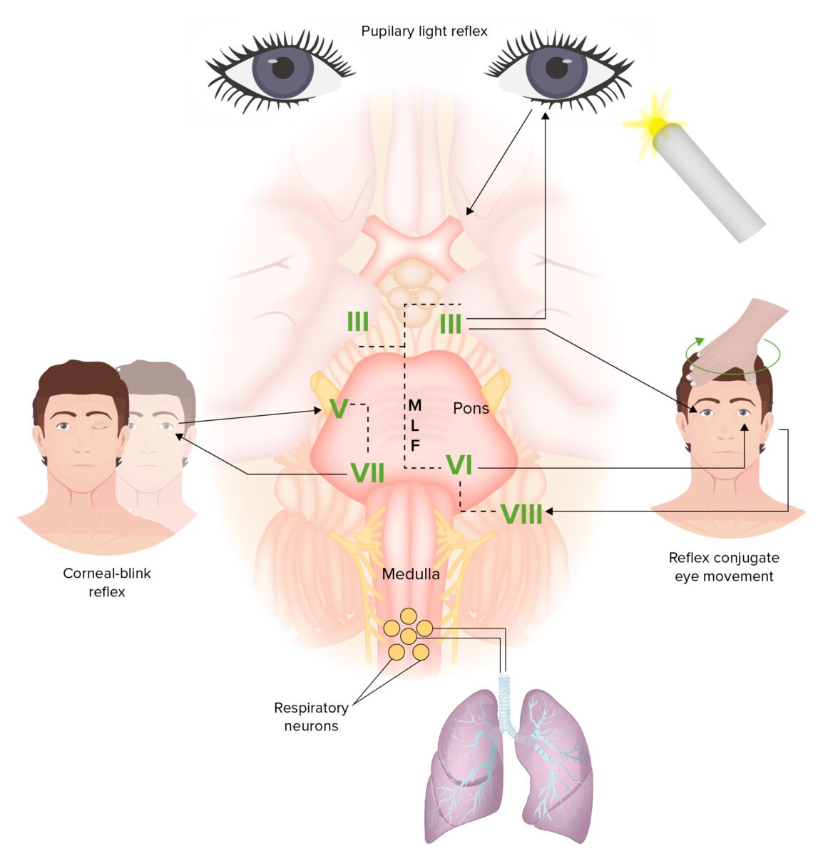
Examination of brain reflexes

Brainstem and reflexes:
The brainstem connects the pathways between the brain and the spinal cord. Functions controlled by the brainstem include the sleep-wake cycle, brainstem reflexes, and regulation of cardiac rhythm and breathing. The afferent limb of the pupillary light reflex is the optic nerve (CN II); the efferent limb is the oculomotor nerve (CN III). The oculocephalic reflex (dolls eye reflex) involves rotation of the head, a motion detected by the vestibular nerve (CN VIII). The signals then reach the vestibular nucleus. Output is then sent to CN III, which innervates the medial rectus, and the abducens nerve (CN VI), which innervates the lateral rectus. Eye movement coordination is via the medial longitudinal fasciculus (MLF), which has fibers connecting the abducens nucleus to the contralateral oculomotor nucleus. The connection is important in performing horizontal conjugate gaze. The corneal reflex is caused by a loop via the sensory trigeminal nerve (CN V) in the cornea, which projects to the facial nucleus, and the facial nerve (CN VII), which exits the facial nucleus and innervates the orbicularis oculi muscle.

Image by Lecturio.

Diagnosis

Diagnosis of coma is strictly clinical and relies on a robust neurologic examination. Further investigation (imaging and labs) are carried out to determine etiology. 

Labs

The laboratory studies to be requested depend on the suspected underlying etiology. Some routine analyses include: 

  • Electrolytes Electrolytes Electrolytes are mineral salts that dissolve in water and dissociate into charged particles called ions, which can be either be positively (cations) or negatively (anions) charged. Electrolytes are distributed in the extracellular and intracellular compartments in different concentrations. Electrolytes are essential for various basic life-sustaining functions. Electrolytes
  • CBC
  • Arterial or venous blood gas analysis
  • Toxicology studies
  • CSF analysis CSF analysis Meningitis: urgent evaluation of suspected infection

Neuroimaging Neuroimaging Non-invasive methods of visualizing the central nervous system, especially the brain, by various imaging modalities. Febrile Infant

CT/MRI to evaluate for:

  • Cerebral perfusion Cerebral Perfusion Syncope abnormalities
  • Intracerebral aneurysm Aneurysm An aneurysm is a bulging, weakened area of a blood vessel that causes an abnormal widening of its diameter > 1.5 times the size of the native vessel. Aneurysms occur more often in arteries than in veins and are at risk of dissection and rupture, which can be life-threatening. Thoracic Aortic Aneurysms or hemorrhage
  • Cerebral edema Cerebral edema Increased intracellular or extracellular fluid in brain tissue. Cytotoxic brain edema (swelling due to increased intracellular fluid) is indicative of a disturbance in cell metabolism, and is commonly associated with hypoxic or ischemic injuries. An increase in extracellular fluid may be caused by increased brain capillary permeability (vasogenic edema), an osmotic gradient, local blockages in interstitial fluid pathways, or by obstruction of CSF flow (e.g., obstructive hydrocephalus). Increased Intracranial Pressure (ICP)
  • Intracranial mass Mass Three-dimensional lesion that occupies a space within the breast Imaging of the Breast

Management

Initial (ABCs)

  • Airway Airway ABCDE Assessment intubation Intubation Peritonsillar Abscess:
  • Breathing:
    • Even with an established airway Airway ABCDE Assessment, persons with coma may have respiratory failure Respiratory failure Respiratory failure is a syndrome that develops when the respiratory system is unable to maintain oxygenation and/or ventilation. Respiratory failure may be acute or chronic and is classified as hypoxemic, hypercapnic, or a combination of the two. Respiratory Failure.
    • Oxygen saturation Oxygen Saturation Basic Procedures should be maintained at  > 90%.
    • Mechanical ventilatory support may be required.
  • Circulation Circulation The movement of the blood as it is pumped through the cardiovascular system. ABCDE Assessment:
    • Persons with coma may be in shock Shock Shock is a life-threatening condition associated with impaired circulation that results in tissue hypoxia. The different types of shock are based on the underlying cause: distributive (↑ cardiac output (CO), ↓ systemic vascular resistance (SVR)), cardiogenic (↓ CO, ↑ SVR), hypovolemic (↓ CO, ↑ SVR), obstructive (↓ CO), and mixed. Types of Shock from a variety of etiologies:
      • Cardiogenic causes
      • Sepsis Sepsis Systemic inflammatory response syndrome with a proven or suspected infectious etiology. When sepsis is associated with organ dysfunction distant from the site of infection, it is called severe sepsis. When sepsis is accompanied by hypotension despite adequate fluid infusion, it is called septic shock. Sepsis and Septic Shock
      • Hypovolemia Hypovolemia Sepsis in Children
    • Adequate vascular access should be established:
      • Peripheral IV
      • Central venous catheter Central Venous Catheter Central venous catheters are IV lines placed into the large central veins for monitoring of central venous pressure (CVP), prolonged drug administration, or administration of parenteral nutrition. The most common sites of insertion are the internal jugular and subclavian veins. Central Venous Catheter
      • Arterial line 
    • Chemical and/or mechanical circulatory support may be required:

Continued management

  • ICU ICU Hospital units providing continuous surveillance and care to acutely ill patients. West Nile Virus admission
  • Continuous monitoring
  • Supportive care
  • Management targeted at underlying etiology:
    • For example, naloxone Naloxone A specific opiate antagonist that has no agonist activity. It is a competitive antagonist at mu, delta, and kappa opioid receptors. Opioid Analgesics should be considered in cases of opioid Opioid Compounds with activity like opiate alkaloids, acting at opioid receptors. Properties include induction of analgesia or narcosis. Constipation overdose.
    • Antibiotics should be started for suspected meningitis Meningitis Meningitis is inflammation of the meninges, the protective membranes of the brain, and spinal cord. The causes of meningitis are varied, with the most common being bacterial or viral infection. The classic presentation of meningitis is a triad of fever, altered mental status, and nuchal rigidity. Meningitis.

Recovery

Recovery is defined as the return of the ability to convincingly and consistently follow commands.

Complications

  • Brain Brain The part of central nervous system that is contained within the skull (cranium). Arising from the neural tube, the embryonic brain is comprised of three major parts including prosencephalon (the forebrain); mesencephalon (the midbrain); and rhombencephalon (the hindbrain). The developed brain consists of cerebrum; cerebellum; and other structures in the brain stem. Nervous System: Anatomy, Structure, and Classification death: irreversible cessation of all brain Brain The part of central nervous system that is contained within the skull (cranium). Arising from the neural tube, the embryonic brain is comprised of three major parts including prosencephalon (the forebrain); mesencephalon (the midbrain); and rhombencephalon (the hindbrain). The developed brain consists of cerebrum; cerebellum; and other structures in the brain stem. Nervous System: Anatomy, Structure, and Classification and brainstem functions (regardless of the cause)
  • Vegetative state:
    • State of wakefulness without awareness of self and the environment
    • Blink responses, primitive motor Motor Neurons which send impulses peripherally to activate muscles or secretory cells. Nervous System: Histology responses, and sleep-wake cycles are present.
  • Persistent vegetative state Persistent vegetative state The term “persistent vegetative state,” also called unresponsive wakefulness, describes the condition of individuals with severe anoxic brain injury who have progressed to a state of wakefulness without any meaningful response to their environment. A persistent vegetative state is distinguished from a coma in that individuals in a persistent vegetative state have intermittent sleep-wake cycles. Persistent Vegetative State:

Prognosis Prognosis A prediction of the probable outcome of a disease based on a individual’s condition and the usual course of the disease as seen in similar situations. Non-Hodgkin Lymphomas

  • The comatose Comatose A profound state of unconsciousness associated with depressed cerebral activity from which the individual cannot be aroused. Coma generally occurs when there is dysfunction or injury involving both cerebral hemispheres or the brain stem reticular formation. Hyponatremia state itself can last for an indefinite period of time.
  • Outcomes depend on the underlying etiology and interpatient variability.
  • Effects of diffuse neuronal injury may linger for the remainder of the individual’s lifetime.

Clinical Relevance

Potential underlying etiologies of coma

  • Loss of cardiac output Cardiac output The volume of blood passing through the heart per unit of time. It is usually expressed as liters (volume) per minute so as not to be confused with stroke volume (volume per beat). Cardiac Mechanics (e.g., massive pulmonary embolism Pulmonary Embolism Pulmonary embolism (PE) is a potentially fatal condition that occurs as a result of intraluminal obstruction of the main pulmonary artery or its branches. The causative factors include thrombi, air, amniotic fluid, and fat. In PE, gas exchange is impaired due to the decreased return of deoxygenated blood to the lungs. Pulmonary Embolism (PE), cardiac arrhythmia, massive MI MI MI is ischemia and death of an area of myocardial tissue due to insufficient blood flow and oxygenation, usually from thrombus formation on a ruptured atherosclerotic plaque in the epicardial arteries. Clinical presentation is most commonly with chest pain, but women and patients with diabetes may have atypical symptoms. Myocardial Infarction)
  • Cerebrovascular accident Cerebrovascular accident An ischemic stroke (also known as cerebrovascular accident) is an acute neurologic injury that occurs as a result of brain ischemia; this condition may be due to cerebral blood vessel occlusion by thrombosis or embolism, or rarely due to systemic hypoperfusion. Ischemic Stroke (e.g., hemorrhagic stroke Hemorrhagic stroke Stroke due to rupture of a weakened blood vessel in the brain (e.g., cerebral hemispheres; cerebellum; subarachnoid space). Subarachnoid Hemorrhage, ischemic stroke Ischemic Stroke An ischemic stroke (also known as cerebrovascular accident) is an acute neurologic injury that occurs as a result of brain ischemia; this condition may be due to cerebral blood vessel occlusion by thrombosis or embolism, or rarely due to systemic hypoperfusion. Ischemic Stroke)
  • Severe hypovolemia Hypovolemia Sepsis in Children (e.g., hemorrhage, sepsis Sepsis Systemic inflammatory response syndrome with a proven or suspected infectious etiology. When sepsis is associated with organ dysfunction distant from the site of infection, it is called severe sepsis. When sepsis is accompanied by hypotension despite adequate fluid infusion, it is called septic shock. Sepsis and Septic Shock)
  • Overdose or toxic exposure (e.g., poisons, drugs, alcohol)
  • Trauma (e.g., fall, motor Motor Neurons which send impulses peripherally to activate muscles or secretory cells. Nervous System: Histology vehicle accident, electrocution)

Differential diagnosis of coma

  • Locked-in syndrome Locked-in syndrome Locked-in syndrome (LIS) is a rare neurological disorder in which patients are awake and conscious but are unable to move their limbs or speak. The disorder is a result of brain injury to the ventral aspect of the pons and caudal ventral midbrain; etiologies include brainstem stroke, tumors, intracranial bleeding, and demyelinating disorders. Locked-in Syndrome: neurologic condition characterized by quadriplegia Quadriplegia Severe or complete loss of motor function in all four limbs which may result from brain diseases; spinal cord diseases; peripheral nervous system diseases; neuromuscular diseases; or rarely muscular diseases. The locked-in syndrome is characterized by quadriplegia in combination with cranial muscle paralysis. Consciousness is spared and the only retained voluntary motor activity may be limited eye movements. This condition is usually caused by a lesion in the upper brain stem which injures the descending corticospinal and corticobulbar tracts. Locked-in Syndrome and bulbar palsy Bulbar Palsy Locked-in Syndrome as a consequence of injury to the brain Brain The part of central nervous system that is contained within the skull (cranium). Arising from the neural tube, the embryonic brain is comprised of three major parts including prosencephalon (the forebrain); mesencephalon (the midbrain); and rhombencephalon (the hindbrain). The developed brain consists of cerebrum; cerebellum; and other structures in the brain stem. Nervous System: Anatomy, Structure, and Classification stem. Consciousness is present, as well as voluntary blinking and vertical eye movements.
  • Akinetic mutism: rare condition characterized by pathologically slowed or nonexistent bodily movement (akinesia) and loss of speech (mutism) in an awake person. The condition stems from injury to the premotor or prefrontal areas.
  • Psychogenic unresponsiveness: also called psychogenic coma. Psychogenic unresponsiveness is the complete lack of responsiveness to external stimuli, stemming from a psychiatric etiology with no organic cause.
  • Neuromuscular paralysis: characterized by acute-onset neuromuscular weakness progressing to paralysis over days to weeks (< 1 month). Bulbar and respiratory muscle involvement is variable Variable Variables represent information about something that can change. The design of the measurement scales, or of the methods for obtaining information, will determine the data gathered and the characteristics of that data. As a result, a variable can be qualitative or quantitative, and may be further classified into subgroups. Types of Variables.
  • Syncope Syncope Syncope is a short-term loss of consciousness and loss of postural stability followed by spontaneous return of consciousness to the previous neurologic baseline without the need for resuscitation. The condition is caused by transient interruption of cerebral blood flow that may be benign or related to a underlying life-threatening condition. Syncope: brief loss of consciousness followed by an immediate return to complete alertness.

References

  1. Huff JS, Tadi P. (2021). Coma. StatPearls. http://www.ncbi.nlm.nih.gov/books/NBK430722/ 
  2. Josephson S, Ropper AH, Hauser SL (2018). Coma. IN: Jameson J, Fauci AS, Kasper DL, Hauser SL, Longo DL, Loscalzo J (Eds.), Harrison’s Principles of Internal Medicine, 20th ed. McGraw-Hill. https://accessmedicine-mhmedical-com.ezproxy.unbosque.edu.co/content.aspx?bookid=2129&sectionid=192032318 
  3. Young GB. (2021). Stupor and coma in adults. UpToDate. Retrieved June 20, 2021, from https://www.uptodate.com/contents/stupor-and-coma-in-adults
  4. Mayer  MD, FCCM, S. A., & Marshall  MD, MS, R. S. (2021). Stupor and coma (S. A. Mayer  MD, FCCM & R. S. Marshall  MD, MS (eds.); pp. 68–87). https://doi.org/http://dx.doi.org/10.1016/B978-0-323-54694-2.00005-0 
  5. Ropper A.H., Samuels M.A., Klein J.P., & Prasad S. (Eds.), (2019). Coma and related disorders of consciousness. Adams and Victor’s Principles of Neurology, 11e. McGraw Hill. https://accessmedicine.mhmedical.com/content.aspx?bookid=1477&sectionid=117186166
  6. Weinhouse G., Young G. (2022). Hypoxic-ischemic brain injury in adults: Evaluation and prognosis. UpToDate. Retrieved Mar 24, 2022 from https://www.uptodate.com/contents/hypoxic-ischemic-brain-injury-in-adults-evaluation-and-prognosis

Create your free account or log in to continue reading!

Sign up now and get free access to Lecturio with concept pages, medical videos, and questions for your medical education.

User Reviews

Unwrap New Skills This Holiday 🎄 Save 30% on all plans now!

Details