Advertisement

Advertisement

Advertisement

Advertisement

Chest Wall: Anatomy

The chest wall consists of skin Skin The skin, also referred to as the integumentary system, is the largest organ of the body. The skin is primarily composed of the epidermis (outer layer) and dermis (deep layer). The epidermis is primarily composed of keratinocytes that undergo rapid turnover, while the dermis contains dense layers of connective tissue. Skin: Structure and Functions, fat, muscles, bones, and cartilage Cartilage Cartilage is a type of connective tissue derived from embryonic mesenchyme that is responsible for structural support, resilience, and the smoothness of physical actions. Perichondrium (connective tissue membrane surrounding cartilage) compensates for the absence of vasculature in cartilage by providing nutrition and support. Cartilage: Histology. The bony structure of the chest wall is composed of the ribs, sternum, and thoracic vertebrae Thoracic vertebrae A group of twelve vertebrae connected to the ribs that support the upper trunk region. Vertebral Column: Anatomy. The chest wall serves as armor for the vital intrathoracic organs and provides the stability necessary for the movement of the shoulders and arms. The interconnections between the bones, cartilage Cartilage Cartilage is a type of connective tissue derived from embryonic mesenchyme that is responsible for structural support, resilience, and the smoothness of physical actions. Perichondrium (connective tissue membrane surrounding cartilage) compensates for the absence of vasculature in cartilage by providing nutrition and support. Cartilage: Histology, and muscles allow for the rhythmic expansion and reduction of the chest wall during breathing, which facilitates changes in intrathoracic pressure to allow expansion of the lungs Lungs Lungs are the main organs of the respiratory system. Lungs are paired viscera located in the thoracic cavity and are composed of spongy tissue. The primary function of the lungs is to oxygenate blood and eliminate CO2. Lungs: Anatomy during inspiration Inspiration Ventilation: Mechanics of Breathing. The extrinsic muscles have 2 bony attachments; the intrinsic muscles only attach to the thoracic skeleton.

Last updated: Nov 19, 2024

Editorial responsibility: Stanley Oiseth, Lindsay Jones, Evelin Maza

Advertisement

Advertisement

Advertisement

Advertisement

Advertisement

Advertisement

Introduction

Embryology

  • The thorax develops from the embryonic neural tube Neural tube A tube of ectodermal tissue in an embryo that will give rise to the central nervous system, including the spinal cord and the brain. Lumen within the neural tube is called neural canal which gives rise to the central canal of the spinal cord and the ventricles of the brain. Gastrulation and Neurulation.
  • The neural tube Neural tube A tube of ectodermal tissue in an embryo that will give rise to the central nervous system, including the spinal cord and the brain. Lumen within the neural tube is called neural canal which gives rise to the central canal of the spinal cord and the ventricles of the brain. Gastrulation and Neurulation consists of all 3 embryonic layers:
  • The chest wall develops from the mesoderm Mesoderm The middle germ layer of an embryo derived from three paired mesenchymal aggregates along the neural tube. Gastrulation and Neurulation.

Components of the chest wall

  • 2 openings: superior and inferior thoracic apertures
  • The thoracic skeleton is formed by:
    • 12 ribs 
    • 12 thoracic vertebrae Thoracic vertebrae A group of twelve vertebrae connected to the ribs that support the upper trunk region. Vertebral Column: Anatomy 
    • Cartilage Cartilage Cartilage is a type of connective tissue derived from embryonic mesenchyme that is responsible for structural support, resilience, and the smoothness of physical actions. Perichondrium (connective tissue membrane surrounding cartilage) compensates for the absence of vasculature in cartilage by providing nutrition and support. Cartilage: Histology 
    • Sternum
  • Joints:
    • Intervertebral disk joint: occupies space between vertebrae and absorbs pressure on the spine Spine The human spine, or vertebral column, is the most important anatomical and functional axis of the human body. It consists of 7 cervical vertebrae, 12 thoracic vertebrae, and 5 lumbar vertebrae and is limited cranially by the skull and caudally by the sacrum. Vertebral Column: Anatomy
    • Costovertebral joint: connects the proximal end of the ribs with corresponding thoracic vertebrae Thoracic vertebrae A group of twelve vertebrae connected to the ribs that support the upper trunk region. Vertebral Column: Anatomy
    • Costochondral joint: connects the ribs to costal cartilages
    • Sternocostal joint: connects the costal cartilages of the 1st 7 ribs to the sternum
    • Sternoclavicular joint Sternoclavicular Joint Examination of the Upper Limbs: connects the clavicle Clavicle A bone on the ventral side of the shoulder girdle, which in humans is commonly called the collar bone. Clavicle Fracture to the sternum
    • Interchondral joint: connects costal cartilages of ribs 7–10
  • Muscles:
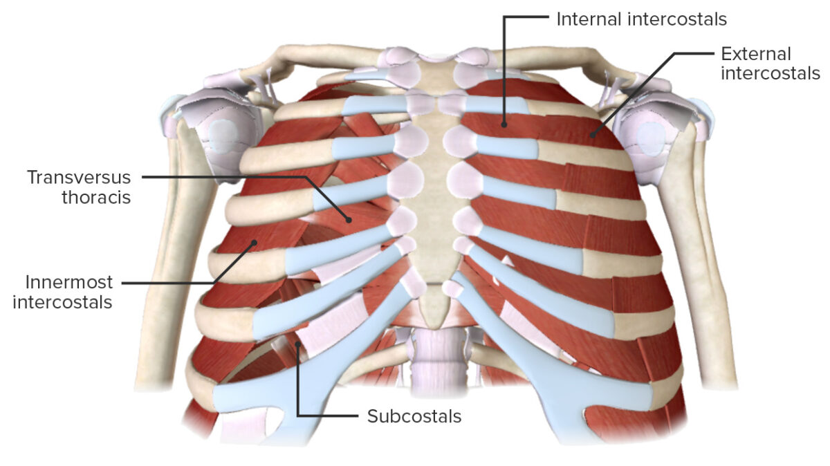
    • Intrinsic thoracic muscles: connect only to the thoracic skeleton:
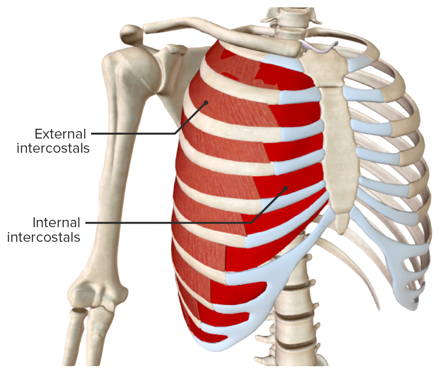
      • External intercostal muscles
      • Internal intercostal muscles
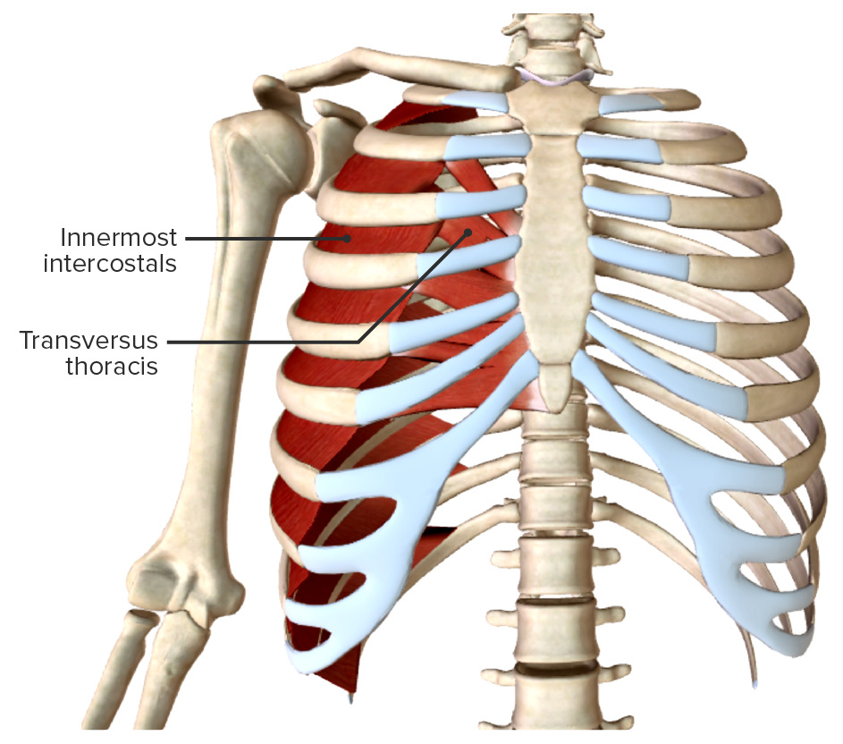
      • Innermost intercostal muscles
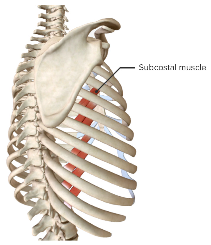
      • Subcostal (posteriorly) muscles
      • Transversus thoracis muscles (anteriorly)
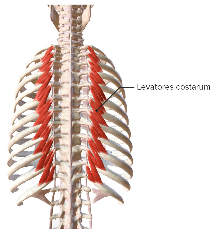
      • Levatores costarum muscles
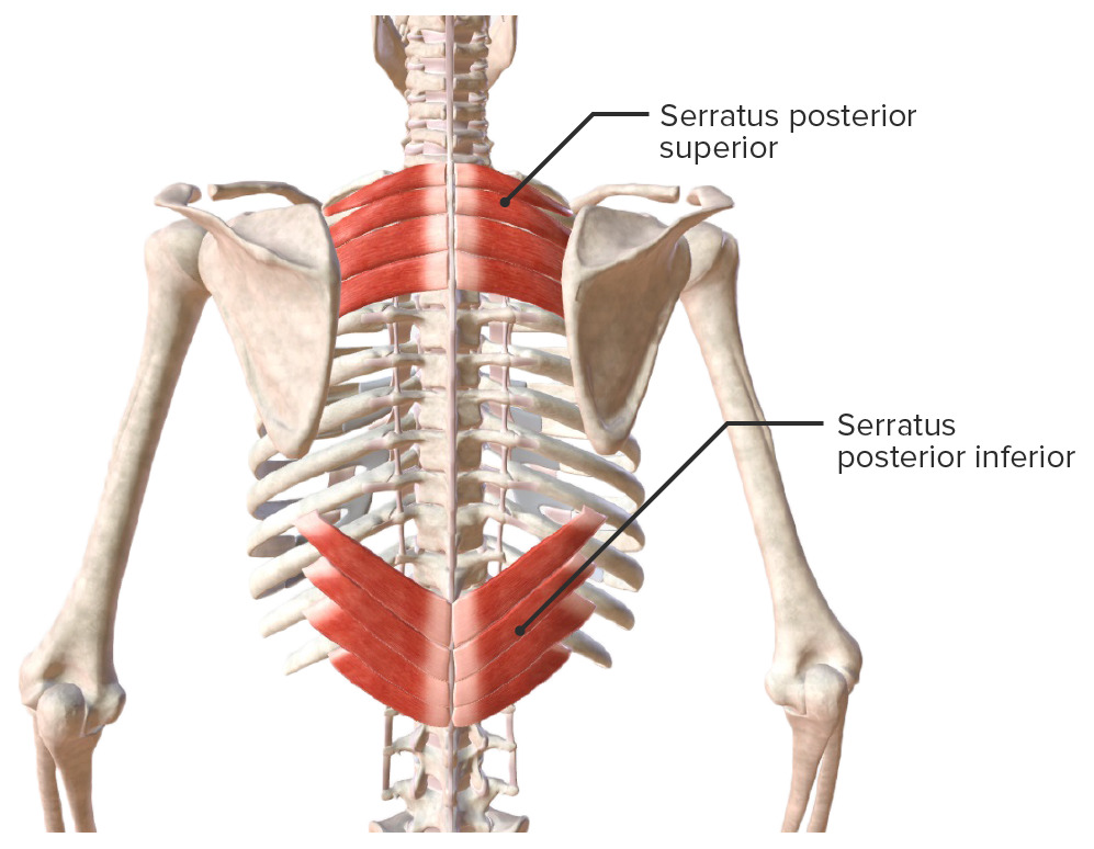
      • Serratus posterior superior and inferior muscles
    • Extrinsic thoracic muscles: connect to other parts of the skeleton:
  • Breasts Breasts The breasts are found on the anterior thoracic wall and consist of mammary glands surrounded by connective tissue. The mammary glands are modified apocrine sweat glands that produce milk, which serves as nutrition for infants. Breasts are rudimentary and usually nonfunctioning in men. Breasts: Anatomy: composed of:

Thoracic Skeleton

Ribs

The thoracic skeleton consists of 12 pairs of ribs.

Classification:

  • Vertebrosternal (true ribs):
    • Ribs 1–7
    • Connect directly to the sternum via the costal cartilages
  • Vertebrochondral (false ribs):
    • Ribs 8–10
    • Connect indirectly to the sternum via the costal arch
    • The costal cartilages join together to form the costal arch.
    • The costal arch connects to the sternum.
  • Vertebral (false ribs):
    • Ribs 11–12
    • Also known as floating ribs because the distal end has no attachment

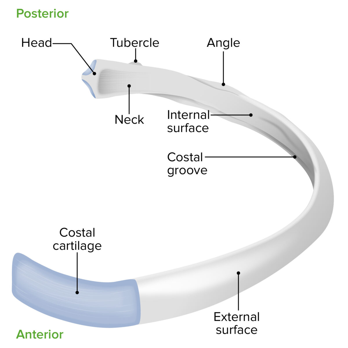
Parts of each rib: 

  • Head:
    • Articulates with 2 successive thoracic vertebrae Thoracic vertebrae A group of twelve vertebrae connected to the ribs that support the upper trunk region. Vertebral Column: Anatomy
    • Exception: Ribs 1, 10, 11, and 12 articulate with 1 vertebra.
  • Neck Neck The part of a human or animal body connecting the head to the rest of the body. Peritonsillar Abscess
  • Tubercle:
    • Articulates with the transverse process of associated vertebra
    • Exception: Ribs 11 and 12 have no transverse process articulation.
  • Angle
  • Costal groove:
    • Located along the inferior aspect of each rib
    • Contains the intercostal neurovascular bundle

Intercostal spaces:

The spaces between the ribs are the intercostal spaces:
  • The intercostal space height is greater anteriorly than posteriorly.
  • The intercostal space between the upper ribs is greater than the lower ribs.
Anatomy of a rib

Anatomy of a rib

Image by Lecturio.

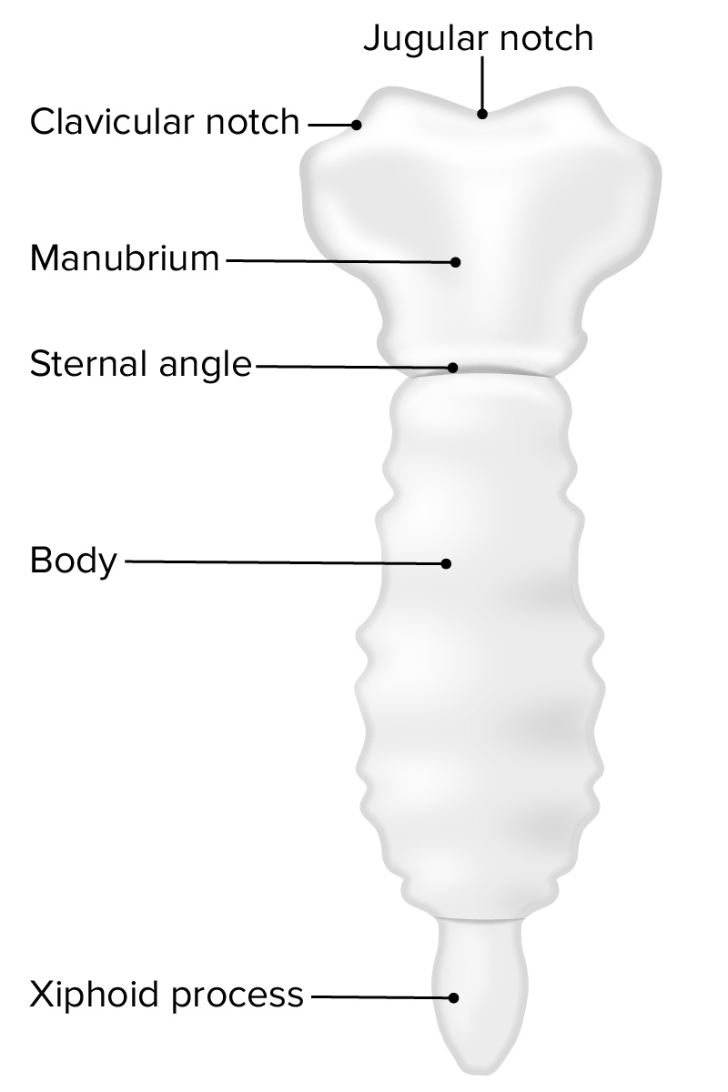
Sternum

Consists of the following parts:

  • Manubrium:
    • Located at the level of the T3 T3 A T3 thyroid hormone normally synthesized and secreted by the thyroid gland in much smaller quantities than thyroxine (T4). Most T3 is derived from peripheral monodeiodination of T4 at the 5′ position of the outer ring of the iodothyronine nucleus. The hormone finally delivered and used by the tissues is mainly t3. Thyroid Hormones and T4 T4 The major hormone derived from the thyroid gland. Thyroxine is synthesized via the iodination of tyrosines (monoiodotyrosine) and the coupling of iodotyrosines (diiodotyrosine) in the thyroglobulin. Thyroxine is released from thyroglobulin by proteolysis and secreted into the blood. Thyroxine is peripherally deiodinated to form triiodothyronine which exerts a broad spectrum of stimulatory effects on cell metabolism. Thyroid Hormones vertebral bodies
    • The thickest of the sternum components
    • A palpable landmark is the jugular, or suprasternal, notch.
  • Body (corpus)
  • Xiphoid process:
    • Cartilaginous in young people
    • Ossified in adulthood
  • Sternal angle: located between the manubrium and the body
Anatomy of the sternum

Anatomy of the sternum

Image by Lecturio.

Related videos

Intrinsic Muscles

Intrinsic muscles attach only to the thorax. The intrinsic muscles receive blood supply from the intercostal arteries Arteries Arteries are tubular collections of cells that transport oxygenated blood and nutrients from the heart to the tissues of the body. The blood passes through the arteries in order of decreasing luminal diameter, starting in the largest artery (the aorta) and ending in the small arterioles. Arteries are classified into 3 types: large elastic arteries, medium muscular arteries, and small arteries and arterioles. Arteries: Histology and are innervated by the intercostal nerves.

External intercostal muscles:

Internal intercostal muscles:

Innermost intercostal muscles:

  • Separated from internal intercostals by intercostal neurovascular bundles
  • Oriented similarly to the internal intercostals
  • Stabilize intercostal spaces during respiratory movements and assist expiration Expiration Ventilation: Mechanics of Breathing

Subcostal muscles:

  • Thin muscles found on the inner surface of the posterior thoracic wall 
  • Bridge 2–3 intercostal spaces
  • Pull the ribs inferiorly during forced exhalation and support the thoracic cage

Transversus thoracis muscles:

  • Run from the posterior surface of the lower sternum and xiphoid process to costal cartilages 2–6
  • Depress ribs

Levatores costarum muscles:

  • Run from the transverse processes of C7–T11 to the superior surfaces of the inferior ribs
  • Elevate the rib cage

Serratus posterior muscles:

  • Serratus posterior superior:
    • Runs from the spinous processes of C7– T3 T3 A T3 thyroid hormone normally synthesized and secreted by the thyroid gland in much smaller quantities than thyroxine (T4). Most T3 is derived from peripheral monodeiodination of T4 at the 5′ position of the outer ring of the iodothyronine nucleus. The hormone finally delivered and used by the tissues is mainly t3. Thyroid Hormones to the costal angle of ribs 2–5
    • Elevates ribs 2–5
  • Serratus posterior inferior:
    • Runs from the spinous processes of T11–L2 to the lower margin of ribs 9–12 
    • Depresses ribs 9–12

Extrinsic muscles

In contrast to the intrinsic muscles, the extrinsic muscles attach to the thorax and other areas of the body.

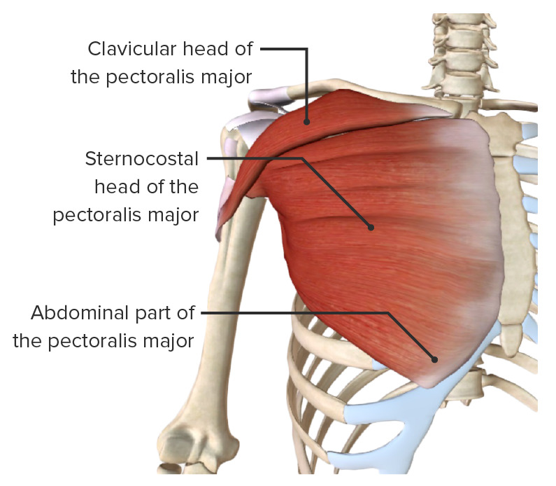
Pectoralis major muscle:

  • Located on the anterior surface of the thoracic cage
  • Divided into parts:
    • Clavicular part: originates from the anterior surface of the medial half of the clavicle Clavicle A bone on the ventral side of the shoulder girdle, which in humans is commonly called the collar bone. Clavicle Fracture
    • Sternocostal part: originates from the anterior surface of the sternum and costal cartilages 1–6
    • Abdominal part Abdominal part Esophagus: Anatomy: originates from an anterior layer of the rectus sheath
  • All 3 parts insert into the medial crest of the intertubercular groove.
  • Function:
  • Blood supply: pectoral branch of the thoracoacromial trunk
  • Innervation: medial and lateral pectoral nerves
Pectoralis major muscle

Pectoralis major muscle

Image by BioDigital, edited by Lecturio
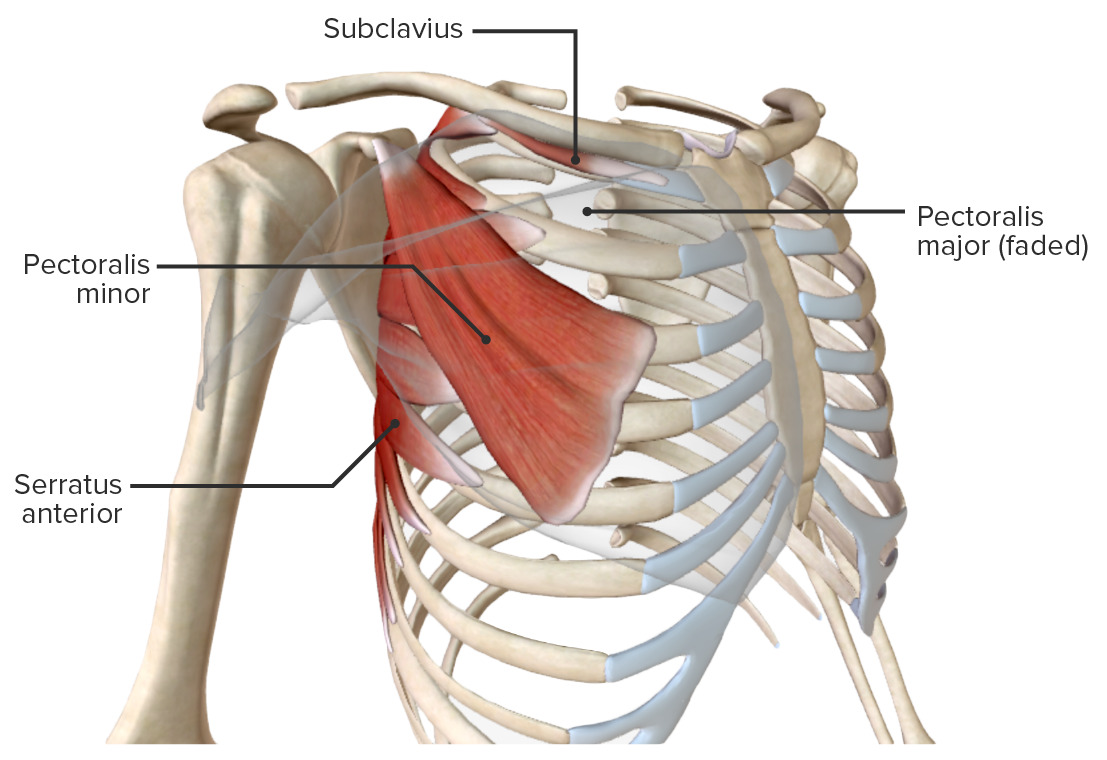
Pectoralis minor muscle:

  • Runs from ribs 3–5 to the medial border of the coracoid process of the scapula
  • Function:
    • Anteroinferior displacement Displacement The process by which an emotional or behavioral response that is appropriate for one situation appears in another situation for which it is inappropriate. Defense Mechanisms of the scapula
    • Stabilization of the scapula on the chest wall
  • Blood supply: pectoral branch of the thoracoacromial trunk
  • Innervation: medial pectoral nerve
Extrinsic muscles of the chest wall

Extrinsic muscles of the chest wall

Image by Lecturio.

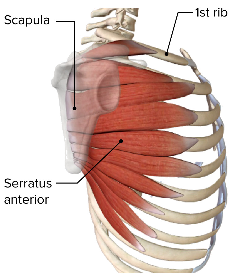
Serratus anterior muscle:

  • Runs from ribs 1–9 to the anterior surface and medial border of the scapula
  • A “serrated” or “saw-toothed” appearance
  • Function:
    • Anterolateral displacement Displacement The process by which an emotional or behavioral response that is appropriate for one situation appears in another situation for which it is inappropriate. Defense Mechanisms and rotation Rotation Motion of an object in which either one or more points on a line are fixed. It is also the motion of a particle about a fixed point. X-rays of the scapula
    • Stabilization of the scapula against the thoracic wall
  • Blood supply:
    • Superior and lateral thoracic arteries Arteries Arteries are tubular collections of cells that transport oxygenated blood and nutrients from the heart to the tissues of the body. The blood passes through the arteries in order of decreasing luminal diameter, starting in the largest artery (the aorta) and ending in the small arterioles. Arteries are classified into 3 types: large elastic arteries, medium muscular arteries, and small arteries and arterioles. Arteries: Histology
    • Thoracodorsal artery
  • Innervation: long thoracic nerve Long thoracic nerve Axilla and Brachial Plexus: Anatomy
The serratus anterior muscle

Serratus anterior muscle

Image by BioDigital, edited by Lecturio
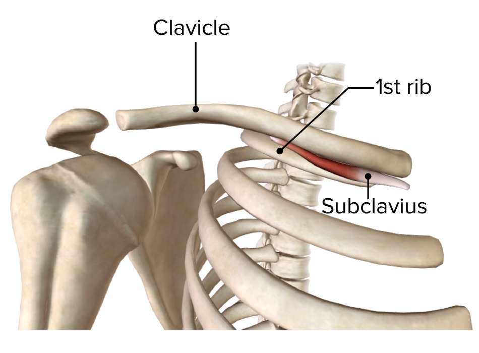
Subclavius Subclavius Muscles of the Neck: Anatomy muscle:

  • Runs from the inferior surface of the clavicle Clavicle A bone on the ventral side of the shoulder girdle, which in humans is commonly called the collar bone. Clavicle Fracture to the 1st rib
  • Function: depression of the clavicle Clavicle A bone on the ventral side of the shoulder girdle, which in humans is commonly called the collar bone. Clavicle Fracture and elevation of the 1st rib
  • Blood supply: clavicular branch of the thoracoacromial trunk
  • Innervation: subclavian nerve
The subclavius muscle

Subclavius muscle

Image by BioDigital, edited by Lecturio
Clinical Relevance

  • Pectus excavatum Pectus Excavatum Cardiovascular Examination (also known as sunken/ funnel chest Funnel Chest Cardiovascular Examination): the most common congenital deformity Deformity Examination of the Upper Limbs of the chest, accounting for 90% of cases. Pectus excavatum Pectus Excavatum Cardiovascular Examination is characterized by the abnormal development of several ribs and the sternum, producing a caved-in appearance of the anterior chest wall. Men are predominantly affected (the ratio of men to women is 3:1). The appearance of the defect and the symptoms vary widely. Young individuals are usually asymptomatic. Symptoms may later include chest pain Pain An unpleasant sensation induced by noxious stimuli which are detected by nerve endings of nociceptive neurons. Pain: Types and Pathways and dyspnea Dyspnea Dyspnea is the subjective sensation of breathing discomfort. Dyspnea is a normal manifestation of heavy physical or psychological exertion, but also may be caused by underlying conditions (both pulmonary and extrapulmonary). Dyspnea. Treatment is surgical with very good results.
  • Pectus carinatum Pectus carinatum A developmental anomaly characterized by abnormal anterior protrusion of the sternum and adjacent costal cartilage. Cardiovascular Examination: the 2nd most common deformity Deformity Examination of the Upper Limbs of the chest wall in children ( family history Family History Adult Health Maintenance in 25% of cases). Associated conditions include Marfan syndrome Marfan syndrome Marfan syndrome is a genetic condition with autosomal dominant inheritance. Marfan syndrome affects the elasticity of connective tissues throughout the body, most notably in the cardiovascular, ocular, and musculoskeletal systems. Marfan Syndrome, Noonan syndrome Noonan syndrome A genetically heterogeneous, multifaceted disorder characterized by short stature, webbed neck, ptosis, skeletal malformations, hypertelorism, hormonal imbalance, cryptorchidism, multiple cardiac abnormalities (most commonly including pulmonary valve stenosis), and some degree of intellectual disability. The phenotype bears similarities to that of turner syndrome that occurs only in females and has its basis in a 45, X karyotype abnormality. Noonan syndrome occurs in both males and females with a normal karyotype (46, XX and 46, xy). Mutations in a several genes (ptpn11, kras, sos1, nf1 and raf1) have been associated the ns phenotype. Mutations in ptpn11 are the most common. Leopard syndrome, a disorder that has clinical features overlapping those of noonan syndrome, is also due to mutations in ptpn11. In addition, there is overlap with the syndrome called neurofibromatosis-noonan syndrome due to mutations in nf1. Hypogonadism, mitral valve Mitral valve The valve between the left atrium and left ventricle of the heart. Heart: Anatomy prolapse, and congenital heart disease. Individuals may be asymptomatic; however, the disease may lead to cardiorespiratory compromise due to increased rigidity Rigidity Continuous involuntary sustained muscle contraction which is often a manifestation of basal ganglia diseases. When an affected muscle is passively stretched, the degree of resistance remains constant regardless of the rate at which the muscle is stretched. This feature helps to distinguish rigidity from muscle spasticity. Megacolon of the chest wall, which impairs normal respiration Respiration The act of breathing with the lungs, consisting of inhalation, or the taking into the lungs of the ambient air, and of exhalation, or the expelling of the modified air which contains more carbon dioxide than the air taken in. Nose Anatomy (External & Internal) (especially upon exertion). The result is alveolar hypoventilation and hypoxemia Hypoxemia Neonatal Respiratory Distress Syndrome, leading to cor pulmonale Cor Pulmonale Cor pulmonale is right ventricular (RV) dysfunction caused by lung disease that results in pulmonary artery hypertension. The most common cause of cor pulmonale is chronic obstructive pulmonary disease. Dyspnea is the usual presenting symptom. Cor Pulmonale (right heart failure Heart Failure A heterogeneous condition in which the heart is unable to pump out sufficient blood to meet the metabolic need of the body. Heart failure can be caused by structural defects, functional abnormalities (ventricular dysfunction), or a sudden overload beyond its capacity. Chronic heart failure is more common than acute heart failure which results from sudden insult to cardiac function, such as myocardial infarction. Total Anomalous Pulmonary Venous Return (TAPVR)). Emphysema Emphysema Enlargement of air spaces distal to the terminal bronchioles where gas-exchange normally takes place. This is usually due to destruction of the alveolar wall. Pulmonary emphysema can be classified by the location and distribution of the lesions. Chronic Obstructive Pulmonary Disease (COPD) and recurrent respiratory infections Infections Invasion of the host organism by microorganisms or their toxins or by parasites that can cause pathological conditions or diseases. Chronic Granulomatous Disease are common. Orthotic bracing is the 1st line of treatment and has very good results, especially in younger individuals. 
  • Ankylosing spondylitis Ankylosing spondylitis Ankylosing spondylitis (also known as Bechterew’s disease or Marie-Strümpell disease) is a seronegative spondyloarthropathy characterized by chronic and indolent inflammation of the axial skeleton. Severe disease can lead to fusion and rigidity of the spine. Ankylosing Spondylitis: a seronegative spondyloarthropathy Spondyloarthropathy Ankylosing Spondylitis characterized by chronic and indolent inflammation Inflammation Inflammation is a complex set of responses to infection and injury involving leukocytes as the principal cellular mediators in the body’s defense against pathogenic organisms. Inflammation is also seen as a response to tissue injury in the process of wound healing. The 5 cardinal signs of inflammation are pain, heat, redness, swelling, and loss of function. Inflammation of the axial Axial Computed Tomography (CT) skeleton, which may involve the thoracic spine Spine The human spine, or vertebral column, is the most important anatomical and functional axis of the human body. It consists of 7 cervical vertebrae, 12 thoracic vertebrae, and 5 lumbar vertebrae and is limited cranially by the skull and caudally by the sacrum. Vertebral Column: Anatomy. Severe disease can lead to fusion and rigidity Rigidity Continuous involuntary sustained muscle contraction which is often a manifestation of basal ganglia diseases. When an affected muscle is passively stretched, the degree of resistance remains constant regardless of the rate at which the muscle is stretched. This feature helps to distinguish rigidity from muscle spasticity. Megacolon of the spine Spine The human spine, or vertebral column, is the most important anatomical and functional axis of the human body. It consists of 7 cervical vertebrae, 12 thoracic vertebrae, and 5 lumbar vertebrae and is limited cranially by the skull and caudally by the sacrum. Vertebral Column: Anatomy. Ankylosing spondylitis Ankylosing spondylitis Ankylosing spondylitis (also known as Bechterew’s disease or Marie-Strümpell disease) is a seronegative spondyloarthropathy characterized by chronic and indolent inflammation of the axial skeleton. Severe disease can lead to fusion and rigidity of the spine. Ankylosing Spondylitis is most often seen in young men and is strongly associated with HLA-B27 positivity. Individuals have morning stiffness, decreased range of motion Range of motion The distance and direction to which a bone joint can be extended. Range of motion is a function of the condition of the joints, muscles, and connective tissues involved. Joint flexibility can be improved through appropriate muscle strength exercises. Examination of the Upper Limbs of the spine Spine The human spine, or vertebral column, is the most important anatomical and functional axis of the human body. It consists of 7 cervical vertebrae, 12 thoracic vertebrae, and 5 lumbar vertebrae and is limited cranially by the skull and caudally by the sacrum. Vertebral Column: Anatomy, and progressive back pain Pain An unpleasant sensation induced by noxious stimuli which are detected by nerve endings of nociceptive neurons. Pain: Types and Pathways, which improves with activity. Most individuals are managed with physical therapy Physical Therapy Becker Muscular Dystrophy and NSAIDs NSAIDS Primary vs Secondary Headaches. Severe cases may require tumor Tumor Inflammation necrosis Necrosis The death of cells in an organ or tissue due to disease, injury or failure of the blood supply. Ischemic Cell Damage factor-alpha inhibitors or surgery.

References

  1. Cataletto, M.E. (2019). Pectus Carinatum. Medscape. Retrieved August 20, 2021, from https://emedicine.medscape.com/article/1003047-overview
  2. Graeber, G.M., Nazim, M. (2007). The anatomy of the ribs and the sternum and their relationship to chest wall structure and function. Thorac Surg Clin. 17(4), 473–89, vi. https://pubmed.ncbi.nlm.nih.gov/18271162/
  3. Hebra, A. (2018). Pectus Excavatum. Medscape. Retrieved August 20, 2021, from https://emedicine.medscape.com/article/1004953-overview
  4. Kudzinskas, A., Callahan, A.L. (2021). Anatomy, Thorax. Treasure Island (FL): StatPearls Publishing.
  5. Miller, J.I. Jr. (2007). Muscles of the chest wall. Thorac Surg Clin. 17(4), 463–72. https://pubmed.ncbi.nlm.nih.gov/18271161/
  6. Pourtaheri, N. (2016). Chest Wall Anatomy. Medscape. Retrieved August 20, 2021, from https://reference.medscape.com/article/2151800-overview

Create your free account or log in to continue reading!

Sign up now and get free access to Lecturio with concept pages, medical videos, and questions for your medical education.

User Reviews

Unwrap New Skills This Holiday 🎄 Save 30% on all plans now!

Details