Advertisement

Advertisement

Advertisement

Advertisement

Cellular Adaptation

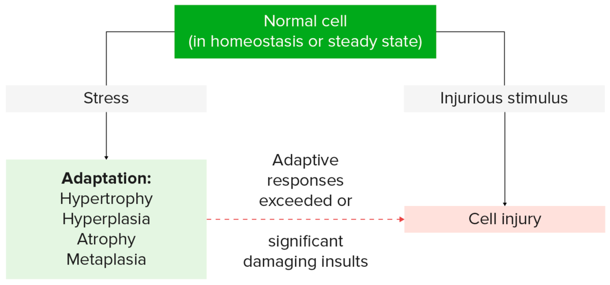
In order to cope with their environment, cells undergo structural and functional changes. These cellular adaptations are reversible responses that allow cells to survive and continue to adequately function. Adaptive processes consist of increased cellular size and function (hypertrophy), increase in cell number (hyperplasia), decrease in cell size and metabolic activity (atrophy), or a change in the phenotype Phenotype The complete genetic complement contained in the DNA of a set of chromosomes in a human. The length of the human genome is about 3 billion base pairs. Basic Terms of Genetics of the cells (metaplasia). If the stress or stimulus is removed, the cell can return to its original state. However, when the limits of the adaptive responses are exceeded, cellular function is adversely affected, leading to cellular injury.

Last updated: May 17, 2024

Editorial responsibility: Stanley Oiseth, Lindsay Jones, Evelin Maza

Advertisement

Advertisement

Advertisement

Advertisement

Advertisement

Advertisement

Definition

Adaptation:

  • Reversible changes in number, size, phenotype Phenotype The complete genetic complement contained in the DNA of a set of chromosomes in a human. The length of the human genome is about 3 billion base pairs. Basic Terms of Genetics, or cell function in response to physiologic and pathologic changes in the environment
  • Allows cell survival and continued cellular function in an altered steady state Steady state Enzyme Kinetics
  • Results from:
    • Increased demand 
    • Changes in vascular supply, nutrients, or stimulation
    • Chronic irritation
Cellular response to stress and stimuli

Cellular response to stress and stimuli

Image by Lecturio.
Hypertrophy

Overview

  • Cells increase in size 
  • No additional new cells
  • ↑ In cellular size → ↑ in size of affected organ
  • Can occur along with hyperplasia
  • Triggered by:
    • Hormonal signaling
    • Functional demand
  • Types:
    • Physiologic hypertrophy
    • Pathologic hypertrophy

Physiologic hypertrophy

  • Due to:
    • Increased functional demand
    • Hormonal stimulation and effect of growth factors
  • Clinical correlation Correlation Determination of whether or not two variables are correlated. This means to study whether an increase or decrease in one variable corresponds to an increase or decrease in the other variable. Causality, Validity, and Reliability:
    • Uterine growth in pregnancy Pregnancy The status during which female mammals carry their developing young (embryos or fetuses) in utero before birth, beginning from fertilization to birth. Pregnancy: Diagnosis, Physiology, and Care
    • Skeletal muscle growth after exercise

Pathologic hypertrophy

  • Due to increased workload 
  • Clinical correlation Correlation Determination of whether or not two variables are correlated. This means to study whether an increase or decrease in one variable corresponds to an increase or decrease in the other variable. Causality, Validity, and Reliability: left ventricular hypertrophy Ventricular Hypertrophy Tetralogy of Fallot
    • Hypertrophy of the wall of the left ventricle 
    • Occurs in hypertension Hypertension Hypertension, or high blood pressure, is a common disease that manifests as elevated systemic arterial pressures. Hypertension is most often asymptomatic and is found incidentally as part of a routine physical examination or during triage for an unrelated medical encounter. Hypertension
    • Persistently increased burden → degradation and death of myocytes Myocytes Mature contractile cells, commonly known as myocytes, that form one of three kinds of muscle. The three types of muscle cells are skeletal, cardiac, and smooth. They are derived from embryonic (precursor) muscle cells called myoblasts. Muscle Tissue: Histology cardiac failure Cardiac failure Congestive heart failure refers to the inability of the heart to supply the body with normal cardiac output to meet metabolic needs. Echocardiography can confirm the diagnosis and give information about the ejection fraction. Heart Failure
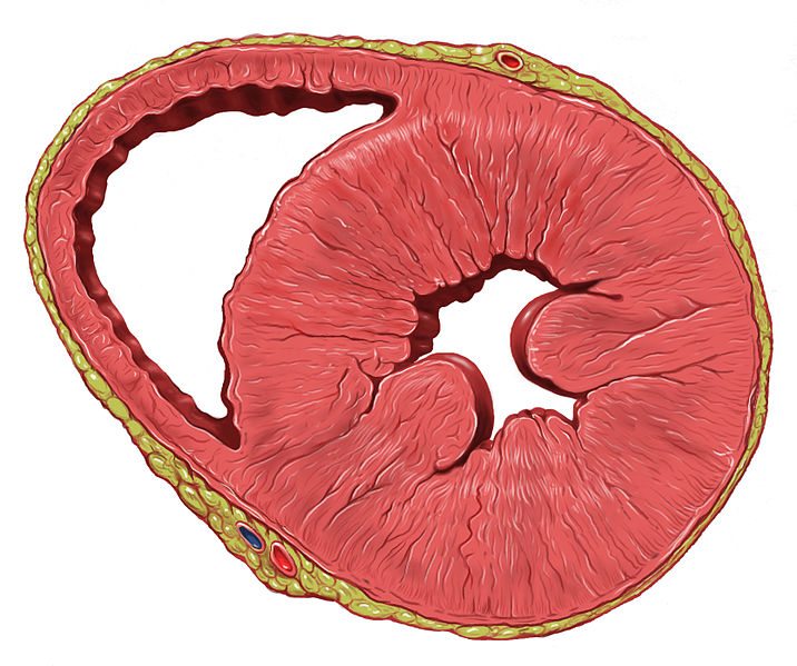
Heart left ventricular hypertrophy

Cross-section of a heart with left ventricular hypertrophy: Note the thickened ventricular walls and associated reduced ventricular cavity.

Image: “Heart left ventricular hypertrophy short axis view” by Patrick J. Lynch, medical illustrator. License: CC BY 2.5
Mechanism

  • Increase in cellular protein production resulting from:
    • Action of growth factors
    • Switch in gene Gene A category of nucleic acid sequences that function as units of heredity and which code for the basic instructions for the development, reproduction, and maintenance of organisms. Basic Terms of Genetics expression
  • Enlargement of cells or organs has a limit Limit A value (e.g., pressure or time) that should not be exceeded and which is specified by the operator to protect the lung Invasive Mechanical Ventilation.
  • Regressive changes or cellular death occur when this limit Limit A value (e.g., pressure or time) that should not be exceeded and which is specified by the operator to protect the lung Invasive Mechanical Ventilation is reached and/or stress is not relieved.

Related videos

Hyperplasia

Overview

  • Cells increase in number in response to a stimulus.
  • Can occur simultaneously with hypertrophy 
  • Occurs only if cells are capable of dividing (cardiac myocytes Myocytes Mature contractile cells, commonly known as myocytes, that form one of three kinds of muscle. The three types of muscle cells are skeletal, cardiac, and smooth. They are derived from embryonic (precursor) muscle cells called myoblasts. Muscle Tissue: Histology and neurons Neurons The basic cellular units of nervous tissue. Each neuron consists of a body, an axon, and dendrites. Their purpose is to receive, conduct, and transmit impulses in the nervous system. Nervous System: Histology in the brain Brain The part of central nervous system that is contained within the skull (cranium). Arising from the neural tube, the embryonic brain is comprised of three major parts including prosencephalon (the forebrain); mesencephalon (the midbrain); and rhombencephalon (the hindbrain). The developed brain consists of cerebrum; cerebellum; and other structures in the brain stem. Nervous System: Anatomy, Structure, and Classification do not undergo hyperplasia)
  • ↑ In cellular number → ↑ in size of affected organ 
  • Types:
    • Physiologic hyperplasia
    • Pathologic hyperplasia
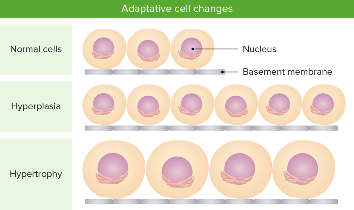
Adaptive cell changes

Illustration of the difference between hypertrophy (increase in size of cells) and hyperplasia (increase in number of cells)

Image by Lecturio.
Physiologic hyperplasia

  • Results from a normal stressor
  • Brought on by an increase in hormones Hormones Hormones are messenger molecules that are synthesized in one part of the body and move through the bloodstream to exert specific regulatory effects on another part of the body. Hormones play critical roles in coordinating cellular activities throughout the body in response to the constant changes in both the internal and external environments. Hormones: Overview and Types and growth factors to enhance functional capacity of the organ
  • Hormonal hyperplasia: 
    • Clinical correlation Correlation Determination of whether or not two variables are correlated. This means to study whether an increase or decrease in one variable corresponds to an increase or decrease in the other variable. Causality, Validity, and Reliability: glandular epithelium Glandular epithelium Glandular epithelia, composed of epithelial tissue, are specialized structures that play a role in the production and release of enzymes, hormones, sweat, oil, and mucus in organisms. The secretion and release of these substances are prompted by either external or internal stimuli. Glandular Epithelium: Histology of mammary glands in puberty Puberty Puberty is a complex series of physical, psychosocial, and cognitive transitions usually experienced by adolescents (11-19 years of age). Puberty is marked by a growth in stature and the development of secondary sexual characteristics, achievement of fertility, and changes in most body systems. Puberty and pregnancy Pregnancy The status during which female mammals carry their developing young (embryos or fetuses) in utero before birth, beginning from fertilization to birth. Pregnancy: Diagnosis, Physiology, and Care
  • Compensatory hyperplasia:
    • Results from damage or resection 
    • Clinical correlation Correlation Determination of whether or not two variables are correlated. This means to study whether an increase or decrease in one variable corresponds to an increase or decrease in the other variable. Causality, Validity, and Reliability:
      • Restoration of a part of the liver Liver The liver is the largest gland in the human body. The liver is found in the superior right quadrant of the abdomen and weighs approximately 1.5 kilograms. Its main functions are detoxification, metabolism, nutrient storage (e.g., iron and vitamins), synthesis of coagulation factors, formation of bile, filtration, and storage of blood. Liver: Anatomy after it has been resected
      • Bone marrow Bone marrow The soft tissue filling the cavities of bones. Bone marrow exists in two types, yellow and red. Yellow marrow is found in the large cavities of large bones and consists mostly of fat cells and a few primitive blood cells. Red marrow is a hematopoietic tissue and is the site of production of erythrocytes and granular leukocytes. Bone marrow is made up of a framework of connective tissue containing branching fibers with the frame being filled with marrow cells. Bone Marrow: Composition and Hematopoiesis hyperplasia in response to acute bleeding
      • Partial nephrectomy Nephrectomy Excision of kidney. Renal Cell Carcinoma leads to hyperplasia of the remaining kidney.

Pathologic hyperplasia

  • Results from excessive or inappropriate stimulation of hormones Hormones Hormones are messenger molecules that are synthesized in one part of the body and move through the bloodstream to exert specific regulatory effects on another part of the body. Hormones play critical roles in coordinating cellular activities throughout the body in response to the constant changes in both the internal and external environments. Hormones: Overview and Types or growth factors
  • Clinical correlation Correlation Determination of whether or not two variables are correlated. This means to study whether an increase or decrease in one variable corresponds to an increase or decrease in the other variable. Causality, Validity, and Reliability:
    • Prolonged estrogen Estrogen Compounds that interact with estrogen receptors in target tissues to bring about the effects similar to those of estradiol. Estrogens stimulate the female reproductive organs, and the development of secondary female sex characteristics. Estrogenic chemicals include natural, synthetic, steroidal, or non-steroidal compounds. Ovaries: Anatomy exposure → endometrial hyperplasia Endometrial Hyperplasia Benign proliferation of the endometrium in the uterus. Endometrial hyperplasia is classified by its cytology and glandular tissue. There are simple, complex (adenomatous without atypia), and atypical hyperplasia representing also the ascending risk of becoming malignant. Endometrial Hyperplasia and Endometrial Cancer → leading to endometrial bleeding
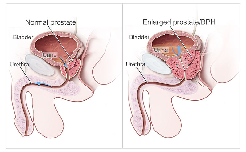
    • Androgens Androgens Androgens are naturally occurring steroid hormones responsible for development and maintenance of the male sex characteristics, including penile, scrotal, and clitoral growth, development of sexual hair, deepening of the voice, and musculoskeletal growth. Androgens and Antiandrogens benign Benign Fibroadenoma prostatic hyperplasia → bladder outlet obstruction Bladder Outlet Obstruction Benign Prostatic Hyperplasia
    • Papillomavirus → epithelial hyperplasia → warts Warts Benign epidermal proliferations or tumors; some are viral in origin. Female Genitourinary Examination and mucosal lesions

Mechanism

  • Results from stimulation by growth factors → mature cells proliferate or new cells form from tissue stem cells
  • Hyperplasia can stop once the stimulus is removed.
  • Unrestricted stimulation → persistent proliferation and increased likelihood of genetic aberrations → increased risk of cancer

Related videos

Atrophy

Overview

  • Decrease in size and number of cells, resulting in reduced metabolic activity
  • If a sufficient number of cells undergo this process, the whole organ becomes atrophic.
  • 2 types of atrophy:
    • Physiologic
    • Pathologic

Physiologic atrophy

  • Results from a normal stimulus 
  • Clinical correlation Correlation Determination of whether or not two variables are correlated. This means to study whether an increase or decrease in one variable corresponds to an increase or decrease in the other variable. Causality, Validity, and Reliability:
    • Uterine involution: reduction of the size of the uterus Uterus The uterus, cervix, and fallopian tubes are part of the internal female reproductive system. The uterus has a thick wall made of smooth muscle (the myometrium) and an inner mucosal layer (the endometrium). The most inferior portion of the uterus is the cervix, which connects the uterine cavity to the vagina. Uterus, Cervix, and Fallopian Tubes: Anatomy after giving birth
    • Thyroglossal duct Thyroglossal duct Thyroid Gland: Anatomy and notochord Notochord A cartilaginous rod of mesodermal cells at the dorsal midline of all chordate embryos. In lower vertebrates, notochord is the backbone of support. In the higher vertebrates, notochord is a transient structure, and segments of the vertebral column will develop around it. Notochord is also a source of midline signals that pattern surrounding tissues including the neural tube development. Gastrulation and Neurulation atrophy during embryonic development

Pathologic atrophy

  • Can be localized or generalized
  • Causes and clinical correlation Correlation Determination of whether or not two variables are correlated. This means to study whether an increase or decrease in one variable corresponds to an increase or decrease in the other variable. Causality, Validity, and Reliability:
    • Loss of workload or disuse atrophy: bedridden patients Patients Individuals participating in the health care system for the purpose of receiving therapeutic, diagnostic, or preventive procedures. Clinician–Patient Relationship → prolonged disuse → skeletal muscle fibers decrease in number and size
    • Loss of innervation or denervation atrophy:  spinal muscular atrophy Spinal Muscular Atrophy Spinal muscular atrophy (SMA) is a spectrum of autosomal recessive syndromes characterized by progressive proximal muscle weakness and atrophy, possibly due to degeneration of the anterior horn cells in the spinal cord and motor nuclei in the lower brainstem. Spinal Muscular Atrophy (SMA), degeneration of the anterior horn Anterior horn One of three central columns of the spinal cord. It is composed of gray matter spinal laminae VIII and ix. Brown-Séquard Syndrome of the spinal cord Spinal cord The spinal cord is the major conduction pathway connecting the brain to the body; it is part of the CNS. In cross section, the spinal cord is divided into an H-shaped area of gray matter (consisting of synapsing neuronal cell bodies) and a surrounding area of white matter (consisting of ascending and descending tracts of myelinated axons). Spinal Cord: Anatomy from a defect in the survival motor Motor Neurons which send impulses peripherally to activate muscles or secretory cells. Nervous System: Histology neuron gene Gene A category of nucleic acid sequences that function as units of heredity and which code for the basic instructions for the development, reproduction, and maintenance of organisms. Basic Terms of Genetics → damaged nerve → skeletal muscle atrophy
    • Decrease in blood supply:  atherosclerosis Atherosclerosis Atherosclerosis is a common form of arterial disease in which lipid deposition forms a plaque in the blood vessel walls. Atherosclerosis is an incurable disease, for which there are clearly defined risk factors that often can be reduced through a change in lifestyle and behavior of the patient. Atherosclerosis → chronic ischemia Ischemia A hypoperfusion of the blood through an organ or tissue caused by a pathologic constriction or obstruction of its blood vessels, or an absence of blood circulation. Ischemic Cell Damage → aging brain Brain The part of central nervous system that is contained within the skull (cranium). Arising from the neural tube, the embryonic brain is comprised of three major parts including prosencephalon (the forebrain); mesencephalon (the midbrain); and rhombencephalon (the hindbrain). The developed brain consists of cerebrum; cerebellum; and other structures in the brain stem. Nervous System: Anatomy, Structure, and Classification or senile atrophy → cognitive decline
    • Lack of nutrition: protein-calorie malnutrition Malnutrition Malnutrition is a clinical state caused by an imbalance or deficiency of calories and/or micronutrients and macronutrients. The 2 main manifestations of acute severe malnutrition are marasmus (total caloric insufficiency) and kwashiorkor (protein malnutrition with characteristic edema). Malnutrition in children in resource-limited countries → skeletal muscle proteins Proteins Linear polypeptides that are synthesized on ribosomes and may be further modified, crosslinked, cleaved, or assembled into complex proteins with several subunits. The specific sequence of amino acids determines the shape the polypeptide will take, during protein folding, and the function of the protein. Energy Homeostasis are utilized as a source of energy → muscle wasting Muscle Wasting Duchenne Muscular Dystrophy 
    • Loss of endocrine stimulation:  menopause Menopause Menopause is a physiologic process in women characterized by the permanent cessation of menstruation that occurs after the loss of ovarian activity. Menopause can only be diagnosed retrospectively, after 12 months without menstrual bleeding. Menopause → loss of estrogen Estrogen Compounds that interact with estrogen receptors in target tissues to bring about the effects similar to those of estradiol. Estrogens stimulate the female reproductive organs, and the development of secondary female sex characteristics. Estrogenic chemicals include natural, synthetic, steroidal, or non-steroidal compounds. Ovaries: Anatomy → vaginal atrophy 
    • Pressure:  tumor Tumor Inflammation compresses adjacent tissue → pressure disrupts cells and compromises the blood supply → atrophy of the surrounding tissues 
  • Note: Hypoplasia Hypoplasia Hypoplastic Left Heart Syndrome (HLHS) is a condition of smaller-than-normal organ or tissue resulting from an abnormal or incomplete development. The tissue involved was never normal in size.
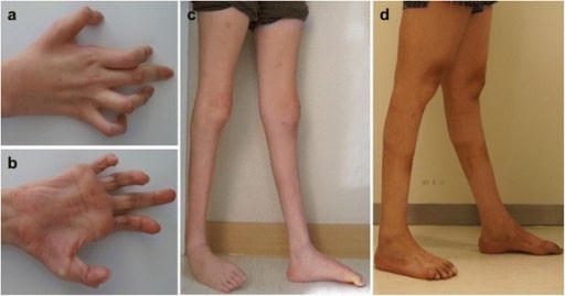
Mpv17 mutation

Photos showing the hands and legs of patients with axonal sensorimotor polyneuropathy:
Hands, patient 1 (a) and 2 (b). Right hand shows severe, atrophied, intrinsic muscle and flexion deformities of interphalangeal joints.
Lower extremities, patient 1 (c) and 2 (d). Severe bilateral distal muscle atrophies with ankle joint deformities are observed in both patients.
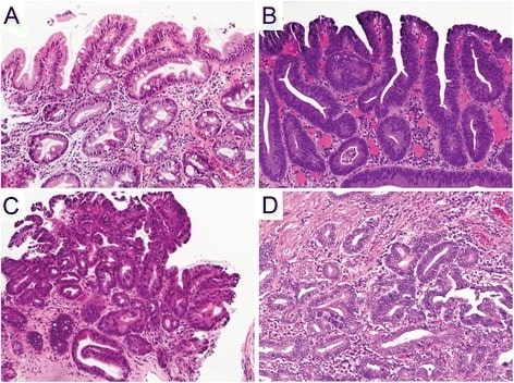
Image: “Hand and leg pictures of patients” by Department of Biochemistry, Ewha Womans University School of Medicine, Seoul, Korea. License: CC BY 4.0

Mechanism

  • Characterized by a decrease in protein synthesis Synthesis Polymerase Chain Reaction (PCR) and an increase in protein degradation
  • Ubiquitin-proteasome pathway:
    • Ubiquitin ligases Ligases A class of enzymes that catalyze the formation of a bond between two substrate molecules, coupled with the hydrolysis of a pyrophosphate bond in ATP or a similar energy donor. Basics of Enzymes are activated, resulting in attachment of ubiquitin to cellular proteins Proteins Linear polypeptides that are synthesized on ribosomes and may be further modified, crosslinked, cleaved, or assembled into complex proteins with several subunits. The specific sequence of amino acids determines the shape the polypeptide will take, during protein folding, and the function of the protein. Energy Homeostasis.
    • These proteins Proteins Linear polypeptides that are synthesized on ribosomes and may be further modified, crosslinked, cleaved, or assembled into complex proteins with several subunits. The specific sequence of amino acids determines the shape the polypeptide will take, during protein folding, and the function of the protein. Energy Homeostasis become targets for degradation.
  • Atrophy is also accompanied by autophagy:
    • Self-degradative process in which a cell eats its contents
    • Marked by the presence of autophagic vacuoles: contain lysosome-degraded proteins Proteins Linear polypeptides that are synthesized on ribosomes and may be further modified, crosslinked, cleaved, or assembled into complex proteins with several subunits. The specific sequence of amino acids determines the shape the polypeptide will take, during protein folding, and the function of the protein. Energy Homeostasis, pathogens, and cellular organelles Organelles A cell is a complex unit that performs several complex functions. An organelle is a specialized subunit within a cell that fulfills a specific role or function. Organelles are enclosed within their own lipid bilayers or are unbound by membranes. The Cell: Organelles 
    • Some cell debris resists autophagy, persisting as residual bodies
    • Lipofuscin Lipofuscin A naturally occurring lipid pigment with histochemical characteristics similar to ceroid. It accumulates in various normal tissues and apparently increases in quantity with age. Macular Degeneration granules:
      • An example of residual bodies, seen in cells as yellow-brown pigment (“wear-and-tear” pigment)
      • When increased in a tissue, causes brownish discoloration ( brown atrophy Brown Atrophy Cellular Accumulations)
  • If atrophy persistently reduces vascular supply:
    • Irreversible cell injury Cell injury The cell undergoes a variety of changes in response to injury, which may or may not lead to cell death. Injurious stimuli trigger the process of cellular adaptation, whereby cells respond to withstand the harmful changes in their environment. Overwhelmed adaptive mechanisms lead to cell injury. Mild stimuli produce reversible injury. If the stimulus is severe or persistent, injury becomes irreversible. Cell Injury and Death occurs.
    • Cells die by apoptosis Apoptosis A regulated cell death mechanism characterized by distinctive morphologic changes in the nucleus and cytoplasm, including the endonucleolytic cleavage of genomic DNA, at regularly spaced, internucleosomal sites, I.e., DNA fragmentation. It is genetically-programmed and serves as a balance to mitosis in regulating the size of animal tissues and in mediating pathologic processes associated with tumor growth. Ischemic Cell Damage, a regulated mechanism of cell death Cell death Injurious stimuli trigger the process of cellular adaptation, whereby cells respond to withstand the harmful changes in their environment. Overwhelmed adaptive mechanisms lead to cell injury. Mild stimuli produce reversible injury. If the stimulus is severe or persistent, injury becomes irreversible. Apoptosis is programmed cell death, a mechanism with both physiologic and pathologic effects. Cell Injury and Death, eliminating irreparably damaged cells.

Metaplasia

Overview

  • Differentiated cell types are reversibly changed into another cell type.
  • Brought about by chronic irritation 
  • A cell type is replaced by another type that can better withstand or tolerate the adverse environment that triggered the change.
  • Epithelium Epithelium The epithelium is a complex of specialized cellular organizations arranged into sheets and lining cavities and covering the surfaces of the body. The cells exhibit polarity, having an apical and a basal pole. Structures important for the epithelial integrity and function involve the basement membrane, the semipermeable sheet on which the cells rest, and interdigitations, as well as cellular junctions. Surface Epithelium: Histology appears normal but is not in the normal location.
  • Effects:
    • Reduced function
    • Potential malignant transformation Transformation Change brought about to an organism’s genetic composition by unidirectional transfer (transfection; transduction, genetic; conjugation, genetic, etc.) and incorporation of foreign DNA into prokaryotic or eukaryotic cells by recombination of part or all of that DNA into the cell’s genome. Bacteriology of the tissue (when there is a persistence of triggering conditions)
  • Dysplasia:
    • Generally not a normative cellular adaptive process but can be found with metaplastic epithelia
    • Characterized by a disordered pattern of growth, with varying size and shape of the cell and nucleus Nucleus Within a eukaryotic cell, a membrane-limited body which contains chromosomes and one or more nucleoli (cell nucleolus). The nuclear membrane consists of a double unit-type membrane which is perforated by a number of pores; the outermost membrane is continuous with the endoplasmic reticulum. A cell may contain more than one nucleus. The Cell: Organelles
    • Represents a pre-malignant change

Clinical correlation Correlation Determination of whether or not two variables are correlated. This means to study whether an increase or decrease in one variable corresponds to an increase or decrease in the other variable. Causality, Validity, and Reliability

  • Respiratory tract and smoking Smoking Willful or deliberate act of inhaling and exhaling smoke from burning substances or agents held by hand. Interstitial Lung Diseases:
    • Normal ciliated columnar epithelial cells replaced by stratified squamous epithelial cells
    • Squamous epithelial cells: Durable but protective mechanisms (ciliary action and mucus secretion Secretion Coagulation Studies) are lost. 
  • Barrett’s esophagus Esophagus The esophagus is a muscular tube-shaped organ of around 25 centimeters in length that connects the pharynx to the stomach. The organ extends from approximately the 6th cervical vertebra to the 11th thoracic vertebra and can be divided grossly into 3 parts: the cervical part, the thoracic part, and the abdominal part. Esophagus: Anatomy: esophageal squamous epithelial cells replaced by intestinal columnar cells due to acid reflux

Mechanism

  • Reprogramming of local tissue stem cells: Affected cells are driven by external stimuli toward a specific differentiation pathway.
  • The affected organ or tissue is colonized by differentiated cell populations from adjacent sites.
Barrets esophagus

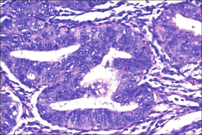
Hematoxylin and eosin staining of the histopathology of Barrett’s esophagus, dysplasia, and adenocarcinoma
A. Non-dysplastic Barrett’s mucosa (columnar cells) characterized by uniform, bland nuclei arranged in a surface monolayer
B. Low-grade dysplasia (nuclear hyperchromasia, elongation, and stratification extending up to the surface epithelium)
C. High-grade dysplasia (increased architectural and cytologic complexity)
D. Intramucosal adenocarcinoma (severe architectural distortion, including angulated glands).

Image: “Hematoxylin & Eosin staining” by Department of Pathology, The Pennsylvania State University College of Medicine, Penn State Milton S. Hershey Medical Center, 500 University Drive, Hershey, PA, 17033, USA. License: CC BY 4.0

Related videos

Clinical Relevance

  • Left ventricular hypertrophy Ventricular Hypertrophy Tetralogy of Fallot and heart failure Heart Failure A heterogeneous condition in which the heart is unable to pump out sufficient blood to meet the metabolic need of the body. Heart failure can be caused by structural defects, functional abnormalities (ventricular dysfunction), or a sudden overload beyond its capacity. Chronic heart failure is more common than acute heart failure which results from sudden insult to cardiac function, such as myocardial infarction. Total Anomalous Pulmonary Venous Return (TAPVR): characterized by hypertrophy of the wall of the left ventricle. The condition is eventually complicated by a reduction in systolic function and/or diastolic dysfunction Diastolic dysfunction Restrictive Cardiomyopathy, leading to heart failure Heart Failure A heterogeneous condition in which the heart is unable to pump out sufficient blood to meet the metabolic need of the body. Heart failure can be caused by structural defects, functional abnormalities (ventricular dysfunction), or a sudden overload beyond its capacity. Chronic heart failure is more common than acute heart failure which results from sudden insult to cardiac function, such as myocardial infarction. Total Anomalous Pulmonary Venous Return (TAPVR). Left ventricular hypertrophy Ventricular Hypertrophy Tetralogy of Fallot regression Regression Corneal Abrasions, Erosion, and Ulcers can be achieved with control of blood pressure, weight loss Weight loss Decrease in existing body weight. Bariatric Surgery, and medications.
  • Endometrial hyperplasia Endometrial Hyperplasia Benign proliferation of the endometrium in the uterus. Endometrial hyperplasia is classified by its cytology and glandular tissue. There are simple, complex (adenomatous without atypia), and atypical hyperplasia representing also the ascending risk of becoming malignant. Endometrial Hyperplasia and Endometrial Cancer and endometrial cancer Endometrial Cancer Endometrial carcinoma (EC) is the most common gynecologic malignancy in the developed world, and it has several histologic types. Endometrioid carcinoma (known as type 1 EC) typically develops from atypical endometrial hyperplasia, is hormonally responsive, and carries a favorable prognosis. Endometrial Hyperplasia and Endometrial Cancer: the abnormal proliferation of endometrial glands relative to the stroma, resulting from prolonged estrogenic stimulation. Patients Patients Individuals participating in the health care system for the purpose of receiving therapeutic, diagnostic, or preventive procedures. Clinician–Patient Relationship present with abnormal uterine bleeding Abnormal Uterine Bleeding Abnormal uterine bleeding is the medical term for abnormalities in the frequency, volume, duration, and regularity of the menstrual cycle. Abnormal uterine bleeding is classified using the acronym PALM-COEIN, with PALM representing the structural causes and COEIN indicating the non-structural causes. Abnormal Uterine Bleeding. Atypical hyperplasia Atypical Hyperplasia Benign Breast Conditions, composed of proliferating glands with nuclear atypia Atypia Fibrocystic Change, is associated with an increased risk of endometrial carcinoma.
  • Brain Brain The part of central nervous system that is contained within the skull (cranium). Arising from the neural tube, the embryonic brain is comprised of three major parts including prosencephalon (the forebrain); mesencephalon (the midbrain); and rhombencephalon (the hindbrain). The developed brain consists of cerebrum; cerebellum; and other structures in the brain stem. Nervous System: Anatomy, Structure, and Classification atrophy and dementia Dementia Major neurocognitive disorders (NCD), also known as dementia, are a group of diseases characterized by decline in a person’s memory and executive function. These disorders are progressive and persistent diseases that are the leading cause of disability among elderly people worldwide. Major Neurocognitive Disorders: Clinical stroke ( ischemia Ischemia A hypoperfusion of the blood through an organ or tissue caused by a pathologic constriction or obstruction of its blood vessels, or an absence of blood circulation. Ischemic Cell Damage or hemorrhage) or cerebral small vessel disease and degenerative processes lead to atrophy of the cerebral structures, from which cognitive and behavioral impairment can develop. Vascular dementia Dementia Major neurocognitive disorders (NCD), also known as dementia, are a group of diseases characterized by decline in a person’s memory and executive function. These disorders are progressive and persistent diseases that are the leading cause of disability among elderly people worldwide. Major Neurocognitive Disorders evolves from reduced cerebrovascular supply. Neuroimaging Neuroimaging Non-invasive methods of visualizing the central nervous system, especially the brain, by various imaging modalities. Febrile Infant shows infarcts and accelerated brain Brain The part of central nervous system that is contained within the skull (cranium). Arising from the neural tube, the embryonic brain is comprised of three major parts including prosencephalon (the forebrain); mesencephalon (the midbrain); and rhombencephalon (the hindbrain). The developed brain consists of cerebrum; cerebellum; and other structures in the brain stem. Nervous System: Anatomy, Structure, and Classification atrophy. Imaging of Alzheimer’s disease, a neurodegenerative disorder, shows focal or generalized brain Brain The part of central nervous system that is contained within the skull (cranium). Arising from the neural tube, the embryonic brain is comprised of three major parts including prosencephalon (the forebrain); mesencephalon (the midbrain); and rhombencephalon (the hindbrain). The developed brain consists of cerebrum; cerebellum; and other structures in the brain stem. Nervous System: Anatomy, Structure, and Classification atrophy.
  • Barrett’s esophagus Esophagus The esophagus is a muscular tube-shaped organ of around 25 centimeters in length that connects the pharynx to the stomach. The organ extends from approximately the 6th cervical vertebra to the 11th thoracic vertebra and can be divided grossly into 3 parts: the cervical part, the thoracic part, and the abdominal part. Esophagus: Anatomy and esophageal adenocarcinoma Esophageal Adenocarcinoma Esophageal Cancer: Barrett’s esophagus Esophagus The esophagus is a muscular tube-shaped organ of around 25 centimeters in length that connects the pharynx to the stomach. The organ extends from approximately the 6th cervical vertebra to the 11th thoracic vertebra and can be divided grossly into 3 parts: the cervical part, the thoracic part, and the abdominal part. Esophagus: Anatomy develops when metaplastic changes occur in the esophagus Esophagus The esophagus is a muscular tube-shaped organ of around 25 centimeters in length that connects the pharynx to the stomach. The organ extends from approximately the 6th cervical vertebra to the 11th thoracic vertebra and can be divided grossly into 3 parts: the cervical part, the thoracic part, and the abdominal part. Esophagus: Anatomy in order to protect itself from gastric acid Gastric acid Hydrochloric acid present in gastric juice. Gastroesophageal Reflux Disease (GERD) reflux. The condition is one of the major risk factors for esophageal cancer Esophageal cancer Esophageal cancer is 1 of the most common causes of cancer-related deaths worldwide. Nearly all esophageal cancers are either adenocarcinoma (commonly affecting the distal esophagus) or squamous cell carcinoma (affecting the proximal two-thirds of the esophagus). Esophageal Cancer. Chronic gastroesophageal reflux disease Gastroesophageal Reflux Disease Gastroesophageal reflux disease (GERD) occurs when the stomach acid frequently flows back into the esophagus. This backwash (acid reflux) can irritate the lining of the esophagus, causing symptoms such as retrosternal burning pain (heartburn). Gastroesophageal Reflux Disease (GERD) ( GERD GERD Gastroesophageal reflux disease (GERD) occurs when the stomach acid frequently flows back into the esophagus. This backwash (acid reflux) can irritate the lining of the esophagus, causing symptoms such as retrosternal burning pain (heartburn). Gastroesophageal Reflux Disease (GERD)) predisposes to dysplasia, or disordered growth and architectural disarray of epithelial cells. Dysplastic changes predispose to the development of adenocarcinoma.

References

  1. Glick, D., Barth, S., MacLeod, K. (2010) Autophagy: cellular and molecular mechanisms. https://www.ncbi.nlm.nih.gov/pmc/articles/PMC2990190/
  2. Kemp W.L., & Burns D.K., & Brown T.G. (Eds.) (2008). Cellular pathology. Pathology: The Big Picture. McGraw-Hill.
  3. McCullough, P., Bakris, G., Forman, J. (2019) Clinical implications and treatment of left ventricular hypertrophy in hypertension. UpToDate. Retrieved 17 Oct 2020, from https://www.uptodate.com/contents/clinical-implications-and-treatment-of-left-ventricular-hypertrophy-in-hypertension
  4. Oakes, S. (2020) Cell injury, cell death and adaptation in Kumar, V., Abbas, A., Aster, J. & Robbins, S. Robbins and Cotran Pathologic Basis of Disease (10th Ed., pp. 33–65). Elsevier, Inc.

Create your free account or log in to continue reading!

Sign up now and get free access to Lecturio with concept pages, medical videos, and questions for your medical education.

User Reviews

Unwrap New Skills This Holiday 🎄 Save 30% on all plans now!

Details