Advertisement

Advertisement

Advertisement

Advertisement

Brown-Séquard Syndrome

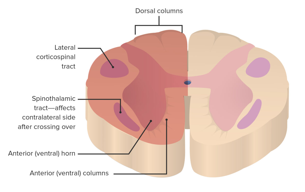
Brown-Séquard syndrome (BSS) is a rare neurologic injury that results from hemisection of the spinal cord Spinal cord The spinal cord is the major conduction pathway connecting the brain to the body; it is part of the CNS. In cross section, the spinal cord is divided into an H-shaped area of gray matter (consisting of synapsing neuronal cell bodies) and a surrounding area of white matter (consisting of ascending and descending tracts of myelinated axons). Spinal Cord: Anatomy, leading to weakness and paralysis of one side of the body and sensory Sensory Neurons which conduct nerve impulses to the central nervous system. Nervous System: Histology loss on the opposite side. This syndrome is most often due to trauma, but it may also occur with disk herniation Herniation Omphalocele, hematoma Hematoma A collection of blood outside the blood vessels. Hematoma can be localized in an organ, space, or tissue. Intussusception, or tumor Tumor Inflammation. Clinical presentation is consistent with ipsilateral damage to the corticospinal tracts Corticospinal Tracts Central Cord Syndrome and posterior columns (weakness, loss of proprioception Proprioception Sensory functions that transduce stimuli received by proprioceptive receptors in joints, tendons, muscles, and the inner ear into neural impulses to be transmitted to the central nervous system. Proprioception provides sense of stationary positions and movements of one's body parts, and is important in maintaining kinesthesia and postural balance. Neurological Examination, and vibration Vibration A continuing periodic change in displacement with respect to a fixed reference. Neurological Examination sensation) below the level of the lesion, and contralateral anterior column Anterior column Spinal Cord: Anatomy symptoms owing to the unilateral involvement of the spinothalamic tract (loss of pain Pain An unpleasant sensation induced by noxious stimuli which are detected by nerve endings of nociceptive neurons. Pain: Types and Pathways and temperature sensation). Diagnosis is confirmed with MRI. Management depends on the etiology and site of injury, and timely intervention is associated with a favorable prognosis Prognosis A prediction of the probable outcome of a disease based on a individual's condition and the usual course of the disease as seen in similar situations. Non-Hodgkin Lymphomas and recovery.
Last updated: Mar 29, 2023

Editorial responsibility: Stanley Oiseth, Lindsay Jones, Evelin Maza

Advertisement

Advertisement

Advertisement

Advertisement

Advertisement

Advertisement

Overview

Definition

Brown-Séquard syndrome (BSS) is a rare neurologic injury that results from hemisection of the spinal cord Spinal cord The spinal cord is the major conduction pathway connecting the brain to the body; it is part of the CNS. In cross section, the spinal cord is divided into an H-shaped area of gray matter (consisting of synapsing neuronal cell bodies) and a surrounding area of white matter (consisting of ascending and descending tracts of myelinated axons). Spinal Cord: Anatomy, leading to weakness and paralysis of one side of the body and sensory Sensory Neurons which conduct nerve impulses to the central nervous system. Nervous System: Histology loss on the opposite side. 

Epidemiology

  • Rare: 2%–4% of spinal cord injuries Spinal cord injuries Spinal cord injuries are complex injuries that involve damage to the neural tissue within the spinal canal. Spinal cord injuries are commonly the result of trauma. Clinical presentation varies depending on the site of injury and on whether the injury is complete or incomplete. Spinal Cord Injuries 
  • Incidence Incidence The number of new cases of a given disease during a given period in a specified population. It also is used for the rate at which new events occur in a defined population. It is differentiated from prevalence, which refers to all cases in the population at a given time. Measures of Disease Frequency: higher in males
  • Average age at occurrence: 40 years

Anatomy

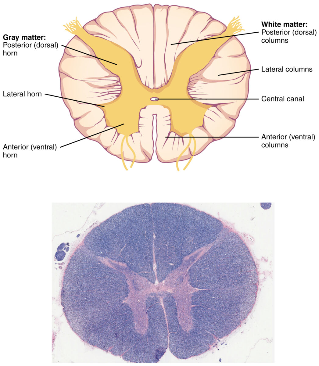
  • In a cross-sectional view, the spinal cord Spinal cord The spinal cord is the major conduction pathway connecting the brain to the body; it is part of the CNS. In cross section, the spinal cord is divided into an H-shaped area of gray matter (consisting of synapsing neuronal cell bodies) and a surrounding area of white matter (consisting of ascending and descending tracts of myelinated axons). Spinal Cord: Anatomy (SC) shows white matter surrounding gray matter Gray matter Region of central nervous system that appears darker in color than the other type, white matter. It is composed of neuronal cell bodies; neuropil; glial cells and capillaries but few myelinated nerve fibers. Cerebral Cortex: Anatomy
  • The gray matter Gray matter Region of central nervous system that appears darker in color than the other type, white matter. It is composed of neuronal cell bodies; neuropil; glial cells and capillaries but few myelinated nerve fibers. Cerebral Cortex: Anatomy is arranged in an H-shaped pattern. 
  • Gray matter Gray matter Region of central nervous system that appears darker in color than the other type, white matter. It is composed of neuronal cell bodies; neuropil; glial cells and capillaries but few myelinated nerve fibers. Cerebral Cortex: Anatomy:
    • Anterior horn 
    • Posterior horn 
    • Lateral horn 
  • White matter: ascending and descending tracts Descending tracts Spinal Cord: Anatomy in the peripheral SC

Etiology

The most frequent cause of BSS is penetrating injury. There are nontraumatic causes as well.

  • Traumatic causes:
    • Knife/stab injury to the cervical region Cervical region Spinal Cord: Anatomy 
    • Gunshot/bullet to the spine Spine The human spine, or vertebral column, is the most important anatomical and functional axis of the human body. It consists of 7 cervical vertebrae, 12 thoracic vertebrae, and 5 lumbar vertebrae and is limited cranially by the skull and caudally by the sacrum. Vertebral Column: Anatomy 
    • Motor Motor Neurons which send impulses peripherally to activate muscles or secretory cells. Nervous System: Histology vehicle accident 
    • Fall resulting in vertebral fracture Fracture A fracture is a disruption of the cortex of any bone and periosteum and is commonly due to mechanical stress after an injury or accident. Open fractures due to trauma can be a medical emergency. Fractures are frequently associated with automobile accidents, workplace injuries, and trauma. Overview of Bone Fractures
  • Nontraumatic causes:
    • Multiple sclerosis Sclerosis A pathological process consisting of hardening or fibrosis of an anatomical structure, often a vessel or a nerve. Wilms Tumor/ demyelination Demyelination Multiple Sclerosis
    • Herniation Herniation Omphalocele of the vertebral disc
    • Primary or metastatic tumors 
    • Spinal epidural hematoma Hematoma A collection of blood outside the blood vessels. Hematoma can be localized in an organ, space, or tissue. Intussusception 
    • Transverse myelitis Transverse myelitis Inflammation which extends horizontally across the spinal cord, believed to be immune-mediated and triggered by infection; associated with signs and symptoms of motor, sensory, and/or autonomic dysfunction. Mononucleosis 
    • Infections Infections Invasion of the host organism by microorganisms or their toxins or by parasites that can cause pathological conditions or diseases. Chronic Granulomatous Disease:
      • TB TB Tuberculosis (TB) is an infectious disease caused by Mycobacterium tuberculosis complex bacteria. The bacteria usually attack the lungs but can also damage other parts of the body. Approximately 30% of people around the world are infected with this pathogen, with the majority harboring a latent infection. Tuberculosis spreads through the air when a person with active pulmonary infection coughs or sneezes. Tuberculosis 
      • Meningitis Meningitis Meningitis is inflammation of the meninges, the protective membranes of the brain, and spinal cord. The causes of meningitis are varied, with the most common being bacterial or viral infection. The classic presentation of meningitis is a triad of fever, altered mental status, and nuchal rigidity. Meningitis 
      • Syphilis Syphilis Syphilis is a bacterial infection caused by the spirochete Treponema pallidum pallidum (T. p. pallidum), which is usually spread through sexual contact. Syphilis has 4 clinical stages: primary, secondary, latent, and tertiary. Syphilis 

Pathophysiology

  • Ascending and descending tracts Descending tracts Spinal Cord: Anatomy on one side of the spinal cord Spinal cord The spinal cord is the major conduction pathway connecting the brain to the body; it is part of the CNS. In cross section, the spinal cord is divided into an H-shaped area of gray matter (consisting of synapsing neuronal cell bodies) and a surrounding area of white matter (consisting of ascending and descending tracts of myelinated axons). Spinal Cord: Anatomy are damaged →
    • Petechial hemorrhages develop in the gray matter Gray matter Region of central nervous system that appears darker in color than the other type, white matter. It is composed of neuronal cell bodies; neuropil; glial cells and capillaries but few myelinated nerve fibers. Cerebral Cortex: Anatomy within an hour after injury → 
    • White matter is affected within 3–4 hours after injury.
  • 24–36 hours after injury → hemorrhagic necrosis Necrosis The death of cells in an organ or tissue due to disease, injury or failure of the blood supply. Ischemic Cell Damage → extensive damage in the myelinated Myelinated Internuclear Ophthalmoplegia fibers and long tracts 
  • Discontinuity in the primary somatosensory cortex Somatosensory cortex Area of the parietal lobe concerned with receiving sensations such as movement, pain, pressure, position, temperature, touch, and vibration. It lies posterior to the central sulcus. Cerebral Cortex: Anatomy and the supplementary motor Motor Neurons which send impulses peripherally to activate muscles or secretory cells. Nervous System: Histology area → clinically decreased sensation

Clinical Presentation and Diagnosis

Brown-Séquard syndrome occurs with hemisection of the spinal cord Spinal cord The spinal cord is the major conduction pathway connecting the brain to the body; it is part of the CNS. In cross section, the spinal cord is divided into an H-shaped area of gray matter (consisting of synapsing neuronal cell bodies) and a surrounding area of white matter (consisting of ascending and descending tracts of myelinated axons). Spinal Cord: Anatomy after an injury or other pathology. The neurologic findings on exam are related to the level of the spinal cord Spinal cord The spinal cord is the major conduction pathway connecting the brain to the body; it is part of the CNS. In cross section, the spinal cord is divided into an H-shaped area of gray matter (consisting of synapsing neuronal cell bodies) and a surrounding area of white matter (consisting of ascending and descending tracts of myelinated axons). Spinal Cord: Anatomy lesion.
Clinical presentation

  • Sensory Sensory Neurons which conduct nerve impulses to the central nervous system. Nervous System: Histology loss:
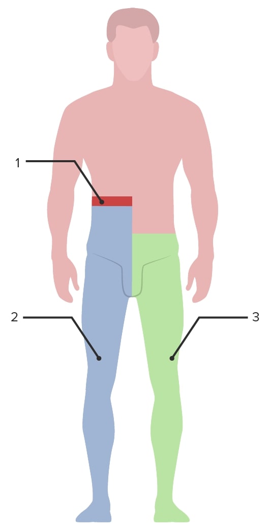
    • Ipsilateral loss of proprioception Proprioception Sensory functions that transduce stimuli received by proprioceptive receptors in joints, tendons, muscles, and the inner ear into neural impulses to be transmitted to the central nervous system. Proprioception provides sense of stationary positions and movements of one’s body parts, and is important in maintaining kinesthesia and postural balance. Neurological Examination and vibratory sensation ( dorsal columns Dorsal Columns Posterior Cord Syndrome) occurs below the level of the lesion (see image).
    • Contralateral loss of pain Pain An unpleasant sensation induced by noxious stimuli which are detected by nerve endings of nociceptive neurons. Pain: Types and Pathways and temperature sensations = spinothalamic tract (STT) beginning approximately 2 levels below the level of the lesion, since the STT continues on the ipsilateral side of the cord before crossing over Crossing over The reciprocal exchange of segments at corresponding positions along pairs of homologous chromosomes by symmetrical breakage and crosswise rejoining forming cross-over sites (holliday junctions) that are resolved during chromosome segregation. Crossing-over typically occurs during meiosis but it may also occur in the absence of meiosis, for example, with bacterial chromosomes, organelle chromosomes, or somatic cell nuclear chromosomes. Basic Terms of Genetics to the contralateral side
  • Motor Motor Neurons which send impulses peripherally to activate muscles or secretory cells. Nervous System: Histology loss: ipsilateral loss of motor Motor Neurons which send impulses peripherally to activate muscles or secretory cells. Nervous System: Histology function below the level of the lesion ( corticospinal tracts Corticospinal Tracts Central Cord Syndrome)
  • Autonomic intact: Unilateral involvement of the descending autonomic fibers does not produce bladder Bladder A musculomembranous sac along the urinary tract. Urine flows from the kidneys into the bladder via the ureters, and is held there until urination. Pyelonephritis and Perinephric Abscess symptoms. 
  • In actuality, most cases of BSS are partial hemisections → varying degrees of sensory Sensory Neurons which conduct nerve impulses to the central nervous system. Nervous System: Histology and motor Motor Neurons which send impulses peripherally to activate muscles or secretory cells. Nervous System: Histology loss depending on the etiology
  • Ipsilateral Horner sign (loss of sympathetic input) if lesion is above T1:
Site of clinical presentation in brown-séquard syndrome

Image representing the clinical findings in Brown-Séquard syndrome:
The green area represents the side of the spinal cord lesion (right side).
1: Level of the lesion
2: Ipsilateral loss of proprioception and vibratory sensation occurs below the level of the lesion (dorsal columns) and motor function (corticospinal tract).
3: Contralateral loss of pain and temperature sensation occurs beginning approximately 2 levels below the level of the lesion (spinothalamic tract).

Image by Lecturio.

Diagnosis

  • History and physical exam:
    • To determine the cause/extent of the damage and neurologic deficits Neurologic Deficits High-Risk Headaches 
    • If the lesion is above T1, Horner syndrome Horner syndrome Horner syndrome is a condition resulting from an interruption of the sympathetic innervation of the eyes. The syndrome is usually idiopathic but can be directly caused by head and neck trauma, cerebrovascular disease, or a tumor of the CNS. Horner Syndrome is seen.
    • Travel history is also essential to rule out infections Infections Invasion of the host organism by microorganisms or their toxins or by parasites that can cause pathological conditions or diseases. Chronic Granulomatous Disease
  • Laboratory analysis:
    • Testing to rule out infectious etiologies Infectious Etiologies High-Risk Headaches, if indicated
    • Consider TB TB Tuberculosis (TB) is an infectious disease caused by Mycobacterium tuberculosis complex bacteria. The bacteria usually attack the lungs but can also damage other parts of the body. Approximately 30% of people around the world are infected with this pathogen, with the majority harboring a latent infection. Tuberculosis spreads through the air when a person with active pulmonary infection coughs or sneezes. Tuberculosis as a cause of BSS, if applicable.
  • Imaging: 
    • Plain films are obtained in cases of acute trauma. 
    • MRI is the gold standard. 
    • CT myelography is done if MRI is contraindicated.
    • Angiography Angiography Radiography of blood vessels after injection of a contrast medium. Cardiac Surgery to determine vascular malformations 

Management

Treatment of BSS depends on the etiology and site of injury. Complications are mainly due to penetrating trauma wounds, in untreated cases, or with late intervention. Prognosis Prognosis A prediction of the probable outcome of a disease based on a individual’s condition and the usual course of the disease as seen in similar situations. Non-Hodgkin Lymphomas in BSS is good as compared with other spinal cord Spinal cord The spinal cord is the major conduction pathway connecting the brain to the body; it is part of the CNS. In cross section, the spinal cord is divided into an H-shaped area of gray matter (consisting of synapsing neuronal cell bodies) and a surrounding area of white matter (consisting of ascending and descending tracts of myelinated axons). Spinal Cord: Anatomy syndromes owing to its incomplete involvement of the SC.  

Treatment

  • Wound debridement Debridement The removal of foreign material and devitalized or contaminated tissue from or adjacent to a traumatic or infected lesion until surrounding healthy tissue is exposed. Stevens-Johnson Syndrome
  • Pharmacotherapy:
    • Tetanus Tetanus Tetanus is a bacterial infection caused by Clostridium tetani, a gram-positive obligate anaerobic bacterium commonly found in soil that enters the body through a contaminated wound. C. tetani produces a neurotoxin that blocks the release of inhibitory neurotransmitters and causes prolonged tonic muscle contractions. Tetanus vaccine Vaccine Suspensions of killed or attenuated microorganisms (bacteria, viruses, fungi, protozoa), antigenic proteins, synthetic constructs, or other bio-molecular derivatives, administered for the prevention, amelioration, or treatment of infectious and other diseases. Vaccination in the presence of a wound
    • Antibiotics to prevent infection (with penetrating trauma)
    • Pain Pain An unpleasant sensation induced by noxious stimuli which are detected by nerve endings of nociceptive neurons. Pain: Types and Pathways management with analgesics
    • Baclofen Baclofen A gamma-aminobutyric acid derivative that is a specific agonist of gaba-b receptors. It is used in the treatment of muscle spasticity, especially that due to spinal cord injuries. Its therapeutic effects result from actions at spinal and supraspinal sites, generally the reduction of excitatory transmission. Spasmolytics for muscle spasticity Spasticity Spinal Disk Herniation 
  • Physical therapy Physical Therapy Becker Muscular Dystrophy:
    • Maintain strength of neurologically intact muscles 
    • Prevent damage to joints and maintain their range of motion Range of motion The distance and direction to which a bone joint can be extended. Range of motion is a function of the condition of the joints, muscles, and connective tissues involved. Joint flexibility can be improved through appropriate muscle strength exercises. Examination of the Upper Limbs
    • Prevent decubitus ulcers
    • Promote mobilization or device-assisted ambulation 
    • Establishing a routine for daily activities
  • Surgery:
    • Indications: BSS due to trauma accompanied by:
    • Procedures:
      • Spinal reduction, destabilization, and decompression 
      • Dural repair
  • Spine Spine The human spine, or vertebral column, is the most important anatomical and functional axis of the human body. It consists of 7 cervical vertebrae, 12 thoracic vertebrae, and 5 lumbar vertebrae and is limited cranially by the skull and caudally by the sacrum. Vertebral Column: Anatomy immobilization Immobilization Delirium

Complications

  • Hypotension Hypotension Hypotension is defined as low blood pressure, specifically < 90/60 mm Hg, and is most commonly a physiologic response. Hypotension may be mild, serious, or life threatening, depending on the cause. Hypotension 
  • Spinal shock Shock Shock is a life-threatening condition associated with impaired circulation that results in tissue hypoxia. The different types of shock are based on the underlying cause: distributive (↑ cardiac output (CO), ↓ systemic vascular resistance (SVR)), cardiogenic (↓ CO, ↑ SVR), hypovolemic (↓ CO, ↑ SVR), obstructive (↓ CO), and mixed. Types of Shock: after acute trauma → motor Motor Neurons which send impulses peripherally to activate muscles or secretory cells. Nervous System: Histology paralysis with initial loss but gradual recovery of reflexes (not neurogenic shock Shock Shock is a life-threatening condition associated with impaired circulation that results in tissue hypoxia. The different types of shock are based on the underlying cause: distributive (↑ cardiac output (CO), ↓ systemic vascular resistance (SVR)), cardiogenic (↓ CO, ↑ SVR), hypovolemic (↓ CO, ↑ SVR), obstructive (↓ CO), and mixed. Types of Shock)
  • Depression 
  • Pulmonary embolism Pulmonary Embolism Pulmonary embolism (PE) is a potentially fatal condition that occurs as a result of intraluminal obstruction of the main pulmonary artery or its branches. The causative factors include thrombi, air, amniotic fluid, and fat. In PE, gas exchange is impaired due to the decreased return of deoxygenated blood to the lungs. Pulmonary Embolism: due to prolonged immobility and deep vein thrombosis Thrombosis Formation and development of a thrombus or blood clot in the blood vessel. Epidemic Typhus 
  • Pulmonary and urinary tract Urinary tract The urinary tract is located in the abdomen and pelvis and consists of the kidneys, ureters, urinary bladder, and urethra. The structures permit the excretion of urine from the body. Urine flows from the kidneys through the ureters to the urinary bladder and out through the urethra. Urinary Tract: Anatomy infections Infections Invasion of the host organism by microorganisms or their toxins or by parasites that can cause pathological conditions or diseases. Chronic Granulomatous Disease 

Prognosis Prognosis A prediction of the probable outcome of a disease based on a individual’s condition and the usual course of the disease as seen in similar situations. Non-Hodgkin Lymphomas

  • Recovery can occur up to 2 years after injury. 
  • Duration of recovery depends on the site and severity of the injury.  
  • Most individuals regain functional walking ability. 

Differential Diagnosis

  • Ischemic stroke Ischemic Stroke An ischemic stroke (also known as cerebrovascular accident) is an acute neurologic injury that occurs as a result of brain ischemia; this condition may be due to cerebral blood vessel occlusion by thrombosis or embolism, or rarely due to systemic hypoperfusion. Ischemic Stroke: also known as a cerebrovascular accident Cerebrovascular accident An ischemic stroke (also known as cerebrovascular accident) is an acute neurologic injury that occurs as a result of brain ischemia; this condition may be due to cerebral blood vessel occlusion by thrombosis or embolism, or rarely due to systemic hypoperfusion. Ischemic Stroke. Ischemic stroke Ischemic Stroke An ischemic stroke (also known as cerebrovascular accident) is an acute neurologic injury that occurs as a result of brain ischemia; this condition may be due to cerebral blood vessel occlusion by thrombosis or embolism, or rarely due to systemic hypoperfusion. Ischemic Stroke is an acute neurologic injury resulting from brain ischemia Brain Ischemia Localized reduction of blood flow to brain tissue due to arterial obstruction or systemic hypoperfusion. This frequently occurs in conjunction with brain hypoxia. Prolonged ischemia is associated with brain infarction. Ischemic Stroke. The clinical presentation includes neurologic symptoms with varying degrees of motor Motor Neurons which send impulses peripherally to activate muscles or secretory cells. Nervous System: Histology and sensory Sensory Neurons which conduct nerve impulses to the central nervous system. Nervous System: Histology loss corresponding to the area of the brain Brain The part of central nervous system that is contained within the skull (cranium). Arising from the neural tube, the embryonic brain is comprised of three major parts including prosencephalon (the forebrain); mesencephalon (the midbrain); and rhombencephalon (the hindbrain). The developed brain consists of cerebrum; cerebellum; and other structures in the brain stem. Nervous System: Anatomy, Structure, and Classification affected and the extent of the tissue damage. Diagnosis is made by physical examination and imaging. Management is ideally with thrombolytic therapy to restore blood flow Blood flow Blood flow refers to the movement of a certain volume of blood through the vasculature over a given unit of time (e.g., mL per minute). Vascular Resistance, Flow, and Mean Arterial Pressure if the time frame and clinical situation permit.
  • Syringomyelia Syringomyelia Longitudinal cavities in the spinal cord, most often in the cervical region, which may extend for multiple spinal levels. The cavities are lined by dense, gliogenous tissue and may be associated with spinal cord neoplasms; spinal cord traumatic injuries; and vascular malformations. Syringomyelia is marked clinically by pain and paresthesia, muscular atrophy of the hands, and analgesia with thermoanesthesia of the hands and arms, but with the tactile sense preserved (sensory dissociation). Lower extremity spasticity and incontinence may also develop. Central Cord Syndrome: formation of a fluid-filled cyst within the spinal cord Spinal cord The spinal cord is the major conduction pathway connecting the brain to the body; it is part of the CNS. In cross section, the spinal cord is divided into an H-shaped area of gray matter (consisting of synapsing neuronal cell bodies) and a surrounding area of white matter (consisting of ascending and descending tracts of myelinated axons). Spinal Cord: Anatomy, called a syrinx, that can expand and elongate over time. Damage to the spinal cord Spinal cord The spinal cord is the major conduction pathway connecting the brain to the body; it is part of the CNS. In cross section, the spinal cord is divided into an H-shaped area of gray matter (consisting of synapsing neuronal cell bodies) and a surrounding area of white matter (consisting of ascending and descending tracts of myelinated axons). Spinal Cord: Anatomy may result in sensory Sensory Neurons which conduct nerve impulses to the central nervous system. Nervous System: Histology and motor Motor Neurons which send impulses peripherally to activate muscles or secretory cells. Nervous System: Histology deficits related to the level affected. This often includes a cape-like bilateral loss of pain Pain An unpleasant sensation induced by noxious stimuli which are detected by nerve endings of nociceptive neurons. Pain: Types and Pathways and temperature sensation along the upper chest and arms. Diagnosis is made by MRI imaging and management is with surgical intervention in individuals with severe symptoms. 
  • Anterior cord syndrome Anterior cord syndrome Anterior cord syndrome (ACS) is an incomplete cord syndrome predominantly affecting the anterior (ventral) …” of the spinal cord while sparing the dorsal columns. Anterior cord syndrome can be caused by occlusion of the anterior spinal artery or by trauma, which results in disc herniation and bone fragments disrupting the spinal cord. Anterior Cord Syndrome (ACS): incomplete cord syndrome to the anterior (ventral) ⅔ of the spinal cord Spinal cord The spinal cord is the major conduction pathway connecting the brain to the body; it is part of the CNS. In cross section, the spinal cord is divided into an H-shaped area of gray matter (consisting of synapsing neuronal cell bodies) and a surrounding area of white matter (consisting of ascending and descending tracts of myelinated axons). Spinal Cord: Anatomy while sparing the dorsal columns Dorsal Columns Posterior Cord Syndrome. Anterior cord syndrome Anterior cord syndrome Anterior cord syndrome (ACS) is an incomplete cord syndrome predominantly affecting the anterior (ventral) …” of the spinal cord while sparing the dorsal columns. Anterior cord syndrome can be caused by occlusion of the anterior spinal artery or by trauma, which results in disc herniation and bone fragments disrupting the spinal cord. Anterior Cord Syndrome can be caused by occlusion of the anterior spinal artery Anterior Spinal Artery Anterior Cord Syndrome or trauma causing disk herniation Herniation Omphalocele and bone Bone Bone is a compact type of hardened connective tissue composed of bone cells, membranes, an extracellular mineralized matrix, and central bone marrow. The 2 primary types of bone are compact and spongy. Bones: Structure and Types fragments that disrupt the spinal cord Spinal cord The spinal cord is the major conduction pathway connecting the brain to the body; it is part of the CNS. In cross section, the spinal cord is divided into an H-shaped area of gray matter (consisting of synapsing neuronal cell bodies) and a surrounding area of white matter (consisting of ascending and descending tracts of myelinated axons). Spinal Cord: Anatomy. Clinical manifestations are loss of motor Motor Neurons which send impulses peripherally to activate muscles or secretory cells. Nervous System: Histology and sensory Sensory Neurons which conduct nerve impulses to the central nervous system. Nervous System: Histology function below the level of injury. Diagnosis of ACS is by clinical exam and neuroimaging Neuroimaging Non-invasive methods of visualizing the central nervous system, especially the brain, by various imaging modalities. Febrile Infant with MRI. Management is directed at resolving the underlying cause.
  • Central cord syndrome Central Cord Syndrome Central cord syndrome (CCS) is a neurological syndrome caused by an injury to the center of the spinal cord, affecting the spinothalamic tracts ((STTs) sensory) and medial aspect of the corticospinal tracts ((CSTs) motor), most often due to trauma in patients with cervical spondylosis. Central Cord Syndrome: neurologic syndrome caused by injury to the center of the spinal cord Spinal cord The spinal cord is the major conduction pathway connecting the brain to the body; it is part of the CNS. In cross section, the spinal cord is divided into an H-shaped area of gray matter (consisting of synapsing neuronal cell bodies) and a surrounding area of white matter (consisting of ascending and descending tracts of myelinated axons). Spinal Cord: Anatomy, affecting the spinothalamic tracts Spinothalamic tracts A bundle of nerve fibers connecting each posterior horn of the spinal cord to the opposite side of the thalamus, carrying information about pain, temperature, and touch. It is one of two major routes by which afferent spinal nerve fibers carrying sensations of somaesthesia are transmitted to the thalamus. Central Cord Syndrome ( sensory Sensory Neurons which conduct nerve impulses to the central nervous system. Nervous System: Histology) and medial aspect of the corticospinal tracts Corticospinal Tracts Central Cord Syndrome ( motor Motor Neurons which send impulses peripherally to activate muscles or secretory cells. Nervous System: Histology). Clinical manifestations are motor Motor Neurons which send impulses peripherally to activate muscles or secretory cells. Nervous System: Histology deficits in the arms more than the legs and variable Variable Variables represent information about something that can change. The design of the measurement scales, or of the methods for obtaining information, will determine the data gathered and the characteristics of that data. As a result, a variable can be qualitative or quantitative, and may be further classified into subgroups. Types of Variables sensory Sensory Neurons which conduct nerve impulses to the central nervous system. Nervous System: Histology deficits below the level of injury. Diagnosis is made clinically and is supported with neuroimaging Neuroimaging Non-invasive methods of visualizing the central nervous system, especially the brain, by various imaging modalities. Febrile Infant. Definitive management can be medical or surgical depending on the severity of the injury.
  • Multiple sclerosis Sclerosis A pathological process consisting of hardening or fibrosis of an anatomical structure, often a vessel or a nerve. Wilms Tumor: autoimmune disorder Autoimmune Disorder Septic Arthritis wherein the immune system Immune system The body’s defense mechanism against foreign organisms or substances and deviant native cells. It includes the humoral immune response and the cell-mediated response and consists of a complex of interrelated cellular, molecular, and genetic components. Primary Lymphatic Organs targets the myelin protective sheath covering the nerve fibers Nerve Fibers Slender processes of neurons, including the axons and their glial envelopes (myelin sheath). Nerve fibers conduct nerve impulses to and from the central nervous system. Nervous System: Histology. Clinical presentation is with visual symptoms, tremors, unsteady gait Gait Manner or style of walking. Neurological Examination, numb or weak limbs, slurred speech Slurred Speech Cerebellar Disorders, fatigue Fatigue The state of weariness following a period of exertion, mental or physical, characterized by a decreased capacity for work and reduced efficiency to respond to stimuli. Fibromyalgia, and dizziness Dizziness An imprecise term which may refer to a sense of spatial disorientation, motion of the environment, or lightheadedness. Lateral Medullary Syndrome (Wallenberg Syndrome). Diagnosis is made by clinical presentation, MRI imaging, and evoked-potential tests. Management is supportive and targeted toward attacks and slowing progression of the disease.  
  • Decompression sickness (DCS): known informally as “the bends.” Decompression sickness is caused by compression Compression Blunt Chest Trauma and decompression of gases contained in the body during descent and rapid ascent while diving. Clinical presentation of DCS may be nonspecific and variable Variable Variables represent information about something that can change. The design of the measurement scales, or of the methods for obtaining information, will determine the data gathered and the characteristics of that data. As a result, a variable can be qualitative or quantitative, and may be further classified into subgroups. Types of Variables, with time of onset varying from immediately after surfacing to 12 hours later. Diagnosis is made clinically. Management is early supportive therapy and hyperbaric recompression treatment.

References

  1. Shams, S., Arain, A. (2021). Brown Séquard syndrome. StatPearls. Retrieved September 29, 2021, from https://www.ncbi.nlm.nih.gov/books/NBK538135/.
  2. Halvorsen, A., Pettersen, A.L., Nilsen, S.M., Halle, K.K., Schaanning, E.E., Rekand, T. (2019). Epidemiology of traumatic spinal cord injury in Norway in 2012-2016: a registry-based cross-sectional study. Spinal Cord 57:331–338. https://www.nature.com/articles/s41393-018-0225-5.
  3. Hachem, L.D., Ahuja, C.S., Fehlings, M.G. (2017). Assessment and management of acute spinal cord injury: from point of injury to rehabilitation. Journal of Spinal Cord Medicine 40:665–675. https://doi.org/10.1080/10790268.2017.1329076.
  4. Decker, J.E., Hergenroeder, A.C. (2020). Overview of cervical spinal cord and cervical peripheral nerve injuries in the child or adolescent athlete. UpToDate. Retrieved September 30, 2021, from https://www.uptodate.com/contents/overview-of-cervical-spinal-cord-and-cervical-peripheral-nerve-injuries-in-the-child-or-adolescent-athlete.

Create your free account or log in to continue reading!

Sign up now and get free access to Lecturio with concept pages, medical videos, and questions for your medical education.

User Reviews

Unwrap New Skills This Holiday 🎄 Save 30% on all plans now!

Details