Advertisement

Advertisement

Advertisement

Advertisement

Becker Muscular Dystrophy

Becker muscular dystrophy (BMD) is an X-linked recessive X-Linked Recessive Duchenne Muscular Dystrophy genetic disorder that is caused by a mutation Mutation Genetic mutations are errors in DNA that can cause protein misfolding and dysfunction. There are various types of mutations, including chromosomal, point, frameshift, and expansion mutations. Types of Mutations in the DMD DMD Duchenne muscular dystrophy (DMD) is an X-linked recessive genetic disorder that is caused by a mutation in the dmd gene. The mutation leads to the production of abnormal dystrophin, resulting in muscle-fiber destruction and replacement with fatty or fibrous tissue. Duchenne Muscular Dystrophy gene Gene A category of nucleic acid sequences that function as units of heredity and which code for the basic instructions for the development, reproduction, and maintenance of organisms. Basic Terms of Genetics. Abnormal, partially functional muscle dystrophin Dystrophin A muscle protein localized in surface membranes which is the product of the Duchenne/Becker muscular dystrophy gene. Individuals with Duchenne muscular dystrophy usually lack dystrophin completely while those with Becker muscular dystrophy have dystrophin of an altered size. It shares features with other cytoskeletal proteins such as spectrin and alpha-actinin but the precise function of dystrophin is not clear. One possible role might be to preserve the integrity and alignment of the plasma membrane to the myofibrils during muscle contraction and relaxation. Blotting Techniques protein is produced, which leads to progressive muscle weakness and the eventual loss of ambulation. The clinical course is highly variable Variable Variables represent information about something that can change. The design of the measurement scales, or of the methods for obtaining information, will determine the data gathered and the characteristics of that data. As a result, a variable can be qualitative or quantitative, and may be further classified into subgroups. Types of Variables, but symptoms generally occur by adolescence. The diagnosis is based on muscle enzymes Enzymes Enzymes are complex protein biocatalysts that accelerate chemical reactions without being consumed by them. Due to the body's constant metabolic needs, the absence of enzymes would make life unsustainable, as reactions would occur too slowly without these molecules. Basics of Enzymes, genetic testing Genetic Testing Detection of a mutation; genotype; karyotype; or specific alleles associated with genetic traits, heritable diseases, or predisposition to a disease, or that may lead to the disease in descendants. It includes prenatal genetic testing. Myotonic Dystrophies, and muscle biopsy Muscle Biopsy Trichinella/Trichinellosis (if necessary). Management of BMD is supportive and aimed at slowing disease progression and complications. Dilated cardiomyopathy Dilated Cardiomyopathy Dilated cardiomyopathy (DCM) is the most common type of non-ischemic cardiomyopathy and a common cause of heart failure (HF). The cause may be idiopathic, familial, or secondary to a variety of underlying conditions. The disease is characterized by the enlargement of 1 or both ventricles and reduced systolic function. Dilated Cardiomyopathy is the leading cause of death.

Last updated: Jul 11, 2022

Editorial responsibility: Stanley Oiseth, Lindsay Jones, Evelin Maza

Advertisement

Advertisement

Advertisement

Advertisement

Advertisement

Advertisement

Overview

Epidemiology

Becker muscular dystrophy (BMD) is the 2nd most common form of muscular dystrophy.

  • Almost exclusively seen in boys 
  • Worldwide prevalence Prevalence The total number of cases of a given disease in a specified population at a designated time. It is differentiated from incidence, which refers to the number of new cases in the population at a given time. Measures of Disease Frequency: 0.1–1.8 per 10,000 male births

Etiology

Becker muscular dystrophy is caused by non-frameshift deletion or duplication mutations of the DMD DMD Duchenne muscular dystrophy (DMD) is an X-linked recessive genetic disorder that is caused by a mutation in the dmd gene. The mutation leads to the production of abnormal dystrophin, resulting in muscle-fiber destruction and replacement with fatty or fibrous tissue. Duchenne Muscular Dystrophy gene Gene A category of nucleic acid sequences that function as units of heredity and which code for the basic instructions for the development, reproduction, and maintenance of organisms. Basic Terms of Genetics on the short arm Arm The arm, or “upper arm” in common usage, is the region of the upper limb that extends from the shoulder to the elbow joint and connects inferiorly to the forearm through the cubital fossa. It is divided into 2 fascial compartments (anterior and posterior). Arm: Anatomy of the X chromosome X chromosome The female sex chromosome, being the differential sex chromosome carried by half the male gametes and all female gametes in human and other male-heterogametic species. Basic Terms of Genetics.

  • DMD DMD Duchenne muscular dystrophy (DMD) is an X-linked recessive genetic disorder that is caused by a mutation in the dmd gene. The mutation leads to the production of abnormal dystrophin, resulting in muscle-fiber destruction and replacement with fatty or fibrous tissue. Duchenne Muscular Dystrophy is the largest known protein-coding human gene Gene A category of nucleic acid sequences that function as units of heredity and which code for the basic instructions for the development, reproduction, and maintenance of organisms. Basic Terms of Genetics, which places it at an increased risk for mutations.
  • Most cases are inherited in an X-linked recessive X-Linked Recessive Duchenne Muscular Dystrophy pattern from a carrier Carrier Vaccination mother.
  • Some cases occur from de novo mutations.

Pathophysiology

Normal physiology:

  • DMD DMD Duchenne muscular dystrophy (DMD) is an X-linked recessive genetic disorder that is caused by a mutation in the dmd gene. The mutation leads to the production of abnormal dystrophin, resulting in muscle-fiber destruction and replacement with fatty or fibrous tissue. Duchenne Muscular Dystrophy gene Gene A category of nucleic acid sequences that function as units of heredity and which code for the basic instructions for the development, reproduction, and maintenance of organisms. Basic Terms of Genetics encodes for normal dystrophin Dystrophin A muscle protein localized in surface membranes which is the product of the Duchenne/Becker muscular dystrophy gene. Individuals with Duchenne muscular dystrophy usually lack dystrophin completely while those with Becker muscular dystrophy have dystrophin of an altered size. It shares features with other cytoskeletal proteins such as spectrin and alpha-actinin but the precise function of dystrophin is not clear. One possible role might be to preserve the integrity and alignment of the plasma membrane to the myofibrils during muscle contraction and relaxation. Blotting Techniques protein, which is essential for the structural stability of the myofibers.
  • Dystrophin Dystrophin A muscle protein localized in surface membranes which is the product of the Duchenne/Becker muscular dystrophy gene. Individuals with Duchenne muscular dystrophy usually lack dystrophin completely while those with Becker muscular dystrophy have dystrophin of an altered size. It shares features with other cytoskeletal proteins such as spectrin and alpha-actinin but the precise function of dystrophin is not clear. One possible role might be to preserve the integrity and alignment of the plasma membrane to the myofibrils during muscle contraction and relaxation. Blotting Techniques forms a large glycoprotein complex ( dystrophin-associated glycoprotein complex Dystrophin-Associated Glycoprotein Complex Duchenne Muscular Dystrophy):
    • Acts as a mechanical link between the cytoskeleton Cytoskeleton The network of filaments, tubules, and interconnecting filamentous bridges which give shape, structure, and organization to the cytoplasm. The Cell: Cytosol and Cytoskeleton and the extracellular matrix Extracellular matrix A meshwork-like substance found within the extracellular space and in association with the basement membrane of the cell surface. It promotes cellular proliferation and provides a supporting structure to which cells or cell lysates in culture dishes adhere. Hypertrophic and Keloid Scars of muscle 
    • Allows for normal muscle function

Pathophysiology in BMD:

  • A mutation Mutation Genetic mutations are errors in DNA that can cause protein misfolding and dysfunction. There are various types of mutations, including chromosomal, point, frameshift, and expansion mutations. Types of Mutations in DMD DMD Duchenne muscular dystrophy (DMD) is an X-linked recessive genetic disorder that is caused by a mutation in the dmd gene. The mutation leads to the production of abnormal dystrophin, resulting in muscle-fiber destruction and replacement with fatty or fibrous tissue. Duchenne Muscular Dystrophy gene Gene A category of nucleic acid sequences that function as units of heredity and which code for the basic instructions for the development, reproduction, and maintenance of organisms. Basic Terms of Genetics encodes for an abnormal, but partially functional, dystrophin Dystrophin A muscle protein localized in surface membranes which is the product of the Duchenne/Becker muscular dystrophy gene. Individuals with Duchenne muscular dystrophy usually lack dystrophin completely while those with Becker muscular dystrophy have dystrophin of an altered size. It shares features with other cytoskeletal proteins such as spectrin and alpha-actinin but the precise function of dystrophin is not clear. One possible role might be to preserve the integrity and alignment of the plasma membrane to the myofibrils during muscle contraction and relaxation. Blotting Techniques protein.
  • Partially functional dystrophin Dystrophin A muscle protein localized in surface membranes which is the product of the Duchenne/Becker muscular dystrophy gene. Individuals with Duchenne muscular dystrophy usually lack dystrophin completely while those with Becker muscular dystrophy have dystrophin of an altered size. It shares features with other cytoskeletal proteins such as spectrin and alpha-actinin but the precise function of dystrophin is not clear. One possible role might be to preserve the integrity and alignment of the plasma membrane to the myofibrils during muscle contraction and relaxation. Blotting Techniques proteins Proteins Linear polypeptides that are synthesized on ribosomes and may be further modified, crosslinked, cleaved, or assembled into complex proteins with several subunits. The specific sequence of amino acids determines the shape the polypeptide will take, during protein folding, and the function of the protein. Energy Homeostasis → partially functional glycoprotein complex → partially functional mechanical links between the cytoskeleton Cytoskeleton The network of filaments, tubules, and interconnecting filamentous bridges which give shape, structure, and organization to the cytoplasm. The Cell: Cytosol and Cytoskeleton and extracellular matrix Extracellular matrix A meshwork-like substance found within the extracellular space and in association with the basement membrane of the cell surface. It promotes cellular proliferation and provides a supporting structure to which cells or cell lysates in culture dishes adhere. Hypertrophic and Keloid Scars of muscle
  • Partial loss of dystrophin Dystrophin A muscle protein localized in surface membranes which is the product of the Duchenne/Becker muscular dystrophy gene. Individuals with Duchenne muscular dystrophy usually lack dystrophin completely while those with Becker muscular dystrophy have dystrophin of an altered size. It shares features with other cytoskeletal proteins such as spectrin and alpha-actinin but the precise function of dystrophin is not clear. One possible role might be to preserve the integrity and alignment of the plasma membrane to the myofibrils during muscle contraction and relaxation. Blotting Techniques proteins Proteins Linear polypeptides that are synthesized on ribosomes and may be further modified, crosslinked, cleaved, or assembled into complex proteins with several subunits. The specific sequence of amino acids determines the shape the polypeptide will take, during protein folding, and the function of the protein. Energy Homeostasis → ↑ susceptibility to myofiber damage and necrosis Necrosis The death of cells in an organ or tissue due to disease, injury or failure of the blood supply. Ischemic Cell Damage
  • Suboptimal attempts at myofiber regeneration Regeneration The physiological renewal, repair, or replacement of tissue. Wound Healing → replacement of muscle with fibrous Fibrous Fibrocystic Change and adipose tissue Adipose tissue Adipose tissue is a specialized type of connective tissue that has both structural and highly complex metabolic functions, including energy storage, glucose homeostasis, and a multitude of endocrine capabilities. There are three types of adipose tissue, white adipose tissue, brown adipose tissue, and beige or “brite” adipose tissue, which is a transitional form. Adipose Tissue: Histology

Clinical Presentation

Timeline

Becker muscular dystrophy has a highly variable Variable Variables represent information about something that can change. The design of the measurement scales, or of the methods for obtaining information, will determine the data gathered and the characteristics of that data. As a result, a variable can be qualitative or quantitative, and may be further classified into subgroups. Types of Variables presentation.

  • Clinical symptoms may present between 5 and 60 years of age, but most often present by adolescence.
  • Most patients Patients Individuals participating in the health care system for the purpose of receiving therapeutic, diagnostic, or preventive procedures. Clinician–Patient Relationship are nonambulatory by approximately 20 years of age.
  • Death often occurs around the age of 40 and is frequently due to dilated cardiomyopathy Dilated Cardiomyopathy Dilated cardiomyopathy (DCM) is the most common type of non-ischemic cardiomyopathy and a common cause of heart failure (HF). The cause may be idiopathic, familial, or secondary to a variety of underlying conditions. The disease is characterized by the enlargement of 1 or both ventricles and reduced systolic function. Dilated Cardiomyopathy.
  • Patients Patients Individuals participating in the health care system for the purpose of receiving therapeutic, diagnostic, or preventive procedures. Clinician–Patient Relationship with less-severe disease may have a near-normal life span.

Presentation

Becker muscular dystrophy follows a course of slowly progressive muscle weakness in a proximal-to-distal pattern.

Symptoms:

  • Muscle cramping with strenuous activity
  • Difficulty walking and running
  • Difficulty climbing stairs and jumping
  • Easily fatigued
  • Toe walking

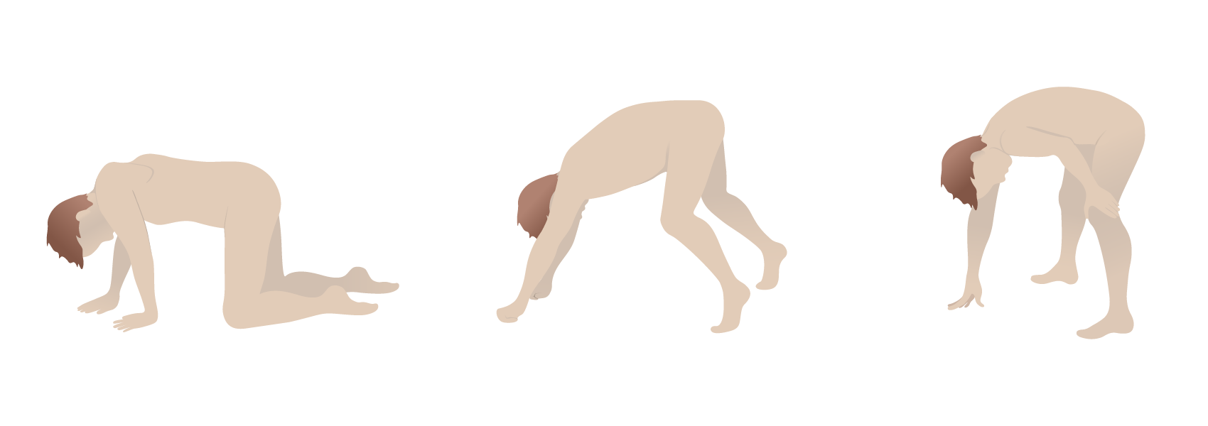
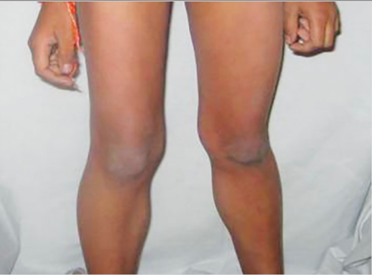
Physical exam findings:

Diagnosis

Muscle enzymes Enzymes Enzymes are complex protein biocatalysts that accelerate chemical reactions without being consumed by them. Due to the body’s constant metabolic needs, the absence of enzymes would make life unsustainable, as reactions would occur too slowly without these molecules. Basics of Enzymes

  • ↑ CK 
    • Typically 5–10 times the upper normal limit Limit A value (e.g., pressure or time) that should not be exceeded and which is specified by the operator to protect the lung Invasive Mechanical Ventilation
    • Peaks at around 10–15 years of age and declines as muscle is replaced by fat and fibrous Fibrous Fibrocystic Change tissue
  • ↑ Aldolase 
  • AST AST Enzymes of the transferase class that catalyze the conversion of l-aspartate and 2-ketoglutarate to oxaloacetate and l-glutamate. Liver Function Tests and ALT ALT An enzyme that catalyzes the conversion of l-alanine and 2-oxoglutarate to pyruvate and l-glutamate. Liver Function Tests

Genetic testing Genetic Testing Detection of a mutation; genotype; karyotype; or specific alleles associated with genetic traits, heritable diseases, or predisposition to a disease, or that may lead to the disease in descendants. It includes prenatal genetic testing. Myotonic Dystrophies

  • Sufficient for the diagnosis
  • Evaluating for DMD DMD Duchenne muscular dystrophy (DMD) is an X-linked recessive genetic disorder that is caused by a mutation in the dmd gene. The mutation leads to the production of abnormal dystrophin, resulting in muscle-fiber destruction and replacement with fatty or fibrous tissue. Duchenne Muscular Dystrophy gene deletions Gene Deletions A genetic rearrangement through loss of segments of DNA or RNA, bringing sequences which are normally separated into close proximity. This deletion may be detected using cytogenetic techniques and can also be inferred from the phenotype, indicating a deletion at one specific locus. Blotting Techniques and duplications
  • Techniques:
    • Multiplex ligation-dependent probe amplification Multiplex Ligation-Dependent Probe Amplification Duchenne Muscular Dystrophy ( MLPA MLPA Duchenne Muscular Dystrophy)
    • FISH FISH A type of in situ hybridization in which target sequences are stained with fluorescent dye so their location and size can be determined using fluorescence microscopy. This staining is sufficiently distinct that the hybridization signal can be seen both in metaphase spreads and in interphase nuclei. Chromosome Testing
    • PCR PCR Polymerase chain reaction (PCR) is a technique that amplifies DNA fragments exponentially for analysis. The process is highly specific, allowing for the targeting of specific genomic sequences, even with minuscule sample amounts. The PCR cycles multiple times through 3 phases: denaturation of the template DNA, annealing of a specific primer to the individual DNA strands, and synthesis/elongation of new DNA molecules. Polymerase Chain Reaction (PCR)

Muscle biopsy Muscle Biopsy Trichinella/Trichinellosis

  • Indicated if genetic testing Genetic Testing Detection of a mutation; genotype; karyotype; or specific alleles associated with genetic traits, heritable diseases, or predisposition to a disease, or that may lead to the disease in descendants. It includes prenatal genetic testing. Myotonic Dystrophies does not confirm a mutation Mutation Genetic mutations are errors in DNA that can cause protein misfolding and dysfunction. There are various types of mutations, including chromosomal, point, frameshift, and expansion mutations. Types of Mutations but there is still a high clinical suspicion
  • Findings: 
    • Necrotic muscle fibers of varying sizes
    • Replacement of muscle with fat and fibrous Fibrous Fibrocystic Change tissue
    • Dystrophin Dystrophin A muscle protein localized in surface membranes which is the product of the Duchenne/Becker muscular dystrophy gene. Individuals with Duchenne muscular dystrophy usually lack dystrophin completely while those with Becker muscular dystrophy have dystrophin of an altered size. It shares features with other cytoskeletal proteins such as spectrin and alpha-actinin but the precise function of dystrophin is not clear. One possible role might be to preserve the integrity and alignment of the plasma membrane to the myofibrils during muscle contraction and relaxation. Blotting Techniques on immunostaining
Histopathological exam of becker muscular dystrophy

Muscle biopsy in Becker muscular dystrophy:
Histopathological examination shows necrotic muscle fibers with replacement by fibrous and adipose tissue (A). Immunostaining shows partial positivity for dystrophin in Becker muscular dystrohy (B) compared with normal muscle (C).

Image: “Histopathological examination” by Jing Miao et al. License: CC BY 4.0

Electromyography

  • Not commonly used for diagnosis, but can help differentiate BMD from other forms of muscle weakness 
  • Shows small polyphasic potentials

Cardiac screening Screening Preoperative Care

  • Electrocardiogram Electrocardiogram An electrocardiogram (ECG) is a graphic representation of the electrical activity of the heart plotted against time. Adhesive electrodes are affixed to the skin surface allowing measurement of cardiac impulses from many angles. The ECG provides 3-dimensional information about the conduction system of the heart, the myocardium, and other cardiac structures. Electrocardiogram (ECG): detects conduction abnormalities  
  • Echocardiogram Echocardiogram Transposition of the Great Vessels: assesses for dilated cardiomyopathy Dilated Cardiomyopathy Dilated cardiomyopathy (DCM) is the most common type of non-ischemic cardiomyopathy and a common cause of heart failure (HF). The cause may be idiopathic, familial, or secondary to a variety of underlying conditions. The disease is characterized by the enlargement of 1 or both ventricles and reduced systolic function. Dilated Cardiomyopathy

Management

No curative treatment exists for BMD. Management is aimed at supportive and palliative care.

General management

  • Multidisciplinary care is required.
  • Physical therapy
  • Mobility Mobility Examination of the Breast aids AIDS Chronic HIV infection and depletion of CD4 cells eventually results in acquired immunodeficiency syndrome (AIDS), which can be diagnosed by the presence of certain opportunistic diseases called AIDS-defining conditions. These conditions include a wide spectrum of bacterial, viral, fungal, and parasitic infections as well as several malignancies and generalized conditions. HIV Infection and AIDS
  • Nutritional support

Medical therapy

  • Corticosteroids Corticosteroids Chorioretinitis:
    • Data in BMD are limited.
    • Goal is to slow the progression of muscle weakness.
  • Investigational therapies:
    • Gene Gene A category of nucleic acid sequences that function as units of heredity and which code for the basic instructions for the development, reproduction, and maintenance of organisms. Basic Terms of Genetics therapy
    • Cell therapy

Surgical management

  • Tendon-release surgery for contractures Contractures Prolonged shortening of the muscle or other soft tissue around a joint, preventing movement of the joint. Wound Healing
  • Treatment of scoliosis Scoliosis Scoliosis is a structural alteration of the vertebral column characterized by a lateral spinal curvature of greater than 10 degrees in the coronal plane. Scoliosis can be classified as idiopathic (in most cases) or secondary to underlying conditions. Scoliosis

Palliative care and advanced planning

  • Patients Patients Individuals participating in the health care system for the purpose of receiving therapeutic, diagnostic, or preventive procedures. Clinician–Patient Relationship and families should be informed of the long-term prognosis Prognosis A prediction of the probable outcome of a disease based on a individual’s condition and the usual course of the disease as seen in similar situations. Non-Hodgkin Lymphomas.
  • Address end-of-life issues End-of-life issues The end of a patient’s life has been a difficult, complex, and often controversial aspect of medicine, because, historically, death has been conceptualized as a “failure” on the physician’s part. As our understanding of death has evolved, so has the physician’s relationship to it, becoming a companion to the patient in their final moments. Moreover, experienced doctors understand that during the last days of a person’s life, the focus must be on maximizing quality of life rather than on prolonging it. End-of-life Issues.
  • Palliative measures can be used at any point in the disease.

Complications

  • Cardiac:
    • Cardiac muscle Cardiac muscle The muscle tissue of the heart. It is composed of striated, involuntary muscle cells connected to form the contractile pump to generate blood flow. Muscle Tissue: Histology fibrosis Fibrosis Any pathological condition where fibrous connective tissue invades any organ, usually as a consequence of inflammation or other injury. Bronchiolitis Obliterans leads to:
      • Dilated cardiomyopathy Dilated Cardiomyopathy Dilated cardiomyopathy (DCM) is the most common type of non-ischemic cardiomyopathy and a common cause of heart failure (HF). The cause may be idiopathic, familial, or secondary to a variety of underlying conditions. The disease is characterized by the enlargement of 1 or both ventricles and reduced systolic function. Dilated Cardiomyopathy
      • Conduction abnormalities and arrhythmias
    • Heart failure Heart Failure A heterogeneous condition in which the heart is unable to pump out sufficient blood to meet the metabolic need of the body. Heart failure can be caused by structural defects, functional abnormalities (ventricular dysfunction), or a sudden overload beyond its capacity. Chronic heart failure is more common than acute heart failure which results from sudden insult to cardiac function, such as myocardial infarction. Total Anomalous Pulmonary Venous Return (TAPVR) due to dilated cardiomyopathy Dilated Cardiomyopathy Dilated cardiomyopathy (DCM) is the most common type of non-ischemic cardiomyopathy and a common cause of heart failure (HF). The cause may be idiopathic, familial, or secondary to a variety of underlying conditions. The disease is characterized by the enlargement of 1 or both ventricles and reduced systolic function. Dilated Cardiomyopathy is the most common cause of death.
    • Cardiac involvement is more common than in Duchenne muscular dystrophy Duchenne muscular dystrophy Duchenne muscular dystrophy (DMD) is an X-linked recessive genetic disorder that is caused by a mutation in the DMD gene. The mutation leads to the production of abnormal dystrophin, resulting in muscle-fiber destruction and replacement with fatty or fibrous tissue. Duchenne Muscular Dystrophy.
  • Respiratory:
    • Progressive impaired pulmonary function due to:
      • Chest wall Chest wall The chest wall consists of skin, fat, muscles, bones, and cartilage. The bony structure of the chest wall is composed of the ribs, sternum, and thoracic vertebrae. The chest wall serves as armor for the vital intrathoracic organs and provides the stability necessary for the movement of the shoulders and arms. Chest Wall: Anatomy and diaphragm Diaphragm The diaphragm is a large, dome-shaped muscle that separates the thoracic cavity from the abdominal cavity. The diaphragm consists of muscle fibers and a large central tendon, which is divided into right and left parts. As the primary muscle of inspiration, the diaphragm contributes 75% of the total inspiratory muscle force. Diaphragm: Anatomy muscle weakness
      • Abnormal posture from scoliosis Scoliosis Scoliosis is a structural alteration of the vertebral column characterized by a lateral spinal curvature of greater than 10 degrees in the coronal plane. Scoliosis can be classified as idiopathic (in most cases) or secondary to underlying conditions. Scoliosis
    • Assisted ventilation Ventilation The total volume of gas inspired or expired per unit of time, usually measured in liters per minute. Ventilation: Mechanics of Breathing may be required.
    • Increased risk for pneumonia Pneumonia Pneumonia or pulmonary inflammation is an acute or chronic inflammation of lung tissue. Causes include infection with bacteria, viruses, or fungi. In more rare cases, pneumonia can also be caused through toxic triggers through inhalation of toxic substances, immunological processes, or in the course of radiotherapy. Pneumonia
  • Orthopedic:
    • Scoliosis Scoliosis Scoliosis is a structural alteration of the vertebral column characterized by a lateral spinal curvature of greater than 10 degrees in the coronal plane. Scoliosis can be classified as idiopathic (in most cases) or secondary to underlying conditions. Scoliosis
    • Fractures secondary to falls
    • Contractures Contractures Prolonged shortening of the muscle or other soft tissue around a joint, preventing movement of the joint. Wound Healing
  • Neurocognitive:
    • Intellectual disability Disability Determination of the degree of a physical, mental, or emotional handicap. The diagnosis is applied to legal qualification for benefits and income under disability insurance and to eligibility for social security and workman’s compensation benefits. ABCDE Assessment
    • Less common than in Duchenne muscular dystrophy Duchenne muscular dystrophy Duchenne muscular dystrophy (DMD) is an X-linked recessive genetic disorder that is caused by a mutation in the DMD gene. The mutation leads to the production of abnormal dystrophin, resulting in muscle-fiber destruction and replacement with fatty or fibrous tissue. Duchenne Muscular Dystrophy

Differential Diagnosis

  • Duchenne muscular dystrophy Duchenne muscular dystrophy Duchenne muscular dystrophy (DMD) is an X-linked recessive genetic disorder that is caused by a mutation in the DMD gene. The mutation leads to the production of abnormal dystrophin, resulting in muscle-fiber destruction and replacement with fatty or fibrous tissue. Duchenne Muscular Dystrophy ( DMD DMD Duchenne muscular dystrophy (DMD) is an X-linked recessive genetic disorder that is caused by a mutation in the dmd gene. The mutation leads to the production of abnormal dystrophin, resulting in muscle-fiber destruction and replacement with fatty or fibrous tissue. Duchenne Muscular Dystrophy): the most common and most severe form of muscular dystrophy. Duchenne muscular dystrophy Duchenne muscular dystrophy Duchenne muscular dystrophy (DMD) is an X-linked recessive genetic disorder that is caused by a mutation in the DMD gene. The mutation leads to the production of abnormal dystrophin, resulting in muscle-fiber destruction and replacement with fatty or fibrous tissue. Duchenne Muscular Dystrophy is due to an X-linked recessive X-Linked Recessive Duchenne Muscular Dystrophy frameshift mutation Frameshift Mutation A type of mutation in which a number of nucleotides deleted from or inserted into a protein coding sequence is not divisible by three, thereby causing an alteration in the reading frames of the entire coding sequence downstream of the mutation. These mutations may be induced by certain types of mutagens or may occur spontaneously. Types of Mutations in the DMD DMD Duchenne muscular dystrophy (DMD) is an X-linked recessive genetic disorder that is caused by a mutation in the dmd gene. The mutation leads to the production of abnormal dystrophin, resulting in muscle-fiber destruction and replacement with fatty or fibrous tissue. Duchenne Muscular Dystrophy gene Gene A category of nucleic acid sequences that function as units of heredity and which code for the basic instructions for the development, reproduction, and maintenance of organisms. Basic Terms of Genetics, which produces abnormal dystrophin Dystrophin A muscle protein localized in surface membranes which is the product of the Duchenne/Becker muscular dystrophy gene. Individuals with Duchenne muscular dystrophy usually lack dystrophin completely while those with Becker muscular dystrophy have dystrophin of an altered size. It shares features with other cytoskeletal proteins such as spectrin and alpha-actinin but the precise function of dystrophin is not clear. One possible role might be to preserve the integrity and alignment of the plasma membrane to the myofibrils during muscle contraction and relaxation. Blotting Techniques protein. The condition is more severe than BMD, with an earlier onset of symptoms, more rapid clinical progression, and shorter life expectancy Life expectancy Based on known statistical data, the number of years which any person of a given age may reasonably expected to live. Population Pyramids. The diagnosis is based on markedly elevated CK, genetic testing Genetic Testing Detection of a mutation; genotype; karyotype; or specific alleles associated with genetic traits, heritable diseases, or predisposition to a disease, or that may lead to the disease in descendants. It includes prenatal genetic testing. Myotonic Dystrophies, and muscle biopsy Muscle Biopsy Trichinella/Trichinellosis. Management is supportive.
  • Facioscapulohumeral dystrophy: an autosomal dominant Autosomal dominant Autosomal inheritance, both dominant and recessive, refers to the transmission of genes from the 22 autosomal chromosomes. Autosomal dominant diseases are expressed when only 1 copy of the dominant allele is inherited. Autosomal Recessive and Autosomal Dominant Inheritance disorder resulting from mutations in DUX4 or SMCHD1 genes Genes A category of nucleic acid sequences that function as units of heredity and which code for the basic instructions for the development, reproduction, and maintenance of organisms. DNA Types and Structure. Both boys and girls are affected. Patients Patients Individuals participating in the health care system for the purpose of receiving therapeutic, diagnostic, or preventive procedures. Clinician–Patient Relationship present with progressive weakness of facial, scapular, and muscles of the upper arm Upper Arm The arm, or “upper arm” in common usage, is the region of the upper limb that extends from the shoulder to the elbow joint and connects inferiorly to the forearm through the cubital fossa. It is divided into 2 fascial compartments (anterior and posterior). Arm: Anatomy by 20 years of age. Cardiac involvement is rare. There is a modest elevation in CK and the diagnosis is confirmed by genetic testing Genetic Testing Detection of a mutation; genotype; karyotype; or specific alleles associated with genetic traits, heritable diseases, or predisposition to a disease, or that may lead to the disease in descendants. It includes prenatal genetic testing. Myotonic Dystrophies. Management is supportive.
  • Congenital Congenital Chorioretinitis muscular dystrophy: a group of mostly autosomal recessive Autosomal recessive Autosomal inheritance, both dominant and recessive, refers to the transmission of genes from the 22 autosomal chromosomes. Autosomal recessive diseases are only expressed when 2 copies of the recessive allele are inherited. Autosomal Recessive and Autosomal Dominant Inheritance genetic disorders. Onset is at birth or in early infancy. Symptoms include hypotonia Hypotonia Duchenne Muscular Dystrophy, delayed motor Motor Neurons which send impulses peripherally to activate muscles or secretory cells. Nervous System: Histology development, progressive muscular weakness, and contractures Contractures Prolonged shortening of the muscle or other soft tissue around a joint, preventing movement of the joint. Wound Healing. Diagnosis is by genetic testing Genetic Testing Detection of a mutation; genotype; karyotype; or specific alleles associated with genetic traits, heritable diseases, or predisposition to a disease, or that may lead to the disease in descendants. It includes prenatal genetic testing. Myotonic Dystrophies, and management is supportive.
  • Limb-Girdle muscular dystrophy: a group of myopathic disorders resulting from varying genetic defects. Both boys and girls are affected. The time of onset for the disease varies and is characterized by slowly progressive atrophy Atrophy Decrease in the size of a cell, tissue, organ, or multiple organs, associated with a variety of pathological conditions such as abnormal cellular changes, ischemia, malnutrition, or hormonal changes. Cellular Adaptation and weakness of the proximal muscles of the hip and shoulder. Cardiac disease is not typical. Diagnosis is confirmed by genetic testing Genetic Testing Detection of a mutation; genotype; karyotype; or specific alleles associated with genetic traits, heritable diseases, or predisposition to a disease, or that may lead to the disease in descendants. It includes prenatal genetic testing. Myotonic Dystrophies. Management is supportive.
  • Myotonic dystrophy: a group of autosomal dominant Autosomal dominant Autosomal inheritance, both dominant and recessive, refers to the transmission of genes from the 22 autosomal chromosomes. Autosomal dominant diseases are expressed when only 1 copy of the dominant allele is inherited. Autosomal Recessive and Autosomal Dominant Inheritance disorders that can affect Affect The feeling-tone accompaniment of an idea or mental representation. It is the most direct psychic derivative of instinct and the psychic representative of the various bodily changes by means of which instincts manifest themselves. Psychiatric Assessment multiple systems. Myotonic dystrophy can be congenital Congenital Chorioretinitis or adult onset. Findings include myotonia Myotonia Prolonged failure of muscle relaxation after contraction. This may occur after voluntary contractions, muscle percussion, or electrical stimulation of the muscle. Myotonia is a characteristic feature of myotonic disorders. Ion Channel Myopathy, distal and facial muscle wasting Muscle Wasting Duchenne Muscular Dystrophy, cataract Cataract Partial or complete opacity on or in the lens or capsule of one or both eyes, impairing vision or causing blindness. The many kinds of cataract are classified by their morphology (size, shape, location) or etiology (cause and time of occurrence). Neurofibromatosis Type 2, intellectual disability Disability Determination of the degree of a physical, mental, or emotional handicap. The diagnosis is applied to legal qualification for benefits and income under disability insurance and to eligibility for social security and workman’s compensation benefits. ABCDE Assessment, and endocrine disorders. The diagnosis is based on genetic testing Genetic Testing Detection of a mutation; genotype; karyotype; or specific alleles associated with genetic traits, heritable diseases, or predisposition to a disease, or that may lead to the disease in descendants. It includes prenatal genetic testing. Myotonic Dystrophies, and management is mainly supportive.
  • Emery-Dreifuss muscular dystrophy: a genetic disease with various modes of inheritance. The disease is characterized by slowly progressive weakness of the upper arms and lower legs, contractures Contractures Prolonged shortening of the muscle or other soft tissue around a joint, preventing movement of the joint. Wound Healing, and cardiac abnormalities in the 1st or 2nd decade of life. Modestly elevated CK may be seen. Genetic testing Genetic Testing Detection of a mutation; genotype; karyotype; or specific alleles associated with genetic traits, heritable diseases, or predisposition to a disease, or that may lead to the disease in descendants. It includes prenatal genetic testing. Myotonic Dystrophies is used to confirm the diagnosis. Management is supportive, and most individuals do not lose the ability to ambulate.
  • Spinal muscular atrophy Atrophy Decrease in the size of a cell, tissue, organ, or multiple organs, associated with a variety of pathological conditions such as abnormal cellular changes, ischemia, malnutrition, or hormonal changes. Cellular Adaptation: a group of autosomal recessive Autosomal recessive Autosomal inheritance, both dominant and recessive, refers to the transmission of genes from the 22 autosomal chromosomes. Autosomal recessive diseases are only expressed when 2 copies of the recessive allele are inherited. Autosomal Recessive and Autosomal Dominant Inheritance disorders leading to degeneration of the anterior horn Anterior horn One of three central columns of the spinal cord. It is composed of gray matter spinal laminae VIII and ix. Brown-Séquard Syndrome cells of the spinal cord Spinal cord The spinal cord is the major conduction pathway connecting the brain to the body; it is part of the CNS. In cross section, the spinal cord is divided into an H-shaped area of gray matter (consisting of synapsing neuronal cell bodies) and a surrounding area of white matter (consisting of ascending and descending tracts of myelinated axons). Spinal Cord: Anatomy and brainstem. The clinical presentation varies. More severe forms of spinal muscular atrophy Atrophy Decrease in the size of a cell, tissue, organ, or multiple organs, associated with a variety of pathological conditions such as abnormal cellular changes, ischemia, malnutrition, or hormonal changes. Cellular Adaptation present in infancy to early childhood with progressive weakness, muscle atrophy Atrophy Decrease in the size of a cell, tissue, organ, or multiple organs, associated with a variety of pathological conditions such as abnormal cellular changes, ischemia, malnutrition, or hormonal changes. Cellular Adaptation, gross motor Gross Motor Developmental Milestones and Normal Growth delay, tongue Tongue The tongue, on the other hand, is a complex muscular structure that permits tasting and facilitates the process of mastication and communication. The blood supply of the tongue originates from the external carotid artery, and the innervation is through cranial nerves. Lips and Tongue: Anatomy atrophy Atrophy Decrease in the size of a cell, tissue, organ, or multiple organs, associated with a variety of pathological conditions such as abnormal cellular changes, ischemia, malnutrition, or hormonal changes. Cellular Adaptation and fasciculations Fasciculations Involuntary contraction of the muscle fibers innervated by a motor unit. Fasciculations may be visualized as a muscle twitch or dimpling under the skin, but usually do not generate sufficient force to move a limb. They may represent a benign condition or occur as a manifestation of motor neuron disease or peripheral nervous system diseases. Polyneuropathy, dysphagia Dysphagia Dysphagia is the subjective sensation of difficulty swallowing. Symptoms can range from a complete inability to swallow, to the sensation of solids or liquids becoming “stuck.” Dysphagia is classified as either oropharyngeal or esophageal, with esophageal dysphagia having 2 sub-types: functional and mechanical. Dysphagia, and respiratory insufficiency. Diagnosis is confirmed based on genetic testing Genetic Testing Detection of a mutation; genotype; karyotype; or specific alleles associated with genetic traits, heritable diseases, or predisposition to a disease, or that may lead to the disease in descendants. It includes prenatal genetic testing. Myotonic Dystrophies. Management is supportive, and life expectancy Life expectancy Based on known statistical data, the number of years which any person of a given age may reasonably expected to live. Population Pyramids varies by the disease type.

References

  1. Nair, D. (2019). Dystrophinopathies. Medcape. Retrieved February 21, 2021, from https://emedicine.medscape.com/article/1173204-overview#a2
  2. National Institutes of Health. (2018). Spinal Muscular Atrophy. https://rarediseases.info.nih.gov/diseases/7674/spinal-muscular-atrophy
  3. Centers for Disease Control and Prevention. (2020). What is Muscular Dystrophy? https://www.cdc.gov/ncbddd/musculardystrophy/facts.html
  4. Darras, B.T. (2021). Duchenne and Becker Muscular Dystrophy. UpToDate. Retrieved February 21, 2021, from https://www.uptodate.com/contents/duchenne-and-becker-muscular-dystrophy-genetics-and-pathogenesis
  5. Darras, B.T. (2020). Duchenne and Becker muscular dystrophy: Clinical features and diagnosis. In Dashe, J.F. (Ed.), Uptodate. Retrieved February 28, 2021, from https://www.uptodate.com/contents/duchenne-and-becker-muscular-dystrophy-clinical-features-and-diagnosis
  6. Darras, B.T. (2020). Duchenne and Becker muscular dystrophy: Management and prognosis. In Dashe, J.F. (Ed.), UpToDate. Retrieved February 28, 2021, from https://www.uptodate.com/contents/duchenne-and-becker-muscular-dystrophy-management-and-prognosis

Create your free account or log in to continue reading!

Sign up now and get free access to Lecturio with concept pages, medical videos, and questions for your medical education.

User Reviews

Unwrap New Skills This Holiday 🎄 Save 30% on all plans now!

Details