Advertisement

Advertisement

Advertisement

Advertisement

Basics of Enzymes

Enzymes are complex protein biocatalysts that accelerate chemical reactions without being consumed by them. Due to the body's constant metabolic needs, the absence of enzymes would make life unsustainable, as reactions would occur too slowly without these molecules. Enzymes have many functions, including macromolecule degradation and synthesis Synthesis Polymerase Chain Reaction (PCR) (catabolism and anabolism), signal transduction Transduction The transfer of bacterial DNA by phages from an infected bacterium to another bacterium. This also refers to the transfer of genes into eukaryotic cells by viruses. This naturally occurring process is routinely employed as a gene transfer technique. Bacteriology, energy generation ( adenosine Adenosine A nucleoside that is composed of adenine and d-ribose. Adenosine or adenosine derivatives play many important biological roles in addition to being components of DNA and RNA. Adenosine itself is a neurotransmitter. Class 5 Antiarrhythmic Drugs triphosphate [ATP]), ion pumps/ active transport Active transport The movement of materials across cell membranes and epithelial layers against an electrochemical gradient, requiring the expenditure of metabolic energy. The Cell: Cell Membrane, defense and clearance reactions (oxidization, reduction, hydrolysis Hydrolysis The process of cleaving a chemical compound by the addition of a molecule of water. Proteins and Peptides), cell regulation, movement ( myosin Myosin A diverse superfamily of proteins that function as translocating proteins. They share the common characteristics of being able to bind actins and hydrolyze mgATP. Myosins generally consist of heavy chains which are involved in locomotion, and light chains which are involved in regulation. Within the structure of myosin heavy chain are three domains: the head, the neck and the tail. The head region of the heavy chain contains the actin binding domain and mgATPase domain which provides energy for locomotion. The neck region is involved in binding the light-chains. The tail region provides the anchoring point that maintains the position of the heavy chain. The superfamily of myosins is organized into structural classes based upon the type and arrangement of the subunits they contain. Skeletal Muscle Contraction ATPase, transport of intracellular substances), and immune responses.

Last updated: Dec 13, 2024

Editorial responsibility: Stanley Oiseth, Lindsay Jones, Evelin Maza

Advertisement

Advertisement

Advertisement

Advertisement

Advertisement

Advertisement

Overview

Definition

An enzyme is a protein that presents active sites which perform reactions by decreasing the activation energy of that reaction.
Enzyme characteristics

  • Complex protein and biocatalyst (catalyst of biological origin)
  • Remains unchanged after the reaction
  • Identified by the presence of the suffix “-ase” (e.g., lactate dehydrogenase Lactate Dehydrogenase Osteosarcoma)
  • Highly specific for particular substrates and products:
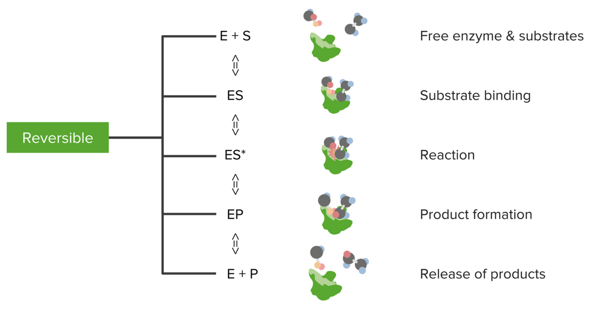
    • Substrate (S): a substance upon which the enzyme acts
    • Enzyme-substrate (ES) complex: temporary molecule formed by the non-covalent binding of the enzyme and substrate via:
    • Product (P): molecule created by the enzymatic reaction
  • pH pH The quantitative measurement of the acidity or basicity of a solution. Acid-Base Balance and temperature may alter enzyme function.
Enzyme kinetics

Relationship between substrates and enzymes, the reversible enzymatic reaction, and product formation and release

Image by Lecturio.
Active site

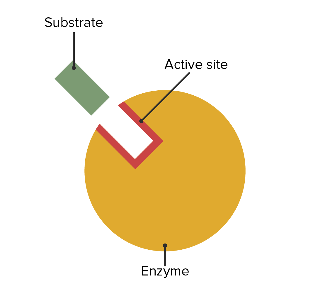
  • Area of an enzyme that binds to specific substrate molecules in order to facilitate a reaction
  • Consists of binding and catalytic sites
  • Binding sites: the area where the the substrate binds
  • Catalytic site: the area that reduces the activation energy (energy required for a reaction to proceed)
Enzyme

The location of the enzymatic active site and its relation with the substrate

Image by Lecturio.

Enzyme Specificity

  • Substrate specificity: Each enzyme can convert only one particular substrate. This is based on the “induced-fit” model, in which the binding site of the enzyme changes to match a particular substrate.
  • Stereospecificity: Substrates must be a specific isomer (e.g., lactate dehydrogenase Lactate Dehydrogenase Osteosarcoma can only convert L-lactate to pyruvate Pyruvate Derivatives of pyruvic acid, including its salts and esters. Glycolysis and not its mirror image, D-lactate).
  • Group specificity: Enzymes react with a specific chemical group (e.g., an amino group) located on the substrate.
  • Reaction specificity: Each enzyme can catalyze only a specific type of reaction (e.g., hydrolysis Hydrolysis The process of cleaving a chemical compound by the addition of a molecule of water. Proteins and Peptides).

Classification

Enzyme naming

The first part of the name describes the substrate. The last part describes the enzyme function. The first part will describe the product if the following are true:

  • The product is biochemically important (e.g., pyruvate kinase Pyruvate kinase Atp:pyruvate 2-o-phosphotransferase. A phosphotransferase that catalyzes reversibly the phosphorylation of pyruvate to phosphoenolpyruvate in the presence of ATP. It has four isozymes (l, r, m1, and m2). Glycolysis).
  • The enzyme is in the ligase category outlined in the table below (e.g., glutamine synthetase Glutamine synthetase Synthesis of Nonessential Amino Acids).
  • An enzyme with the same function already exists and acts upon the substrate (e.g., pyruvate Pyruvate Derivatives of pyruvic acid, including its salts and esters. Glycolysis dehydrogenase and lactate dehydrogenase Lactate Dehydrogenase Osteosarcoma).

Enzyme classification

Main group Catalytic reaction Important subclasses (examples)
Oxidoreductases Transfer of reduction equivalents (1 mole Mole Nevi (singular nevus), also known as “moles,” are benign neoplasms of the skin. Nevus is a non-specific medical term because it encompasses both congenital and acquired lesions, hyper- and hypopigmented lesions, and raised or flat lesions. Nevus/Nevi of electrons); the electron donor is oxidized and increases its charge, the electron acceptor is reduced and decreases its charge Dehydrogenases ( alcohol dehydrogenase Alcohol dehydrogenase A zinc-containing enzyme which oxidizes primary and secondary alcohols or hemiacetals in the presence of nad. In alcoholic fermentation, it catalyzes the final step of reducing an aldehyde to an alcohol in the presence of nadh and hydrogen. Ethanol Metabolism), oxidases (xanthine oxidase Oxidase Neisseria), reductases (glutathione reductase)
Transferases Transfer of entire groups (e.g., amino groups)
  • Aminotransferases ( aspartate Aspartate One of the non-essential amino acids commonly occurring in the l-form. It is found in animals and plants, especially in sugar cane and sugar beets. It may be a neurotransmitter. Synthesis of Nonessential Amino Acids aminotransferase [ AST AST Enzymes of the transferase class that catalyze the conversion of l-aspartate and 2-ketoglutarate to oxaloacetate and l-glutamate. Liver Function Tests])
  • Phosphotransferases Phosphotransferases A rather large group of enzymes comprising not only those transferring phosphate but also diphosphate, nucleotidyl residues, and others. These have also been subdivided according to the acceptor group. Aminoglycosides ( glycogen phosphorylase Glycogen phosphorylase An enzyme that catalyzes the degradation of glycogen in animals by releasing glucose-1-phosphate from the terminal alpha-1, 4-glycosidic bond. This enzyme exists in two forms: an active phosphorylated form ( phosphorylase A) and an inactive un-phosphorylated form (phosphorylase B). Both A and B forms of phosphorylase exist as homodimers. In mammals, the major isozymes of glycogen phosphorylase are found in muscle, liver and brain tissue. Glycogen Metabolism)
Hydrolases Molecular fission with water addition = hydrolytic fission
Lyases Break bonds between 2 carbons, or a carbon atom and oxygen, or carbon and sulfur
Isomerases Conversion of isomeric molecules into each other without changing the molecular formula Cis-trans isomerases (peptidyl-prolyl cis-trans isomerase, phosphoglucoisomerase Phosphoglucoisomerase An aldose-ketose isomerase that catalyzes the reversible interconversion of glucose 6-phosphate and fructose 6-phosphate. In prokaryotic and eukaryotic organisms it plays an essential role in glycolytic and gluconeogenic pathways. In mammalian systems the enzyme is found in the cytoplasm and as a secreted protein. This secreted form of glucose-6-phosphate isomerase has been referred to as autocrine motility factor or neuroleukin, and acts as a cytokine which binds to the autocrine motility factor receptor. Deficiency of the enzyme in humans is an autosomal recessive trait, which results in congenital nonspherocytic hemolytic anemia. Gluconeogenesis)
Ligases Also called synthetases, energy-dependent linkage of compounds (e.g., dependent on ATP)

Isoenzymes, Coenzymes, and Prosthetic Groups

Enzymes can be modified in common ways to allow different organs to have the same activities or for substances outside of the substrate/enzyme/product sequence to influence enzymes.
Isoenzymes

  • Catalyze the same kinds of reactions, but differ slightly in their structure (different amino acid Amino acid Amino acids (AAs) are composed of a central carbon atom attached to a carboxyl group, an amino group, a hydrogen atom, and a side chain (R group). Basics of Amino Acids sequence) and in the organs on which they act
  • Example: glycogen phosphorylase Glycogen phosphorylase An enzyme that catalyzes the degradation of glycogen in animals by releasing glucose-1-phosphate from the terminal alpha-1, 4-glycosidic bond. This enzyme exists in two forms: an active phosphorylated form ( phosphorylase A) and an inactive un-phosphorylated form (phosphorylase B). Both A and B forms of phosphorylase exist as homodimers. In mammals, the major isozymes of glycogen phosphorylase are found in muscle, liver and brain tissue. Glycogen Metabolism in the muscles vs. α-glucosidase in the heart

Coenzymes

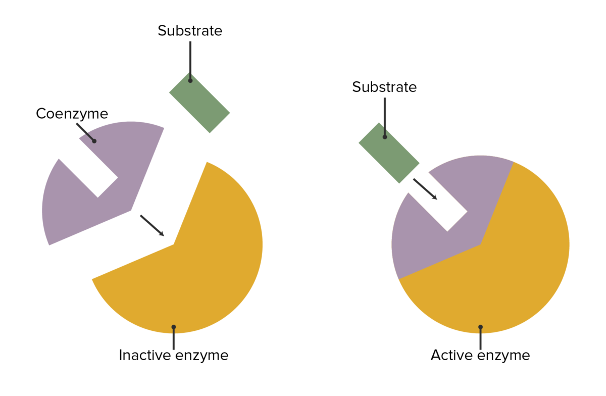
  • Small auxiliary molecules often needed to start the enzymatic reaction:
    • These molecules have the capacity to attach/detach to the enzyme.
    • Serve a variety of functions (e.g., electron transfer, transfer of organic materials)
    • Vitamins often serve as precursors to many organic cofactors.
  • Reusable (not consumed in the reaction)
Coenzyme's role

The interaction between enzymes, coenzymes, and substrate attachment

Image by Lecturio.
Common coenzymes
The following presents a list of common coenzymes and the reaction types with which they are involved.
Coenzyme Associated vitamin Reaction type Enzyme examples
Thiamine Thiamine Also known as thiamine or thiamin, it is a vitamin C12H17N4OSCl of the vitamin B complex that is essential to normal metabolism and nerve function and is widespread in plants and animals Water-soluble Vitamins and their Deficiencies pyrophosphate B1 Oxidative decarboxylation Decarboxylation The removal of a carboxyl group, usually in the form of carbon dioxide, from a chemical compound. Catabolism of Amino Acids
  • Transketolase Transketolase An enzyme of the transferase class that catalyzes the conversion of sedoheptulose 7-phosphate and d-glyceraldehyde 3-phosphate to d-ribose 5-phosphate and d-xylulose 5-phosphate in the pentose phosphate pathway. Pentose Phosphate Pathway
  • Pyruvate Pyruvate Derivatives of pyruvic acid, including its salts and esters. Glycolysis dehydrogenase
  • α-ketoglutarate dehydrogenase
FAD/FADH2 B2 Electron transfer Succinate dehydrogenase Succinate dehydrogenase A flavoprotein containing oxidoreductase that catalyzes the dehydrogenation of succinate to fumarate. In most eukaryotic organisms this enzyme is a component of mitochondrial electron transport complex II. Citric Acid Cycle
NAD NAD+ A coenzyme composed of ribosylnicotinamide 5′-diphosphate coupled to adenosine 5′-phosphate by pyrophosphate linkage. It is found widely in nature and is involved in numerous enzymatic reactions in which it serves as an electron carrier by being alternately oxidized (NAD+) and reduced (NADH). Pentose Phosphate Pathway+/NADP+ B3 Electron transfer Many dehydrogenases
Lipoamide B4 Oxidative decarboxylation Decarboxylation The removal of a carboxyl group, usually in the form of carbon dioxide, from a chemical compound. Catabolism of Amino Acids Pyruvate Pyruvate Derivatives of pyruvic acid, including its salts and esters. Glycolysis dehydrogenase
Coenzyme A (CoA) B5 Acyl group transfer α-ketoglutarate dehydrogenase
Pyridoxal phosphate Phosphate Inorganic salts of phosphoric acid. Electrolytes B6 Transamination Transamination Transamination is the transfer of an amino group from an alpha-AA to an alpha-keto acid, which is an AA with an alpha-keto group (=O) instead of an alpha-amino group (NH2). Catabolism of Amino Acids Alanine Alanine A non-essential amino acid that occurs in high levels in its free state in plasma. It is produced from pyruvate by transamination. It is involved in sugar and acid metabolism, increases immunity, and provides energy for muscle tissue, brain, and the central nervous system. Synthesis of Nonessential Amino Acids transaminase Transaminase A subclass of enzymes of the transferase class that catalyze the transfer of an amino group from a donor (generally an amino acid) to an acceptor (generally a 2-keto acid). Most of these enzymes are pyridoxyl phosphate proteins. Catabolism of Amino Acids ( ALT ALT An enzyme that catalyzes the conversion of l-alanine and 2-oxoglutarate to pyruvate and l-glutamate. Liver Function Tests)
Biotin B7 Carboxyl group transfer Pyruvate carboxylase Pyruvate carboxylase A biotin-dependent enzyme belonging to the ligase family that catalyzes the addition of carbon dioxide to pyruvate. It is occurs in both plants and animals. Gluconeogenesis
Tetrahydrofolate Tetrahydrofolate Sulfonamides and Trimethoprim (THF) B9 Transfer of C1 groups
5-deoxyadenosyl cobalamin Cobalamin A cobalt-containing coordination compound produced by intestinal microorganisms and found also in soil and water. Higher plants do not concentrate vitamin B 12 from the soil and so are a poor source of the substance as compared with animal tissues. Intrinsic factor is important for the assimilation of vitamin B 12. Folate and Vitamin B12 B12 Intramolecular rearrangements

Prosthetic groups

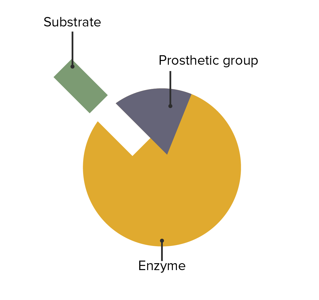
  • Specific non-polypeptide units firmly bound to the enzyme and required for the biological function of some enzymes
  • These molecules are permanently attached to the enzyme.
The interaction between enzymes, prosthetic groups, and substrate attachment.

The interaction between enzymes, prosthetic groups, and substrate attachment

Image by Lecturio.

Clinical Relevance

The following conditions are caused by an enzymatic deficiency:

  • Glucose-6-phosphate dehydrogenase Glucose-6-phosphate dehydrogenase Pentose Phosphate Pathway deficiency: inherited in an X-linked recessive X-Linked Recessive Duchenne Muscular Dystrophy manner. Can cause episodic intravascular hemolytic anemia Hemolytic Anemia Hemolytic anemia (HA) is the term given to a large group of anemias that are caused by the premature destruction/hemolysis of circulating red blood cells (RBCs). Hemolysis can occur within (intravascular hemolysis) or outside the blood vessels (extravascular hemolysis). Hemolytic Anemia when triggered by  infections Infections Invasion of the host organism by microorganisms or their toxins or by parasites that can cause pathological conditions or diseases. Chronic Granulomatous Disease, certain medication, stress, or foods such as fava beans, with all the symptoms and signs of hemolytic anemia Hemolytic Anemia Hemolytic anemia (HA) is the term given to a large group of anemias that are caused by the premature destruction/hemolysis of circulating red blood cells (RBCs). Hemolysis can occur within (intravascular hemolysis) or outside the blood vessels (extravascular hemolysis). Hemolytic Anemia such as jaundice Jaundice Jaundice is the abnormal yellowing of the skin and/or sclera caused by the accumulation of bilirubin. Hyperbilirubinemia is caused by either an increase in bilirubin production or a decrease in the hepatic uptake, conjugation, or excretion of bilirubin. Jaundice, pallor, dyspnea Dyspnea Dyspnea is the subjective sensation of breathing discomfort. Dyspnea is a normal manifestation of heavy physical or psychological exertion, but also may be caused by underlying conditions (both pulmonary and extrapulmonary). Dyspnea, fatigue Fatigue The state of weariness following a period of exertion, mental or physical, characterized by a decreased capacity for work and reduced efficiency to respond to stimuli. Fibromyalgia, and tachycardia Tachycardia Abnormally rapid heartbeat, usually with a heart rate above 100 beats per minute for adults. Tachycardia accompanied by disturbance in the cardiac depolarization (cardiac arrhythmia) is called tachyarrhythmia. Sepsis in Children. It can also produce neonatal jaundice Neonatal jaundice Yellow discoloration of the skin; mucous membrane; and sclera in the newborn. It is a sign of neonatal hyperbilirubinemia. Most cases are transient self-limiting (physiological neonatal jaundice) occurring in the first week of life, but some can be a sign of pathological disorders, particularly liver diseases. Jaundice in newborns.
  • Galactosemia Galactosemia Galactosemia is a disorder caused by defects in galactose metabolism. Galactosemia is an inherited, autosomal-recessive condition, which results in inadequate galactose processing and high blood levels of monosaccharide. The rare disorder often presents in infants with symptoms of lethargy, nausea, vomiting, diarrhea, and jaundice. Galactosemia: most common and severe form of galactosemia Galactosemia Galactosemia is a disorder caused by defects in galactose metabolism. Galactosemia is an inherited, autosomal-recessive condition, which results in inadequate galactose processing and high blood levels of monosaccharide. The rare disorder often presents in infants with symptoms of lethargy, nausea, vomiting, diarrhea, and jaundice. Galactosemia. Produced due to a deficiency of galactose-1 phosphate Phosphate Inorganic salts of phosphoric acid. Electrolytes uridyltransferase. Presents days after birth with clinical manifestations such as lethargy Lethargy A general state of sluggishness, listless, or uninterested, with being tired, and having difficulty concentrating and doing simple tasks. It may be related to depression or drug addiction. Hyponatremia, failure to thrive Failure to Thrive Failure to thrive (FTT), or faltering growth, describes suboptimal weight gain and growth in children. The majority of cases are due to inadequate caloric intake; however, genetic, infectious, and oncological etiologies are also common. Failure to Thrive, jaundice Jaundice Jaundice is the abnormal yellowing of the skin and/or sclera caused by the accumulation of bilirubin. Hyperbilirubinemia is caused by either an increase in bilirubin production or a decrease in the hepatic uptake, conjugation, or excretion of bilirubin. Jaundice, and other features of liver Liver The liver is the largest gland in the human body. The liver is found in the superior right quadrant of the abdomen and weighs approximately 1.5 kilograms. Its main functions are detoxification, metabolism, nutrient storage (e.g., iron and vitamins), synthesis of coagulation factors, formation of bile, filtration, and storage of blood. Liver: Anatomy injury
  • Chronic granulomatous disease Granulomatous disease A defect of leukocyte function in which phagocytic cells ingest but fail to digest bacteria, resulting in recurring bacterial infections with granuloma formation. When chronic granulomatous disease is caused by mutations in the cybb gene, the condition is inherited in an X-linked recessive pattern. When chronic granulomatous disease is caused by cyba, ncf1, ncf2, or ncf4 gene mutations, the condition is inherited in an autosomal recessive pattern. Common Variable Immunodeficiency (CVID): chronic disorder characterized by granuloma formation. Phagocytic cells are unable to produce bactericidal Bactericidal Penicillins superoxide due to a defect in the nicotinamide adenine dinucleotide Nicotinamide adenine dinucleotide A coenzyme composed of ribosylnicotinamide 5′-diphosphate coupled to adenosine 5′-phosphate by pyrophosphate linkage. It is found widely in nature and is involved in numerous enzymatic reactions in which it serves as an electron carrier by being alternately oxidized (NAD+) and reduced (NADH). Pentose Phosphate Pathway phosphate Phosphate Inorganic salts of phosphoric acid. Electrolytes oxidase Oxidase Neisseria in the cells.
  • Lysosomal storage diseases: a group of genetic metabolic disorders caused by lysosomal defects that result in an accumulation of undigested metabolites and cellular death
    • Gaucher’s disease: inherited disorder that leads to the accumulation of undegraded glycolipid substrates in cells due to a deficiency of acid β-glucosidase
    • Krabbe’s disease: Also known as globoid cell leukodystrophy, this condition is produced due to a deficient activity of the galactocerebrosidase Galactocerebrosidase An enzyme that hydrolyzes galactose from ceramide monohexosides. Deficiency of this enzyme may cause globoid cell leukodystrophy. Krabbe Disease, which produces an accumulation of galactosylceramide.
    • Tay-Sachs disease Tay-Sachs disease Tay-Sachs disease is an autosomal recessive lysosomal storage disorder caused by genetic mutations in the hexosaminidase A (HEXA) gene, leading to progressive neurodegeneration. Classic symptoms in infants include rapid degeneration of cognitive and neuromuscular abilities, progressive blindness, and a macular cherry-red spot on physical examination. Tay-Sachs Disease: Produced due to a deficiency of hexosaminidase A Hexosaminidase A A mammalian beta-hexosaminidase isoform that is a heteromeric protein comprised of both hexosaminidase alpha and hexosaminidase beta subunits. Deficiency of hexosaminidase A due to mutations in the gene encoding the hexosaminidase alpha subunit is a case of Tay-sachs disease. Deficiency of hexosaminidase A and hexosaminidase B due to mutations in the gene encoding the hexosaminidase beta subunit is a case of Sandhoff disease. Tay-Sachs Disease, this autosomal recessive Autosomal recessive Autosomal inheritance, both dominant and recessive, refers to the transmission of genes from the 22 autosomal chromosomes. Autosomal recessive diseases are only expressed when 2 copies of the recessive allele are inherited. Autosomal Recessive and Autosomal Dominant Inheritance lipid storage disorder may cause blindness Blindness The inability to see or the loss or absence of perception of visual stimuli. This condition may be the result of eye diseases; optic nerve diseases; optic chiasm diseases; or brain diseases affecting the visual pathways or occipital lobe. Retinopathy of Prematurity, hypotonia Hypotonia Duchenne Muscular Dystrophy, progressive cognitive decline, and seizures Seizures A seizure is abnormal electrical activity of the neurons in the cerebral cortex that can manifest in numerous ways depending on the region of the brain affected. Seizures consist of a sudden imbalance that occurs between the excitatory and inhibitory signals in cortical neurons, creating a net excitation. The 2 major classes of seizures are focal and generalized. Seizures.

References

  1. Berg, J. M., Tymoczko, J. L., & Stryer, L. (2022). Biochemistry (9th ed.). W.H. Freeman and Company.
  2. Ferrier, D. R. (2023). Lippincott Illustrated Reviews: Biochemistry (8th ed.). Wolters Kluwer.
  3. Koolman, J., & Roehm, K. H. (2021). Color Atlas of Biochemistry (3rd ed.). Thieme.
  4. Murray, R. K., Bender, D. A., Botham, K. M., Kennelly, P. J., Rodwell, V. W., & Weil, P. A. (2022). Harper’s Illustrated Biochemistry (32nd ed.). McGraw-Hill Education.
  5. Nelson, D. L., & Cox, M. M. (2021). Lehninger Principles of Biochemistry (8th ed.). W.H. Freeman and Company.
  6. Rodwell, V. W., Bender, D. A., Botham, K. M., Kennelly, P. J., & Weil, P. A. (2021). Harper’s Illustrated Biochemistry (31st ed.). McGraw-Hill Education.
  7. Voet, D., Voet, J. G., & Pratt, C. W. (2021). Fundamentals of Biochemistry: Life at the Molecular Level (6th ed.). John Wiley & Sons.

Create your free account or log in to continue reading!

Sign up now and get free access to Lecturio with concept pages, medical videos, and questions for your medical education.

User Reviews

Unwrap New Skills This Holiday 🎄 Save 30% on all plans now!

Details