Advertisement

Advertisement

Advertisement

Advertisement

Basal Ganglia: Anatomy

Basal ganglia are a group of subcortical nuclear agglomerations involved in movement, and are located deep to the cerebral hemispheres. Basal ganglia include the striatum (caudate nucleus Nucleus Within a eukaryotic cell, a membrane-limited body which contains chromosomes and one or more nucleoli (cell nucleolus). The nuclear membrane consists of a double unit-type membrane which is perforated by a number of pores; the outermost membrane is continuous with the endoplasmic reticulum. A cell may contain more than one nucleus. The Cell: Organelles and putamen), globus pallidus, substantia nigra, and subthalamic nucleus Nucleus Within a eukaryotic cell, a membrane-limited body which contains chromosomes and one or more nucleoli (cell nucleolus). The nuclear membrane consists of a double unit-type membrane which is perforated by a number of pores; the outermost membrane is continuous with the endoplasmic reticulum. A cell may contain more than one nucleus. The Cell: Organelles. The components intricately synapse Synapse The junction between 2 neurons is called a synapse. The synapse allows a neuron to pass an electrical or chemical signal to another neuron or target effector cell. Synapses and Neurotransmission onto each other to promote or antagonize movement.

Last updated: Dec 3, 2024

Editorial responsibility: Stanley Oiseth, Lindsay Jones, Evelin Maza

Advertisement

Advertisement

Advertisement

Advertisement

Advertisement

Advertisement

Development

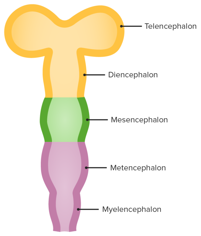
Five secondary brain vesicles

Secondary brain vesicles: The telencephalon develops from the prosencephalon and will go on to form several important structures of the brain, including the cerebral cortex and basal ganglia.

Image by Lecturio.

General Features

Definition

The basal ganglia are a cluster of subcortical nuclei deep to cerebral hemispheres and are involved in the initiation, maintenance, and inhibition of movement.

Major structures

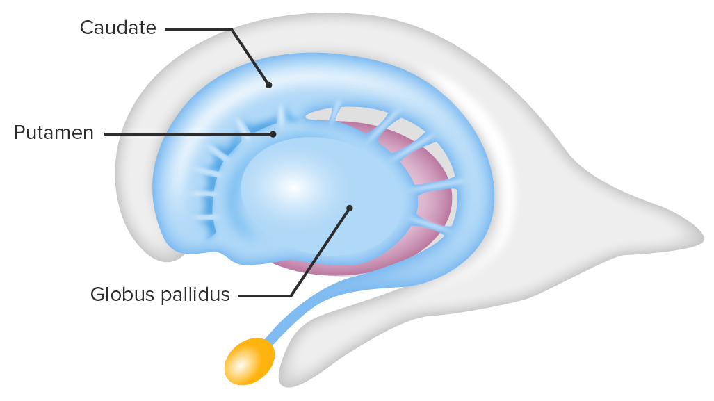
The striatum (neostriatum) is composed of:

  • Caudate nucleus Nucleus Within a eukaryotic cell, a membrane-limited body which contains chromosomes and one or more nucleoli (cell nucleolus). The nuclear membrane consists of a double unit-type membrane which is perforated by a number of pores; the outermost membrane is continuous with the endoplasmic reticulum. A cell may contain more than one nucleus. The Cell: Organelles:
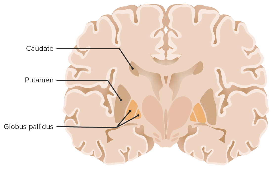
    • An elongated, C-shaped structure
    • Divided into head, body, and tail, which do not have distinct boundaries from each other
    • Runs adjacent to the lateral ventricle
    • The pear-shaped head is continuous with the putamen.
    • The caudate tail tapers and terminates near the amygdala Amygdala Almond-shaped group of basal nuclei anterior to the inferior horn of the lateral ventricle of the temporal lobe. The amygdala is part of the limbic system. Limbic System: Anatomy in the temporal lobe Temporal lobe Lower lateral part of the cerebral hemisphere responsible for auditory, olfactory, and semantic processing. It is located inferior to the lateral fissure and anterior to the occipital lobe. Cerebral Cortex: Anatomy.
  • Putamen:
    • Large nucleus Nucleus Within a eukaryotic cell, a membrane-limited body which contains chromosomes and one or more nucleoli (cell nucleolus). The nuclear membrane consists of a double unit-type membrane which is perforated by a number of pores; the outermost membrane is continuous with the endoplasmic reticulum. A cell may contain more than one nucleus. The Cell: Organelles forming the lateral portion of the basal ganglia
    • Anteriorly and ventrally, the putamen fuses with the head of the caudate.

The lenticular nucleus Nucleus Within a eukaryotic cell, a membrane-limited body which contains chromosomes and one or more nucleoli (cell nucleolus). The nuclear membrane consists of a double unit-type membrane which is perforated by a number of pores; the outermost membrane is continuous with the endoplasmic reticulum. A cell may contain more than one nucleus. The Cell: Organelles is made up of:

  • Globus pallidus (GP):
    • Smaller triangular structure consisting of numerous myelinated Myelinated Internuclear Ophthalmoplegia fibers
    • Has a GP internal segment (GPi) and GP external segment (GPe)
    • Both regions acquire inputs from the putamen and caudate and communicate with the subthalamic nucleus Nucleus Within a eukaryotic cell, a membrane-limited body which contains chromosomes and one or more nucleoli (cell nucleolus). The nuclear membrane consists of a double unit-type membrane which is perforated by a number of pores; the outermost membrane is continuous with the endoplasmic reticulum. A cell may contain more than one nucleus. The Cell: Organelles.
  • Putamen:
    • Larger convex gray mass Mass Three-dimensional lesion that occupies a space within the breast Imaging of the Breast
    • Lies lateral and inferior to the insular cortex

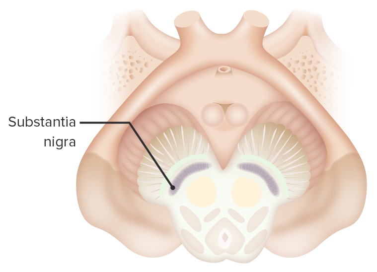
Substantia nigra:

  • Has a ventral portion called the substantia nigra pars reticulata
  • The more dorsal substantia nigra pars compacta Pars compacta A region in the substantia nigra located dorsal to the pars reticulata. Parkinson’s Disease contains the darkly pigmented dopaminergic neurons Neurons The basic cellular units of nervous tissue. Each neuron consists of a body, an axon, and dendrites. Their purpose is to receive, conduct, and transmit impulses in the nervous system. Nervous System: Histology.
  • Degeneration of the dopaminergic neurons Neurons The basic cellular units of nervous tissue. Each neuron consists of a body, an axon, and dendrites. Their purpose is to receive, conduct, and transmit impulses in the nervous system. Nervous System: Histology is an important pathogenic mechanism in Parkinson disease Parkinson disease Parkinson’s disease (PD) is a chronic, progressive neurodegenerative disorder. Although the cause is unknown, several genetic and environmental risk factors are currently being studied. Individuals present clinically with resting tremor, bradykinesia, rigidity, and postural instability. Parkinson’s Disease.

Subthalamic nucleus Nucleus Within a eukaryotic cell, a membrane-limited body which contains chromosomes and one or more nucleoli (cell nucleolus). The nuclear membrane consists of a double unit-type membrane which is perforated by a number of pores; the outermost membrane is continuous with the endoplasmic reticulum. A cell may contain more than one nucleus. The Cell: Organelles:

  • Under the thalamus Thalamus The thalamus is a large, ovoid structure in the dorsal part of the diencephalon that is located between the cerebral cortex and midbrain. It consists of several interconnected nuclei of grey matter separated by the laminae of white matter. The thalamus is the main conductor of information that passes between the cerebral cortex and the periphery, spinal cord, or brain stem. Thalamus: Anatomy lies the spindle- or cigar-shaped subthalamic nucleus Nucleus Within a eukaryotic cell, a membrane-limited body which contains chromosomes and one or more nucleoli (cell nucleolus). The nuclear membrane consists of a double unit-type membrane which is perforated by a number of pores; the outermost membrane is continuous with the endoplasmic reticulum. A cell may contain more than one nucleus. The Cell: Organelles.
  • Unlike the thalamus Thalamus The thalamus is a large, ovoid structure in the dorsal part of the diencephalon that is located between the cerebral cortex and midbrain. It consists of several interconnected nuclei of grey matter separated by the laminae of white matter. The thalamus is the main conductor of information that passes between the cerebral cortex and the periphery, spinal cord, or brain stem. Thalamus: Anatomy, the subthalamic nucleus Nucleus Within a eukaryotic cell, a membrane-limited body which contains chromosomes and one or more nucleoli (cell nucleolus). The nuclear membrane consists of a double unit-type membrane which is perforated by a number of pores; the outermost membrane is continuous with the endoplasmic reticulum. A cell may contain more than one nucleus. The Cell: Organelles is derived embryologically from the midbrain Midbrain The middle of the three primitive cerebral vesicles of the embryonic brain. Without further subdivision, midbrain develops into a short, constricted portion connecting the pons and the diencephalon. Midbrain contains two major parts, the dorsal tectum mesencephali and the ventral tegmentum mesencephali, housing components of auditory, visual, and other sensorimotor systems. Brain Stem: Anatomy rather than the forebrain forebrain The anterior of the three primitive cerebral vesicles of the embryonic brain arising from the neural tube. It subdivides to form diencephalon and telencephalon. Development of the Nervous System and Face.

Functions

  • Significant role in controlling posture and voluntary movement through connections to the:
    • Thalamus Thalamus The thalamus is a large, ovoid structure in the dorsal part of the diencephalon that is located between the cerebral cortex and midbrain. It consists of several interconnected nuclei of grey matter separated by the laminae of white matter. The thalamus is the main conductor of information that passes between the cerebral cortex and the periphery, spinal cord, or brain stem. Thalamus: Anatomy
    • Cerebral cortex Cerebral cortex The cerebral cortex is the largest and most developed part of the human brain and CNS. Occupying the upper part of the cranial cavity, the cerebral cortex has 4 lobes and is divided into 2 hemispheres that are joined centrally by the corpus callosum. Cerebral Cortex: Anatomy
    • Basal nuclei
  • Has connections to limbic system Limbic system The limbic system is a neuronal network that mediates emotion and motivation, while also playing a role in learning and memory. The extended neural network is vital to numerous basic psychological functions and plays an invaluable role in processing and responding to environmental stimuli. Limbic System: Anatomy pathways, which govern the expression of various behaviors and motivational states
  • Plays a major role in making decisions
  • Plays a role in controlling eye movements

Vasculature

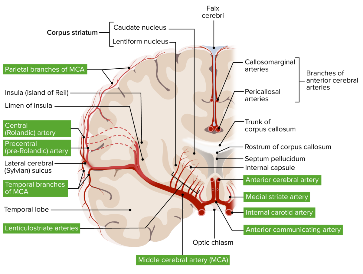
  • Main arterial supply: lenticulostriate arteries Arteries Arteries are tubular collections of cells that transport oxygenated blood and nutrients from the heart to the tissues of the body. The blood passes through the arteries in order of decreasing luminal diameter, starting in the largest artery (the aorta) and ending in the small arterioles. Arteries are classified into 3 types: large elastic arteries, medium muscular arteries, and small arteries and arterioles. Arteries: Histology
    • Perforating arteries Arteries Arteries are tubular collections of cells that transport oxygenated blood and nutrients from the heart to the tissues of the body. The blood passes through the arteries in order of decreasing luminal diameter, starting in the largest artery (the aorta) and ending in the small arterioles. Arteries are classified into 3 types: large elastic arteries, medium muscular arteries, and small arteries and arterioles. Arteries: Histology
    • Arise from:
      • Anterior cerebral artery Anterior cerebral artery Artery formed by the bifurcation of the internal carotid artery. Branches of the anterior cerebral artery supply the caudate nucleus; internal capsule; putamen; septal nuclei; gyrus cinguli; and surfaces of the frontal lobe and parietal lobe. Cerebrovascular System: Anatomy
      • Middle cerebral artery Middle cerebral artery The largest of the cerebral arteries. It trifurcates into temporal, frontal, and parietal branches supplying blood to most of the parenchyma of these lobes in the cerebral cortex. These are the areas involved in motor, sensory, and speech activities. Cerebrovascular System: Anatomy
    • Contain:
      • Lateral lenticulostriate arteries Arteries Arteries are tubular collections of cells that transport oxygenated blood and nutrients from the heart to the tissues of the body. The blood passes through the arteries in order of decreasing luminal diameter, starting in the largest artery (the aorta) and ending in the small arterioles. Arteries are classified into 3 types: large elastic arteries, medium muscular arteries, and small arteries and arterioles. Arteries: Histology
      • Medial lenticulostriate arteries Arteries Arteries are tubular collections of cells that transport oxygenated blood and nutrients from the heart to the tissues of the body. The blood passes through the arteries in order of decreasing luminal diameter, starting in the largest artery (the aorta) and ending in the small arterioles. Arteries are classified into 3 types: large elastic arteries, medium muscular arteries, and small arteries and arterioles. Arteries: Histology
    • Note: The medial striate artery (recurrent artery of Heubner) is often the only angiographically visible artery of this group.
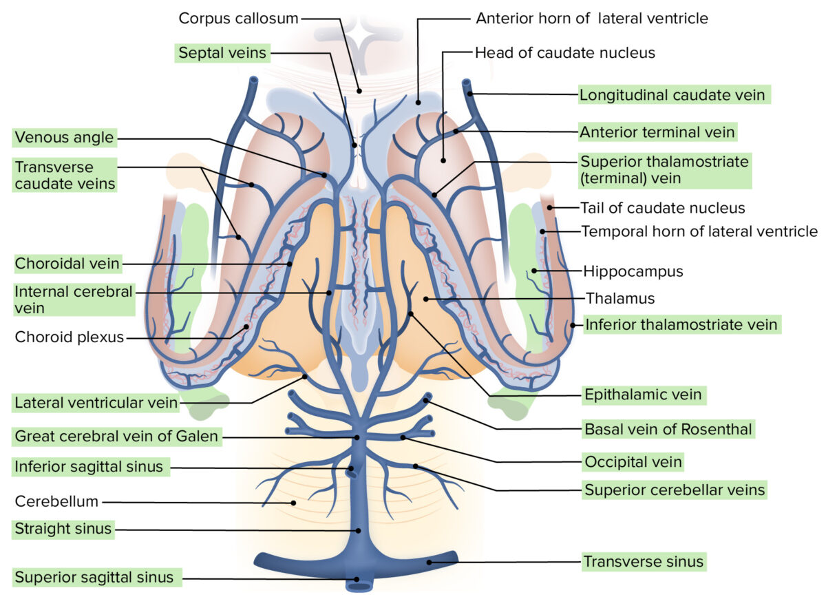
  • Venous drainage: striate branches → internal cerebral and basal veins Veins Veins are tubular collections of cells, which transport deoxygenated blood and waste from the capillary beds back to the heart. Veins are classified into 3 types: small veins/venules, medium veins, and large veins. Each type contains 3 primary layers: tunica intima, tunica media, and tunica adventitia. Veins: Histology → great cerebral vein

Neural Pathways and Intrinsic Relations

Pathways

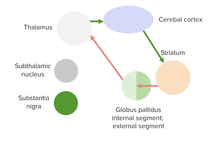
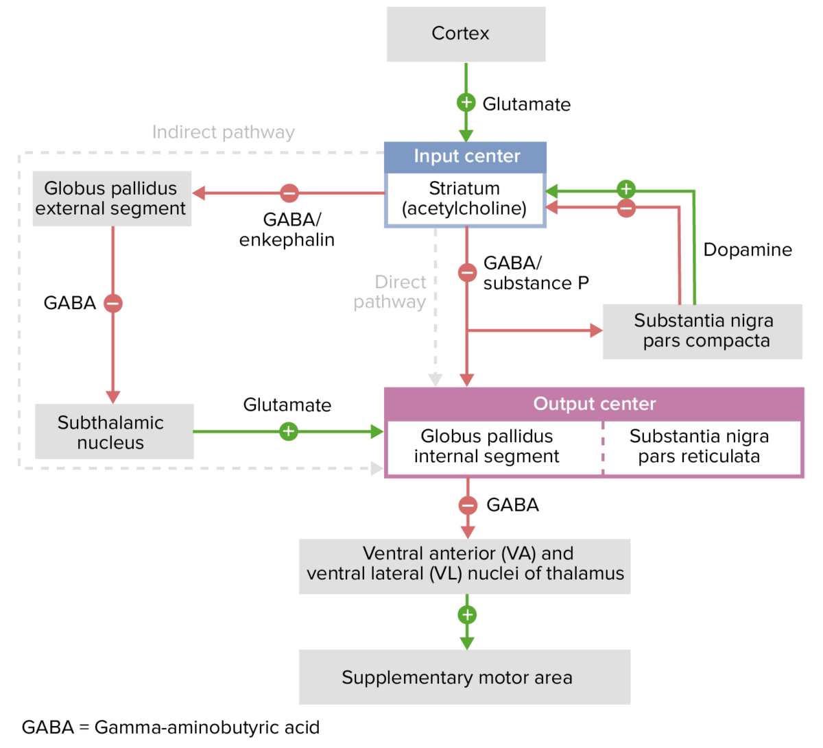
  • Direct (excitatory): striatum → GPi → thalamus Thalamus The thalamus is a large, ovoid structure in the dorsal part of the diencephalon that is located between the cerebral cortex and midbrain. It consists of several interconnected nuclei of grey matter separated by the laminae of white matter. The thalamus is the main conductor of information that passes between the cerebral cortex and the periphery, spinal cord, or brain stem. Thalamus: Anatomy cerebral cortex Cerebral cortex The cerebral cortex is the largest and most developed part of the human brain and CNS. Occupying the upper part of the cranial cavity, the cerebral cortex has 4 lobes and is divided into 2 hemispheres that are joined centrally by the corpus callosum. Cerebral Cortex: Anatomy → feedback to the striatum
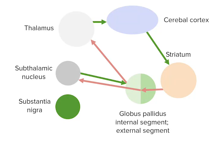
  • Indirect (inhibitory): striatum → GPe → subthalamic nucleus Nucleus Within a eukaryotic cell, a membrane-limited body which contains chromosomes and one or more nucleoli (cell nucleolus). The nuclear membrane consists of a double unit-type membrane which is perforated by a number of pores; the outermost membrane is continuous with the endoplasmic reticulum. A cell may contain more than one nucleus. The Cell: Organelles → GPi → thalamus Thalamus The thalamus is a large, ovoid structure in the dorsal part of the diencephalon that is located between the cerebral cortex and midbrain. It consists of several interconnected nuclei of grey matter separated by the laminae of white matter. The thalamus is the main conductor of information that passes between the cerebral cortex and the periphery, spinal cord, or brain stem. Thalamus: Anatomy cerebral cortex Cerebral cortex The cerebral cortex is the largest and most developed part of the human brain and CNS. Occupying the upper part of the cranial cavity, the cerebral cortex has 4 lobes and is divided into 2 hemispheres that are joined centrally by the corpus callosum. Cerebral Cortex: Anatomy → feedback to the striatum
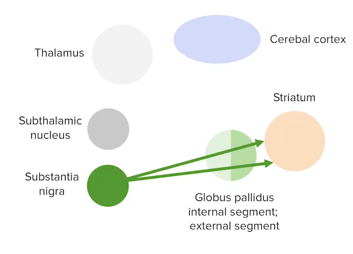
  • Nigrostriatal:
    • Substantia nigra → striatum
    • Directly modulates both the direct and indirect pathways

Intrinsic relations

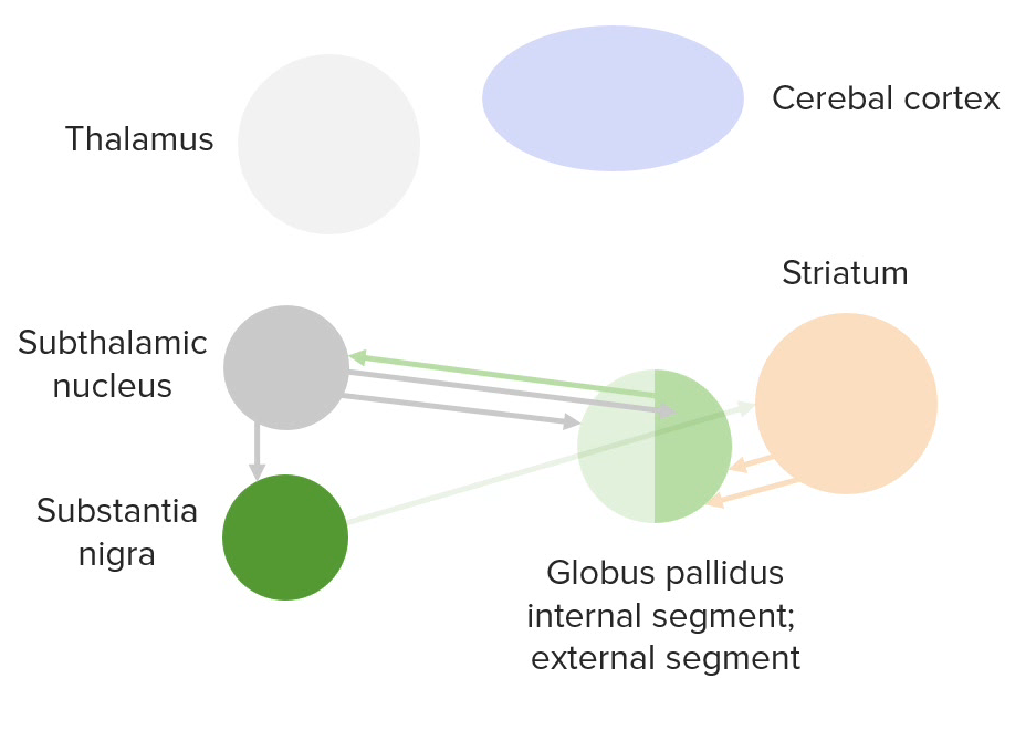
  • Inhibitory (GABAergic):
    • Striatopallidal
    • Striatonigral
    • GPe is inhibitory to the subthalamus.
  • Excitatory:
    • Subthalamic output (glutamatergic)
    • Nigrostriatal (dopaminergic)
A diagram of the intrinsic connections between the basal ganglia

Diagram of the intrinsic connections between the basal ganglia

Image by Lecturio.

Clinical Relevance

  • Parkinson disease Parkinson disease Parkinson’s disease (PD) is a chronic, progressive neurodegenerative disorder. Although the cause is unknown, several genetic and environmental risk factors are currently being studied. Individuals present clinically with resting tremor, bradykinesia, rigidity, and postural instability. Parkinson’s Disease: a degenerative disease of the basal ganglia characterized by a clinical syndrome manifesting with diminished facial expression, bradykinesia Bradykinesia Parkinson’s Disease, festinating gait Gait Manner or style of walking. Neurological Examination (progressively shortened and accelerated steps), cogwheel rigidity Cogwheel Rigidity Parkinson’s Disease, and a “pill-rolling” tremor Tremor Cyclical movement of a body part that can represent either a physiologic process or a manifestation of disease. Intention or action tremor, a common manifestation of cerebellar diseases, is aggravated by movement. In contrast, resting tremor is maximal when there is no attempt at voluntary movement, and occurs as a relatively frequent manifestation of parkinson disease. Myotonic Dystrophies at rest. The finding of Lewy bodies Lewy bodies Intracytoplasmic, eosinophilic, round to elongated inclusions found in vacuoles of injured or fragmented neurons. The presence of lewy bodies is the histological marker of the degenerative changes in lewy body disease and parkinson disease but they may be seen in other neurological conditions. They are typically found in the substantia nigra and locus coeruleus but they are also seen in the basal forebrain, hypothalamic nuclei, and neocortex. Parkinson’s Disease in the brain Brain The part of central nervous system that is contained within the skull (cranium). Arising from the neural tube, the embryonic brain is comprised of three major parts including prosencephalon (the forebrain); mesencephalon (the midbrain); and rhombencephalon (the hindbrain). The developed brain consists of cerebrum; cerebellum; and other structures in the brain stem. Nervous System: Anatomy, Structure, and Classification postmortem is the only confirmation for the disease. Treatment includes supportive physical and emotional care plus medications such as levodopa Levodopa The naturally occurring form of dihydroxyphenylalanine and the immediate precursor of dopamine. Unlike dopamine itself, it can be taken orally and crosses the blood-brain barrier. It is rapidly taken up by dopaminergic neurons and converted to dopamine. It is used for the treatment of parkinsonian disorders and is usually given with agents that inhibit its conversion to dopamine outside of the central nervous system. Parkinson’s Disease Drugs/carbidopa, monoamine oxidase type B inhibitors Monoamine Oxidase Type B Inhibitors Parkinson’s Disease, and dopamine Dopamine One of the catecholamine neurotransmitters in the brain. It is derived from tyrosine and is the precursor to norepinephrine and epinephrine. Dopamine is a major transmitter in the extrapyramidal system of the brain, and important in regulating movement. Receptors and Neurotransmitters of the CNS agonists.
  • Huntington disease Huntington disease Huntington disease (HD) is a progressive neurodegenerative disorder with an autosomal dominant mode of inheritance and poor prognosis. It is caused by cytosine-adenine-guanine (CAG) trinucleotide repeats in the huntingtin gene (HTT). The most common clinical presentation in adulthood is a movement disorder known as chorea: abrupt, involuntary movements of the face, trunk, and limbs. Huntington Disease: a progressive neurodegenerative disorder with an autosomal dominant Autosomal dominant Autosomal inheritance, both dominant and recessive, refers to the transmission of genes from the 22 autosomal chromosomes. Autosomal dominant diseases are expressed when only 1 copy of the dominant allele is inherited. Autosomal Recessive and Autosomal Dominant Inheritance mode of inheritance and poor prognosis Prognosis A prediction of the probable outcome of a disease based on a individual’s condition and the usual course of the disease as seen in similar situations. Non-Hodgkin Lymphomas. The most common clinical presentation in adulthood is chorea Chorea Involuntary, forcible, rapid, jerky movements that may be subtle or become confluent, markedly altering normal patterns of movement. Hypotonia and pendular reflexes are often associated. Conditions which feature recurrent or persistent episodes of chorea as a primary manifestation of disease are referred to as choreatic disorders. Chorea is also a frequent manifestation of basal ganglia diseases. Huntington Disease, characterized by abrupt, involuntary movements of the face, trunk, and limbs. Diagnosis is primarily clinical, often with a positive family history Family History Adult Health Maintenance, and is followed by genetic confirmation. Management is supportive with the goal of maintaining the quality Quality Activities and programs intended to assure or improve the quality of care in either a defined medical setting or a program. The concept includes the assessment or evaluation of the quality of care; identification of problems or shortcomings in the delivery of care; designing activities to overcome these deficiencies; and follow-up monitoring to ensure effectiveness of corrective steps. Quality Measurement and Improvement of life.
  • Hemiballismus: unilateral, hyperkinetic, involuntary movements of the ipsilateral arm Arm The arm, or “upper arm” in common usage, is the region of the upper limb that extends from the shoulder to the elbow joint and connects inferiorly to the forearm through the cubital fossa. It is divided into 2 fascial compartments (anterior and posterior). Arm: Anatomy and/or leg Leg The lower leg, or just “leg” in anatomical terms, is the part of the lower limb between the knee and the ankle joint. The bony structure is composed of the tibia and fibula bones, and the muscles of the leg are grouped into the anterior, lateral, and posterior compartments by extensions of fascia. Leg: Anatomy. Hemiballismus is usually caused by damage to the contralateral subthalamic nucleus Nucleus Within a eukaryotic cell, a membrane-limited body which contains chromosomes and one or more nucleoli (cell nucleolus). The nuclear membrane consists of a double unit-type membrane which is perforated by a number of pores; the outermost membrane is continuous with the endoplasmic reticulum. A cell may contain more than one nucleus. The Cell: Organelles. Causes can also include an ischemic infarct Infarct Area of necrotic cells in an organ, arising mainly from hypoxia and ischemia Ischemic Cell Damage, traumatic brain injury Traumatic brain injury A form of acquired brain injury which occurs when a sudden trauma causes damage to the brain. Le Fort Fractures, neoplasm, or vascular malformation. Antipsychotics or deep brain Brain The part of central nervous system that is contained within the skull (cranium). Arising from the neural tube, the embryonic brain is comprised of three major parts including prosencephalon (the forebrain); mesencephalon (the midbrain); and rhombencephalon (the hindbrain). The developed brain consists of cerebrum; cerebellum; and other structures in the brain stem. Nervous System: Anatomy, Structure, and Classification stimulation can be used to treat severe symptoms, although hemiballismus is usually a self-limiting Self-Limiting Meningitis in Children condition.
  • Tourette syndrome Tourette Syndrome A neuropsychological disorder related to alterations in dopamine metabolism and neurotransmission involving frontal-subcortical neuronal circuits. Both multiple motor and one or more vocal tics need to be present with tics occurring many times a day, nearly daily, over a period of more than one year. The onset is before age 18 and the disturbance is not due to direct physiological effects of a substance or another medical condition. The disturbance causes marked distress or significant impairment in social, occupational, or other important areas of functioning. Tics and Tourette Syndrome: a disorder characterized by at least 1 motor Motor Neurons which send impulses peripherally to activate muscles or secretory cells. Nervous System: Histology and vocal tic. Tourette syndrome Tourette Syndrome A neuropsychological disorder related to alterations in dopamine metabolism and neurotransmission involving frontal-subcortical neuronal circuits. Both multiple motor and one or more vocal tics need to be present with tics occurring many times a day, nearly daily, over a period of more than one year. The onset is before age 18 and the disturbance is not due to direct physiological effects of a substance or another medical condition. The disturbance causes marked distress or significant impairment in social, occupational, or other important areas of functioning. Tics and Tourette Syndrome is potentially related to disinhibition of the limbic and motor Motor Neurons which send impulses peripherally to activate muscles or secretory cells. Nervous System: Histology systems due to disruption of the cortico-striatal-thalamic-cortical (mesolimbic) circuit. Tourette syndrome Tourette Syndrome A neuropsychological disorder related to alterations in dopamine metabolism and neurotransmission involving frontal-subcortical neuronal circuits. Both multiple motor and one or more vocal tics need to be present with tics occurring many times a day, nearly daily, over a period of more than one year. The onset is before age 18 and the disturbance is not due to direct physiological effects of a substance or another medical condition. The disturbance causes marked distress or significant impairment in social, occupational, or other important areas of functioning. Tics and Tourette Syndrome is clinically diagnosed and the treatment involves behavioral therapy, antipsychotics, and treatment of comorbid conditions.
  • Athetosis: a condition characterized by writhing and twisting movements of the limbs, face, and trunk. Important causes include perinatal hypoxia Hypoxia Sub-optimal oxygen levels in the ambient air of living organisms. Ischemic Cell Damage involving the basal ganglia, kernicterus Kernicterus A term used pathologically to describe bilirubin staining of the basal ganglia; brain stem; and cerebellum and clinically to describe a syndrome associated with hyperbilirubinemia. Clinical features include athetosis, muscle spasticity or hypotonia, impaired vertical gaze, and deafness. Nonconjugated bilirubin enters the brain and acts as a neurotoxin, often in association with conditions that impair the blood-brain barrier (e.g., sepsis). This condition occurs primarily in neonates, but may rarely occur in adults. Hyperbilirubinemia of the Newborn caused by severe neonatal jaundice Neonatal jaundice Yellow discoloration of the skin; mucous membrane; and sclera in the newborn. It is a sign of neonatal hyperbilirubinemia. Most cases are transient self-limiting (physiological neonatal jaundice) occurring in the first week of life, but some can be a sign of pathological disorders, particularly liver diseases. Jaundice, Wilson disease Wilson disease Wilson disease (hepatolenticular degeneration) is an autosomal recessive disorder caused by various mutations in the ATP7B gene, which regulates copper transport within hepatocytes. Dysfunction of this transport mechanism leads to abnormal copper accumulations in the liver, brain, eyes, and other organs, with consequent major and variably expressed hepatic, neurologic, and psychiatric disturbances. Wilson Disease, ataxia-telangiectasia Ataxia-telangiectasia Ataxia-telangiectasia, also known as Louis-Bar syndrome, is a neurocutaneous syndrome, which involves multiple systems but mainly affects the neurological system. Ataxia-telangiectasia is an autosomal recessive genetic disorder caused by a mutation in the ATM gene (ATM serine/threonine kinase or the ataxia-telangiectasia mutated gene). Ataxia-telangiectasia, Huntington disease Huntington disease Huntington disease (HD) is a progressive neurodegenerative disorder with an autosomal dominant mode of inheritance and poor prognosis. It is caused by cytosine-adenine-guanine (CAG) trinucleotide repeats in the huntingtin gene (HTT). The most common clinical presentation in adulthood is a movement disorder known as chorea: abrupt, involuntary movements of the face, trunk, and limbs. Huntington Disease, and the use of antipsychotic Antipsychotic Antipsychotics, also called neuroleptics, are used to treat psychotic disorders and alleviate agitation, mania, and aggression. Antipsychotics are notable for their use in treating schizophrenia and bipolar disorder and are divided into 1st-generation antipsychotics (FGAs) and atypical or 2nd-generation antipsychotics. First-Generation Antipsychotics or antiemetic medications.
  • Dystonia Dystonia Dystonia is a hyperkinetic movement disorder characterized by the involuntary contraction of muscles, resulting in abnormal postures or twisting and repetitive movements. Dystonia can present in various ways as may affect many different skeletal muscle groups. Dystonia: abnormal, involuntary muscle contractions that cause postural distortions. Dystonia Dystonia Dystonia is a hyperkinetic movement disorder characterized by the involuntary contraction of muscles, resulting in abnormal postures or twisting and repetitive movements. Dystonia can present in various ways as may affect many different skeletal muscle groups. Dystonia can be generalized, unilateral, or focal. Focal dystonias include torticollis Torticollis A symptom, not a disease, of a twisted neck. In most instances, the head is tipped toward one side and the chin rotated toward the other. The involuntary muscle contractions in the neck region of patients with torticollis can be due to congenital defects, trauma, inflammation, tumors, and neurological or other factors. Cranial Nerve Palsies, which involves the neck muscles Neck muscles The muscles of the neck can be divided into 3 groups: anterior, lateral, and posterior neck muscles. Each of the groups is subdivided according to function and the precise location of the muscles. Muscles of the Neck: Anatomy; blepharospasm, which involves the facial muscles Facial muscles The facial muscles (also called mimetic muscles) control facial expression and are supplied by the facial nerve. Most of them originate from the skull and attach to the skin around the facial openings, which serve as a method to group or classify them. Facial Muscles: Anatomy around the eyes; spasmodic dysphonia Dysphonia Difficulty and/or pain in phonation or speaking. Epiglottitis, which involves the laryngeal muscles Laryngeal muscles The striated muscle groups which move the larynx as a whole or its parts, such as altering tension of the vocal cords, or size of the slit (rima glottidis). Larynx: Anatomy; and writer’s cramp. Dystonia Dystonia Dystonia is a hyperkinetic movement disorder characterized by the involuntary contraction of muscles, resulting in abnormal postures or twisting and repetitive movements. Dystonia can present in various ways as may affect many different skeletal muscle groups. Dystonia is presumed to be caused by basal ganglia dysfunction, although usually no focal lesion is found.

References

  1. Martin, J.H. (2021). The Basal Ganglia. In Neuroanatomy: Text and Atlas, 5e. McGraw Hill. http://accessmedicine.mhmedical.com/content.aspx?aid=1182990257
  2. Young, C.B., Reddy, V., Sonne, J. (2021). Neuroanatomy, Basal Ganglia. StatPearls. Treasure Island (FL): StatPearls Publishing. https://www.ncbi.nlm.nih.gov/books/NBK537141/
  3. Drake, R.L., et al. (2020). Chapter 9, Part VI: Basal Nuclei. In Gray’s Anatomy for Students, 4th Edition, pages e49-51. Churchill Livingstone/Elsevier.
  4. Blumenfeld, H. (2010). Chapter 16, Basal ganglia. In Neuroanatomy through clinical cases (2nd ed.), pages 740-755. Sunderland, Mass.: Sinauer Associates.

Create your free account or log in to continue reading!

Sign up now and get free access to Lecturio with concept pages, medical videos, and questions for your medical education.

User Reviews

Unwrap New Skills This Holiday 🎄 Save 30% on all plans now!

Details