Advertisement

Advertisement

Advertisement

Advertisement

Atelectasis

Atelectasis is the partial or complete collapse of a part of the lung. Atelectasis is almost always a secondary phenomenon from conditions causing bronchial obstruction, external compression External Compression Blunt Chest Trauma, surfactant Surfactant Substances and drugs that lower the surface tension of the mucoid layer lining the pulmonary alveoli. Acute Respiratory Distress Syndrome (ARDS) deficiency, or scarring Scarring Inflammation. Hypoxemia Hypoxemia Neonatal Respiratory Distress Syndrome can occur as a result of blood flowing through unventilated lung segments. Patients Patients Individuals participating in the health care system for the purpose of receiving therapeutic, diagnostic, or preventive procedures. Clinician–Patient Relationship are often asymptomatic. However, dyspnea Dyspnea Dyspnea is the subjective sensation of breathing discomfort. Dyspnea is a normal manifestation of heavy physical or psychological exertion, but also may be caused by underlying conditions (both pulmonary and extrapulmonary). Dyspnea, cough, chest pain Pain An unpleasant sensation induced by noxious stimuli which are detected by nerve endings of nociceptive neurons. Pain: Types and Pathways, and fever Fever Fever is defined as a measured body temperature of at least 38°C (100.4°F). Fever is caused by circulating endogenous and/or exogenous pyrogens that increase levels of prostaglandin E2 in the hypothalamus. Fever is commonly associated with chills, rigors, sweating, and flushing of the skin. Fever can also occur. The diagnosis is made with imaging. Management includes treatment of the underlying etiology, lung expansion exercises, chest physiotherapy Physiotherapy Spinal Stenosis, bronchodilators Bronchodilators Asthma Drugs, and bronchoscopy Bronchoscopy Endoscopic examination, therapy or surgery of the bronchi. Laryngomalacia and Tracheomalacia in select cases.

Last updated: Aug 18, 2022

Editorial responsibility: Stanley Oiseth, Lindsay Jones, Evelin Maza

Advertisement

Advertisement

Advertisement

Advertisement

Advertisement

Advertisement

Definition and Epidemiology

Definition

Atelectasis is the partial or complete collapse of lung tissue.

Epidemiology

  • The incidence Incidence The number of new cases of a given disease during a given period in a specified population. It also is used for the rate at which new events occur in a defined population. It is differentiated from prevalence, which refers to all cases in the population at a given time. Measures of Disease Frequency and prevalence Prevalence The total number of cases of a given disease in a specified population at a designated time. It is differentiated from incidence, which refers to the number of new cases in the population at a given time. Measures of Disease Frequency is not well known.
  • There is no race or sex Sex The totality of characteristics of reproductive structure, functions, phenotype, and genotype, differentiating the male from the female organism. Gender Dysphoria predilection.

Risk factors

  • Premature Premature Childbirth before 37 weeks of pregnancy (259 days from the first day of the mother’s last menstrual period, or 245 days after fertilization). Necrotizing Enterocolitis birth
  • Obesity Obesity Obesity is a condition associated with excess body weight, specifically with the deposition of excessive adipose tissue. Obesity is considered a global epidemic. Major influences come from the western diet and sedentary lifestyles, but the exact mechanisms likely include a mixture of genetic and environmental factors. Obesity
  • Pregnancy Pregnancy The status during which female mammals carry their developing young (embryos or fetuses) in utero before birth, beginning from fertilization to birth. Pregnancy: Diagnosis, Physiology, and Care
  • General anesthesia General anesthesia Procedure in which patients are induced into an unconscious state through use of various medications so that they do not feel pain during surgery. Anesthesiology: History and Basic Concepts
  • Mechanical ventilation Ventilation The total volume of gas inspired or expired per unit of time, usually measured in liters per minute. Ventilation: Mechanics of Breathing
  • Prolonged bed rest
  • Weakened respiratory muscles
  • Restricted chest movement

Related videos

Etiology and Pathophysiology

Classification

Atelectasis is classified on the basis of the underlying pathophysiology.

Obstructive atelectasis

Etiology:

Pathophysiology:

  • Obstruction of a bronchus → reabsorption of gas from the alveoli Alveoli Small polyhedral outpouchings along the walls of the alveolar sacs, alveolar ducts and terminal bronchioles through the walls of which gas exchange between alveolar air and pulmonary capillary blood takes place. Acute Respiratory Distress Syndrome (ARDS) → lung collapse
  • A large amount of lung volume loss → tracheal and mediastinal shift toward the atelectasis
  • Blood perfuses the unventilated lung → hypoxemia Hypoxemia Neonatal Respiratory Distress Syndrome

Nonobstructive atelectasis

Relaxation atelectasis:

  • Etiology:
    • Pleural effusion Pleural Effusion Pleural effusion refers to the accumulation of fluid between the layers of the parietal and visceral pleura. Common causes of this condition include infection, malignancy, autoimmune disorders, or volume overload. Clinical manifestations include chest pain, cough, and dyspnea. Pleural Effusion
    • Pneumothorax Pneumothorax A pneumothorax is a life-threatening condition in which air collects in the pleural space, causing partial or full collapse of the lung. A pneumothorax can be traumatic or spontaneous. Patients present with a sudden onset of sharp chest pain, dyspnea, and diminished breath sounds on exam. Pneumothorax
    • Large emphysematous bulla Bulla Blister filled with fluid, > 1 cm diameter Generalized and Localized Rashes
  • Pathophysiology:
    •  Loss of contact between the parietal Parietal One of a pair of irregularly shaped quadrilateral bones situated between the frontal bone and occipital bone, which together form the sides of the cranium. Skull: Anatomy and visceral pleurae → loss of negative pressure in the pleural space Pleural space The thin serous membrane enveloping the lungs (lung) and lining the thoracic cavity. Pleura consist of two layers, the inner visceral pleura lying next to the pulmonary parenchyma and the outer parietal pleura. Between the two layers is the pleural cavity which contains a thin film of liquid. Pleuritis
    • Lung is not held against the chest wall Chest wall The chest wall consists of skin, fat, muscles, bones, and cartilage. The bony structure of the chest wall is composed of the ribs, sternum, and thoracic vertebrae. The chest wall serves as armor for the vital intrathoracic organs and provides the stability necessary for the movement of the shoulders and arms. Chest Wall: Anatomy → recoils because of its elasticity Elasticity Resistance and recovery from distortion of shape. Skeletal Muscle Contraction → atelectasis

Compressive atelectasis: 

  • Etiology:
    • Chest wall Chest wall The chest wall consists of skin, fat, muscles, bones, and cartilage. The bony structure of the chest wall is composed of the ribs, sternum, and thoracic vertebrae. The chest wall serves as armor for the vital intrathoracic organs and provides the stability necessary for the movement of the shoulders and arms. Chest Wall: Anatomy, pleural, or intraparenchymal masses
    • Collections of pleural fluid
    • Elevated hemidiaphragm
  • Pathophysiology:
    • Space-occupying lesion of the thorax compresses the lung and forces air out of the alveoli Alveoli Small polyhedral outpouchings along the walls of the alveolar sacs, alveolar ducts and terminal bronchioles through the walls of which gas exchange between alveolar air and pulmonary capillary blood takes place. Acute Respiratory Distress Syndrome (ARDS).

Adhesive atelectasis:

Cicatricial atelectasis:

  • Etiology:
    • Chronic tuberculosis Tuberculosis Tuberculosis (TB) is an infectious disease caused by Mycobacterium tuberculosis complex bacteria. The bacteria usually attack the lungs but can also damage other parts of the body. Approximately 30% of people around the world are infected with this pathogen, with the majority harboring a latent infection. Tuberculosis spreads through the air when a person with active pulmonary infection coughs or sneezes. Tuberculosis
    • Fungal infection
    • Radiation Radiation Emission or propagation of acoustic waves (sound), electromagnetic energy waves (such as light; radio waves; gamma rays; or x-rays), or a stream of subatomic particles (such as electrons; neutrons; protons; or alpha particles). Osteosarcoma fibrosis Fibrosis Any pathological condition where fibrous connective tissue invades any organ, usually as a consequence of inflammation or other injury. Bronchiolitis Obliterans
    • Idiopathic Idiopathic Dermatomyositis pulmonary fibrosis Fibrosis Any pathological condition where fibrous connective tissue invades any organ, usually as a consequence of inflammation or other injury. Bronchiolitis Obliterans
    • Necrotizing pneumonia Pneumonia Pneumonia or pulmonary inflammation is an acute or chronic inflammation of lung tissue. Causes include infection with bacteria, viruses, or fungi. In more rare cases, pneumonia can also be caused through toxic triggers through inhalation of toxic substances, immunological processes, or in the course of radiotherapy. Pneumonia
  • Pathophysiology: severe parenchymal scarring Scarring Inflammation → lung contraction

Replacement atelectasis:

  • Etiology: bronchioloalveolar cell carcinoma
  • Pathophysiology: Alveoli Alveoli Small polyhedral outpouchings along the walls of the alveolar sacs, alveolar ducts and terminal bronchioles through the walls of which gas exchange between alveolar air and pulmonary capillary blood takes place. Acute Respiratory Distress Syndrome (ARDS) are filled with tumor Tumor Inflammation.

Rounded atelectasis:

  • Etiology: 
    • Asbestosis Asbestosis A form of pneumoconiosis caused by inhalation of asbestos fibers which elicit potent inflammatory responses in the parenchyma of the lung. The disease is characterized by interstitial fibrosis of the lung, varying from scattered sites to extensive scarring of the alveolar interstitium. Pneumoconiosis
    • Pneumoconiosis Pneumoconiosis Pneumoconiosis is an occupational disease that results from the inhalation and deposition of mineral dusts and other inorganic particles in the lung. It can be categorized according to the type of causative particle involved or by the type of response provoked. Pneumoconiosis
  • Pathophysiology: Lung is trapped by pleural disease.

Clinical Presentation

Symptoms

  • Generally asymptomatic
  • Important cause of fever Fever Fever is defined as a measured body temperature of at least 38°C (100.4°F). Fever is caused by circulating endogenous and/or exogenous pyrogens that increase levels of prostaglandin E2 in the hypothalamus. Fever is commonly associated with chills, rigors, sweating, and flushing of the skin. Fever in the postoperative period Postoperative period The period following a surgical operation. Postoperative Care
  • Most symptoms and signs are determined by:
    • Acuity with which atelectasis occurs
    • Size of the area affected
    • Presence or absence of a complicating infection
  • Rapid, extensive atelectasis can lead to:
    • Sudden onset of dyspnea Dyspnea Dyspnea is the subjective sensation of breathing discomfort. Dyspnea is a normal manifestation of heavy physical or psychological exertion, but also may be caused by underlying conditions (both pulmonary and extrapulmonary). Dyspnea 
    • Cough
    • Chest pain Pain An unpleasant sensation induced by noxious stimuli which are detected by nerve endings of nociceptive neurons. Pain: Types and Pathways on the affected side
  • Slowly developing atelectasis:
    • Asymptomatic 
    • Minor symptoms

Physical exam

  • Vitals:
  • Lungs Lungs Lungs are the main organs of the respiratory system. Lungs are paired viscera located in the thoracic cavity and are composed of spongy tissue. The primary function of the lungs is to oxygenate blood and eliminate CO2. Lungs: Anatomy:
    • Diminished breath sounds on the affected side
    • Dullness to percussion Percussion Act of striking a part with short, sharp blows as an aid in diagnosing the condition beneath the sound obtained. Pulmonary Examination might be elicited (if a large segment is involved).
    • Decreased chest excursion
    • Crackles or wheeze
    • Tracheal deviation Tracheal Deviation Pneumothorax → extensive lung volume loss
  • Skin Skin The skin, also referred to as the integumentary system, is the largest organ of the body. The skin is primarily composed of the epidermis (outer layer) and dermis (deep layer). The epidermis is primarily composed of keratinocytes that undergo rapid turnover, while the dermis contains dense layers of connective tissue. Skin: Structure and Functions: cyanosis Cyanosis A bluish or purplish discoloration of the skin and mucous membranes due to an increase in the amount of deoxygenated hemoglobin in the blood or a structural defect in the hemoglobin molecule. Pulmonary Examination

Complications

  • Pneumonia Pneumonia Pneumonia or pulmonary inflammation is an acute or chronic inflammation of lung tissue. Causes include infection with bacteria, viruses, or fungi. In more rare cases, pneumonia can also be caused through toxic triggers through inhalation of toxic substances, immunological processes, or in the course of radiotherapy. Pneumonia
  • Bronchiectasis Bronchiectasis Bronchiectasis is a chronic disease of the airways that results from permanent bronchial distortion. This results from a continuous cycle of inflammation, bronchial damage and dilation, impaired clearance of secretions, and recurrent infections. Bronchiectasis
  • Respiratory failure Respiratory failure Respiratory failure is a syndrome that develops when the respiratory system is unable to maintain oxygenation and/or ventilation. Respiratory failure may be acute or chronic and is classified as hypoxemic, hypercapnic, or a combination of the two. Respiratory Failure

Diagnosis

Chest X-ray X-ray Penetrating electromagnetic radiation emitted when the inner orbital electrons of an atom are excited and release radiant energy. X-ray wavelengths range from 1 pm to 10 nm. Hard x-rays are the higher energy, shorter wavelength x-rays. Soft x-rays or grenz rays are less energetic and longer in wavelength. The short wavelength end of the x-ray spectrum overlaps the gamma rays wavelength range. The distinction between gamma rays and x-rays is based on their radiation source. Pulmonary Function Tests

Findings:

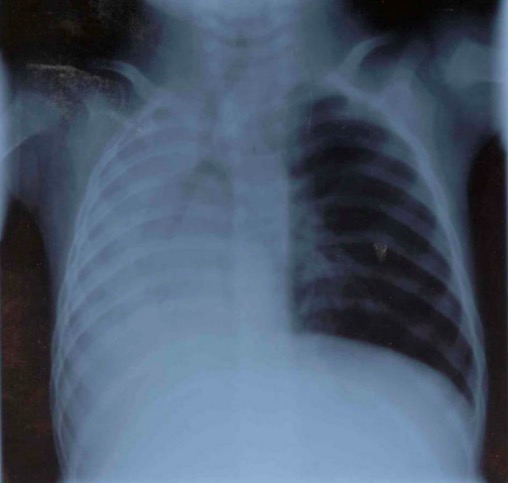
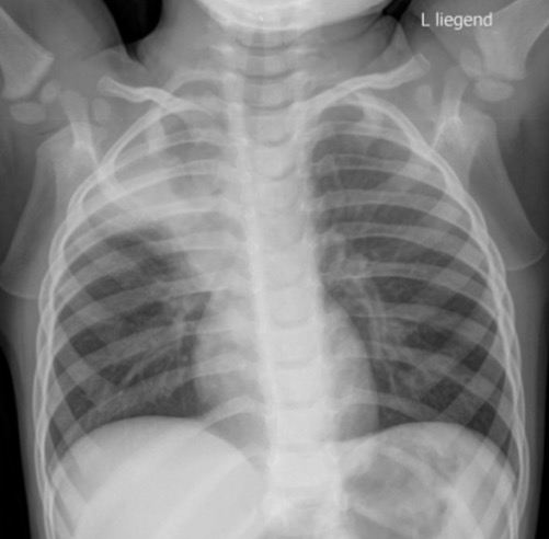
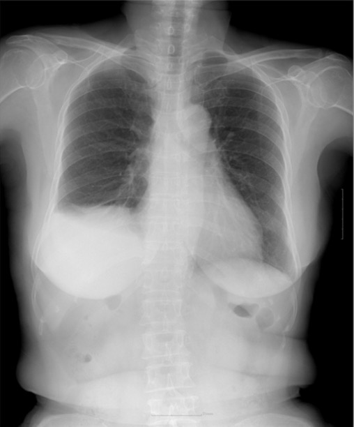
  • Lung opacification:
  • Lung volume loss:
    • Interlobar fissure Fissure A crack or split that extends into the dermis Generalized and Localized Rashes may be displaced.
    • If a large segment of the lung is involved: 
      • Mediastinum Mediastinum The mediastinum is the thoracic area between the 2 pleural cavities. The mediastinum contains vital structures of the circulatory, respiratory, digestive, and nervous systems including the heart and esophagus, and major thoracic vessels. Mediastinum and Great Vessels: Anatomy and tracheal shift toward the affected side.
      • Elevated ipsilateral diaphragm Diaphragm The diaphragm is a large, dome-shaped muscle that separates the thoracic cavity from the abdominal cavity. The diaphragm consists of muscle fibers and a large central tendon, which is divided into right and left parts. As the primary muscle of inspiration, the diaphragm contributes 75% of the total inspiratory muscle force. Diaphragm: Anatomy
      • Compensatory hyperinflation Hyperinflation Imaging of the Lungs and Pleura might be seen on the contralateral side.
  • Identification Identification Defense Mechanisms of foreign bodies

Computed tomography (CT)

Findings are similar to chest X-ray X-ray Penetrating electromagnetic radiation emitted when the inner orbital electrons of an atom are excited and release radiant energy. X-ray wavelengths range from 1 pm to 10 nm. Hard x-rays are the higher energy, shorter wavelength x-rays. Soft x-rays or grenz rays are less energetic and longer in wavelength. The short wavelength end of the x-ray spectrum overlaps the gamma rays wavelength range. The distinction between gamma rays and x-rays is based on their radiation source. Pulmonary Function Tests, but CT may be more sensitive in determining an etiology:
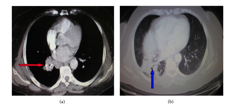
  • Bronchial narrowing due to obstruction may be seen.
  • Consolidation Consolidation Pulmonary Function Tests
  • Pleural effusion Pleural Effusion Pleural effusion refers to the accumulation of fluid between the layers of the parietal and visceral pleura. Common causes of this condition include infection, malignancy, autoimmune disorders, or volume overload. Clinical manifestations include chest pain, cough, and dyspnea. Pleural Effusion
  • Lung mass Mass Three-dimensional lesion that occupies a space within the breast Imaging of the Breast
  • Parenchymal scarring Scarring Inflammation
Lobe atelectasis on ct

CT of the chest showing a right hilar mass (red arrow) compressing the bronchus and leading to right lower lobe atelectasis (blue arrow)

Image: “ Atelectasis” by Department of Pneumonology, Army General Hospital of Athens, 138 Mesogion & Katehaki Avenue, 115 25 Athens, Greece. License: CC BY 3.0

Arterial blood gas Arterial blood gas Respiratory Alkalosis

  • Partial pressure Partial pressure The pressure that would be exerted by one component of a mixture of gases if it were present alone in a container. Gas Exchange of O₂ (PO₂)
  • Normal or ↓ partial pressure Partial pressure The pressure that would be exerted by one component of a mixture of gases if it were present alone in a container. Gas Exchange of CO₂ (PCO₂)

Related videos

Management

General measures

In addition to treating the underlying etiology of atelectasis, the following options can be used for prevention and treatment:

Medical therapy

Bronchoscopy Bronchoscopy Endoscopic examination, therapy or surgery of the bronchi. Laryngomalacia and Tracheomalacia

  • May be considered if other measures fail
  • Diagnostic and therapeutic for potential obstructive causes (e.g., tumor Tumor Inflammation)
Bronchoscopy

Bronchoscopy is a procedure that allows visualization of the airways by way of a bronchoscope.

Image by Lecturio.

Differential Diagnosis

  • Aspiration pneumonitis Pneumonitis Human Herpesvirus 6 and 7: inhalation of either oropharyngeal or gastric contents into the lower airways: This inhalation stimulates an inflammatory response, resulting in cough, dyspnea Dyspnea Dyspnea is the subjective sensation of breathing discomfort. Dyspnea is a normal manifestation of heavy physical or psychological exertion, but also may be caused by underlying conditions (both pulmonary and extrapulmonary). Dyspnea, chest discomfort, and fever Fever Fever is defined as a measured body temperature of at least 38°C (100.4°F). Fever is caused by circulating endogenous and/or exogenous pyrogens that increase levels of prostaglandin E2 in the hypothalamus. Fever is commonly associated with chills, rigors, sweating, and flushing of the skin. Fever. The diagnosis is often clinical and supported with chest X-ray X-ray Penetrating electromagnetic radiation emitted when the inner orbital electrons of an atom are excited and release radiant energy. X-ray wavelengths range from 1 pm to 10 nm. Hard x-rays are the higher energy, shorter wavelength x-rays. Soft x-rays or grenz rays are less energetic and longer in wavelength. The short wavelength end of the x-ray spectrum overlaps the gamma rays wavelength range. The distinction between gamma rays and x-rays is based on their radiation source. Pulmonary Function Tests findings of ground-glass opacities in dependent regions. Management is supportive, and patients Patients Individuals participating in the health care system for the purpose of receiving therapeutic, diagnostic, or preventive procedures. Clinician–Patient Relationship should be monitored for the development of pneumonia Pneumonia Pneumonia or pulmonary inflammation is an acute or chronic inflammation of lung tissue. Causes include infection with bacteria, viruses, or fungi. In more rare cases, pneumonia can also be caused through toxic triggers through inhalation of toxic substances, immunological processes, or in the course of radiotherapy. Pneumonia.
  • Pneumonia Pneumonia Pneumonia or pulmonary inflammation is an acute or chronic inflammation of lung tissue. Causes include infection with bacteria, viruses, or fungi. In more rare cases, pneumonia can also be caused through toxic triggers through inhalation of toxic substances, immunological processes, or in the course of radiotherapy. Pneumonia: infection of the lung parenchyma most often caused by bacteria Bacteria Bacteria are prokaryotic single-celled microorganisms that are metabolically active and divide by binary fission. Some of these organisms play a significant role in the pathogenesis of diseases. Bacteriology or a virus Virus Viruses are infectious, obligate intracellular parasites composed of a nucleic acid core surrounded by a protein capsid. Viruses can be either naked (non-enveloped) or enveloped. The classification of viruses is complex and based on many factors, including type and structure of the nucleoid and capsid, the presence of an envelope, the replication cycle, and the host range. Virology: Patients Patients Individuals participating in the health care system for the purpose of receiving therapeutic, diagnostic, or preventive procedures. Clinician–Patient Relationship present with fever Fever Fever is defined as a measured body temperature of at least 38°C (100.4°F). Fever is caused by circulating endogenous and/or exogenous pyrogens that increase levels of prostaglandin E2 in the hypothalamus. Fever is commonly associated with chills, rigors, sweating, and flushing of the skin. Fever, dyspnea Dyspnea Dyspnea is the subjective sensation of breathing discomfort. Dyspnea is a normal manifestation of heavy physical or psychological exertion, but also may be caused by underlying conditions (both pulmonary and extrapulmonary). Dyspnea, and a productive cough. Chest X-ray X-ray Penetrating electromagnetic radiation emitted when the inner orbital electrons of an atom are excited and release radiant energy. X-ray wavelengths range from 1 pm to 10 nm. Hard x-rays are the higher energy, shorter wavelength x-rays. Soft x-rays or grenz rays are less energetic and longer in wavelength. The short wavelength end of the x-ray spectrum overlaps the gamma rays wavelength range. The distinction between gamma rays and x-rays is based on their radiation source. Pulmonary Function Tests findings usually include lobar consolidation Consolidation Pulmonary Function Tests. Management usually involves empiric antibiotics, which can be tailored if the causative organism is identified. Antivirals are used in cases where a viral cause is suspected.
  • Pleural effusion Pleural Effusion Pleural effusion refers to the accumulation of fluid between the layers of the parietal and visceral pleura. Common causes of this condition include infection, malignancy, autoimmune disorders, or volume overload. Clinical manifestations include chest pain, cough, and dyspnea. Pleural Effusion: accumulation of fluid between the layers of the parietal Parietal One of a pair of irregularly shaped quadrilateral bones situated between the frontal bone and occipital bone, which together form the sides of the cranium. Skull: Anatomy and visceral pleura Visceral pleura Pleura: Anatomy: Symptoms of pleural effusion Pleural Effusion Pleural effusion refers to the accumulation of fluid between the layers of the parietal and visceral pleura. Common causes of this condition include infection, malignancy, autoimmune disorders, or volume overload. Clinical manifestations include chest pain, cough, and dyspnea. Pleural Effusion include chest pain Pain An unpleasant sensation induced by noxious stimuli which are detected by nerve endings of nociceptive neurons. Pain: Types and Pathways, cough, and dyspnea Dyspnea Dyspnea is the subjective sensation of breathing discomfort. Dyspnea is a normal manifestation of heavy physical or psychological exertion, but also may be caused by underlying conditions (both pulmonary and extrapulmonary). Dyspnea, and imaging can confirm its presence. Atelectasis with significant volume loss can appear similar to a massive pleural effusion Pleural Effusion Pleural effusion refers to the accumulation of fluid between the layers of the parietal and visceral pleura. Common causes of this condition include infection, malignancy, autoimmune disorders, or volume overload. Clinical manifestations include chest pain, cough, and dyspnea. Pleural Effusion; however, mediastinal and tracheal shift will be directed away from the effusion. Management depends on the underlying condition and whether the effusion is causing respiratory distress. 
  • Unilateral diaphragm Diaphragm The diaphragm is a large, dome-shaped muscle that separates the thoracic cavity from the abdominal cavity. The diaphragm consists of muscle fibers and a large central tendon, which is divided into right and left parts. As the primary muscle of inspiration, the diaphragm contributes 75% of the total inspiratory muscle force. Diaphragm: Anatomy paralysis: condition in which 1 diaphragm Diaphragm The diaphragm is a large, dome-shaped muscle that separates the thoracic cavity from the abdominal cavity. The diaphragm consists of muscle fibers and a large central tendon, which is divided into right and left parts. As the primary muscle of inspiration, the diaphragm contributes 75% of the total inspiratory muscle force. Diaphragm: Anatomy loses the ability to contract and allow inspiration Inspiration Ventilation: Mechanics of Breathing: Patients Patients Individuals participating in the health care system for the purpose of receiving therapeutic, diagnostic, or preventive procedures. Clinician–Patient Relationship may be asymptomatic or have dyspnea Dyspnea Dyspnea is the subjective sensation of breathing discomfort. Dyspnea is a normal manifestation of heavy physical or psychological exertion, but also may be caused by underlying conditions (both pulmonary and extrapulmonary). Dyspnea. A chest X-ray X-ray Penetrating electromagnetic radiation emitted when the inner orbital electrons of an atom are excited and release radiant energy. X-ray wavelengths range from 1 pm to 10 nm. Hard x-rays are the higher energy, shorter wavelength x-rays. Soft x-rays or grenz rays are less energetic and longer in wavelength. The short wavelength end of the x-ray spectrum overlaps the gamma rays wavelength range. The distinction between gamma rays and x-rays is based on their radiation source. Pulmonary Function Tests will show hemidiaphragmatic elevation. Pulmonary function tests will show a decline in forced vital capacity Vital capacity The volume of air that is exhaled by a maximal expiration following a maximal inspiration. Ventilation: Mechanics of Breathing. Management may not be necessary for asymptomatic cases. Patients Patients Individuals participating in the health care system for the purpose of receiving therapeutic, diagnostic, or preventive procedures. Clinician–Patient Relationship with more significant symptoms may need noninvasive ventilatory support, surgical plication, phrenic nerve Phrenic nerve The motor nerve of the diaphragm. The phrenic nerve fibers originate in the cervical spinal column (mostly C4) and travel through the cervical plexus to the diaphragm. Diaphragm: Anatomy pacing, and management of the underlying cause.
  • Pulmonary embolism Pulmonary Embolism Pulmonary embolism (PE) is a potentially fatal condition that occurs as a result of intraluminal obstruction of the main pulmonary artery or its branches. The causative factors include thrombi, air, amniotic fluid, and fat. In PE, gas exchange is impaired due to the decreased return of deoxygenated blood to the lungs. Pulmonary Embolism: obstruction of the pulmonary arteries Arteries Arteries are tubular collections of cells that transport oxygenated blood and nutrients from the heart to the tissues of the body. The blood passes through the arteries in order of decreasing luminal diameter, starting in the largest artery (the aorta) and ending in the small arterioles. Arteries are classified into 3 types: large elastic arteries, medium muscular arteries, and small arteries and arterioles. Arteries: Histology, most often due to thrombus migration from the deep venous system: Signs and symptoms include pleuritic chest pain Pain An unpleasant sensation induced by noxious stimuli which are detected by nerve endings of nociceptive neurons. Pain: Types and Pathways, dyspnea Dyspnea Dyspnea is the subjective sensation of breathing discomfort. Dyspnea is a normal manifestation of heavy physical or psychological exertion, but also may be caused by underlying conditions (both pulmonary and extrapulmonary). Dyspnea, tachypnea Tachypnea Increased respiratory rate. Pulmonary Examination, and tachycardia Tachycardia Abnormally rapid heartbeat, usually with a heart rate above 100 beats per minute for adults. Tachycardia accompanied by disturbance in the cardiac depolarization (cardiac arrhythmia) is called tachyarrhythmia. Sepsis in Children. Severe cases can result in hemodynamic instability or cardiopulmonary arrest Cardiopulmonary arrest Cardiac arrest is the sudden, complete cessation of cardiac output with hemodynamic collapse. Patients present as pulseless, unresponsive, and apneic. Rhythms associated with cardiac arrest are ventricular fibrillation/tachycardia, asystole, or pulseless electrical activity. Cardiac Arrest. A chest CT with angiography Angiography Radiography of blood vessels after injection of a contrast medium. Cardiac Surgery is the primary method of diagnosis. Management includes oxygenation, anticoagulation Anticoagulation Pulmonary Hypertension Drugs, and thrombolytic therapy for unstable patients Unstable Patients Blunt Chest Trauma.

References

  1. Madappa, T., Sharma, S. (2020). Atelectasis. In Kamangar, N. (Ed.), Medscape. Retrieved April 2, 2021, from https://emedicine.medscape.com/article/296468-overview
  2. Stark, P. (2020). Atelectasis: Types and pathogenesis in adults. In Finlay, G. (Ed.), UpToDate. Retrieved April 2, 2021, from https://www.uptodate.com/contents/atelectasis-types-and-pathogenesis-in-adults
  3. Finder, J. D. (2019). Atelectasis in children. In Hoppin, A. G. (Ed.), UpToDate. Retrieved April 2, 2021, from https://www.uptodate.com/contents/atelectasis-in-children
  4. Coruh, B., Niven, A. S. (2019). Atelectasis. MSD Manual Professional Version. Retrieved April 2, 2021, from https://www.msdmanuals.com/professional/pulmonary-disorders/bronchiectasis-and-atelectasis/atelectasis
  5. Grott, K., Dunlap, J. D. (2021). Atelectasis. StatPearls. Retrieved April 2, 2021, from https://www.ncbi.nlm.nih.gov/books/NBK545316/

Create your free account or log in to continue reading!

Sign up now and get free access to Lecturio with concept pages, medical videos, and questions for your medical education.

User Reviews

Unwrap New Skills This Holiday 🎄 Save 30% on all plans now!

Details