Advertisement

Advertisement

Advertisement

Advertisement

Arm: Anatomy

The arm, or "upper arm" in common usage, is the region of the upper limb that extends from the shoulder to the elbow joint Elbow joint The elbow is the synovial hinge joint between the humerus in the upper arm and the radius and ulna in the forearm. The elbow consists of 3 joints, which form a functional unit enclosed within a single articular capsule. The elbow is the link between the powerful motions of the shoulder and the intricate fine-motor function of the hand. Elbow Joint: Anatomy and connects inferiorly to the forearm Forearm The forearm is the region of the upper limb between the elbow and the wrist. The term "forearm" is used in anatomy to distinguish this area from the arm, a term that is commonly used to describe the entire upper limb. The forearm consists of 2 long bones (the radius and the ulna), the interosseous membrane, and multiple arteries, nerves, and muscles. Forearm: Anatomy through the cubital fossa Cubital Fossa The cubital fossa is the region anterior to the elbow joint. The cubital fossa is seen as the triangular depression between the brachioradialis and pronator teres muscles. The 4 important structures of the cubital fossa (from lateral to medial) are the radial nerve, tendon of the biceps brachii muscle, brachial artery, and median nerve. Cubital Fossa: Anatomy. It is divided into 2 fascial compartments (anterior and posterior) and houses numerous structures, including the humerus; the brachial and profunda brachii arteries Arteries Arteries are tubular collections of cells that transport oxygenated blood and nutrients from the heart to the tissues of the body. The blood passes through the arteries in order of decreasing luminal diameter, starting in the largest artery (the aorta) and ending in the small arterioles. Arteries are classified into 3 types: large elastic arteries, medium muscular arteries, and small arteries and arterioles. Arteries: Histology; the basilic, cephalic, and brachial veins Veins Veins are tubular collections of cells, which transport deoxygenated blood and waste from the capillary beds back to the heart. Veins are classified into 3 types: small veins/venules, medium veins, and large veins. Each type contains 3 primary layers: tunica intima, tunica media, and tunica adventitia. Veins: Histology; the radial, median, ulnar, and musculocutaneous nerves; and the flexor and extensor muscles of the arm.

Last updated: Nov 19, 2024

Editorial responsibility: Stanley Oiseth, Lindsay Jones, Evelin Maza

Advertisement

Advertisement

Advertisement

Advertisement

Advertisement

Advertisement

Bones of the Arm: Humerus

The humerus can be divided into 3 parts: the proximal end, the shaft, and the distal end.
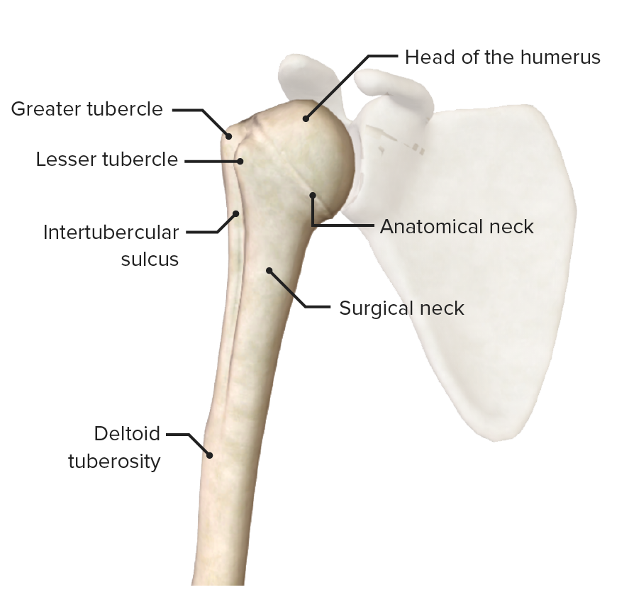
Proximal end

  • Head of the humerus: articulates with the glenoid
  • Anatomical neck Neck The part of a human or animal body connecting the head to the rest of the body. Peritonsillar Abscess
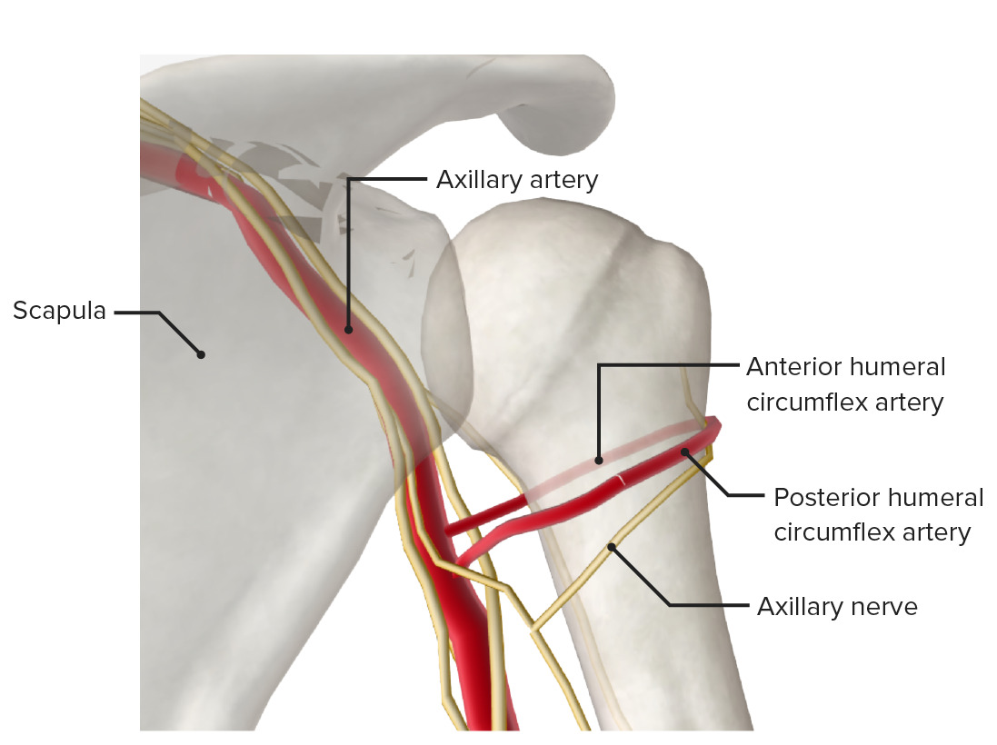
  • Surgical neck Neck The part of a human or animal body connecting the head to the rest of the body. Peritonsillar Abscess:
    • Frequent site of fracture Fracture A fracture is a disruption of the cortex of any bone and periosteum and is commonly due to mechanical stress after an injury or accident. Open fractures due to trauma can be a medical emergency. Fractures are frequently associated with automobile accidents, workplace injuries, and trauma. Overview of Bone Fractures 
    • A fracture Fracture A fracture is a disruption of the cortex of any bone and periosteum and is commonly due to mechanical stress after an injury or accident. Open fractures due to trauma can be a medical emergency. Fractures are frequently associated with automobile accidents, workplace injuries, and trauma. Overview of Bone Fractures here may injure the axillary nerve Axillary nerve Axilla and Brachial Plexus: Anatomy and/or posterior circumflex artery.
  • Greater tubercle: site of attachment of the supraspinatus, infraspinatus, and teres minor rotator cuff tendons
  • Lesser tubercle: site of attachment of the subscapularis rotator cuff tendon
  • Intertubercular sulcus: separates the tubercles and contains the tendon of the long head of the biceps brachii

Shaft

  • Deltoid tuberosity: site of insertion of the deltoid muscle
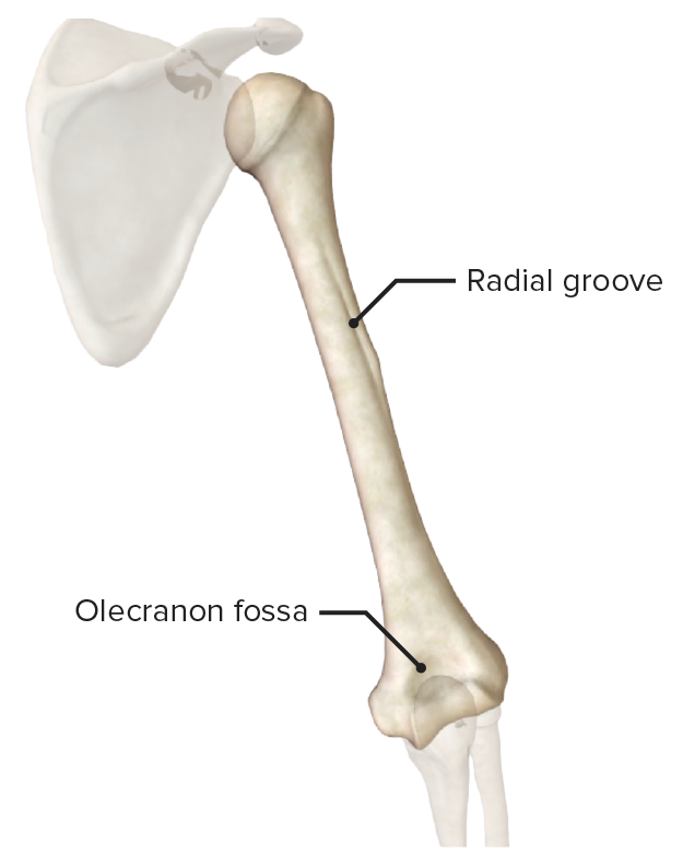
  • Radial or spiral Spiral Computed tomography where there is continuous x-ray exposure to the patient while being transported in a spiral or helical pattern through the beam of irradiation. This provides improved three-dimensional contrast and spatial resolution compared to conventional computed tomography, where data is obtained and computed from individual sequential exposures. Computed Tomography (CT) groove: The radial nerve Radial Nerve A major nerve of the upper extremity. In humans the fibers of the radial nerve originate in the lower cervical and upper thoracic spinal cord (usually C5 to T1), travel via the posterior cord of the brachial plexus, and supply motor innervation to extensor muscles of the arm and cutaneous sensory fibers to extensor regions of the arm and hand. Axilla and Brachial Plexus: Anatomy runs in this groove and may be injured secondary to pressure or trauma, with resultant wrist drop.
  • Medial and lateral supracondylar ridges

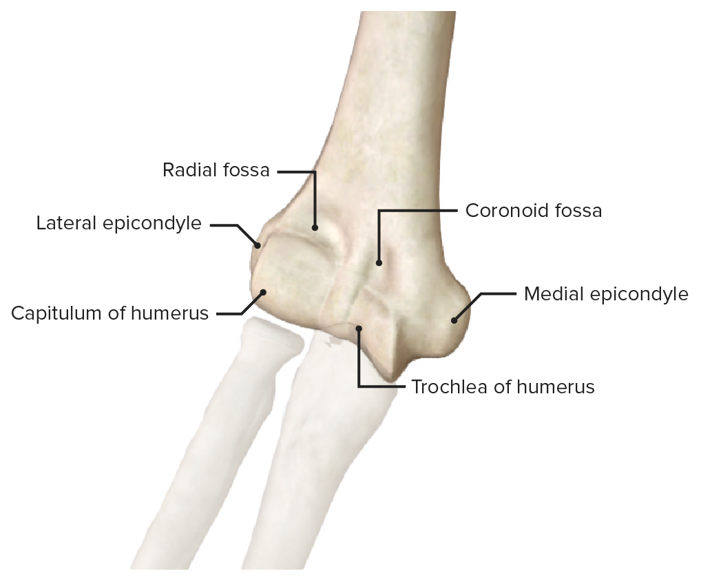
Distal end

  • Trochlea: medial aspect of distal humerus
  • Capitulum: lateral to the trochlea and articulates with the radial head
  • Medial epicondyle: larger of the epicondyles; the ulnar nerve Ulnar Nerve A major nerve of the upper extremity. In humans, the fibers of the ulnar nerve originate in the lower cervical and upper thoracic spinal cord (usually C7 to T1), travel via the medial cord of the brachial plexus, and supply sensory and motor innervation to parts of the hand and forearm. Axilla and Brachial Plexus: Anatomy passes posterior to the medial epicondyle in the ulnar groove
  • Lateral epicondyle: common site of pain Pain An unpleasant sensation induced by noxious stimuli which are detected by nerve endings of nociceptive neurons. Pain: Types and Pathways with “ tennis elbow Tennis elbow A condition characterized by pain in or near the lateral humeral epicondyle or in the forearm extensor muscle mass as a result of unusual strain. It occurs due repetitive stresses on the elbow from activities such as tennis playing. Examination of the Upper Limbs
  • Olecranon, coronoid fossa, and radial fossa: depressions that allow for movement of the elbow during flexion Flexion Examination of the Upper Limbs and extension Extension Examination of the Upper Limbs
Distal end of the humerus

Anterior view of the distal end of the humerus

Image by BioDigital, edited by Lecturio
Related videos

Muscles of the Anterior Compartment of the Arm

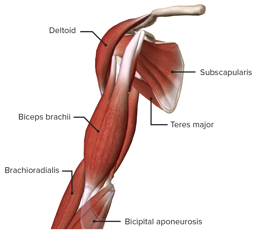
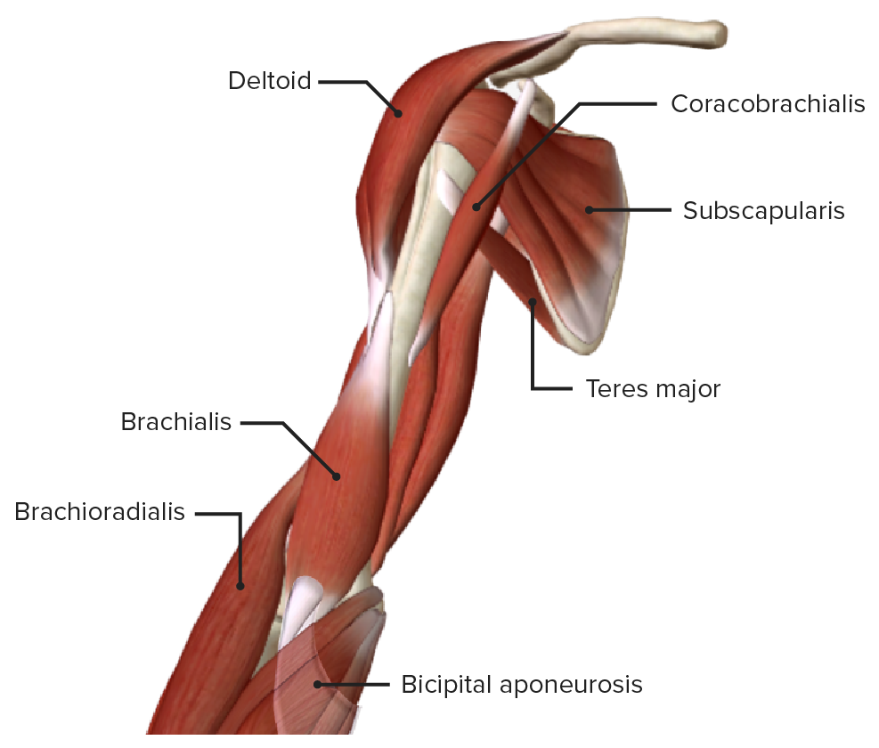
  • The anterior compartment of the arm is also called the flexor compartment, because its muscles are in charge of flexing the forearm Forearm The forearm is the region of the upper limb between the elbow and the wrist. The term “forearm” is used in anatomy to distinguish this area from the arm, a term that is commonly used to describe the entire upper limb. The forearm consists of 2 long bones (the radius and the ulna), the interosseous membrane, and multiple arteries, nerves, and muscles. Forearm: Anatomy toward the upper arm.
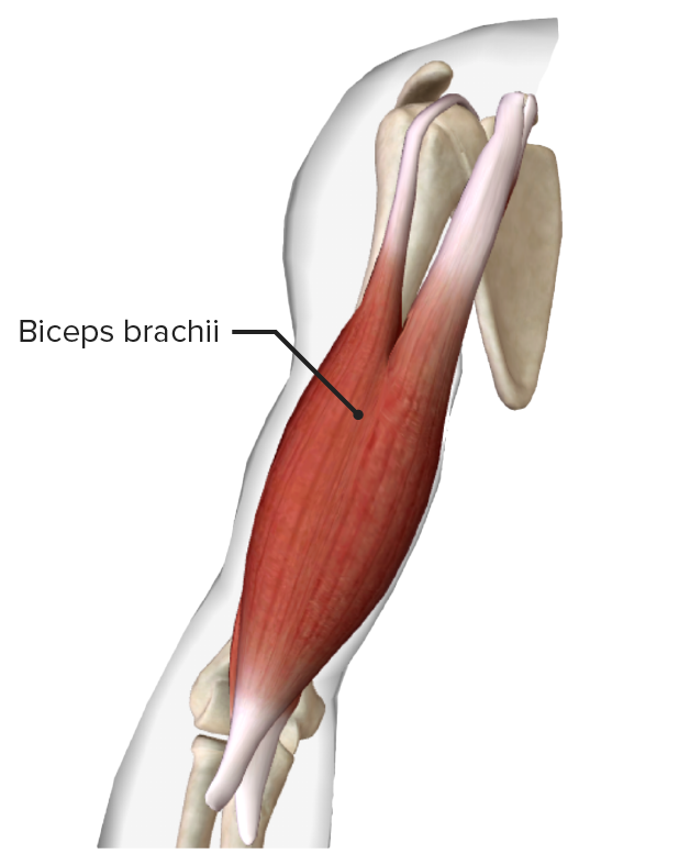
  • Comprises 3 muscles: biceps, brachialis, and coracobrachialis
  • All 3 muscles are innervated by the musculocutaneous nerve Musculocutaneous Nerve A major nerve of the upper extremity. The fibers of the musculocutaneous nerve originate in the lower cervical spinal cord (usually C5 to C7), travel via the lateral cord of the brachial plexus, and supply sensory and motor innervation to the upper arm, elbow, and forearm. Axilla and Brachial Plexus: Anatomy and supplied by the brachial artery Brachial Artery The continuation of the axillary artery; it branches into the radial and ulnar arteries. Cubital Fossa: Anatomy.
  • To recall the muscles of the anterior compartment of the arm, remember “BBC”:
    1. Biceps
    2. Brachialis
    3. Coracobrachialis
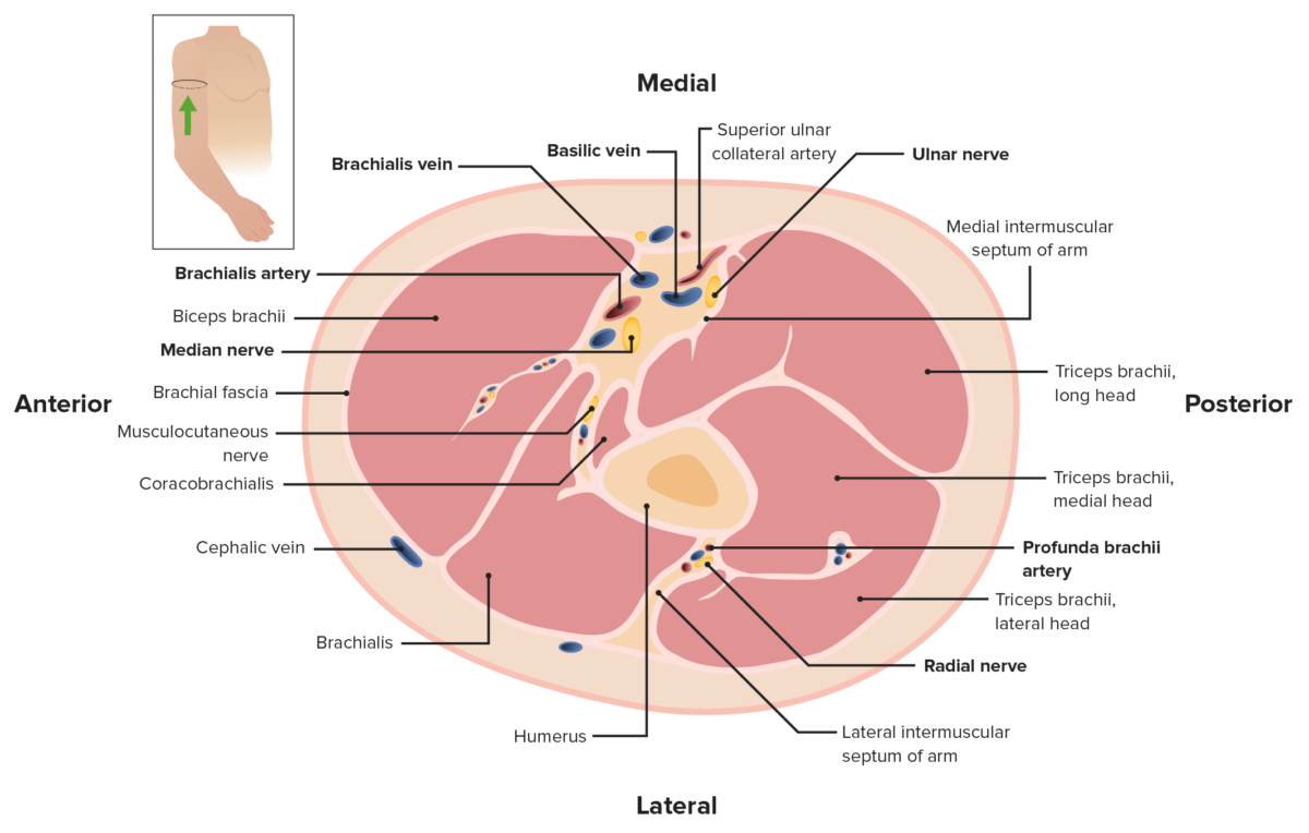
Cross-section of the arm

Cross-section of the arm, featuring the anterior and posterior compartments. Note the brachial fascia enclosing all of the muscles and forming the lateral and medial intermuscular septa.

Image by Lecturio. License: CC BY-NC-SA 4.0
Table: Anterior compartment of the arm: flexors
Muscle Origin Insertion Innervation Function
Biceps brachii
  • Long head: supraglenoid tubercle
  • Short head: coracoid process
Radial tuberosity and forearm Forearm The forearm is the region of the upper limb between the elbow and the wrist. The term “forearm” is used in anatomy to distinguish this area from the arm, a term that is commonly used to describe the entire upper limb. The forearm consists of 2 long bones (the radius and the ulna), the interosseous membrane, and multiple arteries, nerves, and muscles. Forearm: Anatomy fascia Fascia Layers of connective tissue of variable thickness. The superficial fascia is found immediately below the skin; the deep fascia invests muscles, nerves, and other organs. Cellulitis via bicipital aponeurosis Musculocutaneous nerve Musculocutaneous Nerve A major nerve of the upper extremity. The fibers of the musculocutaneous nerve originate in the lower cervical spinal cord (usually C5 to C7), travel via the lateral cord of the brachial plexus, and supply sensory and motor innervation to the upper arm, elbow, and forearm. Axilla and Brachial Plexus: Anatomy (C6)
  • Supinates forearm Forearm The forearm is the region of the upper limb between the elbow and the wrist. The term “forearm” is used in anatomy to distinguish this area from the arm, a term that is commonly used to describe the entire upper limb. The forearm consists of 2 long bones (the radius and the ulna), the interosseous membrane, and multiple arteries, nerves, and muscles. Forearm: Anatomy, and once supinated flexes elbow joint Elbow joint The elbow is the synovial hinge joint between the humerus in the upper arm and the radius and ulna in the forearm. The elbow consists of 3 joints, which form a functional unit enclosed within a single articular capsule. The elbow is the link between the powerful motions of the shoulder and the intricate fine-motor function of the hand. Elbow Joint: Anatomy
  • Flexes shoulder
Brachialis Lateral and medial surface of distal half of humerus Coronoid process and tuberosity of ulna Ulna The inner and longer bone of the forearm. Forearm: Anatomy Musculocutaneous nerve Musculocutaneous Nerve A major nerve of the upper extremity. The fibers of the musculocutaneous nerve originate in the lower cervical spinal cord (usually C5 to C7), travel via the lateral cord of the brachial plexus, and supply sensory and motor innervation to the upper arm, elbow, and forearm. Axilla and Brachial Plexus: Anatomy (C6) Flexes elbow joint Elbow joint The elbow is the synovial hinge joint between the humerus in the upper arm and the radius and ulna in the forearm. The elbow consists of 3 joints, which form a functional unit enclosed within a single articular capsule. The elbow is the link between the powerful motions of the shoulder and the intricate fine-motor function of the hand. Elbow Joint: Anatomy
Coracobrachialis Coracoid process Middle 3rd of humerus Musculocutaneous nerve Musculocutaneous Nerve A major nerve of the upper extremity. The fibers of the musculocutaneous nerve originate in the lower cervical spinal cord (usually C5 to C7), travel via the lateral cord of the brachial plexus, and supply sensory and motor innervation to the upper arm, elbow, and forearm. Axilla and Brachial Plexus: Anatomy (C6) Flexes/adducts shoulder joint Shoulder joint The articulation between the head of the humerus and the glenoid cavity of the scapula. Examination of the Upper Limbs

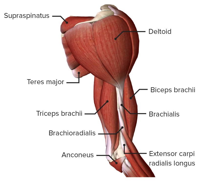
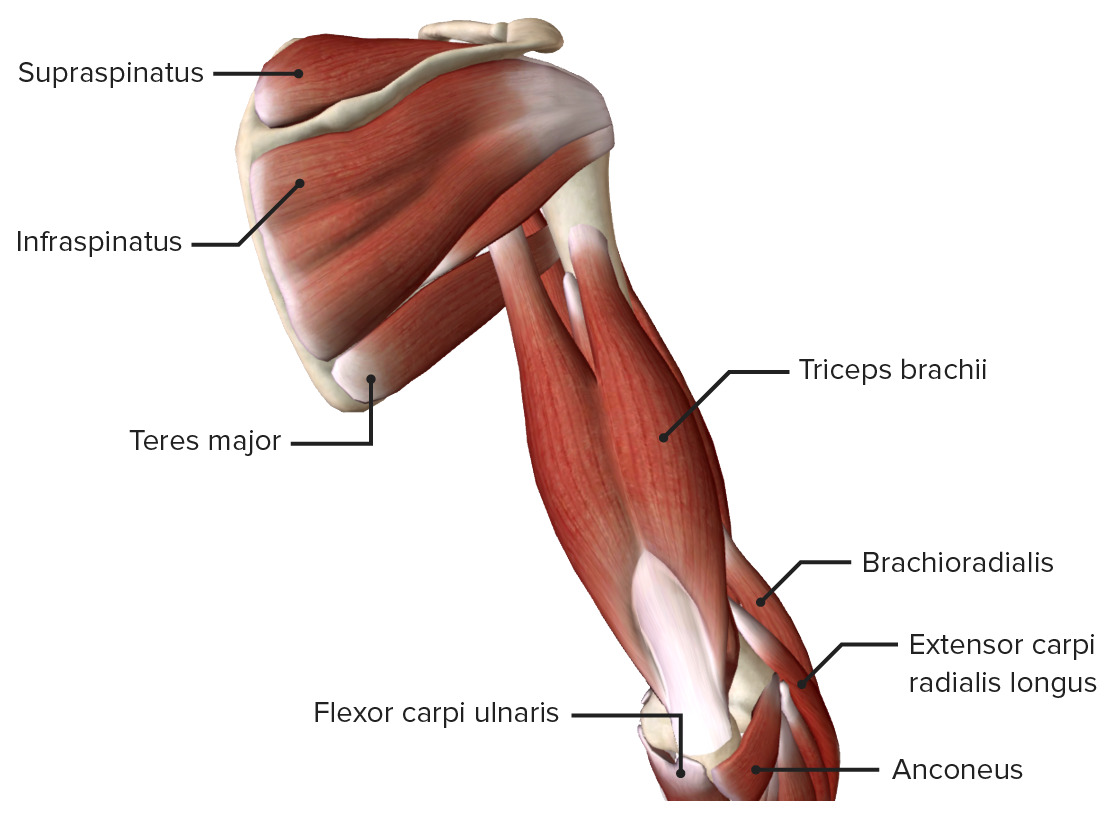
Muscles of the Posterior Compartment of the Arm

  • Also called the extensor compartment because its muscles are in charge of extending the elbow
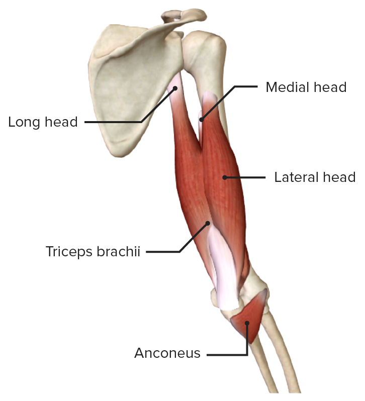
  • The posterior compartment of the arm comprises 2 muscles:
    • Triceps brachii Triceps brachii Elbow Joint: Anatomy
    • Anconeus Anconeus Elbow Joint: Anatomy
      • In some sources considered the “fourth head” of the triceps
      • In some sources considered part of the posterior compartment of the forearm Forearm The forearm is the region of the upper limb between the elbow and the wrist. The term “forearm” is used in anatomy to distinguish this area from the arm, a term that is commonly used to describe the entire upper limb. The forearm consists of 2 long bones (the radius and the ulna), the interosseous membrane, and multiple arteries, nerves, and muscles. Forearm: Anatomy
  • Both muscles are innervated by the radial nerve Radial Nerve A major nerve of the upper extremity. In humans the fibers of the radial nerve originate in the lower cervical and upper thoracic spinal cord (usually C5 to T1), travel via the posterior cord of the brachial plexus, and supply motor innervation to extensor muscles of the arm and cutaneous sensory fibers to extensor regions of the arm and hand. Axilla and Brachial Plexus: Anatomy and supplied by the deep brachial artery Brachial Artery The continuation of the axillary artery; it branches into the radial and ulnar arteries. Cubital Fossa: Anatomy.
Table: Posterior compartment of the arm: extensors
Muscle Origin Insertion Innervation Function
Triceps brachii Triceps brachii Elbow Joint: Anatomy
  • Long head: infraglenoid tubercle
  • Lateral head: superior to radial groove
  • Medial head: inferior to radial groove
Olecranon Radial nerve Radial Nerve A major nerve of the upper extremity. In humans the fibers of the radial nerve originate in the lower cervical and upper thoracic spinal cord (usually C5 to T1), travel via the posterior cord of the brachial plexus, and supply motor innervation to extensor muscles of the arm and cutaneous sensory fibers to extensor regions of the arm and hand. Axilla and Brachial Plexus: Anatomy (C7, C8)
  • Extensor of elbow joint Elbow joint The elbow is the synovial hinge joint between the humerus in the upper arm and the radius and ulna in the forearm. The elbow consists of 3 joints, which form a functional unit enclosed within a single articular capsule. The elbow is the link between the powerful motions of the shoulder and the intricate fine-motor function of the hand. Elbow Joint: Anatomy
  • Long head also extends and externally rotates the shoulder joint Shoulder joint The articulation between the head of the humerus and the glenoid cavity of the scapula. Examination of the Upper Limbs
Anconeus Anconeus Elbow Joint: Anatomy Inserts on the posterior aspect of lateral epicondyle Lateral surface of the olecranon Radial nerve Radial Nerve A major nerve of the upper extremity. In humans the fibers of the radial nerve originate in the lower cervical and upper thoracic spinal cord (usually C5 to T1), travel via the posterior cord of the brachial plexus, and supply motor innervation to extensor muscles of the arm and cutaneous sensory fibers to extensor regions of the arm and hand. Axilla and Brachial Plexus: Anatomy (C7, C8) Assists in extension Extension Examination of the Upper Limbs of the elbow and stabilizes the joint

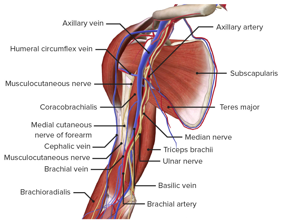
Vessels of the Arm

Table: Topographic location of the vessels of the arm
Medial bicipital groove Lateral bicipital groove
Location Between the biceps brachii muscle Biceps brachii muscle Elbow Joint: Anatomy and the triceps brachii Triceps brachii Elbow Joint: Anatomy Between the biceps brachii muscle Biceps brachii muscle Elbow Joint: Anatomy and the brachial muscle
Arteries Arteries Arteries are tubular collections of cells that transport oxygenated blood and nutrients from the heart to the tissues of the body. The blood passes through the arteries in order of decreasing luminal diameter, starting in the largest artery (the aorta) and ending in the small arterioles. Arteries are classified into 3 types: large elastic arteries, medium muscular arteries, and small arteries and arterioles. Arteries: Histology Brachial artery Brachial Artery The continuation of the axillary artery; it branches into the radial and ulnar arteries. Cubital Fossa: Anatomy Radial collateral artery
Veins Veins Veins are tubular collections of cells, which transport deoxygenated blood and waste from the capillary beds back to the heart. Veins are classified into 3 types: small veins/venules, medium veins, and large veins. Each type contains 3 primary layers: tunica intima, tunica media, and tunica adventitia. Veins: Histology
  • Basilic vein
  • Brachial veins Veins Veins are tubular collections of cells, which transport deoxygenated blood and waste from the capillary beds back to the heart. Veins are classified into 3 types: small veins/venules, medium veins, and large veins. Each type contains 3 primary layers: tunica intima, tunica media, and tunica adventitia. Veins: Histology
  • Radial collateral vein
  • Cephalic vein
Nerves
  • Ulnar nerve Ulnar Nerve A major nerve of the upper extremity. In humans, the fibers of the ulnar nerve originate in the lower cervical and upper thoracic spinal cord (usually C7 to T1), travel via the medial cord of the brachial plexus, and supply sensory and motor innervation to parts of the hand and forearm. Axilla and Brachial Plexus: Anatomy
  • Median nerve Median Nerve A major nerve of the upper extremity. In humans, the fibers of the median nerve originate in the lower cervical and upper thoracic spinal cord (usually C6 to T1), travel via the brachial plexus, and supply sensory and motor innervation to parts of the forearm and hand. Cubital Fossa: Anatomy
  • Medial cutaneous nerve of the forearm Forearm The forearm is the region of the upper limb between the elbow and the wrist. The term “forearm” is used in anatomy to distinguish this area from the arm, a term that is commonly used to describe the entire upper limb. The forearm consists of 2 long bones (the radius and the ulna), the interosseous membrane, and multiple arteries, nerves, and muscles. Forearm: Anatomy
Radial nerve Radial Nerve A major nerve of the upper extremity. In humans the fibers of the radial nerve originate in the lower cervical and upper thoracic spinal cord (usually C5 to T1), travel via the posterior cord of the brachial plexus, and supply motor innervation to extensor muscles of the arm and cutaneous sensory fibers to extensor regions of the arm and hand. Axilla and Brachial Plexus: Anatomy

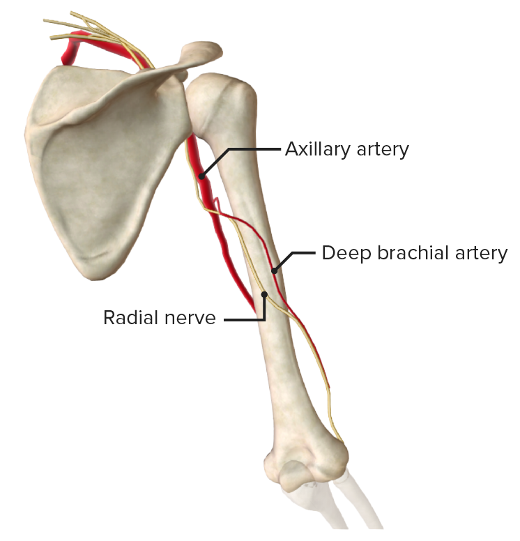
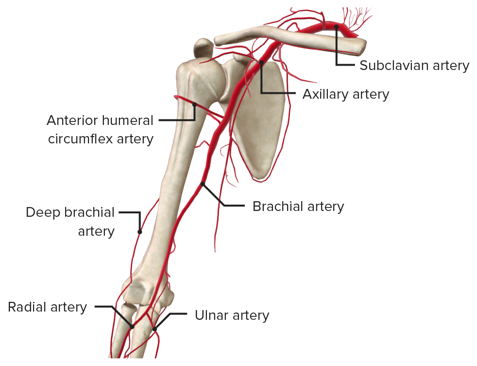
Arterial supply of the arm

The brachial artery Brachial Artery The continuation of the axillary artery; it branches into the radial and ulnar arteries. Cubital Fossa: Anatomy:

  • Begins at the inferior border of the teres major as a continuation of the axillary artery Axillary Artery The continuation of the subclavian artery; it distributes over the upper limb, axilla, chest and shoulder. Axilla and Brachial Plexus: Anatomy
  • Runs primarily between the biceps and brachialis muscles
  • Supplies the anterior compartment
  • Has 1 major branch, the deep brachial artery Brachial Artery The continuation of the axillary artery; it branches into the radial and ulnar arteries. Cubital Fossa: Anatomy
    • Also known as the profunda brachii artery
    • Supplies the posterior compartment
    • Runs in the radial groove with the radial nerve Radial Nerve A major nerve of the upper extremity. In humans the fibers of the radial nerve originate in the lower cervical and upper thoracic spinal cord (usually C5 to T1), travel via the posterior cord of the brachial plexus, and supply motor innervation to extensor muscles of the arm and cutaneous sensory fibers to extensor regions of the arm and hand. Axilla and Brachial Plexus: Anatomy
Arteries of the arm

Arteries of the arm

Image by BioDigital, edited by Lecturio
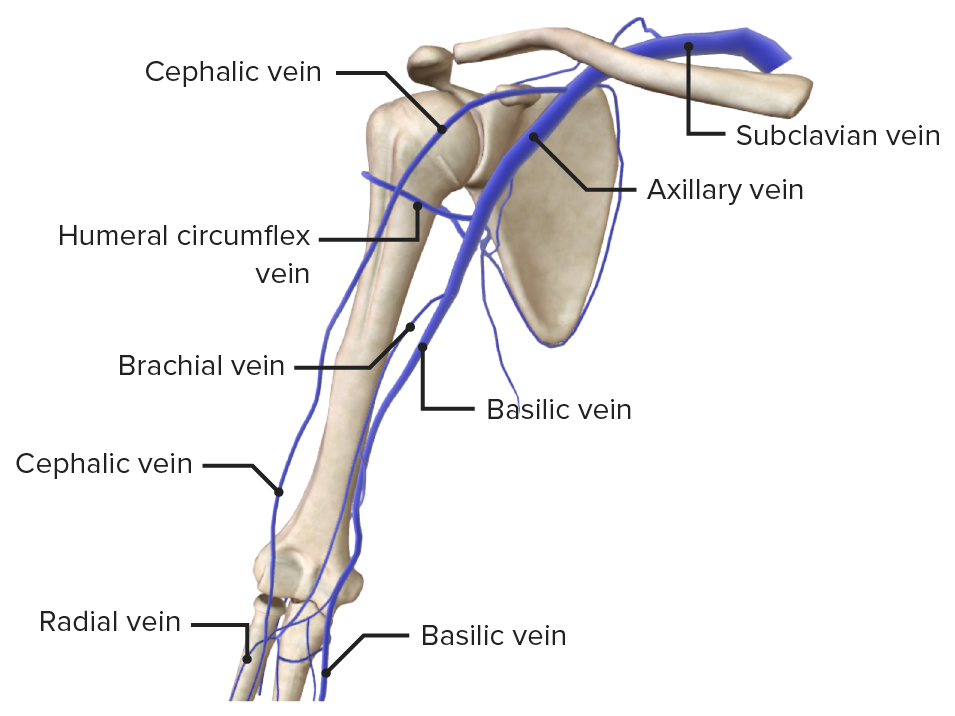
Venous drainage of the arm

  • Basilic vein:
    • Begins distally in the hand Hand The hand constitutes the distal part of the upper limb and provides the fine, precise movements needed in activities of daily living. It consists of 5 metacarpal bones and 14 phalanges, as well as numerous muscles innervated by the median and ulnar nerves. Hand: Anatomy
    • Travels on the medial aspect of the upper arm
    • Combines with the brachial veins Veins Veins are tubular collections of cells, which transport deoxygenated blood and waste from the capillary beds back to the heart. Veins are classified into 3 types: small veins/venules, medium veins, and large veins. Each type contains 3 primary layers: tunica intima, tunica media, and tunica adventitia. Veins: Histology to form the axillary vein Axillary Vein The venous trunk of the upper limb; a continuation of the basilar and brachial veins running from the lower border of the teres major muscle to the outer border of the first rib where it becomes the subclavian vein. Axilla and Brachial Plexus: Anatomy
  • Cephalic vein:
    • Begins distally in the hand Hand The hand constitutes the distal part of the upper limb and provides the fine, precise movements needed in activities of daily living. It consists of 5 metacarpal bones and 14 phalanges, as well as numerous muscles innervated by the median and ulnar nerves. Hand: Anatomy
    • Travels proximally on the antero-lateral aspect of the arm
    • Enters the shoulder through the deltopectoral groove (the groove between the deltoid and pectoralis muscles Pectoralis muscles The pectoralis major and pectoralis minor muscles that make up the upper and fore part of the chest in front of the axilla. Chest Wall: Anatomy), where it empties into the axillary vein Axillary Vein The venous trunk of the upper limb; a continuation of the basilar and brachial veins running from the lower border of the teres major muscle to the outer border of the first rib where it becomes the subclavian vein. Axilla and Brachial Plexus: Anatomy

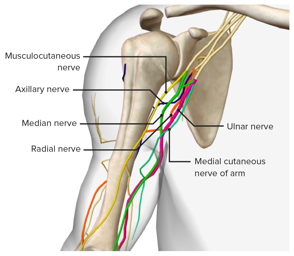
Nerves of the Arm

Nerve Origin Branches Functions
Musculo-cutaneous Terminal branch of lateral cord of brachial plexus Brachial Plexus The large network of nerve fibers which distributes the innervation of the upper extremity. The brachial plexus extends from the neck into the axilla. In humans, the nerves of the plexus usually originate from the lower cervical and the first thoracic spinal cord segments (c5-c8 and T1), but variations are not uncommon. Peripheral Nerve Injuries in the Cervicothoracic Region (C5–C7)
  • Branch to the:
    • Coracobrachialis
    • Biceps
    • Brachialis
    • Humerus
    • Elbow joint Elbow joint The elbow is the synovial hinge joint between the humerus in the upper arm and the radius and ulna in the forearm. The elbow consists of 3 joints, which form a functional unit enclosed within a single articular capsule. The elbow is the link between the powerful motions of the shoulder and the intricate fine-motor function of the hand. Elbow Joint: Anatomy
  • Terminates as the lateral antebrachial cutaneous nerve Lateral antebrachial cutaneous nerve Forearm: Anatomy
  • Motor Motor Neurons which send impulses peripherally to activate muscles or secretory cells. Nervous System: Histology supply of the anterior compartment of the arm
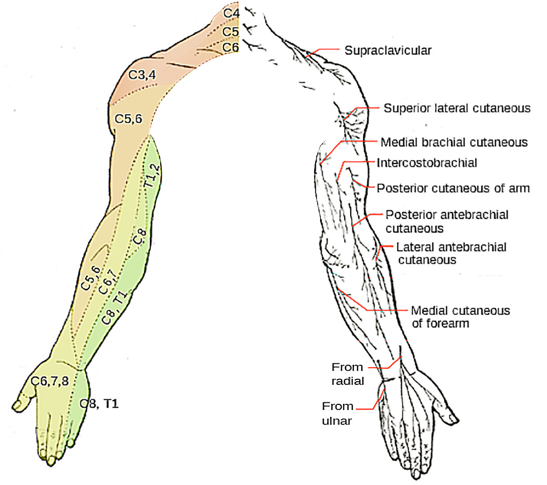
  • Sensory Sensory Neurons which conduct nerve impulses to the central nervous system. Nervous System: Histology supply of the skin Skin The skin, also referred to as the integumentary system, is the largest organ of the body. The skin is primarily composed of the epidermis (outer layer) and dermis (deep layer). The epidermis is primarily composed of keratinocytes that undergo rapid turnover, while the dermis contains dense layers of connective tissue. Skin: Structure and Functions of the lateral forearm Forearm The forearm is the region of the upper limb between the elbow and the wrist. The term “forearm” is used in anatomy to distinguish this area from the arm, a term that is commonly used to describe the entire upper limb. The forearm consists of 2 long bones (the radius and the ulna), the interosseous membrane, and multiple arteries, nerves, and muscles. Forearm: Anatomy
Median Unison of medial and lateral roots of brachial plexus Brachial Plexus The large network of nerve fibers which distributes the innervation of the upper extremity. The brachial plexus extends from the neck into the axilla. In humans, the nerves of the plexus usually originate from the lower cervical and the first thoracic spinal cord segments (c5-c8 and T1), but variations are not uncommon. Peripheral Nerve Injuries in the Cervicothoracic Region (C5–T1)
  • Motor Motor Neurons which send impulses peripherally to activate muscles or secretory cells. Nervous System: Histology supply to the flexor muscles in the forearm Forearm The forearm is the region of the upper limb between the elbow and the wrist. The term “forearm” is used in anatomy to distinguish this area from the arm, a term that is commonly used to describe the entire upper limb. The forearm consists of 2 long bones (the radius and the ulna), the interosseous membrane, and multiple arteries, nerves, and muscles. Forearm: Anatomy (except flexor carpi ulnaris Flexor carpi ulnaris Forearm: Anatomy and the ulnar head of flexor digitorum profundus Flexor digitorum profundus Forearm: Anatomy) and muscles of the thenar eminence and 2 radial lumbricals Lumbricals Hand: Anatomy
  • Sensory Sensory Neurons which conduct nerve impulses to the central nervous system. Nervous System: Histology supply of the palmar and distal dorsal aspects of the lateral 312 digits, adjacent palm, thumb, radial half of 2nd digit, and central palm
Ulnar Terminal branch of the medial cord of the brachial plexus Brachial Plexus The large network of nerve fibers which distributes the innervation of the upper extremity. The brachial plexus extends from the neck into the axilla. In humans, the nerves of the plexus usually originate from the lower cervical and the first thoracic spinal cord segments (c5-c8 and T1), but variations are not uncommon. Peripheral Nerve Injuries in the Cervicothoracic Region (C8, T1)
  • Articular branches to the elbow joint Elbow joint The elbow is the synovial hinge joint between the humerus in the upper arm and the radius and ulna in the forearm. The elbow consists of 3 joints, which form a functional unit enclosed within a single articular capsule. The elbow is the link between the powerful motions of the shoulder and the intricate fine-motor function of the hand. Elbow Joint: Anatomy
  • Muscular branches to the flexor carpi ulnaris Flexor carpi ulnaris Forearm: Anatomy and ulnar part of the flexor digitorum profundus Flexor digitorum profundus Forearm: Anatomy
  • Dorsal sensory Sensory Neurons which conduct nerve impulses to the central nervous system. Nervous System: Histology branch in the hand Hand The hand constitutes the distal part of the upper limb and provides the fine, precise movements needed in activities of daily living. It consists of 5 metacarpal bones and 14 phalanges, as well as numerous muscles innervated by the median and ulnar nerves. Hand: Anatomy
  • Deep terminal branches to the intrinsic hand Hand The hand constitutes the distal part of the upper limb and provides the fine, precise movements needed in activities of daily living. It consists of 5 metacarpal bones and 14 phalanges, as well as numerous muscles innervated by the median and ulnar nerves. Hand: Anatomy muscles
  • Dorsal and palmar cutaneous branches
  • Motor Motor Neurons which send impulses peripherally to activate muscles or secretory cells. Nervous System: Histology supply to the flexors of the forearm Forearm The forearm is the region of the upper limb between the elbow and the wrist. The term “forearm” is used in anatomy to distinguish this area from the arm, a term that is commonly used to describe the entire upper limb. The forearm consists of 2 long bones (the radius and the ulna), the interosseous membrane, and multiple arteries, nerves, and muscles. Forearm: Anatomy and some of the muscles of the hand Hand The hand constitutes the distal part of the upper limb and provides the fine, precise movements needed in activities of daily living. It consists of 5 metacarpal bones and 14 phalanges, as well as numerous muscles innervated by the median and ulnar nerves. Hand: Anatomy (intrinsic)
  • Sensory Sensory Neurons which conduct nerve impulses to the central nervous system. Nervous System: Histology supply to the anterior aspect of 112 ulnar fingers, medial palm, and 1 and a half of the ulnar fingers dorsally
Radial Posterior cord of the brachial plexus Brachial Plexus The large network of nerve fibers which distributes the innervation of the upper extremity. The brachial plexus extends from the neck into the axilla. In humans, the nerves of the plexus usually originate from the lower cervical and the first thoracic spinal cord segments (c5-c8 and T1), but variations are not uncommon. Peripheral Nerve Injuries in the Cervicothoracic Region (C5–T1)
  • Divides at the elbow into:
    • Deep branch, the posterior interosseous nerve, which supplies the wrist extensor and long thumb muscles
    • Superficial branch, which supplies doral skin Skin The skin, also referred to as the integumentary system, is the largest organ of the body. The skin is primarily composed of the epidermis (outer layer) and dermis (deep layer). The epidermis is primarily composed of keratinocytes that undergo rapid turnover, while the dermis contains dense layers of connective tissue. Skin: Structure and Functions of the hand Hand The hand constitutes the distal part of the upper limb and provides the fine, precise movements needed in activities of daily living. It consists of 5 metacarpal bones and 14 phalanges, as well as numerous muscles innervated by the median and ulnar nerves. Hand: Anatomy
  • Provides sensory Sensory Neurons which conduct nerve impulses to the central nervous system. Nervous System: Histology branches for the posterior arm
  • Motor Motor Neurons which send impulses peripherally to activate muscles or secretory cells. Nervous System: Histology supply to the posterior arm muscles (triceps, anconeus Anconeus Elbow Joint: Anatomy, brachioradialis Brachioradialis Forearm: Anatomy) and the posterior forearm Forearm The forearm is the region of the upper limb between the elbow and the wrist. The term “forearm” is used in anatomy to distinguish this area from the arm, a term that is commonly used to describe the entire upper limb. The forearm consists of 2 long bones (the radius and the ulna), the interosseous membrane, and multiple arteries, nerves, and muscles. Forearm: Anatomy muscles
  • Sensory Sensory Neurons which conduct nerve impulses to the central nervous system. Nervous System: Histology supply:
    • Inferior lateral cutaneous nerve of arm
    • Posterior cutaneous nerve of the arm
    • Posterior cutaneous nerve of the forearm Forearm The forearm is the region of the upper limb between the elbow and the wrist. The term “forearm” is used in anatomy to distinguish this area from the arm, a term that is commonly used to describe the entire upper limb. The forearm consists of 2 long bones (the radius and the ulna), the interosseous membrane, and multiple arteries, nerves, and muscles. Forearm: Anatomy
    • Superficial branch of the radial nerve Radial Nerve A major nerve of the upper extremity. In humans the fibers of the radial nerve originate in the lower cervical and upper thoracic spinal cord (usually C5 to T1), travel via the posterior cord of the brachial plexus, and supply motor innervation to extensor muscles of the arm and cutaneous sensory fibers to extensor regions of the arm and hand. Axilla and Brachial Plexus: Anatomy

Clinical Relevance

The following injuries are important conditions related to the upper arm:

  • Proximal humerus fractures: classic patient is an older woman with osteoporosis Osteoporosis Osteoporosis refers to a decrease in bone mass and density leading to an increased number of fractures. There are 2 forms of osteoporosis: primary, which is commonly postmenopausal or senile; and secondary, which is a manifestation of immobilization, underlying medical disorders, or long-term use of certain medications. Osteoporosis who has a low-impact fall onto an outstretched hand Hand The hand constitutes the distal part of the upper limb and provides the fine, precise movements needed in activities of daily living. It consists of 5 metacarpal bones and 14 phalanges, as well as numerous muscles innervated by the median and ulnar nerves. Hand: Anatomy (FOOSH). May cause damage to axillary nerve Axillary nerve Axilla and Brachial Plexus: Anatomy causing decreased function of deltoid and teres minor muscles, with loss of sensation in lateral shoulder.
  • Mid-shaft humerus fractures: may result in injury to the radial nerve Radial Nerve A major nerve of the upper extremity. In humans the fibers of the radial nerve originate in the lower cervical and upper thoracic spinal cord (usually C5 to T1), travel via the posterior cord of the brachial plexus, and supply motor innervation to extensor muscles of the arm and cutaneous sensory fibers to extensor regions of the arm and hand. Axilla and Brachial Plexus: Anatomy within the radial groove. The radial nerve Radial Nerve A major nerve of the upper extremity. In humans the fibers of the radial nerve originate in the lower cervical and upper thoracic spinal cord (usually C5 to T1), travel via the posterior cord of the brachial plexus, and supply motor innervation to extensor muscles of the arm and cutaneous sensory fibers to extensor regions of the arm and hand. Axilla and Brachial Plexus: Anatomy innervates the extensor muscles, and this injury may result in wrist drop.
  • Supracondylar humerus fracture Fracture A fracture is a disruption of the cortex of any bone and periosteum and is commonly due to mechanical stress after an injury or accident. Open fractures due to trauma can be a medical emergency. Fractures are frequently associated with automobile accidents, workplace injuries, and trauma. Overview of Bone Fractures: typically from a FOOSH. If the proximal fragment is displaced anterolaterally, then the radial nerve Radial Nerve A major nerve of the upper extremity. In humans the fibers of the radial nerve originate in the lower cervical and upper thoracic spinal cord (usually C5 to T1), travel via the posterior cord of the brachial plexus, and supply motor innervation to extensor muscles of the arm and cutaneous sensory fibers to extensor regions of the arm and hand. Axilla and Brachial Plexus: Anatomy can be damaged; if it is displaced anteromedially, the median nerve Median Nerve A major nerve of the upper extremity. In humans, the fibers of the median nerve originate in the lower cervical and upper thoracic spinal cord (usually C6 to T1), travel via the brachial plexus, and supply sensory and motor innervation to parts of the forearm and hand. Cubital Fossa: Anatomy and brachial artery Brachial Artery The continuation of the axillary artery; it branches into the radial and ulnar arteries. Cubital Fossa: Anatomy can be injured, possibly resulting in inability to flex wrist or pronate the forearm Forearm The forearm is the region of the upper limb between the elbow and the wrist. The term “forearm” is used in anatomy to distinguish this area from the arm, a term that is commonly used to describe the entire upper limb. The forearm consists of 2 long bones (the radius and the ulna), the interosseous membrane, and multiple arteries, nerves, and muscles. Forearm: Anatomy and/or loss of pulses in the radial and ulnar arteries Arteries Arteries are tubular collections of cells that transport oxygenated blood and nutrients from the heart to the tissues of the body. The blood passes through the arteries in order of decreasing luminal diameter, starting in the largest artery (the aorta) and ending in the small arterioles. Arteries are classified into 3 types: large elastic arteries, medium muscular arteries, and small arteries and arterioles. Arteries: Histology.
  • Anterior shoulder dislocation: the most common dislocation at the shoulder, where the humeral head has been moved to a position in front of the joint; can damage the axillary nerve Axillary nerve Axilla and Brachial Plexus: Anatomy, and (rarely) the axillary artery Axillary Artery The continuation of the subclavian artery; it distributes over the upper limb, axilla, chest and shoulder. Axilla and Brachial Plexus: Anatomy.
  • Tear of the long head of the biceps brachii tendon: Complete tear of the tendon of the long head of the biceps brachii results in a “Popeye muscle” secondary to prominence of the mid-portion of the biceps.
  • Saturday night palsy Palsy paralysis of an area of the body, thus incapable of voluntary movement Cranial Nerve Palsies: compressive neuropathy Neuropathy Leprosy of the radial nerve Radial Nerve A major nerve of the upper extremity. In humans the fibers of the radial nerve originate in the lower cervical and upper thoracic spinal cord (usually C5 to T1), travel via the posterior cord of the brachial plexus, and supply motor innervation to extensor muscles of the arm and cutaneous sensory fibers to extensor regions of the arm and hand. Axilla and Brachial Plexus: Anatomy secondary to prolonged pressure on the nerve; may also result in wrist drop.

References

Drake, R.L., Vogl, A.W., & Mitchell, A.W.M. (2014). Gray’s Anatomy for Students (3rd ed.). Philadelphia, PA:  Churchill Livingstone.

Create your free account or log in to continue reading!

Sign up now and get free access to Lecturio with concept pages, medical videos, and questions for your medical education.

User Reviews

Unwrap New Skills This Holiday 🎄 Save 30% on all plans now!

Details