Advertisement

Advertisement

Advertisement

Advertisement

Ear: Anatomy

The ear is a sensory Sensory Neurons which conduct nerve impulses to the central nervous system. Nervous System: Histology organ responsible for the sense of hearing and balance. Anatomically, the ear can be divided into 3 parts: the outer ear, the middle ear Middle ear The space and structures directly internal to the tympanic membrane and external to the inner ear (labyrinth). Its major components include the auditory ossicles and the eustachian tube that connects the cavity of middle ear (tympanic cavity) to the upper part of the throat. Acute Otitis Media, and the inner ear. The outer ear consists of the auricle and ear canal. The middle ear Middle ear The space and structures directly internal to the tympanic membrane and external to the inner ear (labyrinth). Its major components include the auditory ossicles and the eustachian tube that connects the cavity of middle ear (tympanic cavity) to the upper part of the throat. Acute Otitis Media houses the tympanic structures and ossicles, which are responsible for the detection and initial transmission of sound. Finally, the inner ear contains the bony labyrinth, along with other structures essential for spatial orientation Spatial orientation Change in position or alignment in response to an external stimulus. Psychiatric Assessment, hearing, and balance.

Last updated: Nov 19, 2024

Editorial responsibility: Stanley Oiseth, Lindsay Jones, Evelin Maza

Advertisement

Advertisement

Advertisement

Advertisement

Advertisement

Advertisement

Embryology of the Ear

Development of the ear begins at 3 weeks of gestation and is usually complete by 20 weeks of gestation.
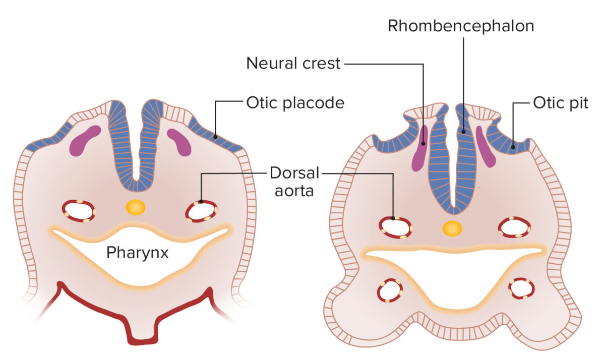
Inner ear

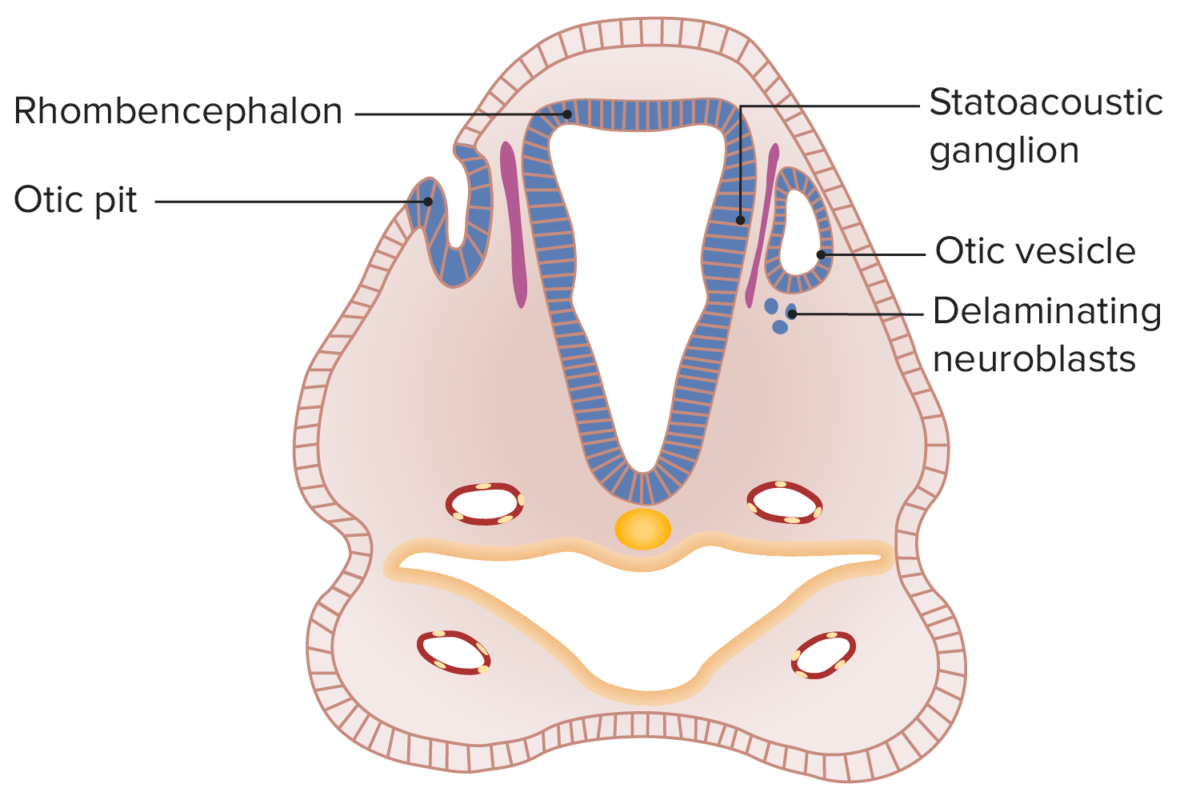
  • Ectoderm Ectoderm The outer of the three germ layers of an embryo. Gastrulation and Neurulation overlying the rhombencephalon Rhombencephalon The posterior of the three primitive cerebral vesicles of an embryonic brain. It consists of myelencephalon, metencephalon, and isthmus rhombencephalon from which develop the major brain stem components, such as medulla oblongata from the myelencephalon, cerebellum and pons from the metencephalon, with the expanded cavity forming the fourth ventricle. Development of the Nervous System and Face thickens to form the otic placode. 
  • The otic placode invaginates to become the otic pit. 
  • The otic pit detaches from surface ectoderm Ectoderm The outer of the three germ layers of an embryo. Gastrulation and Neurulation to form the otic vesicle Vesicle Primary Skin Lesions.  
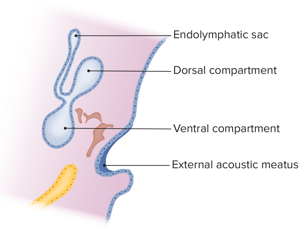
  • The otic vesicle Vesicle Primary Skin Lesions divides into dorsal and ventral compartments.
    • Dorsal compartment gives rise to:
      • Semicircular canals Semicircular canals Three long canals (anterior, posterior, and lateral) of the bony labyrinth. They are set at right angles to each other and are situated posterosuperior to the vestibule of the bony labyrinth (vestibular labyrinth). The semicircular canals have five openings into the vestibule with one shared by the anterior and the posterior canals. Within the canals are the semicircular ducts. Auditory and Vestibular Pathways: Anatomy
      • Utricle Utricle A membranous sac within the vestibular labyrinth of the inner ear. The utricle communicates with the saccule through the utriculosaccular duct from which the endolymphatic duct arises. The utricle and saccule have sensory areas (acoustic maculae) which are innervated by the vestibular nerve. Auditory and Vestibular Pathways: Anatomy
      • Endolymphatic appendage/sac
    • Ventral compartment gives rise to:
      • Cochlea
      • Spiral Spiral Computed tomography where there is continuous x-ray exposure to the patient while being transported in a spiral or helical pattern through the beam of irradiation. This provides improved three-dimensional contrast and spatial resolution compared to conventional computed tomography, where data is obtained and computed from individual sequential exposures. Computed Tomography (CT) of the cochlea
  • Otic vesicle Vesicle Primary Skin Lesions is filled with endolymph Endolymph The lymph fluid found in the membranous labyrinth of the ear. Vertigo, with surrounding mesenchyme that hollows out to create spaces filled with perilymph.
  • Perilymph spaces become the scala tympani and scala vestibuli.
  • Neural crest Neural crest The two longitudinal ridges along the primitive streak appearing near the end of gastrulation during development of nervous system (neurulation). The ridges are formed by folding of neural plate. Between the ridges is a neural groove which deepens as the fold become elevated. When the folds meet at midline, the groove becomes a closed tube, the neural tube. Hirschsprung Disease–derived cells → statoacoustic ganglion → cranial nerve (CN) VIII

Middle ear Middle ear The space and structures directly internal to the tympanic membrane and external to the inner ear (labyrinth). Its major components include the auditory ossicles and the eustachian tube that connects the cavity of middle ear (tympanic cavity) to the upper part of the throat. Acute Otitis Media

  • Develops from the 3 germinal layers
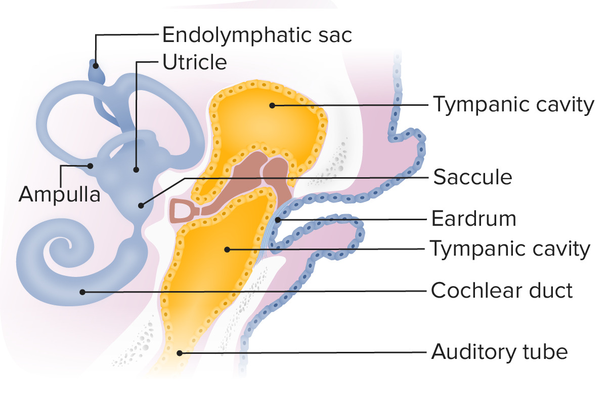
  • Auditory tube extends from the 1st pharyngeal pouch.
  • 1st and 2nd pharyngeal arches Pharyngeal arches The branchial arches, also known as pharyngeal or visceral arches, are embryonic structures seen in the development of vertebrates that serve as precursors for many structures of the face, neck, and head. These arches are composed of a central core of mesoderm, which is covered externally by ectoderm and internally by endoderm. Branchial Apparatus and Aortic Arches give rise to the malleus, incus, and stapes.
  • Auditory tube surrounds the ossicles → tympanic cavity
  • Tympanic membrane
Diagram displaying the auditory tube, which extends from the first pharyngeal pouch

Diagram displaying the auditory tube, which extends from the 1st pharyngeal pouch as it surrounds the ossicles, creating the tympanic cavity

Image by Lecturio.
Outer ear

  • Groove between 1st and 2nd pharyngeal arches Pharyngeal arches The branchial arches, also known as pharyngeal or visceral arches, are embryonic structures seen in the development of vertebrates that serve as precursors for many structures of the face, neck, and head. These arches are composed of a central core of mesoderm, which is covered externally by ectoderm and internally by endoderm. Branchial Apparatus and Aortic Arches → external acoustic meatus
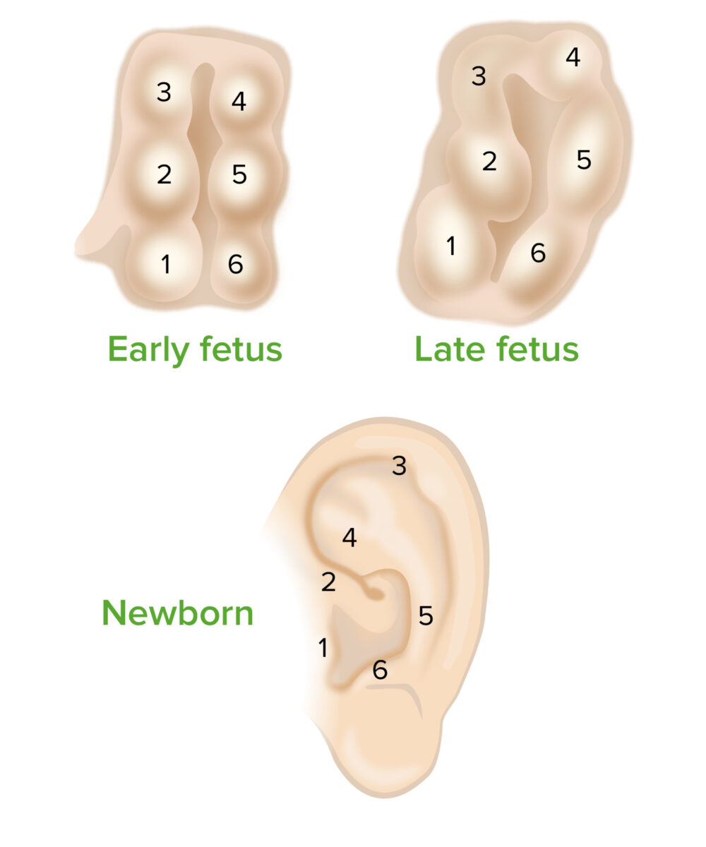
  • Pharyngeal arches Pharyngeal arches The branchial arches, also known as pharyngeal or visceral arches, are embryonic structures seen in the development of vertebrates that serve as precursors for many structures of the face, neck, and head. These arches are composed of a central core of mesoderm, which is covered externally by ectoderm and internally by endoderm. Branchial Apparatus and Aortic Arches → auricular hillocks → external ear/auricle
Six auricular hillocks bulge outward from the pharyngeal arches and shift to form the characteristic shape of the external ear

The 6 auricular hillocks in the fetus bulge outward from the pharyngeal arches and shift to form the characteristic shape of the external ear.

Image by Lecturio.

Related videos

Outer Ear Anatomy

Auricle

The auricle is composed of musculocutaneous tissue and elastic Elastic Connective Tissue: Histology cartilage Cartilage Cartilage is a type of connective tissue derived from embryonic mesenchyme that is responsible for structural support, resilience, and the smoothness of physical actions. Perichondrium (connective tissue membrane surrounding cartilage) compensates for the absence of vasculature in cartilage by providing nutrition and support. Cartilage: Histology covered by skin Skin The skin, also referred to as the integumentary system, is the largest organ of the body. The skin is primarily composed of the epidermis (outer layer) and dermis (deep layer). The epidermis is primarily composed of keratinocytes that undergo rapid turnover, while the dermis contains dense layers of connective tissue. Skin: Structure and Functions. This structure is responsible for collecting and redirecting sound waves into the external auditory meatus.
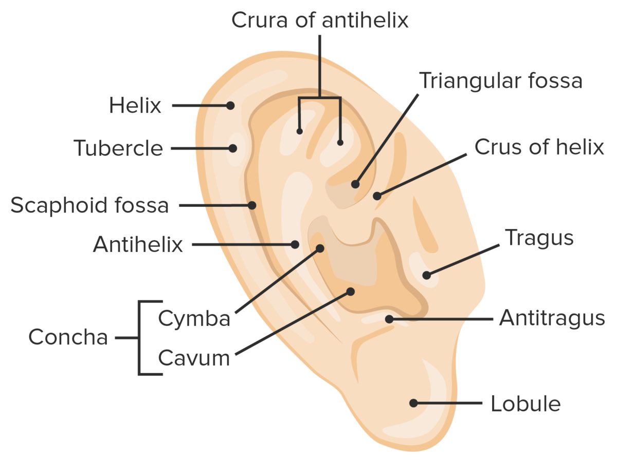
  • Helix: curved outer rim 
  • Antihelix: inner curved rim
  • Tragus Tragus Otitis Externa and antitragus: protrusions that partially obstruct external auditory meatus
  • Concha: hollow region in front of external auditory meatus 
  • Lobule: inferior, fleshy portion of ear
  • Triangular fossa: concave surface of anterosuperior ear
  • Scaphoid fossa: concave surface of posterosuperior ear
Anatomy of the ear

The outer ear, featuring the anatomical landmarks of the auricle

Image by Lecturio.

External auditory meatus

The external auditory meatus is typically 1 in. (2.5 cm) in length and is S-shaped.

  • Lateral ⅓ surrounded by cartilage Cartilage Cartilage is a type of connective tissue derived from embryonic mesenchyme that is responsible for structural support, resilience, and the smoothness of physical actions. Perichondrium (connective tissue membrane surrounding cartilage) compensates for the absence of vasculature in cartilage by providing nutrition and support. Cartilage: Histology (posterosuperior trajectory) 
  • Medial ⅔ surrounded by temporal bone Temporal bone Either of a pair of compound bones forming the lateral (left and right) surfaces and base of the skull which contains the organs of hearing. It is a large bone formed by the fusion of parts: the squamous (the flattened anterior-superior part), the tympanic (the curved anterior-inferior part), the mastoid (the irregular posterior portion), and the petrous (the part at the base of the skull). Jaw and Temporomandibular Joint: Anatomy (anteroinferior trajectory)
  • Surrounding skin Skin The skin, also referred to as the integumentary system, is the largest organ of the body. The skin is primarily composed of the epidermis (outer layer) and dermis (deep layer). The epidermis is primarily composed of keratinocytes that undergo rapid turnover, while the dermis contains dense layers of connective tissue. Skin: Structure and Functions contains ceruminous and sebaceous glands → ear wax

Tympanic membrane

Transmits sound waves from the air to the ossicles → mechanical vibrations
  • Located between outer and middle ear Middle ear The space and structures directly internal to the tympanic membrane and external to the inner ear (labyrinth). Its major components include the auditory ossicles and the eustachian tube that connects the cavity of middle ear (tympanic cavity) to the upper part of the throat. Acute Otitis Media 
  • Attached to fibrocartilaginous ring of temporal bone Temporal bone Either of a pair of compound bones forming the lateral (left and right) surfaces and base of the skull which contains the organs of hearing. It is a large bone formed by the fusion of parts: the squamous (the flattened anterior-superior part), the tympanic (the curved anterior-inferior part), the mastoid (the irregular posterior portion), and the petrous (the part at the base of the skull). Jaw and Temporomandibular Joint: Anatomy
  • 3 layers: cutaneous, fibrous Fibrous Fibrocystic Change, and mucous 
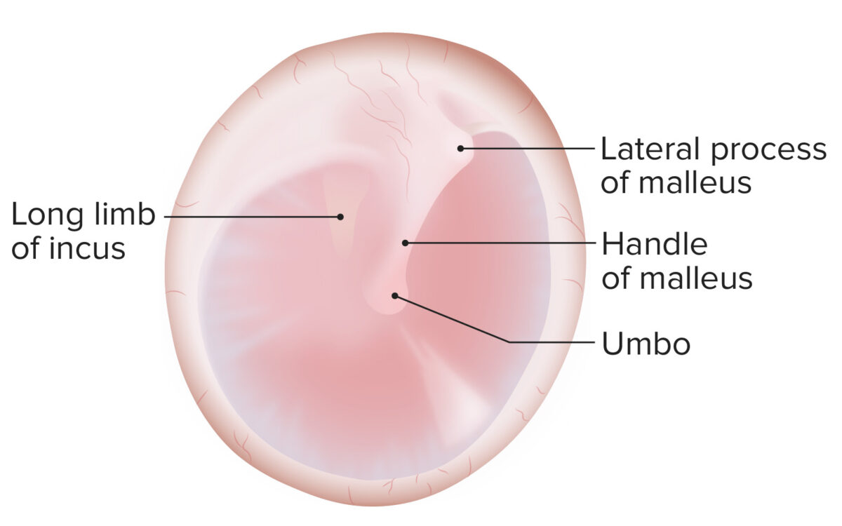
  • Ossicles attach to the tympanic membrane at the umbo.
Tympanic membrane

Tympanic membrane:
Pars flaccida (superior to insertion manubrium) and pars tensa (remainder of tympanic membrane)
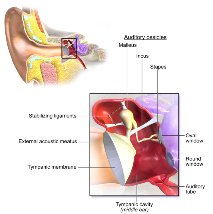
Image by Lecturio.

Related videos

Middle Ear

Tympanic cavity

  • Bony membranous cavity shaped like a 6-sided prism and filled with air
  • Contains the ossicles, muscles, and nerves
  • The roof consists of the tegmental wall, which separates the malleus and incus from the middle cranial fossa Middle cranial fossa The compartment containing the anterior extremities and half the inferior surface of the temporal lobes (temporal lobe) of the cerebral hemispheres. Lying posterior and inferior to the anterior cranial fossa, it is formed by part of the temporal bone and sphenoid bone. It is separated from the posterior cranial fossa by crests formed by the superior borders of the petrous parts of the temporal bones. Skull: Anatomy.
  • Labyrinthine or medial wall contents:
    • Oval window (connects to vestibule of the internal ear)
    • Round window (secondary tympanic membrane) 
    • Promontory
    • Prominence of the facial canal
  • Relations:
    • Lateral wall: tympanic membrane
    • Posterior wall:
      • Mastoid antrum
      • Communicates with mastoid air cells
    • Anterior wall (carotid wall):
      • Thin layer of bone Bone Bone is a compact type of hardened connective tissue composed of bone cells, membranes, an extracellular mineralized matrix, and central bone marrow. The 2 primary types of bone are compact and spongy. Bones: Structure and Types separating the cavity from the carotid artery
      • Pierced by the tympanic branch of the internal carotid artery Internal carotid artery Branch of the common carotid artery which supplies the anterior part of the brain, the eye and its appendages, the forehead and nose. Carotid Arterial System: Anatomy and deep petrosal nerve
      • Contains the auditory tube
    • Floor (jugular wall): separates the cavity from the internal  jugular vein
  • Auditory ossicles:
    • Amplify sound waves and transmit them to the labyrinth of the inner ear
    • Connect the tympanic membrane to the oval window
    • Smallest bones in the human body
    • Lateral → medial: malleus, incus, stapes
Anatomy of the middle ear

The middle ear, featuring the auditory ossicles connecting the tympanic membrane to the oval window

Image: “Anatomy of the middle ear” by BruceBlaus. License: CC BY 3.0

Muscles of the middle ear Middle ear The space and structures directly internal to the tympanic membrane and external to the inner ear (labyrinth). Its major components include the auditory ossicles and the eustachian tube that connects the cavity of middle ear (tympanic cavity) to the upper part of the throat. Acute Otitis Media

Middle ear Middle ear The space and structures directly internal to the tympanic membrane and external to the inner ear (labyrinth). Its major components include the auditory ossicles and the eustachian tube that connects the cavity of middle ear (tympanic cavity) to the upper part of the throat. Acute Otitis Media muscles are responsible for movement of the tympanic membrane.
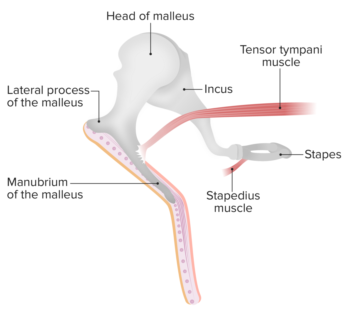
  • Tensor tympani muscle:
    • Origin:
      • Cartilaginous part of eustachian tube
      • Greater wing of the sphenoid
      • Semicanal for tensor tympani (petrous part of temporal bone Temporal bone Either of a pair of compound bones forming the lateral (left and right) surfaces and base of the skull which contains the organs of hearing. It is a large bone formed by the fusion of parts: the squamous (the flattened anterior-superior part), the tympanic (the curved anterior-inferior part), the mastoid (the irregular posterior portion), and the petrous (the part at the base of the skull). Jaw and Temporomandibular Joint: Anatomy)
    • Insertion: superior part of the handle of the malleus
    • Innervation: mandibular branch of the trigeminal nerve Trigeminal nerve The 5th and largest cranial nerve. The trigeminal nerve is a mixed motor and sensory nerve. The larger sensory part forms the ophthalmic, mandibular, and maxillary nerves which carry afferents sensitive to external or internal stimuli from the skin, muscles, and joints of the face and mouth and from the teeth. Most of these fibers originate from cells of the trigeminal ganglion and project to the trigeminal nucleus of the brain stem. The smaller motor part arises from the brain stem trigeminal motor nucleus and innervates the muscles of mastication. The 12 Cranial Nerves: Overview and Functions (CN V3)
    • Function:
      • Pulls the handle of the malleus medially 
      • Tenses the tympanic membrane
  • Stapedius muscle:
    • Origin: cartilaginous/bony margins of eustachian tube
    • Insertion: neck Neck The part of a human or animal body connecting the head to the rest of the body. Peritonsillar Abscess of stapes
    • Innervation: facial nerve Facial nerve The 7th cranial nerve. The facial nerve has two parts, the larger motor root which may be called the facial nerve proper, and the smaller intermediate or sensory root. Together they provide efferent innervation to the muscles of facial expression and to the lacrimal and salivary glands, and convey afferent information for taste from the anterior two-thirds of the tongue and for touch from the external ear. The 12 Cranial Nerves: Overview and Functions (CN VII)
    • Function:
      • Pulls stapes posteriorly  
      • Prevents excessive oscillation of the ossicles
Muscles and ossicles of the middle ear

Muscles and ossicles of the middle ear:
The malleus connects to the tympanic membrane while the incus and stapes are completely within the middle ear. The stapedius muscle prevents excessive oscillation of the ossicles.
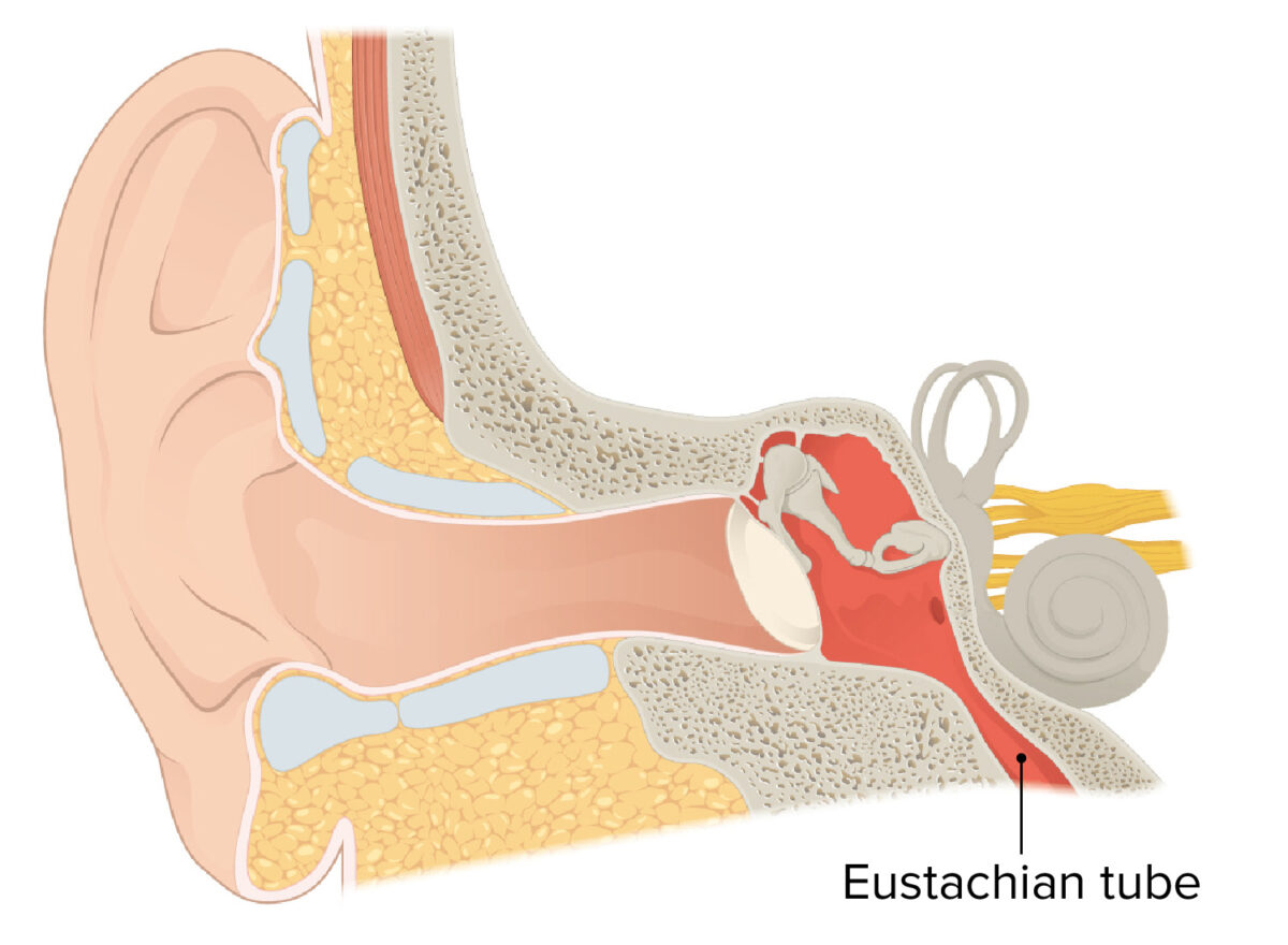
Image by Lecturio.

Eustachian tube (auditory tube)

  • Controls pressure within the middle ear Middle ear The space and structures directly internal to the tympanic membrane and external to the inner ear (labyrinth). Its major components include the auditory ossicles and the eustachian tube that connects the cavity of middle ear (tympanic cavity) to the upper part of the throat. Acute Otitis Media and on both sides of the tympanic membrane
  • Located anterior to the tympanic cavity
  • Connects the middle ear Middle ear The space and structures directly internal to the tympanic membrane and external to the inner ear (labyrinth). Its major components include the auditory ossicles and the eustachian tube that connects the cavity of middle ear (tympanic cavity) to the upper part of the throat. Acute Otitis Media to the nasopharynx Nasopharynx The top portion of the pharynx situated posterior to the nose and superior to the soft palate. The nasopharynx is the posterior extension of the nasal cavities and has a respiratory function. Pharynx: Anatomy
Eustachian tube

The Eustachian tube connects the middle ear with the pharynx:
This connection allows for the equalization of air pressure between the middle ear and the outside world.

Image by Lecturio.

Inner Ear

The inner ear is located within the petrous part of the temporal bone Temporal bone Either of a pair of compound bones forming the lateral (left and right) surfaces and base of the skull which contains the organs of hearing. It is a large bone formed by the fusion of parts: the squamous (the flattened anterior-superior part), the tympanic (the curved anterior-inferior part), the mastoid (the irregular posterior portion), and the petrous (the part at the base of the skull). Jaw and Temporomandibular Joint: Anatomy and contains the organs of hearing and equilibrium Equilibrium Occurs when tumor cells survive the initial elimination attempt These cells are not able to progress, being maintained in a state of dormancy by the adaptive immune system. In this phase, tumor immunogenicity is edited, where T cells keep selectively attacking highly immunogenic tumor cells.This attack leaves other cells with less immunogenicity to potentially develop resistance to the immune response. Cancer Immunotherapy.

Bony labyrinth

The bony labyrinth is filled with perilymph and is separated from the middle ear Middle ear The space and structures directly internal to the tympanic membrane and external to the inner ear (labyrinth). Its major components include the auditory ossicles and the eustachian tube that connects the cavity of middle ear (tympanic cavity) to the upper part of the throat. Acute Otitis Media by the medial wall of the tympanic cavity.
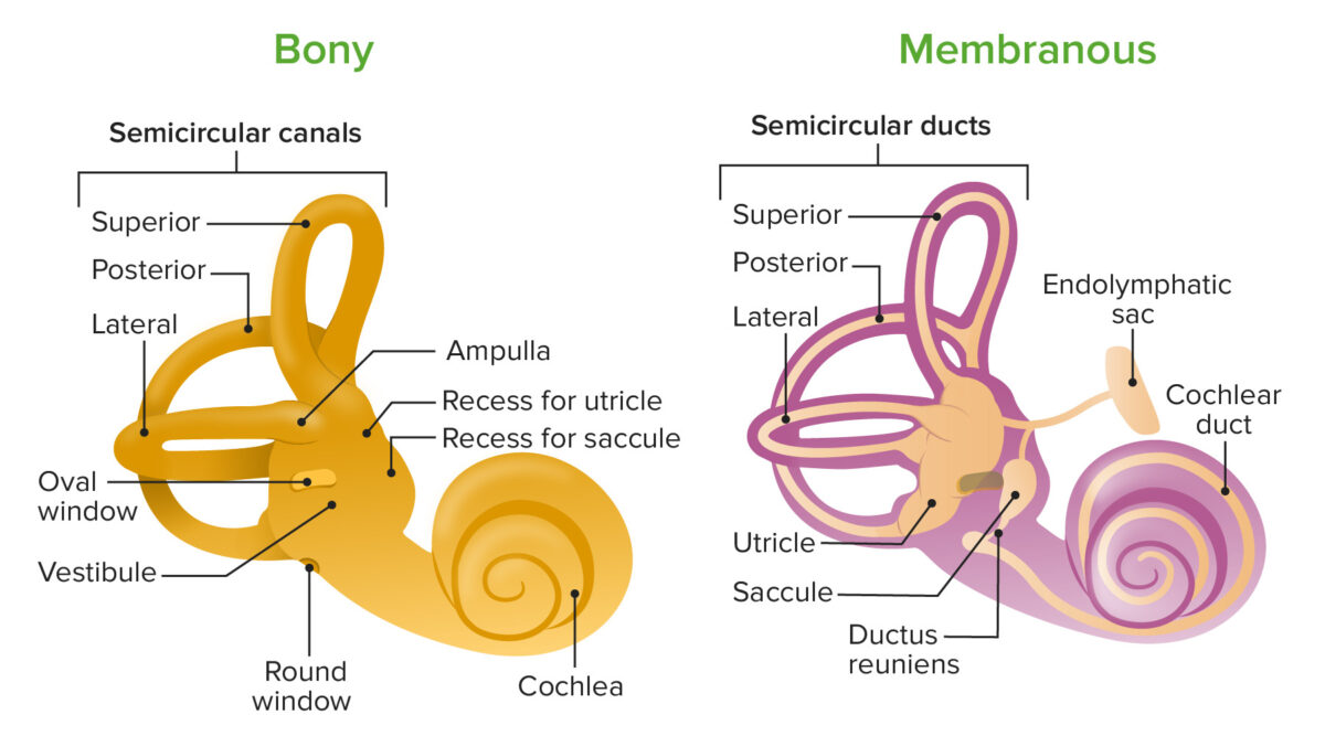
  • Vestibule: central bony cavity:
    • Contains the utricle Utricle A membranous sac within the vestibular labyrinth of the inner ear. The utricle communicates with the saccule through the utriculosaccular duct from which the endolymphatic duct arises. The utricle and saccule have sensory areas (acoustic maculae) which are innervated by the vestibular nerve. Auditory and Vestibular Pathways: Anatomy and saccule Saccule A membranous sac within the vestibular labyrinth of the inner ear. The saccule communicates with cochlear duct through the ductus reuniens, and communicates with utricle through the utriculosaccular duct from which the endolymphatic duct arises. The utricle and saccule have sensory areas (acoustic maculae) which are innervated by the vestibular nerve. Auditory and Vestibular Pathways: Anatomy 
    • Communicates with the middle ear Middle ear The space and structures directly internal to the tympanic membrane and external to the inner ear (labyrinth). Its major components include the auditory ossicles and the eustachian tube that connects the cavity of middle ear (tympanic cavity) to the upper part of the throat. Acute Otitis Media in 3 directions:
      • Laterally through the oval window
      • Anteriorly with the cochlea
      • Posteriorly with the semicircular canals Semicircular canals Three long canals (anterior, posterior, and lateral) of the bony labyrinth. They are set at right angles to each other and are situated posterosuperior to the vestibule of the bony labyrinth (vestibular labyrinth). The semicircular canals have five openings into the vestibule with one shared by the anterior and the posterior canals. Within the canals are the semicircular ducts. Auditory and Vestibular Pathways: Anatomy 
  • Semicircular canals Semicircular canals Three long canals (anterior, posterior, and lateral) of the bony labyrinth. They are set at right angles to each other and are situated posterosuperior to the vestibule of the bony labyrinth (vestibular labyrinth). The semicircular canals have five openings into the vestibule with one shared by the anterior and the posterior canals. Within the canals are the semicircular ducts. Auditory and Vestibular Pathways: Anatomy: 3 canals at 90-degree angles to each other:
  • Cochlea: spiral-shaped bony cavity:
    • Spiral Spiral Computed tomography where there is continuous x-ray exposure to the patient while being transported in a spiral or helical pattern through the beam of irradiation. This provides improved three-dimensional contrast and spatial resolution compared to conventional computed tomography, where data is obtained and computed from individual sequential exposures. Computed Tomography (CT) lamina: superior scala vestibuli and inferior scala tympani
    • Central bony column = modiolus
Bony and membranous labyrinths

Diagram of the bony and membranous labyrinths, featuring their anatomical landmarks:
Note that the cochlear duct is found within the membranous labyrinth and the utricle and saccule are found within the vestibule.

Image by Lecturio.

Membranous labyrinth

The membranous labyrinth is filled with endolymph Endolymph The lymph fluid found in the membranous labyrinth of the ear. Vertigo and is composed of the vestibular and cochlear labyrinths.
  • Vestibular labyrinth:
    • Comprised of the utricle Utricle A membranous sac within the vestibular labyrinth of the inner ear. The utricle communicates with the saccule through the utriculosaccular duct from which the endolymphatic duct arises. The utricle and saccule have sensory areas (acoustic maculae) which are innervated by the vestibular nerve. Auditory and Vestibular Pathways: Anatomy, saccule Saccule A membranous sac within the vestibular labyrinth of the inner ear. The saccule communicates with cochlear duct through the ductus reuniens, and communicates with utricle through the utriculosaccular duct from which the endolymphatic duct arises. The utricle and saccule have sensory areas (acoustic maculae) which are innervated by the vestibular nerve. Auditory and Vestibular Pathways: Anatomy, and 3 membranous semicircular ducts
    • Contains sensory Sensory Neurons which conduct nerve impulses to the central nervous system. Nervous System: Histology organs for balance
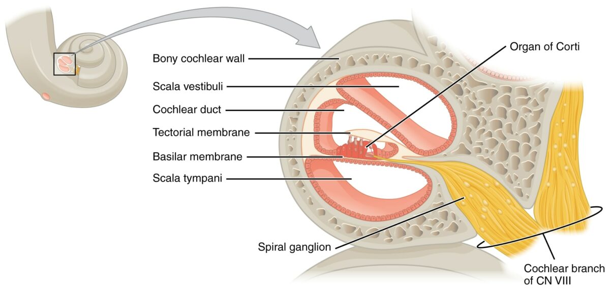
  • Cochlear labyrinth:
    • Sensory Sensory Neurons which conduct nerve impulses to the central nervous system. Nervous System: Histology organ for hearing
    • Scala vestibuli:
      • Contains perilymph
      • Anterior chamber Anterior chamber The space in the eye, filled with aqueous humor, bounded anteriorly by the cornea and a small portion of the sclera and posteriorly by a small portion of the ciliary body, the iris, and that part of the crystalline lens which presents through the pupil. Eye: Anatomy up which sound travels
    • Scala tympani:
    • Scala media (cochlear duct):
      • Contains endolymph Endolymph The lymph fluid found in the membranous labyrinth of the ear. Vertigo
      • Located between scala vestibuli and scala tympani
      • Houses organ of Corti Organ of Corti The spiral epithelium containing sensory auditory hair cells and supporting cells in the cochlea. Organ of corti, situated on the basilar membrane and overlaid by a gelatinous tectorial membrane, converts sound-induced mechanical waves to neural impulses to the brain. Auditory and Vestibular Pathways: Anatomy
  • Organ of Corti Organ of Corti The spiral epithelium containing sensory auditory hair cells and supporting cells in the cochlea. Organ of corti, situated on the basilar membrane and overlaid by a gelatinous tectorial membrane, converts sound-induced mechanical waves to neural impulses to the brain. Auditory and Vestibular Pathways: Anatomy:
    • Spiral Spiral Computed tomography where there is continuous x-ray exposure to the patient while being transported in a spiral or helical pattern through the beam of irradiation. This provides improved three-dimensional contrast and spatial resolution compared to conventional computed tomography, where data is obtained and computed from individual sequential exposures. Computed Tomography (CT) organ within cochlear labyrinth
    • Contains mechanoreceptors called hair cells Hair cells Auditory sensory cells of organ of corti, usually placed in one row medially to the core of spongy bone (the modiolus). Inner hair cells are in fewer numbers than the outer auditory hair cells, and their stereocilia are approximately twice as thick as those of the outer hair cells. Auditory and Vestibular Pathways: Anatomy
    • Hair cells Hair cells Auditory sensory cells of organ of corti, usually placed in one row medially to the core of spongy bone (the modiolus). Inner hair cells are in fewer numbers than the outer auditory hair cells, and their stereocilia are approximately twice as thick as those of the outer hair cells. Auditory and Vestibular Pathways: Anatomy determine the movement and position of the head in space.
    • Types I (bulbous) and II (elongated) hair cells Hair cells Auditory sensory cells of organ of corti, usually placed in one row medially to the core of spongy bone (the modiolus). Inner hair cells are in fewer numbers than the outer auditory hair cells, and their stereocilia are approximately twice as thick as those of the outer hair cells. Auditory and Vestibular Pathways: Anatomy
      • Both types have stereocilia of varying lengths at the apex. 
      • Stereocilia respond to movement of endolymph Endolymph The lymph fluid found in the membranous labyrinth of the ear. Vertigo
      • Kinocilium: single cilium at lateral portion of hair cell, defines polarity of the cell
      • Hair cells Hair cells Auditory sensory cells of organ of corti, usually placed in one row medially to the core of spongy bone (the modiolus). Inner hair cells are in fewer numbers than the outer auditory hair cells, and their stereocilia are approximately twice as thick as those of the outer hair cells. Auditory and Vestibular Pathways: Anatomy in the organ of Corti Organ of Corti The spiral epithelium containing sensory auditory hair cells and supporting cells in the cochlea. Organ of corti, situated on the basilar membrane and overlaid by a gelatinous tectorial membrane, converts sound-induced mechanical waves to neural impulses to the brain. Auditory and Vestibular Pathways: Anatomy lack kinocilium.
The organ of corti is located within the scala media

The organ of Corti is located within the scala media:
The organ of Corti detects sound in the form of vibration and transmits the information through the cochlear nerve.
CN: cranial nerve
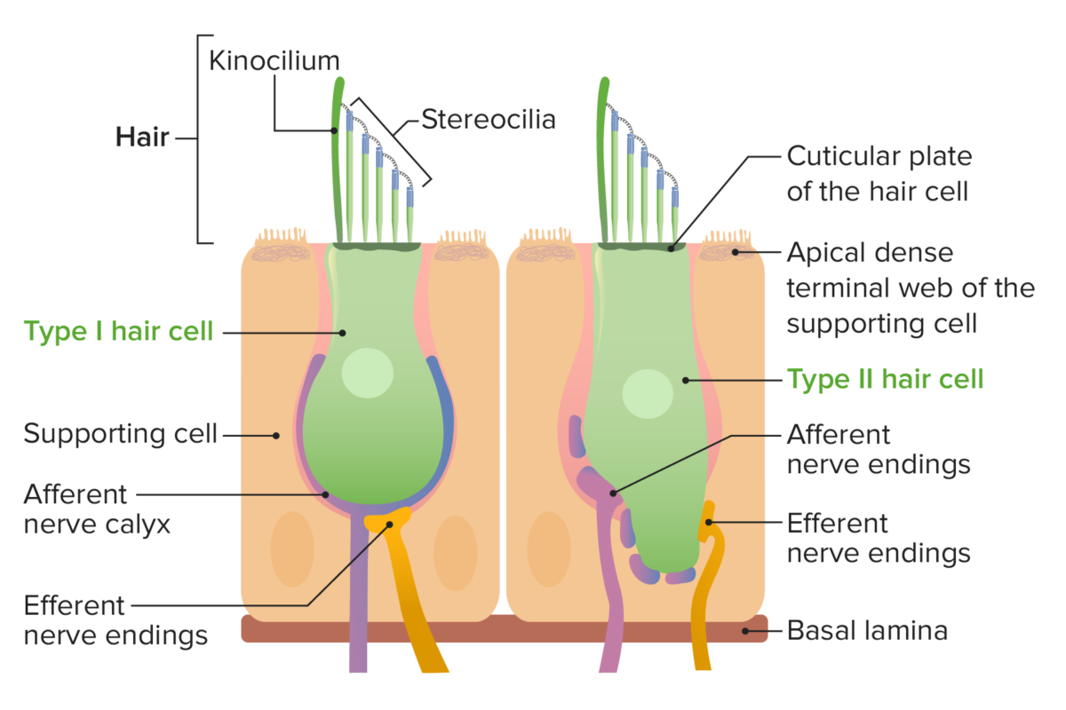
Image by Lecturio.
Hair cells type i and type ii

Types I and II hair cells:
Hair cells determine the movement and position of the head in space.

Image by Lecturio.

Neurovasculature of the Ear

Table: Neurovasculature of the ear
Structure Blood supply Lymphatics Innervation
Auricle
  • Posterior auricular artery
  • Anterior auricular arteries Arteries Arteries are tubular collections of cells that transport oxygenated blood and nutrients from the heart to the tissues of the body. The blood passes through the arteries in order of decreasing luminal diameter, starting in the largest artery (the aorta) and ending in the small arterioles. Arteries are classified into 3 types: large elastic arteries, medium muscular arteries, and small arteries and arterioles. Arteries: Histology
  • Branches of the occipital Occipital Part of the back and base of the cranium that encloses the foramen magnum. Skull: Anatomy artery
Superficial parotid, mastoid, and cervical lymph nodes Lymph Nodes They are oval or bean shaped bodies (1 – 30 mm in diameter) located along the lymphatic system. Lymphatic Drainage System: Anatomy
  • Auriculotemporal branch of the mandibular nerve Mandibular nerve A branch of the trigeminal (5th cranial) nerve. The mandibular nerve carries motor fibers to the muscles of mastication and sensory fibers to the teeth and gingivae, the face in the region of the mandible, and parts of the dura. Jaw and Temporomandibular Joint: Anatomy (CN V3)
  • Cervical plexus Cervical Plexus A network of nerve fibers originating in the upper four cervical spinal cord segments. The cervical plexus distributes cutaneous nerves to parts of the neck, shoulders, and back of the head. It also distributes motor fibers to muscles of the cervical spinal column, infrahyoid muscles, and the diaphragm. Peripheral Nerve Injuries in the Cervicothoracic Region
  • Lesser occipital Occipital Part of the back and base of the cranium that encloses the foramen magnum. Skull: Anatomy nerve
  • Great auricular nerve
  • Auricular branch of the vagus nerve Vagus nerve The 10th cranial nerve. The vagus is a mixed nerve which contains somatic afferents (from skin in back of the ear and the external auditory meatus), visceral afferents (from the pharynx, larynx, thorax, and abdomen), parasympathetic efferents (to the thorax and abdomen), and efferents to striated muscle (of the larynx and pharynx). Pharynx: Anatomy
  • Branches of the facial nerve Facial nerve The 7th cranial nerve. The facial nerve has two parts, the larger motor root which may be called the facial nerve proper, and the smaller intermediate or sensory root. Together they provide efferent innervation to the muscles of facial expression and to the lacrimal and salivary glands, and convey afferent information for taste from the anterior two-thirds of the tongue and for touch from the external ear. The 12 Cranial Nerves: Overview and Functions
Tympanic membrane
  • Branches of the maxillary artery
  • Branch of the posterior auricular artery
  • Inferior tympanic artery
Periauricular lymph nodes Lymph Nodes They are oval or bean shaped bodies (1 – 30 mm in diameter) located along the lymphatic system. Lymphatic Drainage System: Anatomy
  • Lateral side: branches of the auriculotemporal nerve and auricular branch of CN X
  • Medial side: CN IX
Tympanic cavity
  • Anterior tympanic artery
  • Posterior, inferior, anterior, and superior tympanic arteries Arteries Arteries are tubular collections of cells that transport oxygenated blood and nutrients from the heart to the tissues of the body. The blood passes through the arteries in order of decreasing luminal diameter, starting in the largest artery (the aorta) and ending in the small arterioles. Arteries are classified into 3 types: large elastic arteries, medium muscular arteries, and small arteries and arterioles. Arteries: Histology
  • Mastoid branch of the occipital Occipital Part of the back and base of the cranium that encloses the foramen magnum. Skull: Anatomy artery
Retroauricular cervical lymph nodes Lymph Nodes They are oval or bean shaped bodies (1 – 30 mm in diameter) located along the lymphatic system. Lymphatic Drainage System: Anatomy
  • Tympanic cavity: tympanic nerve (branch of CN IX)
  • Muscles: tensor tympani nerve (CN V) and stapedius nerve (CN VII)
Bony labyrinth
  • Anterior tympanic artery
  • Stylomastoid artery
  • Petrosal artery
Parotid, mastoid, and superficial cervical lymph nodes Lymph Nodes They are oval or bean shaped bodies (1 – 30 mm in diameter) located along the lymphatic system. Lymphatic Drainage System: Anatomy
  • Hearing: cochlear nerve from spiral Spiral Computed tomography where there is continuous x-ray exposure to the patient while being transported in a spiral or helical pattern through the beam of irradiation. This provides improved three-dimensional contrast and spatial resolution compared to conventional computed tomography, where data is obtained and computed from individual sequential exposures. Computed Tomography (CT) ganglion
  • Balance: vestibular nerve from the semicircular ducts, utricle Utricle A membranous sac within the vestibular labyrinth of the inner ear. The utricle communicates with the saccule through the utriculosaccular duct from which the endolymphatic duct arises. The utricle and saccule have sensory areas (acoustic maculae) which are innervated by the vestibular nerve. Auditory and Vestibular Pathways: Anatomy, and saccule Saccule A membranous sac within the vestibular labyrinth of the inner ear. The saccule communicates with cochlear duct through the ductus reuniens, and communicates with utricle through the utriculosaccular duct from which the endolymphatic duct arises. The utricle and saccule have sensory areas (acoustic maculae) which are innervated by the vestibular nerve. Auditory and Vestibular Pathways: Anatomy
  • Cochlear and vestibular nerves unite and form the vestibulocochlear nerve Vestibulocochlear nerve The 8th cranial nerve. The vestibulocochlear nerve has a cochlear part (cochlear nerve) which is concerned with hearing and a vestibular part (vestibular nerve) which mediates the sense of balance and head position. The fibers of the cochlear nerve originate from neurons of the spiral ganglion and project to the cochlear nuclei (cochlear nucleus). The fibers of the vestibular nerve arise from neurons of scarpa’s ganglion and project to the vestibular nuclei. The 12 Cranial Nerves: Overview and Functions (CN VIII)
Membranous labyrinth Labyrinthine artery (branch of the basilar artery)

Clinical Relevance

  • Otitis externa Otitis externa Otitis externa (also known as external otitis or swimmer’s ear) is an infection of the external auditory canal that is most often caused by acute bacterial infection and is frequently associated with hot, humid weather and water exposure. Patients commonly present with ear pain, pruritus, discharge, and hearing loss. Otitis Externa: inflammation Inflammation Inflammation is a complex set of responses to infection and injury involving leukocytes as the principal cellular mediators in the body’s defense against pathogenic organisms. Inflammation is also seen as a response to tissue injury in the process of wound healing. The 5 cardinal signs of inflammation are pain, heat, redness, swelling, and loss of function. Inflammation of the external acoustic meatus. Otitis externa Otitis externa Otitis externa (also known as external otitis or swimmer’s ear) is an infection of the external auditory canal that is most often caused by acute bacterial infection and is frequently associated with hot, humid weather and water exposure. Patients commonly present with ear pain, pruritus, discharge, and hearing loss. Otitis Externa is a bacterial infection that most commonly occurs in children. The typical causative organisms are Pseudomonas Pseudomonas Pseudomonas is a non-lactose-fermenting, gram-negative bacillus that produces pyocyanin, which gives it a characteristic blue-green color. Pseudomonas is found ubiquitously in the environment, as well as in moist reservoirs, such as hospital sinks and respiratory equipment. Pseudomonas and Staphylococcus aureus Staphylococcus aureus Potentially pathogenic bacteria found in nasal membranes, skin, hair follicles, and perineum of warm-blooded animals. They may cause a wide range of infections and intoxications. Brain Abscess. Presentation is with exquisite tenderness on manipulation of the external ear. Treatment is with antibiotic eardrops.
  • Acute otitis media Acute Otitis Media Acute otitis media is an infection in the middle ear characterized by mucosal inflammation and retention of fluid. The most common pathogens are Streptococcus pneumoniae, Haemophilus influenzae, and Moraxella catarrhalis. The condition can present with fever, otalgia, and diminished hearing. Acute Otitis Media: acute inflammation Acute Inflammation Inflammation of the middle ear Middle ear The space and structures directly internal to the tympanic membrane and external to the inner ear (labyrinth). Its major components include the auditory ossicles and the eustachian tube that connects the cavity of middle ear (tympanic cavity) to the upper part of the throat. Acute Otitis Media due to viral or bacterial infections Infections Invasion of the host organism by microorganisms or their toxins or by parasites that can cause pathological conditions or diseases. Chronic Granulomatous Disease. The condition is most common in children ages 6–18 months. Presentation is with fever Fever Fever is defined as a measured body temperature of at least 38°C (100.4°F). Fever is caused by circulating endogenous and/or exogenous pyrogens that increase levels of prostaglandin E2 in the hypothalamus. Fever is commonly associated with chills, rigors, sweating, and flushing of the skin. Fever, symptoms of upper respiratory tract infection, and ear pain Ear Pain Acute Otitis Media. The majority of cases are caused by viruses Viruses Minute infectious agents whose genomes are composed of DNA or RNA, but not both. They are characterized by a lack of independent metabolism and the inability to replicate outside living host cells. Virology, but antibiotics should be initiated if a bacterial etiology is suspected.
  • Vertigo Vertigo Vertigo is defined as the perceived sensation of rotational motion while remaining still. A very common complaint in primary care and the ER, vertigo is more frequently experienced by women and its prevalence increases with age. Vertigo is classified into peripheral or central based on its etiology. Vertigo: sensation of movement between oneself and surroundings. Vertigo Vertigo Vertigo is defined as the perceived sensation of rotational motion while remaining still. A very common complaint in primary care and the ER, vertigo is more frequently experienced by women and its prevalence increases with age. Vertigo is classified into peripheral or central based on its etiology. Vertigo is not limited to a feeling of rotation Rotation Motion of an object in which either one or more points on a line are fixed. It is also the motion of a particle about a fixed point. X-rays but may include other forms of movement, such as upward lifting, swaying, rocking, or nonsystematic movement. It is usually a symptom of an underlying condition such as labyrinthitis, Ménière disease Ménière disease Ménière disease is a condition characterized by episodes of vertigo, tinnitus, and hearing loss, likely caused by endolymphatic hydrops of the labyrinthine system in the inner ear. The risk factors include a family history of Ménière disease, preexisting autoimmune disorders, allergies, and trauma to the head or ear. Ménière Disease, or an adverse reaction to medications.
  • Ménière disease Ménière disease Ménière disease is a condition characterized by episodes of vertigo, tinnitus, and hearing loss, likely caused by endolymphatic hydrops of the labyrinthine system in the inner ear. The risk factors include a family history of Ménière disease, preexisting autoimmune disorders, allergies, and trauma to the head or ear. Ménière Disease: characterized by a symptom triad of vertigo Vertigo Vertigo is defined as the perceived sensation of rotational motion while remaining still. A very common complaint in primary care and the ER, vertigo is more frequently experienced by women and its prevalence increases with age. Vertigo is classified into peripheral or central based on its etiology. Vertigo, tinnitus Tinnitus A nonspecific symptom of hearing disorder characterized by the sensation of buzzing, ringing, clicking, pulsations, and other noises in the ear. Objective tinnitus refers to noises generated from within the ear or adjacent structures that can be heard by other individuals. The term subjective tinnitus is used when the sound is audible only to the affected individual. Tinnitus may occur as a manifestation of cochlear diseases; vestibulocochlear nerve diseases; intracranial hypertension; craniocerebral trauma; and other conditions. Cranial Nerve Palsies, and hearing impairment Hearing impairment Hearing loss, also known as hearing impairment, is any degree of impairment in the ability to apprehend sound as determined by audiometry to be below normal hearing thresholds. Clinical presentation may occur at birth or as a gradual loss of hearing with age, including a short-term or sudden loss at any point. Hearing Loss. The episodes of vertigo Vertigo Vertigo is defined as the perceived sensation of rotational motion while remaining still. A very common complaint in primary care and the ER, vertigo is more frequently experienced by women and its prevalence increases with age. Vertigo is classified into peripheral or central based on its etiology. Vertigo in Ménière disease Ménière disease Ménière disease is a condition characterized by episodes of vertigo, tinnitus, and hearing loss, likely caused by endolymphatic hydrops of the labyrinthine system in the inner ear. The risk factors include a family history of Ménière disease, preexisting autoimmune disorders, allergies, and trauma to the head or ear. Ménière Disease can last from a few minutes to several hours, and the hearing impairment Hearing impairment Hearing loss, also known as hearing impairment, is any degree of impairment in the ability to apprehend sound as determined by audiometry to be below normal hearing thresholds. Clinical presentation may occur at birth or as a gradual loss of hearing with age, including a short-term or sudden loss at any point. Hearing Loss mostly affects low frequencies. Treatment is geared toward nausea Nausea An unpleasant sensation in the stomach usually accompanied by the urge to vomit. Common causes are early pregnancy, sea and motion sickness, emotional stress, intense pain, food poisoning, and various enteroviruses. Antiemetics prevention and physical therapy Physical Therapy Becker Muscular Dystrophy to maintain physical functionality.
  • Hearing loss Hearing loss Hearing loss, also known as hearing impairment, is any degree of impairment in the ability to apprehend sound as determined by audiometry to be below normal hearing thresholds. Clinical presentation may occur at birth or as a gradual loss of hearing with age, including a short-term or sudden loss at any point. Hearing Loss: hearing impairments are classified into conductive hearing loss Conductive hearing loss Hearing loss due to interference with the mechanical reception or amplification of sound to the cochlea. The interference is in the outer or middle ear involving the ear canal; tympanic membrane; or ear ossicles. Hearing Loss and sensorineural hearing loss Sensorineural hearing loss Hearing loss resulting from damage to the cochlea and the sensorineural elements which lie internally beyond the oval and round windows. These elements include the auditory nerve and its connections in the brainstem. Hearing Loss. Conductive hearing loss Conductive hearing loss Hearing loss due to interference with the mechanical reception or amplification of sound to the cochlea. The interference is in the outer or middle ear involving the ear canal; tympanic membrane; or ear ossicles. Hearing Loss stems from damage to the middle ear Middle ear The space and structures directly internal to the tympanic membrane and external to the inner ear (labyrinth). Its major components include the auditory ossicles and the eustachian tube that connects the cavity of middle ear (tympanic cavity) to the upper part of the throat. Acute Otitis Media, which prevents the proper transmission of sound. In sensorineural hearing loss Sensorineural hearing loss Hearing loss resulting from damage to the cochlea and the sensorineural elements which lie internally beyond the oval and round windows. These elements include the auditory nerve and its connections in the brainstem. Hearing Loss, the hearing organ is damaged.
  • Tinnitus Tinnitus A nonspecific symptom of hearing disorder characterized by the sensation of buzzing, ringing, clicking, pulsations, and other noises in the ear. Objective tinnitus refers to noises generated from within the ear or adjacent structures that can be heard by other individuals. The term subjective tinnitus is used when the sound is audible only to the affected individual. Tinnitus may occur as a manifestation of cochlear diseases; vestibulocochlear nerve diseases; intracranial hypertension; craniocerebral trauma; and other conditions. Cranial Nerve Palsies: perception Perception The process by which the nature and meaning of sensory stimuli are recognized and interpreted. Psychiatric Assessment of noise or “ringing in the ears.” Tinnitus Tinnitus A nonspecific symptom of hearing disorder characterized by the sensation of buzzing, ringing, clicking, pulsations, and other noises in the ear. Objective tinnitus refers to noises generated from within the ear or adjacent structures that can be heard by other individuals. The term subjective tinnitus is used when the sound is audible only to the affected individual. Tinnitus may occur as a manifestation of cochlear diseases; vestibulocochlear nerve diseases; intracranial hypertension; craniocerebral trauma; and other conditions. Cranial Nerve Palsies is a common condition that is experienced in the absence of an external auditory stimulus. The sound perceived is described as whistling, hissing, buzzing, ringing, and/or pulsating. Tinnitus Tinnitus A nonspecific symptom of hearing disorder characterized by the sensation of buzzing, ringing, clicking, pulsations, and other noises in the ear. Objective tinnitus refers to noises generated from within the ear or adjacent structures that can be heard by other individuals. The term subjective tinnitus is used when the sound is audible only to the affected individual. Tinnitus may occur as a manifestation of cochlear diseases; vestibulocochlear nerve diseases; intracranial hypertension; craniocerebral trauma; and other conditions. Cranial Nerve Palsies may be related to underlying conditions such as Ménière disease Ménière disease Ménière disease is a condition characterized by episodes of vertigo, tinnitus, and hearing loss, likely caused by endolymphatic hydrops of the labyrinthine system in the inner ear. The risk factors include a family history of Ménière disease, preexisting autoimmune disorders, allergies, and trauma to the head or ear. Ménière Disease, ear and sinus infections Infections Invasion of the host organism by microorganisms or their toxins or by parasites that can cause pathological conditions or diseases. Chronic Granulomatous Disease, and brain Brain The part of central nervous system that is contained within the skull (cranium). Arising from the neural tube, the embryonic brain is comprised of three major parts including prosencephalon (the forebrain); mesencephalon (the midbrain); and rhombencephalon (the hindbrain). The developed brain consists of cerebrum; cerebellum; and other structures in the brain stem. Nervous System: Anatomy, Structure, and Classification tumors.

References

  1. Bhatt RA. (2020). Ear anatomy: overview, embryology, gross anatomy. Medscape. https://emedicine.medscape.com/article/1948907-overview
  2. ONeill OJ, Brett K, Frank AJ. (2021). Middle ear barotrauma. StatPearls. https://www.ncbi.nlm.nih.gov/books/NBK499851/
  3. Szymanski, A. Geiger, Z. (2017). Anatomy, head and neck, ear. StatPearls. https://europepmc.org/article/nbk/nbk470359

Create your free account or log in to continue reading!

Sign up now and get free access to Lecturio with concept pages, medical videos, and questions for your medical education.

User Reviews

Unwrap New Skills This Holiday 🎄 Save 30% on all plans now!

Details