Advertisement

Advertisement

Advertisement

Advertisement

Anal Cancer

Anal cancer accounts for 2.7% of all gastrointestinal tract cancers. Squamous cell carcinoma Squamous cell carcinoma Cutaneous squamous cell carcinoma (cSCC) is caused by malignant proliferation of atypical keratinocytes. This condition is the 2nd most common skin malignancy and usually affects sun-exposed areas of fair-skinned patients. The cancer presents as a firm, erythematous, keratotic plaque or papule. Squamous Cell Carcinoma (SCC) is the most common type of anal cancer. The patient can present with rectal bleeding (most common), change in bowel habits, perianal pruritic mass Mass Three-dimensional lesion that occupies a space within the breast Imaging of the Breast, or perianal painful ulceration Ulceration Corneal Abrasions, Erosion, and Ulcers. The diagnosis is established via biopsy Biopsy Removal and pathologic examination of specimens from the living body. Ewing Sarcoma. Staging Staging Methods which attempt to express in replicable terms the extent of the neoplasm in the patient. Grading, Staging, and Metastasis is done through imaging studies. Depending on the stage, treatment options include chemoradiotherapy and/or surgery. If treated early, anal cancer has a favorable prognosis Prognosis A prediction of the probable outcome of a disease based on a individual's condition and the usual course of the disease as seen in similar situations. Non-Hodgkin Lymphomas.

Last updated: Apr 11, 2023

Editorial responsibility: Stanley Oiseth, Lindsay Jones, Evelin Maza

Advertisement

Advertisement

Advertisement

Advertisement

Advertisement

Advertisement

Overview

Definition

Anal cancers are cancers arising in the anal canal or anal margin.

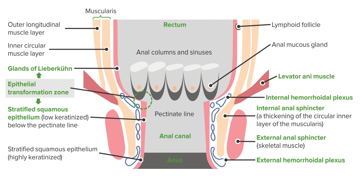
Anatomy:

  • Anal canal:
  • Anal margin (verge):
    • Where anal canal connects to the skin Skin The skin, also referred to as the integumentary system, is the largest organ of the body. The skin is primarily composed of the epidermis (outer layer) and dermis (deep layer). The epidermis is primarily composed of keratinocytes that undergo rapid turnover, while the dermis contains dense layers of connective tissue. Skin: Structure and Functions
    • Skin Skin The skin, also referred to as the integumentary system, is the largest organ of the body. The skin is primarily composed of the epidermis (outer layer) and dermis (deep layer). The epidermis is primarily composed of keratinocytes that undergo rapid turnover, while the dermis contains dense layers of connective tissue. Skin: Structure and Functions in the anal verge: perianal skin Skin The skin, also referred to as the integumentary system, is the largest organ of the body. The skin is primarily composed of the epidermis (outer layer) and dermis (deep layer). The epidermis is primarily composed of keratinocytes that undergo rapid turnover, while the dermis contains dense layers of connective tissue. Skin: Structure and Functions

Histology:

  • Types of mucosa (from proximal to distal area):
    • Glandular
    • Transitional
    • Nonkeratinizing squamous (devoid of epidermal appendages; above the dentate line Dentate line Rectum and Anal Canal: Anatomy)
    • Keratinizing squamous: in the anal canal below the dentate line Dentate line Rectum and Anal Canal: Anatomy, and in the perianal skin Skin The skin, also referred to as the integumentary system, is the largest organ of the body. The skin is primarily composed of the epidermis (outer layer) and dermis (deep layer). The epidermis is primarily composed of keratinocytes that undergo rapid turnover, while the dermis contains dense layers of connective tissue. Skin: Structure and Functions
  • Pectinate/ dentate line Dentate line Rectum and Anal Canal: Anatomy: area of transition from glandular to squamous mucosa
  • Clinical significance:
    • Tumors arising from the glandular area: adenocarcinoma
    • Tumors arising from the transitional and squamous mucosa: squamous cell carcinoma Squamous cell carcinoma Cutaneous squamous cell carcinoma (cSCC) is caused by malignant proliferation of atypical keratinocytes. This condition is the 2nd most common skin malignancy and usually affects sun-exposed areas of fair-skinned patients. The cancer presents as a firm, erythematous, keratotic plaque or papule. Squamous Cell Carcinoma (SCC) (SCC)
    • Tumors arising from the keratinizing squamous area and skin Skin The skin, also referred to as the integumentary system, is the largest organ of the body. The skin is primarily composed of the epidermis (outer layer) and dermis (deep layer). The epidermis is primarily composed of keratinocytes that undergo rapid turnover, while the dermis contains dense layers of connective tissue. Skin: Structure and Functions: perianal skin Skin The skin, also referred to as the integumentary system, is the largest organ of the body. The skin is primarily composed of the epidermis (outer layer) and dermis (deep layer). The epidermis is primarily composed of keratinocytes that undergo rapid turnover, while the dermis contains dense layers of connective tissue. Skin: Structure and Functions cancers, often SCC (same treatment approach as anal canal cancers)
Rectum and anal canal schematic

Anatomy of the anal canal

Image by Lecturio.

Epidemiology

  • 2.7% of all digestive tract cancers
  • Incidence Incidence The number of new cases of a given disease during a given period in a specified population. It also is used for the rate at which new events occur in a defined population. It is differentiated from prevalence, which refers to all cases in the population at a given time. Measures of Disease Frequency: 8,500 cases per year in the United States
  • Rising incidence Incidence The number of new cases of a given disease during a given period in a specified population. It also is used for the rate at which new events occur in a defined population. It is differentiated from prevalence, which refers to all cases in the population at a given time. Measures of Disease Frequency in young Black men and older (> 60) women
  • More common in women

Etiology

Risk factors for anal cancer:

  • Older age: > 50 years
  • Multiple sexual partners
  • Receptive anal intercourse Anal intercourse Hemorrhoids
  • History of vulvar, vaginal, or cervical cancers in women
  • Human papillomavirus Human papillomavirus Human papillomavirus (HPV) is a nonenveloped, circular, double-stranded DNA virus belonging to the Papillomaviridae family. Humans are the only reservoir, and transmission occurs through close skin-to-skin or sexual contact. Human papillomaviruses infect basal epithelial cells and can affect cell-regulatory proteins to result in cell proliferation. Papillomavirus (HPV) ( HPV HPV Human papillomavirus (HPV) is a nonenveloped, circular, double-stranded DNA virus belonging to the Papillomaviridae family. Humans are the only reservoir, and transmission occurs through close skin-to-skin or sexual contact. Human papillomaviruses infect basal epithelial cells and can affect cell-regulatory proteins to result in cell proliferation. Papillomavirus (HPV)): subclinical infection Subclinical infection Toxoplasma/Toxoplasmosis or condylomas
  • Herpes simplex Herpes Simplex A group of acute infections caused by herpes simplex virus type 1 or type 2 that is characterized by the development of one or more small fluid-filled vesicles with a raised erythematous base on the skin or mucous membrane. It occurs as a primary infection or recurs due to a reactivation of a latent infection. Congenital TORCH Infections virus Virus Viruses are infectious, obligate intracellular parasites composed of a nucleic acid core surrounded by a protein capsid. Viruses can be either naked (non-enveloped) or enveloped. The classification of viruses is complex and based on many factors, including type and structure of the nucleoid and capsid, the presence of an envelope, the replication cycle, and the host range. Virology infection ( HSV HSV Herpes simplex virus (HSV) is a double-stranded DNA virus belonging to the family Herpesviridae. Herpes simplex virus commonly causes recurrent infections involving the skin and mucosal surfaces, including the mouth, lips, eyes, and genitals. Herpes Simplex Virus 1 and 2)
  • Chlamydia Chlamydia Chlamydiae are obligate intracellular gram-negative bacteria. They lack a peptidoglycan layer and are best visualized using Giemsa stain. The family of Chlamydiaceae comprises 3 pathogens that can infect humans: Chlamydia trachomatis, Chlamydia psittaci, and Chlamydia pneumoniae. Chlamydia in women
  • Gonorrhea Gonorrhea Gonorrhea is a sexually transmitted infection (STI) caused by the gram-negative bacteria Neisseria gonorrhoeae (N. gonorrhoeae). Gonorrhea may be asymptomatic but commonly manifests as cervicitis or urethritis with less common presentations such as proctitis, conjunctivitis, or pharyngitis. Gonorrhea in men
  • Human immunodeficiency Immunodeficiency Chédiak-Higashi Syndrome virus Virus Viruses are infectious, obligate intracellular parasites composed of a nucleic acid core surrounded by a protein capsid. Viruses can be either naked (non-enveloped) or enveloped. The classification of viruses is complex and based on many factors, including type and structure of the nucleoid and capsid, the presence of an envelope, the replication cycle, and the host range. Virology ( HIV HIV Anti-HIV Drugs) infection
  • Immunosuppression (disease or medications)
  • Smoking Smoking Willful or deliberate act of inhaling and exhaling smoke from burning substances or agents held by hand. Interstitial Lung Diseases

Pathophysiology and Clinical Presentation

Pathophysiology

  • Carcinogenesis Carcinogenesis The origin, production or development of cancer through genotypic and phenotypic changes which upset the normal balance between cell proliferation and cell death. Carcinogenesis generally requires a constellation of steps, which may occur quickly or over a period of many years. Carcinogenesis of anal SCC:
    • Inflammatory response of the anal epithelium Epithelium The epithelium is a complex of specialized cellular organizations arranged into sheets and lining cavities and covering the surfaces of the body. The cells exhibit polarity, having an apical and a basal pole. Structures important for the epithelial integrity and function involve the basement membrane, the semipermeable sheet on which the cells rest, and interdigitations, as well as cellular junctions. Surface Epithelium: Histology develops most commonly as a response to infection by HPV HPV Human papillomavirus (HPV) is a nonenveloped, circular, double-stranded DNA virus belonging to the Papillomaviridae family. Humans are the only reservoir, and transmission occurs through close skin-to-skin or sexual contact. Human papillomaviruses infect basal epithelial cells and can affect cell-regulatory proteins to result in cell proliferation. Papillomavirus (HPV).
    • Inflammation Inflammation Inflammation is a complex set of responses to infection and injury involving leukocytes as the principal cellular mediators in the body’s defense against pathogenic organisms. Inflammation is also seen as a response to tissue injury in the process of wound healing. The 5 cardinal signs of inflammation are pain, heat, redness, swelling, and loss of function. Inflammation progresses to the development of anal intraepithelia neoplasia (AIN):
      • AIN 1: corresponds to low-grade squamous epithelial lesion (LSIL)
      • AIN 2 and 3: high-grade SIL (HSIL); pre-malignant
      • HSIL can progress to invasive SCC.
  • The progression to invasive cancer is promoted by HIV HIV Anti-HIV Drugs/immunosuppression and other risk factors (multiple HPV HPV Human papillomavirus (HPV) is a nonenveloped, circular, double-stranded DNA virus belonging to the Papillomaviridae family. Humans are the only reservoir, and transmission occurs through close skin-to-skin or sexual contact. Human papillomaviruses infect basal epithelial cells and can affect cell-regulatory proteins to result in cell proliferation. Papillomavirus (HPV) types, high-risk sexual behavior).

Histologic types

  • Anal canal: located above the anal verge
    • SCC:
    • Adenocarcinoma:
      • Rare
      • Arises from glandular elements in the anal canal
      • Natural history and treatment similar to rectal adenocarcinomas
    • Mucosal melanoma Melanoma Melanoma is a malignant tumor arising from melanocytes, the melanin-producing cells of the epidermis. These tumors are most common in fair-skinned individuals with a history of excessive sun exposure and sunburns. Melanoma:
      • 3rd-most common site for primary malignant melanoma Melanoma Melanoma is a malignant tumor arising from melanocytes, the melanin-producing cells of the epidermis. These tumors are most common in fair-skinned individuals with a history of excessive sun exposure and sunburns. Melanoma after skin Skin The skin, also referred to as the integumentary system, is the largest organ of the body. The skin is primarily composed of the epidermis (outer layer) and dermis (deep layer). The epidermis is primarily composed of keratinocytes that undergo rapid turnover, while the dermis contains dense layers of connective tissue. Skin: Structure and Functions and eyes 
      • Arises from melanocytes Melanocytes Mammalian pigment cells that produce melanins, pigments found mainly in the epidermis, but also in the eyes and the hair, by a process called melanogenesis. Coloration can be altered by the number of melanocytes or the amount of pigment produced and stored in the organelles called melanosomes. The large non-mammalian melanin-containing cells are called melanophores. Skin: Structure and Functions found in the transitional zone of the anal canal
      • Aggressive: distant metastases common at the time of diagnosis
  • Anal margin:below the anal verge (perianal skin Skin The skin, also referred to as the integumentary system, is the largest organ of the body. The skin is primarily composed of the epidermis (outer layer) and dermis (deep layer). The epidermis is primarily composed of keratinocytes that undergo rapid turnover, while the dermis contains dense layers of connective tissue. Skin: Structure and Functions cancers)
    • SCC
    • Bowen’s disease: SCC in situ (pre-malignant)
    • Paget’s disease of the anus (intraepithelial adenocarcinoma):
      • Primary: sweat gland morphology
      • Secondary: extension Extension Examination of the Upper Limbs from adenocarcinoma of the rectum Rectum The rectum and anal canal are the most terminal parts of the lower GI tract/large intestine that form a functional unit and control defecation. Fecal continence is maintained by several important anatomic structures including rectal folds, anal valves, the sling-like puborectalis muscle, and internal and external anal sphincters. Rectum and Anal Canal: Anatomy or perianal glands
    • Malignant melanoma Melanoma Melanoma is a malignant tumor arising from melanocytes, the melanin-producing cells of the epidermis. These tumors are most common in fair-skinned individuals with a history of excessive sun exposure and sunburns. Melanoma of anal margin: similar to other cutaneous melanomas

Lymphatic drainage of anal cancers

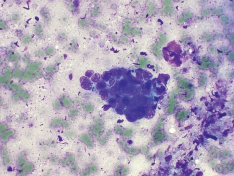
Squamous cell carcinoma of the anal canal

Squamous cell carcinoma of the anal canal:
Endoscopic US-guided fine-needle aspiration reveals poorly differentiated squamous cell carcinoma of the anal canal. Peripheral nuclear palisading cells are seen. (H&E staining, 40x)

Image: “f4-cmo-2009-059” by Leblanc J, Kongkam P. License: CC BY 3.0

Clinical presentation

  • Asymptomatic (25%)
  • Bleeding per rectum Rectum The rectum and anal canal are the most terminal parts of the lower GI tract/large intestine that form a functional unit and control defecation. Fecal continence is maintained by several important anatomic structures including rectal folds, anal valves, the sling-like puborectalis muscle, and internal and external anal sphincters. Rectum and Anal Canal: Anatomy (most common initial presentation)
  • A mass Mass Three-dimensional lesion that occupies a space within the breast Imaging of the Breast around the anus that may invade deeper tissues
  • Perianal ulceration Ulceration Corneal Abrasions, Erosion, and Ulcers and pruritus Pruritus An intense itching sensation that produces the urge to rub or scratch the skin to obtain relief. Atopic Dermatitis (Eczema)
  • Tenderness in the anal area
  • Fecal/flatus incontinence
  • Obstipation Obstipation Large Bowel Obstruction
  • Thrombosed hemorrhoid Thrombosed hemorrhoid Hemorrhoids (a possible manifestation of anal melanoma Melanoma Melanoma is a malignant tumor arising from melanocytes, the melanin-producing cells of the epidermis. These tumors are most common in fair-skinned individuals with a history of excessive sun exposure and sunburns. Melanoma)

Diagnosis and Staging

Clinical findings

  • History:
    • Typical clinical presentation
    • History of sexually transmitted diseases and other risk factors
  • Physical examination:
    • Large tumors or anal margin tumors may be directly visualized.
    • Digital rectal exam ( DRE DRE A physical examination in which the qualified health care worker inserts a lubricated, gloved finger of one hand into the rectum and may use the other hand to press on the lower abdomen or pelvic area to palpate for abnormalities in the lower rectum, and nearby organs or tissues. The method is commonly used to check the lower rectum, the prostate gland in men, and the uterus and ovaries in women. Prostate Cancer Screening): may feel hard, immovable, non-tender lump 
    • Anoscopy Anoscopy Anal Fissure: If a mass Mass Three-dimensional lesion that occupies a space within the breast Imaging of the Breast is seen, biopsy Biopsy Removal and pathologic examination of specimens from the living body. Ewing Sarcoma can be performed.
    • Inguinal lymphadenopathy Inguinal Lymphadenopathy Lymphadenopathy
    • Gynecologic exam should be performed in women (looking for concomitant genital lesions/ condylomas).

Diagnostic tests Diagnostic tests Diagnostic tests are important aspects in making a diagnosis. Some of the most important epidemiological values of diagnostic tests include sensitivity and specificity, false positives and false negatives, positive and negative predictive values, likelihood ratios, and pre-test and post-test probabilities. Epidemiological Values of Diagnostic Tests

  • Biopsy Biopsy Removal and pathologic examination of specimens from the living body. Ewing Sarcoma:
  • Laboratory studies:
    • Complete blood cell count (CBC) may show anemia Anemia Anemia is a condition in which individuals have low Hb levels, which can arise from various causes. Anemia is accompanied by a reduced number of RBCs and may manifest with fatigue, shortness of breath, pallor, and weakness. Subtypes are classified by the size of RBCs, chronicity, and etiology. Anemia: Overview and Types.
    • HIV HIV Anti-HIV Drugs, HPV HPV Human papillomavirus (HPV) is a nonenveloped, circular, double-stranded DNA virus belonging to the Papillomaviridae family. Humans are the only reservoir, and transmission occurs through close skin-to-skin or sexual contact. Human papillomaviruses infect basal epithelial cells and can affect cell-regulatory proteins to result in cell proliferation. Papillomavirus (HPV), and HSV HSV Herpes simplex virus (HSV) is a double-stranded DNA virus belonging to the family Herpesviridae. Herpes simplex virus commonly causes recurrent infections involving the skin and mucosal surfaces, including the mouth, lips, eyes, and genitals. Herpes Simplex Virus 1 and 2 testing should be considered depending on individual history.
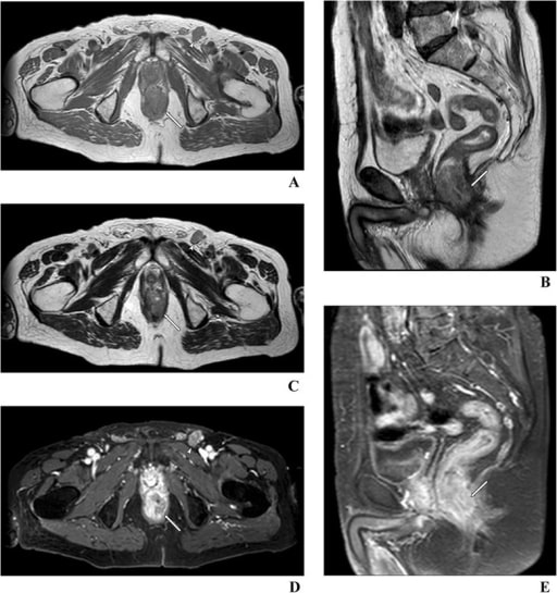
  • Imaging for diagnosis and staging Staging Methods which attempt to express in replicable terms the extent of the neoplasm in the patient. Grading, Staging, and Metastasis evaluation:
    • Factors evaluated:
    • The following are utilized to determine the stage of disease:
      • Endoscopic ultrasound (EUS)
      • Computed tomography (CT) scan
      • Magnetic resonance imaging (MRI)
      • Positron-emission tomography ( PET PET An imaging technique that combines a positron-emission tomography (PET) scanner and a ct X ray scanner. This establishes a precise anatomic localization in the same session. Nuclear Imaging)

Staging Staging Methods which attempt to express in replicable terms the extent of the neoplasm in the patient. Grading, Staging, and Metastasis

Table: Anal cancer TNM staging Staging Methods which attempt to express in replicable terms the extent of the neoplasm in the patient. Grading, Staging, and Metastasis
Primary tumor Tumor Inflammation (T) Regional lymph nodes Lymph Nodes They are oval or bean shaped bodies (1 – 30 mm in diameter) located along the lymphatic system. Lymphatic Drainage System: Anatomy (N) Distant metastasis Metastasis The transfer of a neoplasm from one organ or part of the body to another remote from the primary site. Grading, Staging, and Metastasis (M)
Tx: primary tumor Tumor Inflammation not assessed Nx: regional lymph nodes Lymph Nodes They are oval or bean shaped bodies (1 – 30 mm in diameter) located along the lymphatic system. Lymphatic Drainage System: Anatomy cannot be assessed M0: no distant metastasis Metastasis The transfer of a neoplasm from one organ or part of the body to another remote from the primary site. Grading, Staging, and Metastasis
T0: no evidence of primary tumor Tumor Inflammation N0: no regional lymph Lymph The interstitial fluid that is in the lymphatic system. Secondary Lymphatic Organs node metastasis Metastasis The transfer of a neoplasm from one organ or part of the body to another remote from the primary site. Grading, Staging, and Metastasis M1: distant metastasis Metastasis The transfer of a neoplasm from one organ or part of the body to another remote from the primary site. Grading, Staging, and Metastasis
Tis: high-grade squamous intraepithelial lesion N1: metastasis Metastasis The transfer of a neoplasm from one organ or part of the body to another remote from the primary site. Grading, Staging, and Metastasis in inguinal, mesorectal, internal iliac, or external iliac nodes
T1: tumor Tumor Inflammation ≤ 2 cm
T2: tumor Tumor Inflammation > 2 cm but ≤ 5 cm
T3 T3 A T3 thyroid hormone normally synthesized and secreted by the thyroid gland in much smaller quantities than thyroxine (T4). Most T3 is derived from peripheral monodeiodination of T4 at the 5′ position of the outer ring of the iodothyronine nucleus. The hormone finally delivered and used by the tissues is mainly t3. Thyroid Hormones: tumor Tumor Inflammation > 5 cm
T4 T4 The major hormone derived from the thyroid gland. Thyroxine is synthesized via the iodination of tyrosines (monoiodotyrosine) and the coupling of iodotyrosines (diiodotyrosine) in the thyroglobulin. Thyroxine is released from thyroglobulin by proteolysis and secreted into the blood. Thyroxine is peripherally deiodinated to form triiodothyronine which exerts a broad spectrum of stimulatory effects on cell metabolism. Thyroid Hormones: tumor Tumor Inflammation of any size invading adjacent organs, such as the vagina Vagina The vagina is the female genital canal, extending from the vulva externally to the cervix uteri internally. The structures have sexual, reproductive, and urinary functions and a rich blood supply, mainly arising from the internal iliac artery. Vagina, Vulva, and Pelvic Floor: Anatomy, urethra Urethra A tube that transports urine from the urinary bladder to the outside of the body in both the sexes. It also has a reproductive function in the male by providing a passage for sperm. Urinary Tract: Anatomy, or bladder Bladder A musculomembranous sac along the urinary tract. Urine flows from the kidneys into the bladder via the ureters, and is held there until urination. Pyelonephritis and Perinephric Abscess
Source: American Joint Commitee on Cancer (AJCC), 8th edition
Table: Stages of anal cancer
Stage T N M
0 Tis N0 M0
I T1 N0 M0
IIA T2 N0 M0
IIB T3 T3 A T3 thyroid hormone normally synthesized and secreted by the thyroid gland in much smaller quantities than thyroxine (T4). Most T3 is derived from peripheral monodeiodination of T4 at the 5′ position of the outer ring of the iodothyronine nucleus. The hormone finally delivered and used by the tissues is mainly t3. Thyroid Hormones N0 M0
IIIA T1–2 N1 M0
IIIB T4 T4 The major hormone derived from the thyroid gland. Thyroxine is synthesized via the iodination of tyrosines (monoiodotyrosine) and the coupling of iodotyrosines (diiodotyrosine) in the thyroglobulin. Thyroxine is released from thyroglobulin by proteolysis and secreted into the blood. Thyroxine is peripherally deiodinated to form triiodothyronine which exerts a broad spectrum of stimulatory effects on cell metabolism. Thyroid Hormones N0 M0
IIIC T3 T3 A T3 thyroid hormone normally synthesized and secreted by the thyroid gland in much smaller quantities than thyroxine (T4). Most T3 is derived from peripheral monodeiodination of T4 at the 5′ position of the outer ring of the iodothyronine nucleus. The hormone finally delivered and used by the tissues is mainly t3. Thyroid Hormones–4 N1 M0
IV Any T Any N M1

Management

Management of SCC

  • Localized (stage IIII):
    • Chemoradiotherapy:
      • Preferred; provides cure and preserves anal sphincter
      • Fluorouracil Fluorouracil A pyrimidine analog that is an antineoplastic antimetabolite. It interferes with DNA synthesis by blocking the thymidylate synthetase conversion of deoxyuridylic acid to thymidylic acid. Bowen Disease and Erythroplasia of Queyrat + mitomycin Mitomycin A group of methylazirinopyrroloindolediones obtained from certain streptomyces strains. They are very toxic antibiotics used as antineoplastic agents in some solid tumors. Porfiromycin and mitomycin are the most useful members of the group. Antitumor Antibiotics + radiotherapy
    • Local excision:
      • For small (< 1 cm) superficial tumors
      • < 3 mm MM Multiple myeloma (MM) is a malignant condition of plasma cells (activated B lymphocytes) primarily seen in the elderly. Monoclonal proliferation of plasma cells results in cytokine-driven osteoclastic activity and excessive secretion of IgG antibodies. Multiple Myeloma basement membrane Basement membrane A darkly stained mat-like extracellular matrix (ecm) that separates cell layers, such as epithelium from endothelium or a layer of connective tissue. The ecm layer that supports an overlying epithelium or endothelium is called basal lamina. Basement membrane (bm) can be formed by the fusion of either two adjacent basal laminae or a basal lamina with an adjacent reticular lamina of connective tissue. Bm, composed mainly of type IV collagen; glycoprotein laminin; and proteoglycan, provides barriers as well as channels between interacting cell layers. Thin Basement Membrane Nephropathy (TBMN) invasion
      • < 7 mm MM Multiple myeloma (MM) is a malignant condition of plasma cells (activated B lymphocytes) primarily seen in the elderly. Monoclonal proliferation of plasma cells results in cytokine-driven osteoclastic activity and excessive secretion of IgG antibodies. Multiple Myeloma horizontal spread
    • Inguinal lymphadenectomy:
      • For persistent/recurrent inguinal lymph Lymph The interstitial fluid that is in the lymphatic system. Secondary Lymphatic Organs node disease after chemoradiation
      • Additional radiation Radiation Emission or propagation of acoustic waves (sound), electromagnetic energy waves (such as light; radio waves; gamma rays; or x-rays), or a stream of subatomic particles (such as electrons; neutrons; protons; or alpha particles). Osteosarcoma to inguinal region Inguinal region Anterior Abdominal Wall: Anatomy also considered
  • Stage IV (metastatic diseases): 
    • Systemic chemotherapy Chemotherapy Osteosarcoma
    • Immunotherapy ( pembrolizumab Pembrolizumab Squamous Cell Carcinoma (SCC), nivolumab Nivolumab A genetically engineered, fully humanized immunoglobulin g4 monoclonal antibody that binds to the pd-1 receptor, activating an immune response to tumor cells. It is used as monotherapy or in combination with ipilimumab for the treatment of advanced malignant melanoma. It is also used in the treatment of advanced or recurring non-small cell lung cancer; renal cell carcinoma; and Hodgkin’s lymphoma. Melanoma)
    • Liver Liver The liver is the largest gland in the human body. The liver is found in the superior right quadrant of the abdomen and weighs approximately 1.5 kilograms. Its main functions are detoxification, metabolism, nutrient storage (e.g., iron and vitamins), synthesis of coagulation factors, formation of bile, filtration, and storage of blood. Liver: Anatomy resection may play a role in patients Patients Individuals participating in the health care system for the purpose of receiving therapeutic, diagnostic, or preventive procedures. Clinician–Patient Relationship with localized liver Liver The liver is the largest gland in the human body. The liver is found in the superior right quadrant of the abdomen and weighs approximately 1.5 kilograms. Its main functions are detoxification, metabolism, nutrient storage (e.g., iron and vitamins), synthesis of coagulation factors, formation of bile, filtration, and storage of blood. Liver: Anatomy metastases.
  • Recurrent/persistent disease after chemoradiotherapy: 
    • Needs to be confirmed with biopsy Biopsy Removal and pathologic examination of specimens from the living body. Ewing Sarcoma
    • Surgery (abdominoperineal resection (APR)): Patients Patients Individuals participating in the health care system for the purpose of receiving therapeutic, diagnostic, or preventive procedures. Clinician–Patient Relationship end up with a permanent colostomy.
    • Salvage chemoradiotherapy can be considered to avoid colostomy.
  • Surveillance Surveillance Developmental Milestones and Normal Growth:
    • DRE DRE A physical examination in which the qualified health care worker inserts a lubricated, gloved finger of one hand into the rectum and may use the other hand to press on the lower abdomen or pelvic area to palpate for abnormalities in the lower rectum, and nearby organs or tissues. The method is commonly used to check the lower rectum, the prostate gland in men, and the uterus and ovaries in women. Prostate Cancer Screening, anoscopy Anoscopy Anal Fissure, and inguinal lymph Lymph The interstitial fluid that is in the lymphatic system. Secondary Lymphatic Organs node palpation Palpation Application of fingers with light pressure to the surface of the body to determine consistency of parts beneath in physical diagnosis; includes palpation for determining the outlines of organs. Dermatologic Examination:
      • Starts 8–12 weeks post-therapy
      • Every 3–6 months for 5 years
      • It may take up to 26 weeks post-treatment to show complete response.
    • CT scan or MRI of chest/abdomen/ pelvis Pelvis The pelvis consists of the bony pelvic girdle, the muscular and ligamentous pelvic floor, and the pelvic cavity, which contains viscera, vessels, and multiple nerves and muscles. The pelvic girdle, composed of 2 “hip” bones and the sacrum, is a ring-like bony structure of the axial skeleton that links the vertebral column with the lower extremities. Pelvis: Anatomy: Consider annually for 3 years.

Management of other cancer types

  • Adenocarcinoma:
    • Approach similar to rectal adenocarcinomas
    • APR or local excision for small/early tumors
    • Systemic chemotherapy Chemotherapy Osteosarcoma for metastatic disease
  • Melanoma Melanoma Melanoma is a malignant tumor arising from melanocytes, the melanin-producing cells of the epidermis. These tumors are most common in fair-skinned individuals with a history of excessive sun exposure and sunburns. Melanoma:
    • Local excision with adjuvant Adjuvant Substances that augment, stimulate, activate, potentiate, or modulate the immune response at either the cellular or humoral level. The classical agents (freund’s adjuvant, bcg, corynebacterium parvum, et al.) contain bacterial antigens. Some are endogenous (e.g., histamine, interferon, transfer factor, tuftsin, interleukin-1). Their mode of action is either non-specific, resulting in increased immune responsiveness to a wide variety of antigens, or antigen-specific, i.e., affecting a restricted type of immune response to a narrow group of antigens. The therapeutic efficacy of many biological response modifiers is related to their antigen-specific immunoadjuvanticity. Vaccination radiation Radiation Emission or propagation of acoustic waves (sound), electromagnetic energy waves (such as light; radio waves; gamma rays; or x-rays), or a stream of subatomic particles (such as electrons; neutrons; protons; or alpha particles). Osteosarcoma
    • APR for more advanced disease
    • Systemic chemotherapy Chemotherapy Osteosarcoma for metastatic disease

Prognosis and Complications

Prognosis Prognosis A prediction of the probable outcome of a disease based on a individual’s condition and the usual course of the disease as seen in similar situations. Non-Hodgkin Lymphomas

  • 5-year survival of SCC:
    • T1: 86%
    • T2: 86%
    • T3 T3 A T3 thyroid hormone normally synthesized and secreted by the thyroid gland in much smaller quantities than thyroxine (T4). Most T3 is derived from peripheral monodeiodination of T4 at the 5′ position of the outer ring of the iodothyronine nucleus. The hormone finally delivered and used by the tissues is mainly t3. Thyroid Hormones: 60%
    • T4 T4 The major hormone derived from the thyroid gland. Thyroxine is synthesized via the iodination of tyrosines (monoiodotyrosine) and the coupling of iodotyrosines (diiodotyrosine) in the thyroglobulin. Thyroxine is released from thyroglobulin by proteolysis and secreted into the blood. Thyroxine is peripherally deiodinated to form triiodothyronine which exerts a broad spectrum of stimulatory effects on cell metabolism. Thyroid Hormones: 45%
    • N0: 76%
    • Node-positive: 54%
    • For all stages with complete response to chemo/ radiation Radiation Emission or propagation of acoustic waves (sound), electromagnetic energy waves (such as light; radio waves; gamma rays; or x-rays), or a stream of subatomic particles (such as electrons; neutrons; protons; or alpha particles). Osteosarcoma (70%–80% of patients Patients Individuals participating in the health care system for the purpose of receiving therapeutic, diagnostic, or preventive procedures. Clinician–Patient Relationship): 65%
  • 5-year survival of adenocarcinoma: 55% when treated surgically with no distant metastases
  • 5-year survival of melanoma Melanoma Melanoma is a malignant tumor arising from melanocytes, the melanin-producing cells of the epidermis. These tumors are most common in fair-skinned individuals with a history of excessive sun exposure and sunburns. Melanoma:

Complications

  • Usually related to treatment
  • Chemotherapy Chemotherapy Osteosarcoma:
    •  Bowel dysfunction
    •   Nausea Nausea An unpleasant sensation in the stomach usually accompanied by the urge to vomit. Common causes are early pregnancy, sea and motion sickness, emotional stress, intense pain, food poisoning, and various enteroviruses. Antiemetics/ vomiting Vomiting The forcible expulsion of the contents of the stomach through the mouth. Hypokalemia
  • Radiation Radiation Emission or propagation of acoustic waves (sound), electromagnetic energy waves (such as light; radio waves; gamma rays; or x-rays), or a stream of subatomic particles (such as electrons; neutrons; protons; or alpha particles). Osteosarcoma:
    • Radiation Radiation Emission or propagation of acoustic waves (sound), electromagnetic energy waves (such as light; radio waves; gamma rays; or x-rays), or a stream of subatomic particles (such as electrons; neutrons; protons; or alpha particles). Osteosarcoma proctitis Proctitis Inflammation of the mucous membrane of the rectum, the distal end of the large intestine. Chronic Granulomatous Disease ( pain Pain An unpleasant sensation induced by noxious stimuli which are detected by nerve endings of nociceptive neurons. Pain: Types and Pathways, rectal bleeding, diarrhea Diarrhea Diarrhea is defined as ≥ 3 watery or loose stools in a 24-hour period. There are a multitude of etiologies, which can be classified based on the underlying mechanism of disease. The duration of symptoms (acute or chronic) and characteristics of the stools (e.g., watery, bloody, steatorrheic, mucoid) can help guide further diagnostic evaluation. Diarrhea)
    • Sexual dysfunction Sexual dysfunction Physiological disturbances in normal sexual performance in either the male or the female. Sexual Physiology (men and women)
  • Surgery:
    • Strictures
    • Perianal fistulas
  • Wound infections Infections Invasion of the host organism by microorganisms or their toxins or by parasites that can cause pathological conditions or diseases. Chronic Granulomatous Disease

Differential Diagnosis

  • Chancroid Chancroid Chancroid is a highly transmissible STD caused by Haemophilus ducreyi. The disease presents with painful ulcer(s) on the genital tract (termed chancroid or “soft chancre”). Up to 50% of patients will develop painful inguinal lymphadenopathy. Chancroid: bacterial infection that causes open sores around the genitals. Caused by Haemophilus ducreyi Haemophilus ducreyi A species of Haemophilus that appears to be the pathogen or causative agent of the sexually transmitted disease, chancroid. Haemophilus. Chancroid Chancroid Chancroid is a highly transmissible STD caused by Haemophilus ducreyi. The disease presents with painful ulcer(s) on the genital tract (termed chancroid or “soft chancre”). Up to 50% of patients will develop painful inguinal lymphadenopathy. Chancroid is a sexually transmitted disease Sexually Transmitted Disease Sexually transmitted diseases (STDs) are infections that spread either by vaginal intercourse, anal sex, or oral sex. Symptoms and signs may include vaginal discharge, penile discharge, dysuria, skin lesions (e.g., warts, ulcers) on or around the genitals, and pelvic pain. Some infections can lead to infertility and chronic debilitating disease. Sexually Transmitted Infections (STIs) that can also spread through skin-to-skin contact with an infected person.
  • Anal fissure Fissure A crack or split that extends into the dermis Generalized and Localized Rashes or fistula Fistula Abnormal communication most commonly seen between two internal organs, or between an internal organ and the surface of the body. Anal Fistula: a fissure Fissure A crack or split that extends into the dermis Generalized and Localized Rashes is a small tear in the anal mucosa. The most common presenting symptom is anal pain Pain An unpleasant sensation induced by noxious stimuli which are detected by nerve endings of nociceptive neurons. Pain: Types and Pathways. A fistula Fistula Abnormal communication most commonly seen between two internal organs, or between an internal organ and the surface of the body. Anal Fistula is an abnormal connection between anal/rectal mucosa and perianal skin Skin The skin, also referred to as the integumentary system, is the largest organ of the body. The skin is primarily composed of the epidermis (outer layer) and dermis (deep layer). The epidermis is primarily composed of keratinocytes that undergo rapid turnover, while the dermis contains dense layers of connective tissue. Skin: Structure and Functions. The condition is usually a consequence of recurrent/chronic infection, and one of the manifestations of Crohn’s disease.
  • Hemorrhoids Hemorrhoids Hemorrhoids are normal vascular cushions in the anal canal composed of dilated vascular tissue, smooth muscle, and connective tissue. They do not cause issues unless they are enlarged, inflamed, thrombosed, or prolapsed. Patients often present with rectal bleeding of bright red blood, or they may have pain, perianal pruritus, or a palpable mass. Hemorrhoids: engorged/ varicose veins Varicose veins Enlarged and tortuous veins. Chronic Venous Insufficiency of the anal canal and lower rectum Rectum The rectum and anal canal are the most terminal parts of the lower GI tract/large intestine that form a functional unit and control defecation. Fecal continence is maintained by several important anatomic structures including rectal folds, anal valves, the sling-like puborectalis muscle, and internal and external anal sphincters. Rectum and Anal Canal: Anatomy. Can present as a bluish anal mass Mass Three-dimensional lesion that occupies a space within the breast Imaging of the Breast Hemorrhoids Hemorrhoids Hemorrhoids are normal vascular cushions in the anal canal composed of dilated vascular tissue, smooth muscle, and connective tissue. They do not cause issues unless they are enlarged, inflamed, thrombosed, or prolapsed. Patients often present with rectal bleeding of bright red blood, or they may have pain, perianal pruritus, or a palpable mass. Hemorrhoids can be painful (if they originate below the dentate line Dentate line Rectum and Anal Canal: Anatomy) or painless (above the dentate line Dentate line Rectum and Anal Canal: Anatomy).
  • Condylomata acuminatum: also referred to as anogenital warts Warts Benign epidermal proliferations or tumors; some are viral in origin. Female Genitourinary Examination; caused by HPV HPV Human papillomavirus (HPV) is a nonenveloped, circular, double-stranded DNA virus belonging to the Papillomaviridae family. Humans are the only reservoir, and transmission occurs through close skin-to-skin or sexual contact. Human papillomaviruses infect basal epithelial cells and can affect cell-regulatory proteins to result in cell proliferation. Papillomavirus (HPV) infection. Usually present as soft, skin-colored, fleshy lesions around the anus and genitals.
  • Psoriasis Psoriasis Psoriasis is a common T-cell-mediated inflammatory skin condition. The etiology is unknown, but is thought to be due to genetic inheritance and environmental triggers. There are 4 major subtypes, with the most common form being chronic plaque psoriasis. Psoriasis: usually presents as red or purple, typically non-scaly pruritic lesions. May be confused with yeast Yeast A general term for single-celled rounded fungi that reproduce by budding. Brewers’ and bakers’ yeasts are saccharomyces cerevisiae; therapeutic dried yeast is yeast, dried. Mycology or parasitic infection. Can also cause rectal bleeding and pain Pain An unpleasant sensation induced by noxious stimuli which are detected by nerve endings of nociceptive neurons. Pain: Types and Pathways with defecation Defecation The normal process of elimination of fecal material from the rectum. Gastrointestinal Motility.

References

  1. Babiker, H.M, Kashyap S, Mehta S.R. et al. (2020). Anal Cancer. https://www.ncbi.nlm.nih.gov/books/NBK441891/
  2. Parra R.S., Ribeiro de Almeida A.L, Badiale, G.B. et al. (2010). Melanoma of the anal canal. Retrieved November 18 2020, from DOI: 10.1590/s1807-59322010001000026
  3. Ryan D.P., Willett C.G. (2020). Classification and epidemiology of anal cancer. Retrieved 21 November 2020, from https://www.uptodate.com/contents/classification-and-epidemiology-of-anal-cancer?search=anal%20cancer&source=search_result&selectedTitle=3~124&usage_type=default&display_rank=3
  4. Ryan D.P., Willett C.G. (2020). Treatment of anal cancer. Retrieved 21 November 2020, from https://www.uptodate.com/contents/treatment-of-anal-cancer?search=anal%20cancer&source=search_result&selectedTitle=1~124&usage_type=default&display_rank=1
  5. Ryan D.P., Willett C.G. (2020). Clinical features and staging of anal cancer. Retrieved 21 November 2020, from https://www.uptodate.com/contents/clinical-features-and-staging-of-anal-cancer?search=anal%20cancer&source=search_result&selectedTitle=2~124&usage_type=default&display_rank=2
  6. Wegner R.E., White R.J, Hasan S. et al. (2019). Anal Adenocarcinoma: Treatment outcomes and trends in a rare disease entity. onlinelibrary.wiley.com/doi/full/10.1002/cam4.2076

Create your free account or log in to continue reading!

Sign up now and get free access to Lecturio with concept pages, medical videos, and questions for your medical education.

User Reviews

Unwrap New Skills This Holiday 🎄 Save 30% on all plans now!

Details