Advertisement

Advertisement

Advertisement

Advertisement

Adrenal Glands: Anatomy

The adrenal glands are a pair of retroperitoneal Retroperitoneal Peritoneum: Anatomy endocrine glands Endocrine glands Ductless glands that secrete hormones directly into the blood circulation. These hormones influence the metabolism and other functions of cells in the body. Glandular Epithelium: Histology located above the kidneys Kidneys The kidneys are a pair of bean-shaped organs located retroperitoneally against the posterior wall of the abdomen on either side of the spine. As part of the urinary tract, the kidneys are responsible for blood filtration and excretion of water-soluble waste in the urine. Kidneys: Anatomy. The outer parenchyma is called the adrenal cortex and has 3 distinct zones, each with its own secretory products. The outer zona glomerulosa secretes mineralocorticoids Mineralocorticoids Mineralocorticoids are a drug class within the corticosteroid family and fludrocortisone is the primary medication within this class. Fludrocortisone is a fluorinated analog of cortisone. The fluorine moiety protects the drug from isoenzyme inactivation in the kidney, allowing it to exert its mineralocorticoid effect. Mineralocorticoids (primarily aldosterone Aldosterone A hormone secreted by the adrenal cortex that regulates electrolyte and water balance by increasing the renal retention of sodium and the excretion of potassium. Hyperkalemia); the middle zona fasciculata secretes glucocorticoids Glucocorticoids Glucocorticoids are a class within the corticosteroid family. Glucocorticoids are chemically and functionally similar to endogenous cortisol. There are a wide array of indications, which primarily benefit from the antiinflammatory and immunosuppressive effects of this class of drugs. Glucocorticoids (primarily cortisol Cortisol Glucocorticoids); and the innermost zona reticularis secretes androgens Androgens Androgens are naturally occurring steroid hormones responsible for development and maintenance of the male sex characteristics, including penile, scrotal, and clitoral growth, development of sexual hair, deepening of the voice, and musculoskeletal growth. Androgens and Antiandrogens. Beneath the cortex lies the adrenal medulla, which secretes catecholamines Catecholamines A general class of ortho-dihydroxyphenylalkylamines derived from tyrosine. Adrenal Hormones involved in the fight-or-flight response. The medullary cells function more like neurosecretory postsynaptic neurons Postsynaptic neurons Autonomic Nervous System: Anatomy rather than traditional endocrine cells. The adrenal glands have a rich vascular supply and complex relationships with their surrounding organs.

Last updated: Nov 19, 2024

Editorial responsibility: Stanley Oiseth, Lindsay Jones, Evelin Maza

Advertisement

Advertisement

Advertisement

Advertisement

Advertisement

Advertisement

Overview

Location

There are 2 adrenal glands in the body and each gland is in close association with a kidney. The adrenal glands are:
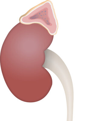
  • Retroperitoneal Retroperitoneal Peritoneum: Anatomy structures
  • Located on the superomedial aspect of each kidney
  • Within the renal fascia Fascia Layers of connective tissue of variable thickness. The superficial fascia is found immediately below the skin; the deep fascia invests muscles, nerves, and other organs. Cellulitis and perirenal fat
  • Separated from the kidneys Kidneys The kidneys are a pair of bean-shaped organs located retroperitoneally against the posterior wall of the abdomen on either side of the spine. As part of the urinary tract, the kidneys are responsible for blood filtration and excretion of water-soluble waste in the urine. Kidneys: Anatomy by a fibrous Fibrous Fibrocystic Change tissue
Adrenal gland

Adrenal gland located on the superior medial aspect of each kidney

Image by Lecturio.

Borders/anatomic relations

  • Inferior to both glands is the kidney.
  • Right adrenal gland:
    • Anteromedial: inferior vena cava Inferior vena cava The venous trunk which receives blood from the lower extremities and from the pelvic and abdominal organs. Mediastinum and Great Vessels: Anatomy
    • Lateral: liver Liver The liver is the largest gland in the human body. The liver is found in the superior right quadrant of the abdomen and weighs approximately 1.5 kilograms. Its main functions are detoxification, metabolism, nutrient storage (e.g., iron and vitamins), synthesis of coagulation factors, formation of bile, filtration, and storage of blood. Liver: Anatomy
    • Posterior: diaphragm Diaphragm The diaphragm is a large, dome-shaped muscle that separates the thoracic cavity from the abdominal cavity. The diaphragm consists of muscle fibers and a large central tendon, which is divided into right and left parts. As the primary muscle of inspiration, the diaphragm contributes 75% of the total inspiratory muscle force. Diaphragm: Anatomy
  • Left adrenal gland:
    • Anteriorly:
      • Tail of the pancreas Pancreas The pancreas lies mostly posterior to the stomach and extends across the posterior abdominal wall from the duodenum on the right to the spleen on the left. This organ has both exocrine and endocrine tissue. Pancreas: Anatomy
      • Spleen Spleen The spleen is the largest lymphoid organ in the body, located in the LUQ of the abdomen, superior to the left kidney and posterior to the stomach at the level of the 9th-11th ribs just below the diaphragm. The spleen is highly vascular and acts as an important blood filter, cleansing the blood of pathogens and damaged erythrocytes. Spleen: Anatomy
      • Stomach Stomach The stomach is a muscular sac in the upper left portion of the abdomen that plays a critical role in digestion. The stomach develops from the foregut and connects the esophagus with the duodenum. Structurally, the stomach is C-shaped and forms a greater and lesser curvature and is divided grossly into regions: the cardia, fundus, body, and pylorus. Stomach: Anatomy
    • Medially: abdominal aorta Abdominal Aorta The aorta from the diaphragm to the bifurcation into the right and left common iliac arteries. Posterior Abdominal Wall: Anatomy
    • Inferiorly: splenic vein and artery
    • Posterior: diaphragm Diaphragm The diaphragm is a large, dome-shaped muscle that separates the thoracic cavity from the abdominal cavity. The diaphragm consists of muscle fibers and a large central tendon, which is divided into right and left parts. As the primary muscle of inspiration, the diaphragm contributes 75% of the total inspiratory muscle force. Diaphragm: Anatomy

Development

  • Adrenal cortex:
    • Derives from the mesoderm Mesoderm The middle germ layer of an embryo derived from three paired mesenchymal aggregates along the neural tube. Gastrulation and Neurulation
    • The primitive fetal cortex develops from the mesothelial cells on the dorsal wall of the primitive coelom.
    • Fetal cortex:
      • Actively produces steroids Steroids A group of polycyclic compounds closely related biochemically to terpenes. They include cholesterol, numerous hormones, precursors of certain vitamins, bile acids, alcohols (sterols), and certain natural drugs and poisons. Steroids have a common nucleus, a fused, reduced 17-carbon atom ring system, cyclopentanoperhydrophenanthrene. Most steroids also have two methyl groups and an aliphatic side-chain attached to the nucleus. Benign Liver Tumors during gestation
      • Involutes after birth
    • Definitive cortex (with mature cortical zones) develops slowly after birth.
  • Adrenal medulla:
  • Changes in size:
    • At birth: The adrenal gland is ⅓ the size of the kidney.
    • Adulthood: The adrenal gland is 1/30th the size of the kidney.

Gross Anatomy

General structure

The adrenal glands are composed of 2 functionally separate layers: the adrenal cortex and the adrenal medulla. Each layer has its own embryologic origins, anatomy, histology, and functions.

  • Right: triangular in shape
  • Left: semilunar in shape

Adrenal cortex

Adrenal medulla

  • Central portion of the gland
  • Only about 10%‒20% of the gland by volume
  • Red-brown in color

Microscopic Anatomy

Adrenal cortex

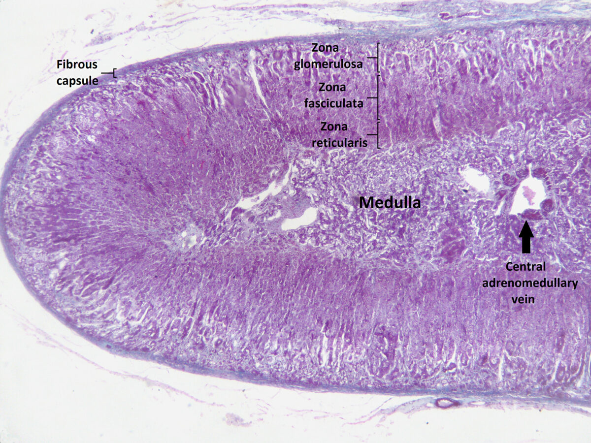
The adrenal cortex has 3 distinct zones, and each zone has its own regulation and secretory products. These 3 zones (from external to internal) are:
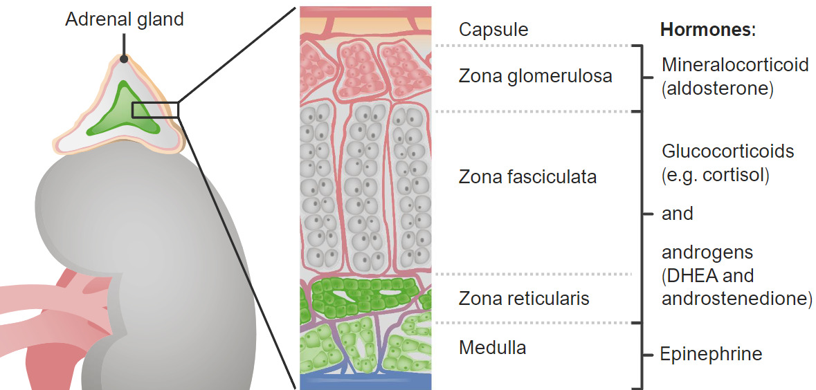
  • Zona glomerulosa:
    • Secretes mineralocorticoids Mineralocorticoids Mineralocorticoids are a drug class within the corticosteroid family and fludrocortisone is the primary medication within this class. Fludrocortisone is a fluorinated analog of cortisone. The fluorine moiety protects the drug from isoenzyme inactivation in the kidney, allowing it to exert its mineralocorticoid effect. Mineralocorticoids (mainly aldosterone Aldosterone A hormone secreted by the adrenal cortex that regulates electrolyte and water balance by increasing the renal retention of sodium and the excretion of potassium. Hyperkalemia)
    • Cells are arranged in circular clusters.
  • Zona fasciculata:
    • Secretes glucocorticoids Glucocorticoids Glucocorticoids are a class within the corticosteroid family. Glucocorticoids are chemically and functionally similar to endogenous cortisol. There are a wide array of indications, which primarily benefit from the antiinflammatory and immunosuppressive effects of this class of drugs. Glucocorticoids (mainly cortisol Cortisol Glucocorticoids)
    • Cells are arranged in columns.
  • Zona reticularis:
    • Secretes androgens Androgens Androgens are naturally occurring steroid hormones responsible for development and maintenance of the male sex characteristics, including penile, scrotal, and clitoral growth, development of sexual hair, deepening of the voice, and musculoskeletal growth. Androgens and Antiandrogens (e.g., dehydroepiandrosterone Dehydroepiandrosterone A major C19 steroid produced by the adrenal cortex. It is also produced in small quantities in the testis and the ovary. Dehydroepiandrosterone (DHEA) can be converted to testosterone; androstenedione; estradiol; and estrone. Androgens and Antiandrogens ( DHEA DHEA A major C19 steroid produced by the adrenal cortex. It is also produced in small quantities in the testis and the ovary. Dehydroepiandrosterone (dhea) can be converted to testosterone; androstenedione; estradiol; and estrone. Androgens and Antiandrogens) and androstenedione Androstenedione A delta-4 C19 steroid that is produced not only in the testis, but also in the ovary and the adrenal cortex. Depending on the tissue type, androstenedione can serve as a precursor to testosterone as well as estrone and estradiol. Androgens and Antiandrogens)
    • Cells are in a reticular arrangement.

Adrenal medulla

  • Cells within the medulla are modified postganglionic sympathetic neurons Neurons The basic cellular units of nervous tissue. Each neuron consists of a body, an axon, and dendrites. Their purpose is to receive, conduct, and transmit impulses in the nervous system. Nervous System: Histology that form synapses with preganglionic sympathetic nerve fibers Nerve Fibers Slender processes of neurons, including the axons and their glial envelopes (myelin sheath). Nerve fibers conduct nerve impulses to and from the central nervous system. Nervous System: Histology.
  • Made up of chromaffin cells Chromaffin cells Cells that store epinephrine secretory vesicles. During times of stress, the nervous system signals the vesicles to secrete their hormonal content. Their name derives from their ability to stain a brownish color with chromic salts. Characteristically, they are located in the adrenal medulla and paraganglia of the sympathetic nervous system. Adrenal Hormones:
    • Neurosecretory “postsynaptic” cells (thought to be kept in a state of incomplete differentiation by cortisol Cortisol Glucocorticoids)
    • Secrete catecholamines Catecholamines A general class of ortho-dihydroxyphenylalkylamines derived from tyrosine. Adrenal Hormones:
      • Epinephrine Epinephrine The active sympathomimetic hormone from the adrenal medulla. It stimulates both the alpha- and beta- adrenergic systems, causes systemic vasoconstriction and gastrointestinal relaxation, stimulates the heart, and dilates bronchi and cerebral vessels. Sympathomimetic Drugs and norepinephrine Norepinephrine Precursor of epinephrine that is secreted by the adrenal medulla and is a widespread central and autonomic neurotransmitter. Norepinephrine is the principal transmitter of most postganglionic sympathetic fibers, and of the diffuse projection system in the brain that arises from the locus ceruleus. Receptors and Neurotransmitters of the CNS
      • Responsible for the fight-or-flight response
    • Can be described as an endocrine sympathetic ganglion (lack axons Axons Nerve fibers that are capable of rapidly conducting impulses away from the neuron cell body. Nervous System: Histology)

Neurovasculature

Arterial supply

Adrenal glands are highly vascular organs.
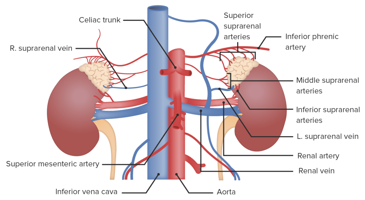
  • Blood supply (to both the cortex and medulla): about 50‒60 branches from the following 3 major arteries Arteries Arteries are tubular collections of cells that transport oxygenated blood and nutrients from the heart to the tissues of the body. The blood passes through the arteries in order of decreasing luminal diameter, starting in the largest artery (the aorta) and ending in the small arterioles. Arteries are classified into 3 types: large elastic arteries, medium muscular arteries, and small arteries and arterioles. Arteries: Histology entering the glands:
  • Medullary blood supply:
    • Some capillaries Capillaries Capillaries are the primary structures in the circulatory system that allow the exchange of gas, nutrients, and other materials between the blood and the extracellular fluid (ECF). Capillaries are the smallest of the blood vessels. Because a capillary diameter is so small, only 1 RBC may pass through at a time. Capillaries: Histology pass through all 3 layers of the cortex and into the medulla. The medulla can be influenced by secretions of the cortex (especially glucocorticoids Glucocorticoids Glucocorticoids are a class within the corticosteroid family. Glucocorticoids are chemically and functionally similar to endogenous cortisol. There are a wide array of indications, which primarily benefit from the antiinflammatory and immunosuppressive effects of this class of drugs. Glucocorticoids).
    • Some arteriole branches go straight from the capsule Capsule An envelope of loose gel surrounding a bacterial cell which is associated with the virulence of pathogenic bacteria. Some capsules have a well-defined border, whereas others form a slime layer that trails off into the medium. Most capsules consist of relatively simple polysaccharides but there are some bacteria whose capsules are made of polypeptides. Bacteroides into the medulla.
  • Blood flow Blood flow Blood flow refers to the movement of a certain volume of blood through the vasculature over a given unit of time (e.g., mL per minute). Vascular Resistance, Flow, and Mean Arterial Pressure: approximately 10 mL/min
  • Adrenocorticotropic hormone Adrenocorticotropic hormone An anterior pituitary hormone that stimulates the adrenal cortex and its production of corticosteroids. Acth is a 39-amino acid polypeptide of which the n-terminal 24-amino acid segment is identical in all species and contains the adrenocorticotropic activity. Upon further tissue-specific processing, acth can yield alpha-msh and corticotropin-like intermediate lobe peptide (clip). Adrenal Hormones (ACTH) increases blood flow Blood flow Blood flow refers to the movement of a certain volume of blood through the vasculature over a given unit of time (e.g., mL per minute). Vascular Resistance, Flow, and Mean Arterial Pressure to the adrenal glands.
  • Adrenal capillaries Capillaries Capillaries are the primary structures in the circulatory system that allow the exchange of gas, nutrients, and other materials between the blood and the extracellular fluid (ECF). Capillaries are the smallest of the blood vessels. Because a capillary diameter is so small, only 1 RBC may pass through at a time. Capillaries: Histology are fenestrated → better absorption Absorption Absorption involves the uptake of nutrient molecules and their transfer from the lumen of the GI tract across the enterocytes and into the interstitial space, where they can be taken up in the venous or lymphatic circulation. Digestion and Absorption of secretary products

Venous drainage

  • Typically drain via a single vein on each side: suprarenal (adrenal) veins Veins Veins are tubular collections of cells, which transport deoxygenated blood and waste from the capillary beds back to the heart. Veins are classified into 3 types: small veins/venules, medium veins, and large veins. Each type contains 3 primary layers: tunica intima, tunica media, and tunica adventitia. Veins: Histology
  • Specialized characteristics of suprarenal veins Veins Veins are tubular collections of cells, which transport deoxygenated blood and waste from the capillary beds back to the heart. Veins are classified into 3 types: small veins/venules, medium veins, and large veins. Each type contains 3 primary layers: tunica intima, tunica media, and tunica adventitia. Veins: Histology:
    • Lack a tunica media Tunica media The middle layer of blood vessel walls, composed principally of thin, cylindrical, smooth muscle cells and elastic tissue. It accounts for the bulk of the wall of most arteries. The smooth muscle cells are arranged in circular layers around the vessel, and the thickness of the coat varies with the size of the vessel. Arteries: Histology in some segments → chromaffin cells Chromaffin cells Cells that store epinephrine secretory vesicles. During times of stress, the nervous system signals the vesicles to secrete their hormonal content. Their name derives from their ability to stain a brownish color with chromic salts. Characteristically, they are located in the adrenal medulla and paraganglia of the sympathetic nervous system. Adrenal Hormones are present right next to the tunica intima Tunica intima The innermost layer of an artery or vein, made up of one layer of endothelial cells and supported by an internal elastic lamina. Arteries: Histologysecrete their content quickly/directly into the blood
    • Smooth muscle of the tunica media Tunica media The middle layer of blood vessel walls, composed principally of thin, cylindrical, smooth muscle cells and elastic tissue. It accounts for the bulk of the wall of most arteries. The smooth muscle cells are arranged in circular layers around the vessel, and the thickness of the coat varies with the size of the vessel. Arteries: Histology is arranged longitudinally → can contract and expel its content quickly
Vascular supply to the adrenal glands

Vascular supply to the adrenal glands

Image by Lecturio.

Lymphatic drainage

  • Drained via 2 lymphatic plexuses:
    • 1 in the medulla
    • 1 deep in the adrenal capsule Capsule An envelope of loose gel surrounding a bacterial cell which is associated with the virulence of pathogenic bacteria. Some capsules have a well-defined border, whereas others form a slime layer that trails off into the medium. Most capsules consist of relatively simple polysaccharides but there are some bacteria whose capsules are made of polypeptides. Bacteroides
  • Drains into:
    • Lateral aortic lymph nodes Lymph Nodes They are oval or bean shaped bodies (1 – 30 mm in diameter) located along the lymphatic system. Lymphatic Drainage System: Anatomy
    • Paraaortic nodes near the origin of the renal artery Renal artery A branch of the abdominal aorta which supplies the kidneys, adrenal glands and ureters. Glomerular Filtration
    • Thoracic duct Thoracic Duct The largest lymphatic vessel that passes through the chest and drains into the subclavian vein. Lymphatic Drainage System: Anatomy (some lymphatics traverse the diaphragm Diaphragm The diaphragm is a large, dome-shaped muscle that separates the thoracic cavity from the abdominal cavity. The diaphragm consists of muscle fibers and a large central tendon, which is divided into right and left parts. As the primary muscle of inspiration, the diaphragm contributes 75% of the total inspiratory muscle force. Diaphragm: Anatomy)

Innervation

  • Visceral fibers (traversing the retroperitoneal Retroperitoneal Peritoneum: Anatomy autonomic ganglia) from:
    • Vagus nerve Vagus nerve The 10th cranial nerve. The vagus is a mixed nerve which contains somatic afferents (from skin in back of the ear and the external auditory meatus), visceral afferents (from the pharynx, larynx, thorax, and abdomen), parasympathetic efferents (to the thorax and abdomen), and efferents to striated muscle (of the larynx and pharynx). Pharynx: Anatomy
    • Phrenic nerve Phrenic nerve The motor nerve of the diaphragm. The phrenic nerve fibers originate in the cervical spinal column (mostly C4) and travel through the cervical plexus to the diaphragm. Diaphragm: Anatomy
    • Greater and lesser splanchnic nerves
  • Preganglionic sympathetic fibers synapse Synapse The junction between 2 neurons is called a synapse. The synapse allows a neuron to pass an electrical or chemical signal to another neuron or target effector cell. Synapses and Neurotransmission directly with cells in the adrenal medulla.

Function

Functions of the adrenal cortex hormones Adrenal Cortex Hormones Adrenal Hormones

The adrenal cortex secretes hormones Hormones Hormones are messenger molecules that are synthesized in one part of the body and move through the bloodstream to exert specific regulatory effects on another part of the body. Hormones play critical roles in coordinating cellular activities throughout the body in response to the constant changes in both the internal and external environments. Hormones: Overview and Types that are involved in the regulation of fluids, electrolytes Electrolytes Electrolytes are mineral salts that dissolve in water and dissociate into charged particles called ions, which can be either be positively (cations) or negatively (anions) charged. Electrolytes are distributed in the extracellular and intracellular compartments in different concentrations. Electrolytes are essential for various basic life-sustaining functions. Electrolytes, available energy, and inflammation Inflammation Inflammation is a complex set of responses to infection and injury involving leukocytes as the principal cellular mediators in the body’s defense against pathogenic organisms. Inflammation is also seen as a response to tissue injury in the process of wound healing. The 5 cardinal signs of inflammation are pain, heat, redness, swelling, and loss of function. Inflammation, and in the development of sexual characteristics.

  • Mineralocorticoids Mineralocorticoids Mineralocorticoids are a drug class within the corticosteroid family and fludrocortisone is the primary medication within this class. Fludrocortisone is a fluorinated analog of cortisone. The fluorine moiety protects the drug from isoenzyme inactivation in the kidney, allowing it to exert its mineralocorticoid effect. Mineralocorticoids: aldosterone Aldosterone A hormone secreted by the adrenal cortex that regulates electrolyte and water balance by increasing the renal retention of sodium and the excretion of potassium. Hyperkalemia
    • ↑ Blood pressure (↑ water reabsorption from the kidneys Kidneys The kidneys are a pair of bean-shaped organs located retroperitoneally against the posterior wall of the abdomen on either side of the spine. As part of the urinary tract, the kidneys are responsible for blood filtration and excretion of water-soluble waste in the urine. Kidneys: Anatomy)
    • ↑ Serum Na+ (↓ urinary excretion of Na+)
    • ↓ Serum K+ (↑ urinary excretion of K+)
    • ↑ Serum pH pH The quantitative measurement of the acidity or basicity of a solution. Acid-Base Balance (↑ urinary excretion of H+)
  • Glucocorticoids Glucocorticoids Glucocorticoids are a class within the corticosteroid family. Glucocorticoids are chemically and functionally similar to endogenous cortisol. There are a wide array of indications, which primarily benefit from the antiinflammatory and immunosuppressive effects of this class of drugs. Glucocorticoids: cortisol Cortisol Glucocorticoids, corticosterone Corticosterone An adrenocortical steroid that has modest but significant activities as a mineralocorticoid and a glucocorticoid. Adrenal Hormones
    • ↑ Immediate available energy through:
      • Fat and protein catabolism → ↑ amino acid Amino acid Amino acids (AAs) are composed of a central carbon atom attached to a carboxyl group, an amino group, a hydrogen atom, and a side chain (R group). Basics of Amino Acids and lipid levels in blood
      • Gluconeogenesis Gluconeogenesis Gluconeogenesis is the process of making glucose from noncarbohydrate precursors. This metabolic pathway is more than just a reversal of glycolysis. Gluconeogenesis provides the body with glucose not obtained from food, such as during a fasting period. The production of glucose is critical for organs and cells that cannot use fat for fuel. Gluconeogenesis
      • Appetite stimulation
    • RBCs RBCs Erythrocytes, or red blood cells (RBCs), are the most abundant cells in the blood. While erythrocytes in the fetus are initially produced in the yolk sac then the liver, the bone marrow eventually becomes the main site of production. Erythrocytes: Histology
    • ↑ Serum calcium Calcium A basic element found in nearly all tissues. It is a member of the alkaline earth family of metals with the atomic symbol ca, atomic number 20, and atomic weight 40. Calcium is the most abundant mineral in the body and combines with phosphorus to form calcium phosphate in the bones and teeth. It is essential for the normal functioning of nerves and muscles and plays a role in blood coagulation (as factor IV) and in many enzymatic processes. Electrolytes from bone resorption Bone resorption Bone loss due to osteoclastic activity. Bones: Remodeling and Healing (leads to ↓ bone Bone Bone is a compact type of hardened connective tissue composed of bone cells, membranes, an extracellular mineralized matrix, and central bone marrow. The 2 primary types of bone are compact and spongy. Bones: Structure and Types mineral density over time)
    • Anti-inflammatory effects:
      • Inflammation Inflammation Inflammation is a complex set of responses to infection and injury involving leukocytes as the principal cellular mediators in the body’s defense against pathogenic organisms. Inflammation is also seen as a response to tissue injury in the process of wound healing. The 5 cardinal signs of inflammation are pain, heat, redness, swelling, and loss of function. Inflammation
      • ↓ Blood vessel permeability
      • Long-term exposure suppresses the immune system Immune system The body’s defense mechanism against foreign organisms or substances and deviant native cells. It includes the humoral immune response and the cell-mediated response and consists of a complex of interrelated cellular, molecular, and genetic components. Primary Lymphatic Organs.
  • Androgens Androgens Androgens are naturally occurring steroid hormones responsible for development and maintenance of the male sex characteristics, including penile, scrotal, and clitoral growth, development of sexual hair, deepening of the voice, and musculoskeletal growth. Androgens and Antiandrogens: DHEA DHEA A major C19 steroid produced by the adrenal cortex. It is also produced in small quantities in the testis and the ovary. Dehydroepiandrosterone (dhea) can be converted to testosterone; androstenedione; estradiol; and estrone. Androgens and Antiandrogens and androstenedione Androstenedione A delta-4 C19 steroid that is produced not only in the testis, but also in the ovary and the adrenal cortex. Depending on the tissue type, androstenedione can serve as a precursor to testosterone as well as estrone and estradiol. Androgens and Antiandrogens

Functions of the adrenal medulla hormones Hormones Hormones are messenger molecules that are synthesized in one part of the body and move through the bloodstream to exert specific regulatory effects on another part of the body. Hormones play critical roles in coordinating cellular activities throughout the body in response to the constant changes in both the internal and external environments. Hormones: Overview and Types

The adrenal medulla secretes the catecholamines Catecholamines A general class of ortho-dihydroxyphenylalkylamines derived from tyrosine. Adrenal Hormones epinephrine Epinephrine The active sympathomimetic hormone from the adrenal medulla. It stimulates both the alpha- and beta- adrenergic systems, causes systemic vasoconstriction and gastrointestinal relaxation, stimulates the heart, and dilates bronchi and cerebral vessels. Sympathomimetic Drugs and norepinephrine Norepinephrine Precursor of epinephrine that is secreted by the adrenal medulla and is a widespread central and autonomic neurotransmitter. Norepinephrine is the principal transmitter of most postganglionic sympathetic fibers, and of the diffuse projection system in the brain that arises from the locus ceruleus. Receptors and Neurotransmitters of the CNS, which stimulate the fight-or-flight response. The effects include:

  • ↑ Blood pressure
  • Heart rate Heart rate The number of times the heart ventricles contract per unit of time, usually per minute. Cardiac Physiology
  • Circulation Circulation The movement of the blood as it is pumped through the cardiovascular system. ABCDE Assessment to skeletal muscles Skeletal muscles A subtype of striated muscle, attached by tendons to the skeleton. Skeletal muscles are innervated and their movement can be consciously controlled. They are also called voluntary muscles. Muscle Tissue: Histology
  • Respiration Respiration The act of breathing with the lungs, consisting of inhalation, or the taking into the lungs of the ambient air, and of exhalation, or the expelling of the modified air which contains more carbon dioxide than the air taken in. Nose Anatomy (External & Internal) and bronchodilation
  • ↑ Blood glucose Glucose A primary source of energy for living organisms. It is naturally occurring and is found in fruits and other parts of plants in its free state. It is used therapeutically in fluid and nutrient replacement. Lactose Intolerance levels by:
    • Stimulating glycogenolysis Glycogenolysis The release of glucose from glycogen by glycogen phosphorylase (phosphorolysis). The released glucose-1-phosphate is then converted to glucose-6-phosphate by phosphoglucomutase before entering glycolysis. Glycogenolysis is stimulated by glucagon or epinephrine via the activation of phosphorylase kinase. Glycogen Metabolism
    • Stimulating gluconeogenesis Gluconeogenesis Gluconeogenesis is the process of making glucose from noncarbohydrate precursors. This metabolic pathway is more than just a reversal of glycolysis. Gluconeogenesis provides the body with glucose not obtained from food, such as during a fasting period. The production of glucose is critical for organs and cells that cannot use fat for fuel. Gluconeogenesis
    • Inhibiting insulin Insulin Insulin is a peptide hormone that is produced by the beta cells of the pancreas. Insulin plays a role in metabolic functions such as glucose uptake, glycolysis, glycogenesis, lipogenesis, and protein synthesis. Exogenous insulin may be needed for individuals with diabetes mellitus, in whom there is a deficiency in endogenous insulin or increased insulin resistance. Insulin release
  • Digestion Digestion Digestion refers to the process of the mechanical and chemical breakdown of food into smaller particles, which can then be absorbed and utilized by the body. Digestion and Absorption

Related videos

Clinical Relevance

Several clinical conditions can result from abnormalities of the adrenal hormones Adrenal hormones There are 2 primary portions of the adrenal glands, the adrenal medulla and the adrenal cortex. The adrenal medulla is the inner portion of the gland, secreting epinephrine and, to a lesser degree, norepinephrine. The adrenal cortex is the outer portion of the gland and secretes mineralocorticoids, glucocorticoids, and androgens. Adrenal Hormones. The most important conditions include:

  • Pheochromocytoma Pheochromocytoma Pheochromocytoma is a catecholamine-secreting tumor derived from chromaffin cells. The majority of tumors originate in the adrenal medulla, but they may also arise from sympathetic ganglia (also referred to as paraganglioma). Symptoms are associated with excessive catecholamine production and commonly include hypertension, tachycardia, headache, and sweating. Pheochromocytoma: rare catecholamine-secreting tumors that arise from the chromaffin cells Chromaffin cells Cells that store epinephrine secretory vesicles. During times of stress, the nervous system signals the vesicles to secrete their hormonal content. Their name derives from their ability to stain a brownish color with chromic salts. Characteristically, they are located in the adrenal medulla and paraganglia of the sympathetic nervous system. Adrenal Hormones of the adrenal medulla. Individuals typically present with treatment-resistant hypertension Hypertension Hypertension, or high blood pressure, is a common disease that manifests as elevated systemic arterial pressures. Hypertension is most often asymptomatic and is found incidentally as part of a routine physical examination or during triage for an unrelated medical encounter. Hypertension, episodic headaches, sweating, and tachycardia Tachycardia Abnormally rapid heartbeat, usually with a heart rate above 100 beats per minute for adults. Tachycardia accompanied by disturbance in the cardiac depolarization (cardiac arrhythmia) is called tachyarrhythmia. Sepsis in Children. Diagnosis is made via adrenal imaging studies and laboratory assessment that shows elevated serum catecholamines Catecholamines A general class of ortho-dihydroxyphenylalkylamines derived from tyrosine. Adrenal Hormones. About 90% of cases are benign Benign Fibroadenoma, and surgical resection is the only curative treatment.
  • Adrenal insufficiency Adrenal Insufficiency Conditions in which the production of adrenal corticosteroids falls below the requirement of the body. Adrenal insufficiency can be caused by defects in the adrenal glands, the pituitary gland, or the hypothalamus. Adrenal Insufficiency and Addison Disease: inadequate production of glucocorticoids Glucocorticoids Glucocorticoids are a class within the corticosteroid family. Glucocorticoids are chemically and functionally similar to endogenous cortisol. There are a wide array of indications, which primarily benefit from the antiinflammatory and immunosuppressive effects of this class of drugs. Glucocorticoids, mineralocorticoids Mineralocorticoids Mineralocorticoids are a drug class within the corticosteroid family and fludrocortisone is the primary medication within this class. Fludrocortisone is a fluorinated analog of cortisone. The fluorine moiety protects the drug from isoenzyme inactivation in the kidney, allowing it to exert its mineralocorticoid effect. Mineralocorticoids, and adrenal androgens Androgens Androgens are naturally occurring steroid hormones responsible for development and maintenance of the male sex characteristics, including penile, scrotal, and clitoral growth, development of sexual hair, deepening of the voice, and musculoskeletal growth. Androgens and Antiandrogens. Diagnosis is made by measuring cortisol Cortisol Glucocorticoids and ACTH levels and with ACTH stimulation testing. Management involves glucocorticoid and mineralocorticoid replacement therapy.
  • Congenital adrenal hyperplasia Hyperplasia An increase in the number of cells in a tissue or organ without tumor formation. It differs from hypertrophy, which is an increase in bulk without an increase in the number of cells. Cellular Adaptation: a group of autosomal recessive Autosomal recessive Autosomal inheritance, both dominant and recessive, refers to the transmission of genes from the 22 autosomal chromosomes. Autosomal recessive diseases are only expressed when 2 copies of the recessive allele are inherited. Autosomal Recessive and Autosomal Dominant Inheritance disorders that cause a deficiency of an enzyme (most commonly 21-hydroxylase deficiency 21-hydroxylase deficiency Congenital Adrenal Hyperplasia) needed for cortisol Cortisol Glucocorticoids, aldosterone Aldosterone A hormone secreted by the adrenal cortex that regulates electrolyte and water balance by increasing the renal retention of sodium and the excretion of potassium. Hyperkalemia, and androgen synthesis Synthesis Polymerase Chain Reaction (PCR). Clinical manifestations include ambiguous genitalia Ambiguous Genitalia Primary Amenorrhea in genotypic females, salt wasting, and hypoglycemia Hypoglycemia Hypoglycemia is an emergency condition defined as a serum glucose level ≤ 70 mg/dL (≤ 3.9 mmol/L) in diabetic patients. In nondiabetic patients, there is no specific or defined limit for normal serum glucose levels, and hypoglycemia is defined mainly by its clinical features. Hypoglycemia. A less severe form known as nonclassical congenital adrenal hyperplasia Nonclassical Congenital Adrenal Hyperplasia Secondary Amenorrhea also exists, which typically presents in puberty Puberty Puberty is a complex series of physical, psychosocial, and cognitive transitions usually experienced by adolescents (11-19 years of age). Puberty is marked by a growth in stature and the development of secondary sexual characteristics, achievement of fertility, and changes in most body systems. Puberty. Lifelong glucocorticoid replacement is needed.
  • Cushing’s syndrome: a condition resulting from chronic exposure to excess glucocorticoids Glucocorticoids Glucocorticoids are a class within the corticosteroid family. Glucocorticoids are chemically and functionally similar to endogenous cortisol. There are a wide array of indications, which primarily benefit from the antiinflammatory and immunosuppressive effects of this class of drugs. Glucocorticoids. Etiologies include chronic glucocorticoid intake, increased adrenal secretion Secretion Coagulation Studies of cortisol Cortisol Glucocorticoids, or increased pituitary Pituitary A small, unpaired gland situated in the sella turcica. It is connected to the hypothalamus by a short stalk which is called the infundibulum. Hormones: Overview and Types or ectopic secretion Secretion Coagulation Studies of ACTH. Typical clinical features include central obesity Central Obesity Cushing Syndrome, thin and bruisable skin Skin The skin, also referred to as the integumentary system, is the largest organ of the body. The skin is primarily composed of the epidermis (outer layer) and dermis (deep layer). The epidermis is primarily composed of keratinocytes that undergo rapid turnover, while the dermis contains dense layers of connective tissue. Skin: Structure and Functions, abdominal striae, secondary hypertension Secondary hypertension Hypertension, hyperglycemia Hyperglycemia Abnormally high blood glucose level. Diabetes Mellitus, and proximal muscle weakness Proximal Muscle Weakness Lambert-Eaton Myasthenic Syndrome. Diagnosis is by urinary and/or salivary cortisol Cortisol Glucocorticoids testing along with a low dose dexamethasone-suppression test Dexamethasone-Suppression Test Cushing Syndrome, determination of ACTH levels, and imaging.
  • Hyperaldosteronism Hyperaldosteronism Hyperaldosteronism is defined as the increased secretion of aldosterone from the zona glomerulosa of the adrenal cortex. Hyperaldosteronism may be primary (resulting from autonomous secretion), or secondary (resulting from physiological secretion due to stimulation of the RAAS). Classically, hyperaldosteronism presents with hypertension, hypokalemia, and metabolic alkalosis. Hyperaldosteronism: increased secretion Secretion Coagulation Studies of aldosterone Aldosterone A hormone secreted by the adrenal cortex that regulates electrolyte and water balance by increasing the renal retention of sodium and the excretion of potassium. Hyperkalemia from the adrenal cortex. Hyperaldosteronism Hyperaldosteronism Hyperaldosteronism is defined as the increased secretion of aldosterone from the zona glomerulosa of the adrenal cortex. Hyperaldosteronism may be primary (resulting from autonomous secretion), or secondary (resulting from physiological secretion due to stimulation of the RAAS). Classically, hyperaldosteronism presents with hypertension, hypokalemia, and metabolic alkalosis. Hyperaldosteronism may be primary (resulting from autonomous secretion Secretion Coagulation Studies ( Conn syndrome Conn Syndrome Hyperaldosteronism)) or secondary (resulting from physiologic secretion Secretion Coagulation Studies due to stimulation of the RAAS RAAS A blood pressure regulating system of interacting components that include renin; angiotensinogen; angiotensin converting enzyme; angiotensin i; angiotensin ii; and angiotensinase. Renin, an enzyme produced in the kidney, acts on angiotensinogen, an alpha-2 globulin produced by the liver, forming angiotensin I. Angiotensin-converting enzyme, contained in the lung, acts on angiotensin I in the plasma converting it to angiotensin II, an extremely powerful vasoconstrictor. Angiotensin II causes contraction of the arteriolar and renal vascular smooth muscle, leading to retention of salt and water in the kidney and increased arterial blood pressure. In addition, angiotensin II stimulates the release of aldosterone from the adrenal cortex, which in turn also increases salt and water retention in the kidney. Angiotensin-converting enzyme also breaks down bradykinin, a powerful vasodilator and component of the kallikrein-kinin system. Adrenal Hormones) and presents with hypertension Hypertension Hypertension, or high blood pressure, is a common disease that manifests as elevated systemic arterial pressures. Hypertension is most often asymptomatic and is found incidentally as part of a routine physical examination or during triage for an unrelated medical encounter. Hypertension, hypokalemia Hypokalemia Hypokalemia is defined as plasma potassium (K+) concentration < 3.5 mEq/L. Homeostatic mechanisms maintain plasma concentration between 3.5-5.2 mEq/L despite marked variation in dietary intake. Hypokalemia can be due to renal losses, GI losses, transcellular shifts, or poor dietary intake. Hypokalemia, and metabolic alkalosis Alkalosis A pathological condition that removes acid or adds base to the body fluids. Respiratory Alkalosis. Diagnosis involves measuring plasma Plasma The residual portion of blood that is left after removal of blood cells by centrifugation without prior blood coagulation. Transfusion Products aldosterone Aldosterone A hormone secreted by the adrenal cortex that regulates electrolyte and water balance by increasing the renal retention of sodium and the excretion of potassium. Hyperkalemia and renin Renin A highly specific (leu-leu) endopeptidase that generates angiotensin I from its precursor angiotensinogen, leading to a cascade of reactions which elevate blood pressure and increase sodium retention by the kidney in the renin-angiotensin system. Renal Sodium and Water Regulation activity along with imaging to identify hormone-secreting tumors. Management involves the use of aldosterone Aldosterone A hormone secreted by the adrenal cortex that regulates electrolyte and water balance by increasing the renal retention of sodium and the excretion of potassium. Hyperkalemia receptor Receptor Receptors are proteins located either on the surface of or within a cell that can bind to signaling molecules known as ligands (e.g., hormones) and cause some type of response within the cell. Receptors antagonists and surgical excision of any aldosterone-secreting tumors.
  • Hypoaldosteronism Hypoaldosteronism Hypoaldosteronism is a hormonal disorder characterized by low levels of aldosterone. These low levels can be caused by decreased aldosterone production or a peripheral resistance to aldosterone. When hypoaldosteronism occurs as a result of an acquired decrease in renin production, the condition is more commonly referred to as renal tubular acidosis (RTA) type 4. Hypoaldosteronism: a condition resulting from low levels of aldosterone Aldosterone A hormone secreted by the adrenal cortex that regulates electrolyte and water balance by increasing the renal retention of sodium and the excretion of potassium. Hyperkalemia. Hypoaldosteronism Hypoaldosteronism Hypoaldosteronism is a hormonal disorder characterized by low levels of aldosterone. These low levels can be caused by decreased aldosterone production or a peripheral resistance to aldosterone. When hypoaldosteronism occurs as a result of an acquired decrease in renin production, the condition is more commonly referred to as renal tubular acidosis (RTA) type 4. Hypoaldosteronism can be caused by decreased aldosterone Aldosterone A hormone secreted by the adrenal cortex that regulates electrolyte and water balance by increasing the renal retention of sodium and the excretion of potassium. Hyperkalemia production or peripheral resistance Resistance Physiologically, the opposition to flow of air caused by the forces of friction. As a part of pulmonary function testing, it is the ratio of driving pressure to the rate of air flow. Ventilation: Mechanics of Breathing to aldosterone Aldosterone A hormone secreted by the adrenal cortex that regulates electrolyte and water balance by increasing the renal retention of sodium and the excretion of potassium. Hyperkalemia. When hypoaldosteronism Hypoaldosteronism Hypoaldosteronism is a hormonal disorder characterized by low levels of aldosterone. These low levels can be caused by decreased aldosterone production or a peripheral resistance to aldosterone. When hypoaldosteronism occurs as a result of an acquired decrease in renin production, the condition is more commonly referred to as renal tubular acidosis (RTA) type 4. Hypoaldosteronism occurs as a result of an acquired decrease in renin Renin A highly specific (leu-leu) endopeptidase that generates angiotensin I from its precursor angiotensinogen, leading to a cascade of reactions which elevate blood pressure and increase sodium retention by the kidney in the renin-angiotensin system. Renal Sodium and Water Regulation production from the kidneys Kidneys The kidneys are a pair of bean-shaped organs located retroperitoneally against the posterior wall of the abdomen on either side of the spine. As part of the urinary tract, the kidneys are responsible for blood filtration and excretion of water-soluble waste in the urine. Kidneys: Anatomy, the condition is referred to as renal tubular acidosis Acidosis A pathologic condition of acid accumulation or depletion of base in the body. The two main types are respiratory acidosis and metabolic acidosis, due to metabolic acid build up. Respiratory Acidosis ( RTA RTA Renal tubular acidosis (RTA) is an imbalance in physiologic pH caused by the kidney’s inability to acidify urine to maintain blood pH at physiologic levels. Renal tubular acidosis exist in multiple types, including distal RTA (type 1), proximal RTA (type 2), and hyperkalemic RTA (type 4). Renal Tubular Acidosis) type 4 Type 4 Spinal Muscular Atrophy. Most cases are asymptomatic and diagnosed when routine lab evaluation reveals hyperkalemia Hyperkalemia Hyperkalemia is defined as a serum potassium (K+) concentration >5.2 mEq/L. Homeostatic mechanisms maintain the serum K+ concentration between 3.5 and 5.2 mEq/L, despite marked variation in dietary intake. Hyperkalemia can be due to a variety of causes, which include transcellular shifts, tissue breakdown, inadequate renal excretion, and drugs. Hyperkalemia and mild hyperchloremic metabolic acidosis Hyperchloremic Metabolic Acidosis Potassium-sparing Diuretics, prompting further work-up.

References

  1. Roman, S. (2020). Surgical anatomy of the adrenal glands. UpToDate. Retrieved Aug 16, 2021, from https://www.uptodate.com/contents/surgical-anatomy-of-the-adrenal-glands 
  2. Megha, R. (2021). Anatomy, Abdomen and pelvis, adrenal glands (suprarenal glands). StatPearls. Retrieved Aug 16, 2021, from https://www.statpearls.com/articlelibrary/viewarticle/666/ 
  3. Ovalle, W., Nahirney, P. (2020). Netter’s Essential Histology: With Correlated Histopathology (Netter Basic Science) (3rd ed.). Elsevier.

Create your free account or log in to continue reading!

Sign up now and get free access to Lecturio with concept pages, medical videos, and questions for your medical education.

User Reviews

Unwrap New Skills This Holiday 🎄 Save 30% on all plans now!

Details