Advertisement

Advertisement

Advertisement

Advertisement

ABCDE Assessment

The airway, breathing, and circulation, disability and exposure (ABCDE) assessment is the mainstay management approach used in managing critically ill patients Patients Individuals participating in the health care system for the purpose of receiving therapeutic, diagnostic, or preventive procedures. Clinician–Patient Relationship. The ABCDEs are the essential 1st steps to perform in many situations including unresponsive patients Patients Individuals participating in the health care system for the purpose of receiving therapeutic, diagnostic, or preventive procedures. Clinician–Patient Relationship, cardiac arrests, and critical medical or trauma patients Patients Individuals participating in the health care system for the purpose of receiving therapeutic, diagnostic, or preventive procedures. Clinician–Patient Relationship. For the trauma patient, ABCDE is included in the primary survey Primary Survey Thoracic Trauma in Children, the initial evaluation, and management of injuries.

Last updated: Jan 17, 2023

Editorial responsibility: Stanley Oiseth, Lindsay Jones, Evelin Maza

Advertisement

Advertisement

Advertisement

Advertisement

Advertisement

Advertisement

Primary Survey

A primary survey Primary Survey Thoracic Trauma in Children is the initial evaluation used to identify and manage life-threatening injuries in a trauma patient.

The components of the primary survey Primary Survey Thoracic Trauma in Children are:
  • Airway
  • Breathing
  • Circulation
  • Disability
  • Exposure

Airway

Purpose

  • Establish airway patency.
  • Assess the patient’s ability to protect the airway.
  • Manage the airway.
  • Spinal immobilization Immobilization Delirium: using a backboard and rigid cervical collar

Causes of airway compromise

  • Traumatic injury
  • Patient’s tongue Tongue The tongue, on the other hand, is a complex muscular structure that permits tasting and facilitates the process of mastication and communication. The blood supply of the tongue originates from the external carotid artery, and the innervation is through cranial nerves. Lips and Tongue: Anatomy
  • Foreign body Foreign Body Foreign Body Aspiration 
  • Vomit, blood, and secretions
  • Localized swelling Swelling Inflammation due to infection or anaphylaxis Anaphylaxis An acute hypersensitivity reaction due to exposure to a previously encountered antigen. The reaction may include rapidly progressing urticaria, respiratory distress, vascular collapse, systemic shock, and death. Type I Hypersensitivity Reaction

Airway assessment Airway Assessment Airway Management

  • If the patient can speak normally → airway is intact.
  • Signs of an unprotected airway:
    • Paradoxical chest and abdominal movements
    • Cyanosis Cyanosis A bluish or purplish discoloration of the skin and mucous membranes due to an increase in the amount of deoxygenated hemoglobin in the blood or a structural defect in the hemoglobin molecule. Pulmonary Examination
    • Abnormal breathing sounds:
    • Presence of an expanding hematoma Hematoma A collection of blood outside the blood vessels. Hematoma can be localized in an organ, space, or tissue. Intussusception
    • Presence of subcutaneous emphysema Subcutaneous emphysema Presence of air or gas in the subcutaneous tissues of the body. Mallory-Weiss Syndrome (Mallory-Weiss Tear)
    • Glasgow Coma Coma Coma is defined as a deep state of unarousable unresponsiveness, characterized by a score of 3 points on the GCS. A comatose state can be caused by a multitude of conditions, making the precise epidemiology and prognosis of coma difficult to determine. Coma Scale Scale Dermatologic Examination ( GCS GCS A scale that assesses the response to stimuli in patients with craniocerebral injuries. The parameters are eye opening, motor response, and verbal response. Coma) < 9

Airway management Airway management An airway, breathing, and circulation (ABC) assessment is the mainstay for evaluating and treating critically ill individuals. The airway assessment helps identify individuals with potential obstruction of the airway, which may benefit from airway management techniques to ensure adequate ventilation and oxygenation. Airway Management

  • Adequate ventilation Ventilation The total volume of gas inspired or expired per unit of time, usually measured in liters per minute. Ventilation: Mechanics of Breathing with 100% oxygen 100% Oxygen Cluster Headaches using a bag and mask
  • Pulse oximetry to monitor oxygen levels
  • Airway suctioning (with a suction catheter) can help remove secretions in an effort to clear the airway.
  • Special maneuvers:
    • Chin Chin The anatomical frontal portion of the mandible, also known as the mentum, that contains the line of fusion of the two separate halves of the mandible (symphysis menti). This line of fusion divides inferiorly to enclose a triangular area called the mental protuberance. On each side, inferior to the second premolar tooth, is the mental foramen for the passage of blood vessels and a nerve. Melasma lift, jaw Jaw The jaw is made up of the mandible, which comprises the lower jaw, and the maxilla, which comprises the upper jaw. The mandible articulates with the temporal bone via the temporomandibular joint (TMJ). The 4 muscles of mastication produce the movements of the TMJ to ensure the efficient chewing of food. Jaw and Temporomandibular Joint: Anatomy thrust
    • Oral (oropharyngeal tube) or nasal (nasopharyngeal tube) airways can be used to temporarily maintain the airway 
  • If maneuvers fail, establish a definitive airway:
    • Endotracheal (orotracheal) intubation Intubation Peritonsillar Abscess
      • Insertion of a tube into the trachea Trachea The trachea is a tubular structure that forms part of the lower respiratory tract. The trachea is continuous superiorly with the larynx and inferiorly becomes the bronchial tree within the lungs. The trachea consists of a support frame of semicircular, or C-shaped, rings made out of hyaline cartilage and reinforced by collagenous connective tissue. Trachea: Anatomy through the mouth (less commonly the nose Nose The nose is the human body’s primary organ of smell and functions as part of the upper respiratory system. The nose may be best known for inhaling oxygen and exhaling carbon dioxide, but it also contributes to other important functions, such as tasting. The anatomy of the nose can be divided into the external nose and the nasal cavity. Nose Anatomy (External & Internal))
      • 1st-line procedure
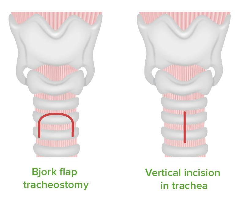
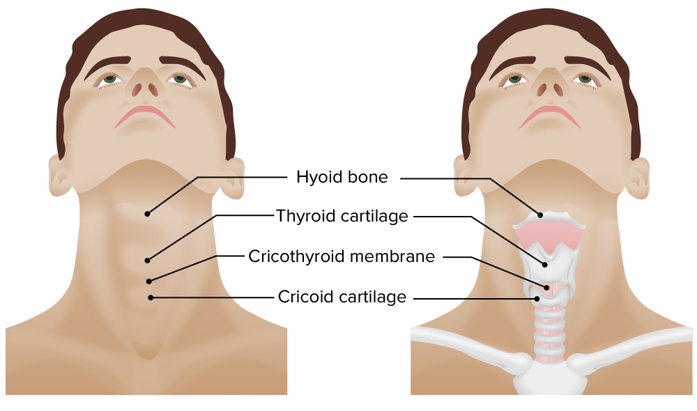
    • Cricothyrotomy
      • Incision of the membrane between the thyroid Thyroid The thyroid gland is one of the largest endocrine glands in the human body. The thyroid gland is a highly vascular, brownish-red gland located in the visceral compartment of the anterior region of the neck. Thyroid Gland: Anatomy cartilage Cartilage Cartilage is a type of connective tissue derived from embryonic mesenchyme that is responsible for structural support, resilience, and the smoothness of physical actions. Perichondrium (connective tissue membrane surrounding cartilage) compensates for the absence of vasculature in cartilage by providing nutrition and support. Cartilage: Histology and cricoid cartilage Cartilage Cartilage is a type of connective tissue derived from embryonic mesenchyme that is responsible for structural support, resilience, and the smoothness of physical actions. Perichondrium (connective tissue membrane surrounding cartilage) compensates for the absence of vasculature in cartilage by providing nutrition and support. Cartilage: Histology
      • Used if endotracheal in severe traumatic injuries, if intubation Intubation Peritonsillar Abscess fails, or if airway is severely swollen ( anaphylaxis Anaphylaxis An acute hypersensitivity reaction due to exposure to a previously encountered antigen. The reaction may include rapidly progressing urticaria, respiratory distress, vascular collapse, systemic shock, and death. Type I Hypersensitivity Reaction!) 
      • Easier to perform than tracheostomy Tracheostomy Surgical formation of an opening into the trachea through the neck, or the opening so created. Laryngomalacia and Tracheomalacia
      • Temporary procedure
    • Tracheotomy Tracheotomy Surgical incision of the trachea. Basic Procedures
      • Incision to the trachea Trachea The trachea is a tubular structure that forms part of the lower respiratory tract. The trachea is continuous superiorly with the larynx and inferiorly becomes the bronchial tree within the lungs. The trachea consists of a support frame of semicircular, or C-shaped, rings made out of hyaline cartilage and reinforced by collagenous connective tissue. Trachea: Anatomy with insertion of a tracheal tube
      • Preferred in pediatric patients Patients Individuals participating in the health care system for the purpose of receiving therapeutic, diagnostic, or preventive procedures. Clinician–Patient Relationship (age < 8) as the cricoid is much smaller

Breathing

Breathing is the next step after the airway has been deemed adequate.
Purpose

  • Detect signs of respiratory distress.
  • Maintain sufficient oxygenation and ventilation Ventilation The total volume of gas inspired or expired per unit of time, usually measured in liters per minute. Ventilation: Mechanics of Breathing.
  • At this step, if breathing problems are found, it may be necessary to perform procedures (i.e., thoracostomy Thoracostomy Surgical procedure involving the creation of an opening (stoma) into the chest cavity for drainage; used in the treatment of pleural effusion; pneumothorax; hemothorax; and empyema. Hemothorax) to correct breathing issues found.

Breathing assessment Breathing Assessment Multitrauma

  • Listen to breath sounds.
  • Count respiratory rate Respiratory rate The number of times an organism breathes with the lungs (respiration) per unit time, usually per minute. Pulmonary Examination:
    • 12–20/min is normal.
    • If respiratory rate Respiratory rate The number of times an organism breathes with the lungs (respiration) per unit time, usually per minute. Pulmonary Examination is ↓ or ↑, consider airway assistance.
  • 40% of cardiac arrest Cardiac arrest Cardiac arrest is the sudden, complete cessation of cardiac output with hemodynamic collapse. Patients present as pulseless, unresponsive, and apneic. Rhythms associated with cardiac arrest are ventricular fibrillation/tachycardia, asystole, or pulseless electrical activity. Cardiac Arrest patients Patients Individuals participating in the health care system for the purpose of receiving therapeutic, diagnostic, or preventive procedures. Clinician–Patient Relationship may have agonal breathing (series of noisy gasps).

Signs of respiratory distress

  • Inspection Inspection Dermatologic Examination:
  • Percussion Percussion Act of striking a part with short, sharp blows as an aid in diagnosing the condition beneath the sound obtained. Pulmonary Examination: hyperresonance ( pneumothorax Pneumothorax A pneumothorax is a life-threatening condition in which air collects in the pleural space, causing partial or full collapse of the lung. A pneumothorax can be traumatic or spontaneous. Patients present with a sudden onset of sharp chest pain, dyspnea, and diminished breath sounds on exam. Pneumothorax) or dullness ( hemothorax Hemothorax A hemothorax is a collection of blood in the pleural cavity. Hemothorax most commonly occurs due to damage to the intercostal arteries or from a lung laceration following chest trauma. Hemothorax can also occur as a complication of disease, or hemothorax may be spontaneous or iatrogenic. Hemothorax)
  • Palpation Palpation Application of fingers with light pressure to the surface of the body to determine consistency of parts beneath in physical diagnosis; includes palpation for determining the outlines of organs. Dermatologic Examination: tracheal shift, subcutaneous emphysema Subcutaneous emphysema Presence of air or gas in the subcutaneous tissues of the body. Mallory-Weiss Syndrome (Mallory-Weiss Tear), flail segments
  • Auscultation: ↓ air entry during auscultation
  • Oxygen saturation Oxygen Saturation Basic Procedures < 88%
  • ↓ end-tidal CO₂ (capnography)

Breathing management

  • Depends on the cause
  • Return to A (airway) to establish definitive airway if there is respiratory distress.
  • Procedures for management of life-threatening breathing conditions:
    • Tube thoracostomy Tube Thoracostomy Surgical procedure involving the creation of an opening (stoma) into the chest cavity for drainage; used in the treatment of pleural effusion; pneumothorax; hemothorax; and empyema. Thoracic Surgery (small incision of the chest wall Chest wall The chest wall consists of skin, fat, muscles, bones, and cartilage. The bony structure of the chest wall is composed of the ribs, sternum, and thoracic vertebrae. The chest wall serves as armor for the vital intrathoracic organs and provides the stability necessary for the movement of the shoulders and arms. Chest Wall: Anatomy is made and a chest tube is inserted) is needed in:
      • Tension pneumothorax Tension Pneumothorax Pneumothorax, open pneumothorax Open Pneumothorax Pneumothorax
      • Flail chest Flail chest Flail chest is a life-threatening traumatic injury that occurs when 3 or more contiguous ribs are fractured in 2 or more different locations. Patients present with chest pain, tachypnea, hypoxia, and paradoxical chest wall movement. Flail Chest
      • Massive hemothorax Hemothorax A hemothorax is a collection of blood in the pleural cavity. Hemothorax most commonly occurs due to damage to the intercostal arteries or from a lung laceration following chest trauma. Hemothorax can also occur as a complication of disease, or hemothorax may be spontaneous or iatrogenic. Hemothorax
    • Pericardiocentesis Pericardiocentesis Puncture and aspiration of fluid from the pericardium. Cardiac Surgery (a needle and small catheter are inserted into the pericardial sac to drain excess fluid) is needed in cardiac tamponade Tamponade Pericardial effusion, usually of rapid onset, exceeding ventricular filling pressures and causing collapse of the heart with a markedly reduced cardiac output. Pericarditis.
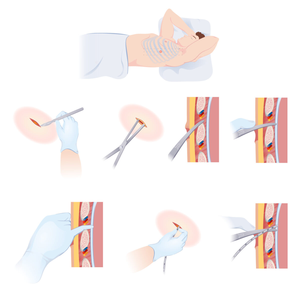
Insertion of a chest tube - thoracostomy

Thoracostomy:
A primary skin incision is made superiorly to the rib to avoid the neurovascular supply that runs inferiorly to the rib. The surgeon tunnels through the subcutaneous tissue and muscle to enter the pleural cavity. Entrance into the pleural cavity is confirmed and the chest tube is placed.

Image by Lecturio.

Circulation

Performed after the airway and breathing have been judged as normal and adequate.

Purpose

  • Determine the effectiveness of the cardiac output Cardiac output The volume of blood passing through the heart per unit of time. It is usually expressed as liters (volume) per minute so as not to be confused with stroke volume (volume per beat). Cardiac Mechanics.
  • Secure adequate tissue perfusion.
  • Treatment of external bleeding

Causes of poor circulation

  • Shock Shock Shock is a life-threatening condition associated with impaired circulation that results in tissue hypoxia. The different types of shock are based on the underlying cause: distributive (↑ cardiac output (CO), ↓ systemic vascular resistance (SVR)), cardiogenic (↓ CO, ↑ SVR), hypovolemic (↓ CO, ↑ SVR), obstructive (↓ CO), and mixed. Types of Shock (including hypovolemia Hypovolemia Sepsis in Children, septic, or anaphylactic shock Shock Shock is a life-threatening condition associated with impaired circulation that results in tissue hypoxia. The different types of shock are based on the underlying cause: distributive (↑ cardiac output (CO), ↓ systemic vascular resistance (SVR)), cardiogenic (↓ CO, ↑ SVR), hypovolemic (↓ CO, ↑ SVR), obstructive (↓ CO), and mixed. Types of Shock)
  • Traumatic injury to arteries Arteries Arteries are tubular collections of cells that transport oxygenated blood and nutrients from the heart to the tissues of the body. The blood passes through the arteries in order of decreasing luminal diameter, starting in the largest artery (the aorta) and ending in the small arterioles. Arteries are classified into 3 types: large elastic arteries, medium muscular arteries, and small arteries and arterioles. Arteries: Histology ( compartment syndrome Compartment Syndrome Compartment syndrome is a surgical emergency usually occurring secondary to trauma. The condition is marked by increased pressure within a compartment that compromises the circulation and function of the tissues within that space. Compartment Syndrome, arterial lacerations, crush injury Crush injury Excessive compression of parts of the body that causes muscle swelling, fracture, and/or neurological disturbances in the affected areas. Crush injury with systemic manifestations is referred to as crush syndrome. Crush Syndrome)
  • Cardiac arrhythmias 
  • Heart failure Heart Failure A heterogeneous condition in which the heart is unable to pump out sufficient blood to meet the metabolic need of the body. Heart failure can be caused by structural defects, functional abnormalities (ventricular dysfunction), or a sudden overload beyond its capacity. Chronic heart failure is more common than acute heart failure which results from sudden insult to cardiac function, such as myocardial infarction. Total Anomalous Pulmonary Venous Return (TAPVR) 
  • Pulmonary embolism Pulmonary Embolism Pulmonary embolism (PE) is a potentially fatal condition that occurs as a result of intraluminal obstruction of the main pulmonary artery or its branches. The causative factors include thrombi, air, amniotic fluid, and fat. In PE, gas exchange is impaired due to the decreased return of deoxygenated blood to the lungs. Pulmonary Embolism

Circulation assessment Circulation Assessment Multitrauma

  • Non-breathing (apneic) patients Patients Individuals participating in the health care system for the purpose of receiving therapeutic, diagnostic, or preventive procedures. Clinician–Patient Relationship:
    • CPR CPR The artificial substitution of heart and lung action as indicated for heart arrest resulting from electric shock, drowning, respiratory arrest, or other causes. The two major components of cardiopulmonary resuscitation are artificial ventilation and closed-chest cardiac massage. Cardiac Arrest (cardiopulmonary resuscitation Resuscitation The restoration to life or consciousness of one apparently dead. . Neonatal Respiratory Distress Syndrome) (function as artificial circulation)
    • Protect the spinal cord Spinal cord The spinal cord is the major conduction pathway connecting the brain to the body; it is part of the CNS. In cross section, the spinal cord is divided into an H-shaped area of gray matter (consisting of synapsing neuronal cell bodies) and a surrounding area of white matter (consisting of ascending and descending tracts of myelinated axons). Spinal Cord: Anatomy with cervical collar
  • Breathing patients Patients Individuals participating in the health care system for the purpose of receiving therapeutic, diagnostic, or preventive procedures. Clinician–Patient Relationship:
    • Blood pressure assessment: indication of the effectiveness of the cardiac output Cardiac output The volume of blood passing through the heart per unit of time. It is usually expressed as liters (volume) per minute so as not to be confused with stroke volume (volume per beat). Cardiac Mechanics, considered ↓ if systolic < 90 mm MM Multiple myeloma (MM) is a malignant condition of plasma cells (activated B lymphocytes) primarily seen in the elderly. Monoclonal proliferation of plasma cells results in cytokine-driven osteoclastic activity and excessive secretion of IgG antibodies. Multiple Myeloma Hg
    • Measure heart rate Heart rate The number of times the heart ventricles contract per unit of time, usually per minute. Cardiac Physiology by palpating the following arteries Arteries Arteries are tubular collections of cells that transport oxygenated blood and nutrients from the heart to the tissues of the body. The blood passes through the arteries in order of decreasing luminal diameter, starting in the largest artery (the aorta) and ending in the small arterioles. Arteries are classified into 3 types: large elastic arteries, medium muscular arteries, and small arteries and arterioles. Arteries: Histology:
      • If carotid pulse Carotid Pulse Cardiovascular Examination is palpable → systolic pressure likely ≥ 60 mm MM Multiple myeloma (MM) is a malignant condition of plasma cells (activated B lymphocytes) primarily seen in the elderly. Monoclonal proliferation of plasma cells results in cytokine-driven osteoclastic activity and excessive secretion of IgG antibodies. Multiple Myeloma Hg
      • If femoral pulse is palpable → systolic pressure likely ≥ 70 mm MM Multiple myeloma (MM) is a malignant condition of plasma cells (activated B lymphocytes) primarily seen in the elderly. Monoclonal proliferation of plasma cells results in cytokine-driven osteoclastic activity and excessive secretion of IgG antibodies. Multiple Myeloma Hg
      • If radial pulse is palpable → systolic pressure likely ≥ 80 mm MM Multiple myeloma (MM) is a malignant condition of plasma cells (activated B lymphocytes) primarily seen in the elderly. Monoclonal proliferation of plasma cells results in cytokine-driven osteoclastic activity and excessive secretion of IgG antibodies. Multiple Myeloma Hg
      • If dorsalis pedis pulse is palpable → systolic pressure likely ≥ 90 mm MM Multiple myeloma (MM) is a malignant condition of plasma cells (activated B lymphocytes) primarily seen in the elderly. Monoclonal proliferation of plasma cells results in cytokine-driven osteoclastic activity and excessive secretion of IgG antibodies. Multiple Myeloma Hg
    • Check skin Skin The skin, also referred to as the integumentary system, is the largest organ of the body. The skin is primarily composed of the epidermis (outer layer) and dermis (deep layer). The epidermis is primarily composed of keratinocytes that undergo rapid turnover, while the dermis contains dense layers of connective tissue. Skin: Structure and Functions (cold, clammy skin Skin The skin, also referred to as the integumentary system, is the largest organ of the body. The skin is primarily composed of the epidermis (outer layer) and dermis (deep layer). The epidermis is primarily composed of keratinocytes that undergo rapid turnover, while the dermis contains dense layers of connective tissue. Skin: Structure and Functions is an indication of hypovolemia Hypovolemia Sepsis in Children).
    • Check capillary refill time Capillary Refill Time Cardiovascular Examination (abnormal greater than 2 seconds).
    • Check urinary output (< 0.5 ml/kg/h considered low).

Circulation management

  • Direct pressure to control any external hemorrhage
  • 2 large-bore IV lines to be placed in all patients Patients Individuals participating in the health care system for the purpose of receiving therapeutic, diagnostic, or preventive procedures. Clinician–Patient Relationship; if not possible, alternatives include:
    • Central access into femoral, jugular, subclavian veins Veins Veins are tubular collections of cells, which transport deoxygenated blood and waste from the capillary beds back to the heart. Veins are classified into 3 types: small veins/venules, medium veins, and large veins. Each type contains 3 primary layers: tunica intima, tunica media, and tunica adventitia. Veins: Histology
    • Intraosseous access
    • Percutaneous and cutdown catheters in the lower limb saphenous vein (less common)
  • Consider mass Mass Three-dimensional lesion that occupies a space within the breast Imaging of the Breast transfusion blood protocol.

Disability and Exposure

Disability assessment Disability Assessment Multitrauma

The goal of disability assessment Disability Assessment Multitrauma is to determine and manage the presence of neurologic injury.

  • Examine pupils → pupil Pupil The pupil is the space within the eye that permits light to project onto the retina. Anatomically located in front of the lens, the pupil’s size is controlled by the surrounding iris. The pupil provides insight into the function of the central and autonomic nervous systems. Pupil: Physiology and Abnormalities dilation suggests ipsilateral brain Brain The part of central nervous system that is contained within the skull (cranium). Arising from the neural tube, the embryonic brain is comprised of three major parts including prosencephalon (the forebrain); mesencephalon (the midbrain); and rhombencephalon (the hindbrain). The developed brain consists of cerebrum; cerebellum; and other structures in the brain stem. Nervous System: Anatomy, Structure, and Classification mass Mass Three-dimensional lesion that occupies a space within the breast Imaging of the Breast or blood collection, causing compression Compression Blunt Chest Trauma on the 3rd cranial nerve 
  • Motor Motor Neurons which send impulses peripherally to activate muscles or secretory cells. Nervous System: Histology and sensory Sensory Neurons which conduct nerve impulses to the central nervous system. Nervous System: Histology examination
  • Assess level of consciousness and mental status through GCS GCS A scale that assesses the response to stimuli in patients with craniocerebral injuries. The parameters are eye opening, motor response, and verbal response. Coma:
    • Patient receives score for best response in each area.
    • Scores in each area are combined to reach a total score of 3–15.
    • ↑ the number → the better the prognosis Prognosis A prediction of the probable outcome of a disease based on a individual’s condition and the usual course of the disease as seen in similar situations. Non-Hodgkin Lymphomas
    • Score ≤ 9 indicates coma Coma Coma is defined as a deep state of unarousable unresponsiveness, characterized by a score of 3 points on the GCS. A comatose state can be caused by a multitude of conditions, making the precise epidemiology and prognosis of coma difficult to determine. Coma and patient needs endotracheal intubation Intubation Peritonsillar Abscess.
  • Life-threatening neurological injuries include:
    • Penetrating cranial injury
    • Intracranial hemorrhage Intracranial hemorrhage Subarachnoid hemorrhage (SAH) is a type of cerebrovascular accident (stroke) resulting from intracranial hemorrhage into the subarachnoid space between the arachnoid and the pia mater layers of the meninges surrounding the brain. Most sahs originate from a saccular aneurysm in the circle of willis but may also occur as a result of trauma, uncontrolled hypertension, vasculitis, anticoagulant use, or stimulant use. Subarachnoid Hemorrhage:
      • Subdural hematomas
      • Epidural hematomas
      • Traumatic subarachnoid hemorrhage Subarachnoid Hemorrhage Subarachnoid hemorrhage (SAH) is a type of cerebrovascular accident (stroke) resulting from intracranial hemorrhage into the subarachnoid space between the arachnoid and the pia mater layers of the meninges surrounding the brain. Most SAHs originate from a saccular aneurysm in the circle of Willis but may also occur as a result of trauma, uncontrolled hypertension, vasculitis, anticoagulant use, or stimulant use. Subarachnoid Hemorrhage
      • Intraparenchymal or intraventricular bleeding
    • Diffuse axonal injury Diffuse axonal injury A relatively common sequela of blunt head injury, characterized by a global disruption of axons throughout the brain. Associated clinical features may include neurobehavioral manifestations; persistent vegetative state; dementia; and other disorders. Head Trauma
    • High spinal cord Spinal cord The spinal cord is the major conduction pathway connecting the brain to the body; it is part of the CNS. In cross section, the spinal cord is divided into an H-shaped area of gray matter (consisting of synapsing neuronal cell bodies) and a surrounding area of white matter (consisting of ascending and descending tracts of myelinated axons). Spinal Cord: Anatomy injury
Table: Glasgow Coma Coma Coma is defined as a deep state of unarousable unresponsiveness, characterized by a score of 3 points on the GCS. A comatose state can be caused by a multitude of conditions, making the precise epidemiology and prognosis of coma difficult to determine. Coma Scale Scale Dermatologic Examination
Feature Response Score
Eye opening Open spontaneously 4
Open to verbal command 3
Open to pain Pain An unpleasant sensation induced by noxious stimuli which are detected by nerve endings of nociceptive neurons. Pain: Types and Pathways 2
No eye opening 1
Verbal response Oriented and appropriate 5
Disoriented but conversant 4
Nonsensical words 3
Moaning 2
Silent 1
Motor Motor Neurons which send impulses peripherally to activate muscles or secretory cells. Nervous System: Histology response Follows commands 6
Localizes pain Pain An unpleasant sensation induced by noxious stimuli which are detected by nerve endings of nociceptive neurons. Pain: Types and Pathways 5
Withdraws to pain Pain An unpleasant sensation induced by noxious stimuli which are detected by nerve endings of nociceptive neurons. Pain: Types and Pathways 4
Flexor posturing 3
Extensor posturing Extensor posturing A condition characterized by abnormal posturing of the limbs that is associated with injury to the brainstem. This may occur as a clinical manifestation or induced experimentally in animals. The extensor reflexes are exaggerated leading to rigid extension of the limbs accompanied by hyperreflexia and opisthotonus. This condition is usually caused by lesions which occur in the region of the brainstem that lies between the red nuclei and the vestibular nuclei. In contrast, decorticate rigidity is characterized by flexion of the elbows and wrists with extension of the legs and feet. The causative lesion for this condition is located above the red nuclei and usually consists of diffuse cerebral damage. Increased Intracranial Pressure (ICP) 2
Flaccid 1

Exposure

The goal of this step is to evaluate and manage negative environmental effects:

  • Completely undress the patient and carry out a full physical exam.
  • Keep the patient in a warm environment ( hypothermia Hypothermia Hypothermia can be defined as a drop in the core body temperature below 35°C (95°F) and is classified into mild, moderate, severe, and profound forms based on the degree of temperature decrease. Hypothermia can trigger Trigger The type of signal that initiates the inspiratory phase by the ventilator Invasive Mechanical Ventilation coagulopathies Coagulopathies Hemothorax).

Secondary Survey

The goal of the secondary survey is to rapidly and thoroughly examine the patient from head to toe to identify all potentially significant injuries.

  • Performed after the primary survey Primary Survey Thoracic Trauma in Children and initial stabilization are completed
  • Examine patient from head to toe, including all orifices (ears, nose Nose The nose is the human body’s primary organ of smell and functions as part of the upper respiratory system. The nose may be best known for inhaling oxygen and exhaling carbon dioxide, but it also contributes to other important functions, such as tasting. The anatomy of the nose can be divided into the external nose and the nasal cavity. Nose Anatomy (External & Internal), mouth, vagina Vagina The vagina is the female genital canal, extending from the vulva externally to the cervix uteri internally. The structures have sexual, reproductive, and urinary functions and a rich blood supply, mainly arising from the internal iliac artery. Vagina, Vulva, and Pelvic Floor: Anatomy, rectum Rectum The rectum and anal canal are the most terminal parts of the lower GI tract/large intestine that form a functional unit and control defecation. Fecal continence is maintained by several important anatomic structures including rectal folds, anal valves, the sling-like puborectalis muscle, and internal and external anal sphincters. Rectum and Anal Canal: Anatomy).
  • Order imaging as needed.
  • Go back to primary survey Primary Survey Thoracic Trauma in Children for reassessment frequently!

Clinical Relevance

The following are conditions that can cause acutely and severely impaired cardiovascular function in a patient:

  • Cardiac arrest Cardiac arrest Cardiac arrest is the sudden, complete cessation of cardiac output with hemodynamic collapse. Patients present as pulseless, unresponsive, and apneic. Rhythms associated with cardiac arrest are ventricular fibrillation/tachycardia, asystole, or pulseless electrical activity. Cardiac Arrest: the loss of cardiac function in a person with or without known cardiac disease. The 4 cardiac rhythms that are known to produce a pulseless cardiac arrest Cardiac arrest Cardiac arrest is the sudden, complete cessation of cardiac output with hemodynamic collapse. Patients present as pulseless, unresponsive, and apneic. Rhythms associated with cardiac arrest are ventricular fibrillation/tachycardia, asystole, or pulseless electrical activity. Cardiac Arrest are ventricular fibrillation Ventricular fibrillation Ventricular fibrillation (VF or V-fib) is a type of ventricular tachyarrhythmia (> 300/min) often preceded by ventricular tachycardia. In this arrhythmia, the ventricle beats rapidly and sporadically. The ventricular contraction is uncoordinated, leading to a decrease in cardiac output and immediate hemodynamic collapse. Ventricular Fibrillation (V-fib), rapid ventricular tachycardia Tachycardia Abnormally rapid heartbeat, usually with a heart rate above 100 beats per minute for adults. Tachycardia accompanied by disturbance in the cardiac depolarization (cardiac arrhythmia) is called tachyarrhythmia. Sepsis in Children, pulseless electrical activity Pulseless electrical activity Electrocardiogram (ECG) shows a cardiac rhythm without a palpable pulse. May be organized (with normal-appearing ECG complexes) or unorganized (no discernible complexes on ecg). From electromechanical dissociation, or no cardiac filling (“empty heart”). Cardiac Arrest, and asystole Asystole No discernible electrical activity, flatline on electrocardiogram (P waves and QRS complexes are not present). Cardiac Arrest. Treatment is with advanced cardiac life support (ACLS), which includes CPR CPR The artificial substitution of heart and lung action as indicated for heart arrest resulting from electric shock, drowning, respiratory arrest, or other causes. The two major components of cardiopulmonary resuscitation are artificial ventilation and closed-chest cardiac massage. Cardiac Arrest and giving epinephrine Epinephrine The active sympathomimetic hormone from the adrenal medulla. It stimulates both the alpha- and beta- adrenergic systems, causes systemic vasoconstriction and gastrointestinal relaxation, stimulates the heart, and dilates bronchi and cerebral vessels. Sympathomimetic Drugs
  • Flail chest Flail chest Flail chest is a life-threatening traumatic injury that occurs when 3 or more contiguous ribs are fractured in 2 or more different locations. Patients present with chest pain, tachypnea, hypoxia, and paradoxical chest wall movement. Flail Chest: a condition that occurs when 3 or more contiguous ribs Ribs A set of twelve curved bones which connect to the vertebral column posteriorly, and terminate anteriorly as costal cartilage. Together, they form a protective cage around the internal thoracic organs. Chest Wall: Anatomy are fractured in 2 or more different locations. Marked by chest pain Pain An unpleasant sensation induced by noxious stimuli which are detected by nerve endings of nociceptive neurons. Pain: Types and Pathways, tachypnea Tachypnea Increased respiratory rate. Pulmonary Examination, hypoxemia Hypoxemia Neonatal Respiratory Distress Syndrome, and paradoxical thoracic wall movement. Often, tube thoracostomy Tube Thoracostomy Surgical procedure involving the creation of an opening (stoma) into the chest cavity for drainage; used in the treatment of pleural effusion; pneumothorax; hemothorax; and empyema. Thoracic Surgery is needed, as the condition is associated with pneumothorax Pneumothorax A pneumothorax is a life-threatening condition in which air collects in the pleural space, causing partial or full collapse of the lung. A pneumothorax can be traumatic or spontaneous. Patients present with a sudden onset of sharp chest pain, dyspnea, and diminished breath sounds on exam. Pneumothorax. Management includes oxygen supplementation, pain Pain An unpleasant sensation induced by noxious stimuli which are detected by nerve endings of nociceptive neurons. Pain: Types and Pathways control, and positive pressure ventilation Positive pressure ventilation Application of positive pressure to the inspiratory phase when the patient has an artificial airway in place and is connected to a ventilator. Flail Chest if respiratory failure Respiratory failure Respiratory failure is a syndrome that develops when the respiratory system is unable to maintain oxygenation and/or ventilation. Respiratory failure may be acute or chronic and is classified as hypoxemic, hypercapnic, or a combination of the two. Respiratory Failure presents.
  • Hemothorax Hemothorax A hemothorax is a collection of blood in the pleural cavity. Hemothorax most commonly occurs due to damage to the intercostal arteries or from a lung laceration following chest trauma. Hemothorax can also occur as a complication of disease, or hemothorax may be spontaneous or iatrogenic. Hemothorax: a collection of blood in the pleural cavity Pleural cavity Paired but separate cavity within the thoracic cavity. It consists of the space between the parietal and visceral pleura and normally contains a capillary layer of serous fluid that lubricates the pleural surfaces. Pleura: Anatomy. The condition usually occurs following chest trauma, which leads to lung laceration Laceration Torn, ragged, mangled wounds. Blunt Chest Trauma or damage to intercostal arteries Arteries Arteries are tubular collections of cells that transport oxygenated blood and nutrients from the heart to the tissues of the body. The blood passes through the arteries in order of decreasing luminal diameter, starting in the largest artery (the aorta) and ending in the small arterioles. Arteries are classified into 3 types: large elastic arteries, medium muscular arteries, and small arteries and arterioles. Arteries: Histology. Symptoms include shortness of breath Shortness of breath Dyspnea is the subjective sensation of breathing discomfort. Dyspnea is a normal manifestation of heavy physical or psychological exertion, but also may be caused by underlying conditions (both pulmonary and extrapulmonary). Dyspnea and chest pain Pain An unpleasant sensation induced by noxious stimuli which are detected by nerve endings of nociceptive neurons. Pain: Types and Pathways. Signs include hypotension Hypotension Hypotension is defined as low blood pressure, specifically < 90/60 mm Hg, and is most commonly a physiologic response. Hypotension may be mild, serious, or life threatening, depending on the cause. Hypotension, tachycardia Tachycardia Abnormally rapid heartbeat, usually with a heart rate above 100 beats per minute for adults. Tachycardia accompanied by disturbance in the cardiac depolarization (cardiac arrhythmia) is called tachyarrhythmia. Sepsis in Children, decreased air entry, tracheal deviation Tracheal Deviation Pneumothorax, and dullness on percussion Percussion Act of striking a part with short, sharp blows as an aid in diagnosing the condition beneath the sound obtained. Pulmonary Examination. Management is by chest tube insertion.
  • Pulmonary contusion Pulmonary Contusion Flail Chest: a traumatic parenchymal lung injury. Patients Patients Individuals participating in the health care system for the purpose of receiving therapeutic, diagnostic, or preventive procedures. Clinician–Patient Relationship present with tachypnea Tachypnea Increased respiratory rate. Pulmonary Examination, tachycardia Tachycardia Abnormally rapid heartbeat, usually with a heart rate above 100 beats per minute for adults. Tachycardia accompanied by disturbance in the cardiac depolarization (cardiac arrhythmia) is called tachyarrhythmia. Sepsis in Children, and hypoxemia Hypoxemia Neonatal Respiratory Distress Syndrome. Computed tomography scan shows patchy alveolar infiltrates not restricted by anatomical borders (non-lobar opacification). Management involves oxygen administration, pain Pain An unpleasant sensation induced by noxious stimuli which are detected by nerve endings of nociceptive neurons. Pain: Types and Pathways control, chest physiotherapy Physiotherapy Spinal Stenosis, and mechanical ventilation Ventilation The total volume of gas inspired or expired per unit of time, usually measured in liters per minute. Ventilation: Mechanics of Breathing in severe cases.
  • Pneumothorax Pneumothorax A pneumothorax is a life-threatening condition in which air collects in the pleural space, causing partial or full collapse of the lung. A pneumothorax can be traumatic or spontaneous. Patients present with a sudden onset of sharp chest pain, dyspnea, and diminished breath sounds on exam. Pneumothorax: an abnormal collection of air in the pleural space Pleural space The thin serous membrane enveloping the lungs (lung) and lining the thoracic cavity. Pleura consist of two layers, the inner visceral pleura lying next to the pulmonary parenchyma and the outer parietal pleura. Between the two layers is the pleural cavity which contains a thin film of liquid. Pleuritis. Physical exam findings include decreased breath sounds, hyperresonance on percussion Percussion Act of striking a part with short, sharp blows as an aid in diagnosing the condition beneath the sound obtained. Pulmonary Examination, tracheal deviation Tracheal Deviation Pneumothorax, mediastinal shift (away from tension pneumothorax Tension Pneumothorax Pneumothorax), decreased tactile vocal fremitus, and distended jugular veins Veins Veins are tubular collections of cells, which transport deoxygenated blood and waste from the capillary beds back to the heart. Veins are classified into 3 types: small veins/venules, medium veins, and large veins. Each type contains 3 primary layers: tunica intima, tunica media, and tunica adventitia. Veins: Histology. Chest X-ray X-ray Penetrating electromagnetic radiation emitted when the inner orbital electrons of an atom are excited and release radiant energy. X-ray wavelengths range from 1 pm to 10 nm. Hard x-rays are the higher energy, shorter wavelength x-rays. Soft x-rays or grenz rays are less energetic and longer in wavelength. The short wavelength end of the x-ray spectrum overlaps the gamma rays wavelength range. The distinction between gamma rays and x-rays is based on their radiation source. Pulmonary Function Tests, ultrasound of chest, and CT all can identify pneumothoraces. Treatment includes emergent needle decompression Needle Decompression Pneumothorax and thoracotomy Thoracotomy Surgical incision into the chest wall. Thoracic Surgery.
  • Cardiac tamponade Tamponade Pericardial effusion, usually of rapid onset, exceeding ventricular filling pressures and causing collapse of the heart with a markedly reduced cardiac output. Pericarditis: an accumulation of fluid in the pericardial space Pericardial Space Pericardial Effusion and Cardiac Tamponade, resulting in reduced ventricular filling Ventricular filling Cardiac Cycle and subsequent hemodynamic compromise. Cardiac tamponade Tamponade Pericardial effusion, usually of rapid onset, exceeding ventricular filling pressures and causing collapse of the heart with a markedly reduced cardiac output. Pericarditis is a severe form of pericardial effusion Pericardial effusion Fluid accumulation within the pericardium. Serous effusions are associated with pericardial diseases. Hemopericardium is associated with trauma. Lipid-containing effusion (chylopericardium) results from leakage of thoracic duct. Severe cases can lead to cardiac tamponade. Pericardial Effusion and Cardiac Tamponade. In the setting of trauma, the effusion is blood. Physical examination findings include Beck’s triad ( hypotension Hypotension Hypotension is defined as low blood pressure, specifically < 90/60 mm Hg, and is most commonly a physiologic response. Hypotension may be mild, serious, or life threatening, depending on the cause. Hypotension, jugular venous distention, and muffled heart sounds Muffled Heart Sounds Pericardial Effusion and Cardiac Tamponade). Treatment is emergent pericardiocentesis Pericardiocentesis Puncture and aspiration of fluid from the pericardium. Cardiac Surgery.

References

  1. Advanced trauma life support (ATLS®): the ninth edition. J Trauma Acute Care Surg. 2013 May;74(5):1363-6. doi: 10.1097/TA. 0b013e31828b82f5.
  2. Sivilotti, M. Initial management of the critically ill adult with an unknown overdose. (2019). UpToDate. Retrieved November 22, 2020 from: https://www.uptodate.com/contents/initial-management-of-the-critically-ill-adult-with-an-unknown-overdose
  3. Thim, T., Krarup, N. H., Grove, E. L., Rohde, C. V., & Løfgren, B. (2012). Initial assessment and treatment with the Airway, Breathing, Circulation, Disability, Exposure (ABCDE) approach. International journal of general medicine, 5, 117–121. https://doi.org/10.2147/IJGM.S28478

Create your free account or log in to continue reading!

Sign up now and get free access to Lecturio with concept pages, medical videos, and questions for your medical education.

User Reviews

Unwrap New Skills This Holiday 🎄 Save 30% on all plans now!

Details