Advertisement

Advertisement

Advertisement

Advertisement

Atypical Parkinsonian Syndromes

Atypical parkinsonian syndromes are a group of neurodegenerative disorders characterized by parkinsonian features although with different pathophysiology. These characteristics include progressive supranuclear palsy Palsy paralysis of an area of the body, thus incapable of voluntary movement Cranial Nerve Palsies (PSP), multiple system atrophy Atrophy Decrease in the size of a cell, tissue, organ, or multiple organs, associated with a variety of pathological conditions such as abnormal cellular changes, ischemia, malnutrition, or hormonal changes. Cellular Adaptation (MSA), and corticobasal degeneration (CBD). Although each syndrome has its own distinctive features, common factors are a rarity. There is a general propensity to affect middle-aged and elderly populations and a lack of definitive management options.

Last updated: Feb 7, 2023

Editorial responsibility: Stanley Oiseth, Lindsay Jones, Evelin Maza

Advertisement

Advertisement

Advertisement

Advertisement

Advertisement

Advertisement

Progressive Supranuclear Palsy

Definition

Progressive supranuclear palsy Palsy paralysis of an area of the body, thus incapable of voluntary movement Cranial Nerve Palsies (PSP; also known as a Parkinson-plus disorder) is a degenerative movement disorder that affects the brain Brain The part of central nervous system that is contained within the skull (cranium). Arising from the neural tube, the embryonic brain is comprised of three major parts including prosencephalon (the forebrain); mesencephalon (the midbrain); and rhombencephalon (the hindbrain). The developed brain consists of cerebrum; cerebellum; and other structures in the brain stem. Nervous System: Anatomy, Structure, and Classification stem, basal ganglia Basal Ganglia Basal ganglia are a group of subcortical nuclear agglomerations involved in movement, and are located deep to the cerebral hemispheres. Basal ganglia include the striatum (caudate nucleus and putamen), globus pallidus, substantia nigra, and subthalamic nucleus. Basal Ganglia: Anatomy, diencephalon Diencephalon The paired caudal parts of the prosencephalon from which the thalamus; hypothalamus; epithalamus; and subthalamus are derived. Development of the Nervous System and Face, and cortex, causing gaze dysfunction, extrapyramidal symptoms Extrapyramidal Symptoms Ataxia-telangiectasia, and cognitive dysfunction.
Epidemiology and etiology

  • Prevalence Prevalence The total number of cases of a given disease in a specified population at a designated time. It is differentiated from incidence, which refers to the number of new cases in the population at a given time. Measures of Disease Frequency: 7 per 100,000 in those > 55 years old
  • Affects 4%–6% of individuals with parkinsonism Parkinsonism West Nile Virus
  • Annual incidence Incidence The number of new cases of a given disease during a given period in a specified population. It also is used for the rate at which new events occur in a defined population. It is differentiated from prevalence, which refers to all cases in the population at a given time. Measures of Disease Frequency: 0.3–0.4 per 100,000 
  • Male predominance
  • 6th to 7th decades of life
  • Unknown etiology
  • Incidence Incidence The number of new cases of a given disease during a given period in a specified population. It also is used for the rate at which new events occur in a defined population. It is differentiated from prevalence, which refers to all cases in the population at a given time. Measures of Disease Frequency ↑ with age

Pathophysiology

  • Hyperphosphorylation of tau protein leads to ↓ affinity for microtubules Microtubules Slender, cylindrical filaments found in the cytoskeleton of plant and animal cells. They are composed of the protein tubulin and are influenced by tubulin modulators. The Cell: Cytosol and Cytoskeleton.
  • Hyperphosphorylated tau protein is resistant to proteolysis and forms aggregates. 
  • As a result, tau protein is accumulated and neurofibrillary tangles Neurofibrillary Tangles Abnormal structures located in various parts of the brain and composed of dense arrays of paired helical filaments (neurofilaments and microtubules). These double helical stacks of transverse subunits are twisted into left-handed ribbon-like filaments that likely incorporate the following proteins: (1) the intermediate filaments: medium- and high-molecular-weight neurofilaments; (2) the microtubule-associated proteins map-2 and tau; (3) actin; and (4) ubiquitins. As one of the hallmarks of alzheimer disease, the neurofibrillary tangles eventually occupy the whole of the cytoplasm in certain classes of cell in the neocortex, hippocampus, brain stem, and diencephalon. The number of these tangles, as seen in post mortem histology, correlates with the degree of dementia during life. Some studies suggest that tangle antigens leak into the systemic circulation both in the course of normal aging and in cases of alzheimer disease. Alzheimer Disease are formed.
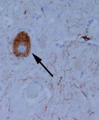
Tangle-like tau protein aggregate in a patient with progressive supranuclear palsy

Tangle-like tau protein aggregate in an individual with progressive supranuclear palsy

Image: “Tangle-like tau protein aggregate in a patient with progressive supranuclear palsy” by Ling et al. License: CC BY 2.0, cropped by Lecturio.

Clinical presentation

History:

  • Insidious onset
  • An akinetic-rigid form of parkinsonism Parkinsonism West Nile Virus
    • Falls and gait Gait Manner or style of walking. Neurological Examination difficulty
    • Insomnia Insomnia Insomnia is a sleep disorder characterized by difficulty in the initiation, maintenance, and consolidation of sleep, leading to impairment of function. Patients may exhibit symptoms such as difficulty falling asleep, disrupted sleep, trouble going back to sleep, early awakenings, and feeling tired upon waking. Insomnia and difficulty maintaining sleep Sleep A readily reversible suspension of sensorimotor interaction with the environment, usually associated with recumbency and immobility. Physiology of Sleep
    • Personality changes

Physical examination:

  • Parkinsonism Parkinsonism West Nile Virus
    • Pseudobulbar palsy Palsy paralysis of an area of the body, thus incapable of voluntary movement Cranial Nerve Palsies: spastic dysarthria Dysarthria Disorders of speech articulation caused by imperfect coordination of pharynx, larynx, tongue, or face muscles. This may result from cranial nerve diseases; neuromuscular diseases; cerebellar diseases; basal ganglia diseases; brain stem diseases; or diseases of the corticobulbar tracts. The cortical language centers are intact in this condition. Wilson Disease and dysphagia Dysphagia Dysphagia is the subjective sensation of difficulty swallowing. Symptoms can range from a complete inability to swallow, to the sensation of solids or liquids becoming “stuck.” Dysphagia is classified as either oropharyngeal or esophageal, with esophageal dysphagia having 2 sub-types: functional and mechanical. Dysphagia
    • Facial dystonia Dystonia Dystonia is a hyperkinetic movement disorder characterized by the involuntary contraction of muscles, resulting in abnormal postures or twisting and repetitive movements. Dystonia can present in various ways as may affect many different skeletal muscle groups. Dystonia: expression of “perpetual surprise”
    • Bradykinesia Bradykinesia Parkinson’s Disease
    • Micrographia
  • Eye movement abnormalities
    • Supranuclear ophthalmoplegia Ophthalmoplegia Paralysis of one or more of the ocular muscles due to disorders of the eye muscles, neuromuscular junction, supporting soft tissue, tendons, or innervation to the muscles. Orbital and Preseptal Cellulitis is the hallmark of PSP.
    • Blepharospasm and eyelid-opening apraxia Apraxia A group of cognitive disorders characterized by the inability to perform previously learned skills that cannot be attributed to deficits of motor or sensory function. The two major subtypes of this condition are ideomotor and ideational apraxia, which refers to loss of the ability to mentally formulate the processes involved with performing an action. For example, dressing apraxia may result from an inability to mentally formulate the act of placing clothes on the body. Apraxias are generally associated with lesions of the dominant parietal lobe and supramarginal gyrus. Cranial Nerve Palsies
    • Eyes are immobile at the end of the disease.
  • “Drunken sailor” gait Gait Manner or style of walking. Neurological Examination
    • Stiff and broad-based gait Gait Manner or style of walking. Neurological Examination with extended knees and trunk
    • Turns by pivoting quickly on their toes, rather than turning “ en EN Erythema nodosum is an immune-mediated panniculitis (inflammation of the subcutaneous fat) caused by a type IV (delayed-type) hypersensitivity reaction. It commonly manifests in young women as tender, erythematous nodules on the shins. Erythema Nodosum bloc”
  • Pyramidal signs
    • Hyperreflexia
    • Babinski signs
  • Cognitive dysfunction
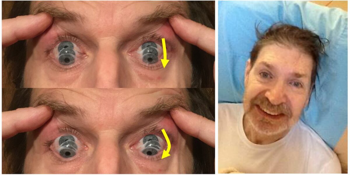
Parkinsonian syndrome - round the houses sign

Left: The photographs show the “round-the-houses” sign, illustrated for a downward saccade. Note the lateral curvature of downward path of eye movement (yellow arrows). The velocity may also be slow.
Right: This face gives many clues to the diagnosis. As shown by this man, there may be retrocollis, raised eyebrows, and frontalis overactivity. People with progressive supranuclear palsy may have a rather fixed smile, with lips drawn back rather than up.

Image: “‘Round-the-houses’ sign” by James B. Rowe, University of Cambridge Department of Clinical Neurosciences, Cambridge, Cambridgeshire, UK. License: CC BY 4.0

Diagnosis

Initial diagnosis requires fulfillment of diagnostic criteria for PSP. Definitive diagnosis can only be made via pathologic examination. 

  • Inclusion criteria:
    • Age at onset: > 40 years
    • Gradual progression
    • Vertical, supranuclear gaze abnormalities 
    • Postural instability with frequent falls 
  • Exclusion criteria (cannot have these):
    • Cortical dementia Dementia Major neurocognitive disorders (NCD), also known as dementia, are a group of diseases characterized by decline in a person’s memory and executive function. These disorders are progressive and persistent diseases that are the leading cause of disability among elderly people worldwide. Major Neurocognitive Disorders similar to Alzheimer’s 
    • Hallucinations Hallucinations Subjectively experienced sensations in the absence of an appropriate stimulus, but which are regarded by the individual as real. They may be of organic origin or associated with mental disorders. Schizophrenia or delusions not secondary to exogenous dopamine Dopamine One of the catecholamine neurotransmitters in the brain. It is derived from tyrosine and is the precursor to norepinephrine and epinephrine. Dopamine is a major transmitter in the extrapyramidal system of the brain, and important in regulating movement. Receptors and Neurotransmitters of the CNS 
    • Neuroimaging Neuroimaging Non-invasive methods of visualizing the central nervous system, especially the brain, by various imaging modalities. Febrile Infant suggestive of structural abnormality
    • Asymmetric parkinsonian signs

Testing

  • Neuroimaging Neuroimaging Non-invasive methods of visualizing the central nervous system, especially the brain, by various imaging modalities. Febrile Infant with MRI:
    • Atrophy Atrophy Decrease in the size of a cell, tissue, organ, or multiple organs, associated with a variety of pathological conditions such as abnormal cellular changes, ischemia, malnutrition, or hormonal changes. Cellular Adaptation of the midbrain Midbrain The middle of the three primitive cerebral vesicles of the embryonic brain. Without further subdivision, midbrain develops into a short, constricted portion connecting the pons and the diencephalon. Midbrain contains two major parts, the dorsal tectum mesencephali and the ventral tegmentum mesencephali, housing components of auditory, visual, and other sensorimotor systems. Brain Stem: Anatomy, quadrigeminal plate, pons Pons The front part of the hindbrain (rhombencephalon) that lies between the medulla and the midbrain (mesencephalon) ventral to the cerebellum. It is composed of two parts, the dorsal and the ventral. The pons serves as a relay station for neural pathways between the cerebellum to the cerebrum. Brain Stem: Anatomy, and cerebellum Cerebellum The cerebellum, Latin for “little brain,” is located in the posterior cranial fossa, dorsal to the pons and midbrain, and its principal role is in the coordination of movements. The cerebellum consists of 3 lobes on either side of its 2 hemispheres and is connected in the middle by the vermis. Cerebellum: Anatomy
    • Degeneration of the red nucleus Nucleus Within a eukaryotic cell, a membrane-limited body which contains chromosomes and one or more nucleoli (cell nucleolus). The nuclear membrane consists of a double unit-type membrane which is perforated by a number of pores; the outermost membrane is continuous with the endoplasmic reticulum. A cell may contain more than one nucleus. The Cell: Organelles
    • Enlargement of the aqueduct and 3rd ventricle
    • Atrophy Atrophy Decrease in the size of a cell, tissue, organ, or multiple organs, associated with a variety of pathological conditions such as abnormal cellular changes, ischemia, malnutrition, or hormonal changes. Cellular Adaptation of the frontal Frontal The bone that forms the frontal aspect of the skull. Its flat part forms the forehead, articulating inferiorly with the nasal bone and the cheek bone on each side of the face. Skull: Anatomy and temporal lobes indicate advanced disease
    • Hummingbird sign: Midsagittal cuts of midbrain Midbrain The middle of the three primitive cerebral vesicles of the embryonic brain. Without further subdivision, midbrain develops into a short, constricted portion connecting the pons and the diencephalon. Midbrain contains two major parts, the dorsal tectum mesencephali and the ventral tegmentum mesencephali, housing components of auditory, visual, and other sensorimotor systems. Brain Stem: Anatomy look like a hummingbird because of atrophy Atrophy Decrease in the size of a cell, tissue, organ, or multiple organs, associated with a variety of pathological conditions such as abnormal cellular changes, ischemia, malnutrition, or hormonal changes. Cellular Adaptation.
  • Labs:
    • No diagnostic tests Diagnostic tests Diagnostic tests are important aspects in making a diagnosis. Some of the most important epidemiological values of diagnostic tests include sensitivity and specificity, false positives and false negatives, positive and negative predictive values, likelihood ratios, and pre-test and post-test probabilities. Epidemiological Values of Diagnostic Tests
    • Labs to rule out other conditions
Mri and datscan (nuclear imaging test that allows doctors to view the brain's dopamine levels) features in psp

MRI and DaTscan (nuclear imaging test that allows doctors to view the brain’s dopamine levels) features in progressive supranuclear palsy (PSP)

Image: “MRI and DaTscan features in progressive supranuclear palsy” by James B. Rowe, University of Cambridge Department of Clinical Neurosciences, Cambridge, Cambridgeshire, UK. License: CC BY 4.0

Management

  • No definitive treatment 
  • Medical therapy:
    • Levodopa Levodopa The naturally occurring form of dihydroxyphenylalanine and the immediate precursor of dopamine. Unlike dopamine itself, it can be taken orally and crosses the blood-brain barrier. It is rapidly taken up by dopaminergic neurons and converted to dopamine. It is used for the treatment of parkinsonian disorders and is usually given with agents that inhibit its conversion to dopamine outside of the central nervous system. Parkinson’s Disease Drugs/carbidopa for parkinsonian symptoms
    • Injections of botulinum toxin Botulinum toxin Toxic proteins produced from the species Clostridium botulinum. The toxins are synthesized as a single peptide chain which is processed into a mature protein consisting of a heavy chain and light chain joined via a disulfide bond. The botulinum toxin light chain is a zinc-dependent protease which is released from the heavy chain upon endocytosis into presynaptic nerve endings. Once inside the cell the botulinum toxin light chain cleaves specific snare proteins which are essential for secretion of acetylcholine by synaptic vesicles. This inhibition of acetylcholine release results in muscular paralysis. Botulism can be used for focal dystonia Focal Dystonia Dystonia.

Prognosis Prognosis A prediction of the probable outcome of a disease based on a individual’s condition and the usual course of the disease as seen in similar situations. Non-Hodgkin Lymphomas

  • Rapid disease progression
  • Significant reduction in quality Quality Activities and programs intended to assure or improve the quality of care in either a defined medical setting or a program. The concept includes the assessment or evaluation of the quality of care; identification of problems or shortcomings in the delivery of care; designing activities to overcome these deficiencies; and follow-up monitoring to ensure effectiveness of corrective steps. Quality Measurement and Improvement of life and dependency within 3–4 years
  • Death usually occurs within 6 years after diagnosis.
  • Dysphagia Dysphagia Dysphagia is the subjective sensation of difficulty swallowing. Symptoms can range from a complete inability to swallow, to the sensation of solids or liquids becoming “stuck.” Dysphagia is classified as either oropharyngeal or esophageal, with esophageal dysphagia having 2 sub-types: functional and mechanical. Dysphagia is associated with ↓ survival.

Multiple System Atrophy

Definition

Multiple system atrophy Atrophy Decrease in the size of a cell, tissue, organ, or multiple organs, associated with a variety of pathological conditions such as abnormal cellular changes, ischemia, malnutrition, or hormonal changes. Cellular Adaptation (MSA) is an adult-onset, sporadic Sporadic Selective IgA Deficiency, rapidly progressive, multisystem, fatal neurodegenerative disease characterized by parkinsonian features, cerebellar, autonomic, and urogenital dysfunction; and corticospinal disorders.
Epidemiology and etiology

  • Incidence Incidence The number of new cases of a given disease during a given period in a specified population. It also is used for the rate at which new events occur in a defined population. It is differentiated from prevalence, which refers to all cases in the population at a given time. Measures of Disease Frequency: 3 per 100,000
  • Prevalence Prevalence The total number of cases of a given disease in a specified population at a designated time. It is differentiated from incidence, which refers to the number of new cases in the population at a given time. Measures of Disease Frequency: 2–5 cases per 100,000
  • Mean age at onset: 54 years
  • Unknown etiology, but trauma has been suggested.

Pathophysiology

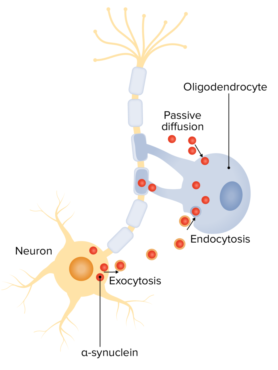
  • Spreading of aberrant α-synuclein from neurons Neurons The basic cellular units of nervous tissue. Each neuron consists of a body, an axon, and dendrites. Their purpose is to receive, conduct, and transmit impulses in the nervous system. Nervous System: Histology to glial cells Glial Cells The non-neuronal cells of the nervous system. They not only provide physical support, but also respond to injury, regulate the ionic and chemical composition of the extracellular milieu, participate in the blood-brain barrier and blood-retinal barrier, form the myelin insulation of nervous pathways, guide neuronal migration during development, and exchange metabolites with neurons. Neuroglia have high-affinity transmitter uptake systems, voltage-dependent and transmitter-gated ion channels, and can release transmitters, but their role in signaling (as in many other functions) is unclear. Nervous System: Histology → chronic alterations in glial cells Glial Cells The non-neuronal cells of the nervous system. They not only provide physical support, but also respond to injury, regulate the ionic and chemical composition of the extracellular milieu, participate in the blood-brain barrier and blood-retinal barrier, form the myelin insulation of nervous pathways, guide neuronal migration during development, and exchange metabolites with neurons. Neuroglia have high-affinity transmitter uptake systems, voltage-dependent and transmitter-gated ion channels, and can release transmitters, but their role in signaling (as in many other functions) is unclear. Nervous System: Histology → impaired trophic function between oligodendrocytes and axons Axons Nerve fibers that are capable of rapidly conducting impulses away from the neuron cell body. Nervous System: Histology →  secondary neuronal damage + extensive myelin degeneration
  • Glial and myelin dysfunction occur.
  • An inflammatory cascade is induced, and secondary neurodegeneration follows.
Alpha-synuclein in the pathogenesis of msa

Alpha-synuclein in the pathogenesis of multiple system atrophy (MSA):
Neuron-oligodendrocyte interaction mechanisms potentially involved in α-syn accumulation: oligodendroglial α-syn uptake from surrounding neurons and extracellular environment through endocytosis and passive transmembrane diffusion

Image by Lecturio.

Clinical presentation

History:

Physical examination:

  • In parkinsonism-predominant cases:
    • Akinesia/ bradykinesia Bradykinesia Parkinson’s Disease
    • Rigidity Rigidity Continuous involuntary sustained muscle contraction which is often a manifestation of basal ganglia diseases. When an affected muscle is passively stretched, the degree of resistance remains constant regardless of the rate at which the muscle is stretched. This feature helps to distinguish rigidity from muscle spasticity. Megacolon
    • Postural instability
    • Postural and action tremor Tremor Cyclical movement of a body part that can represent either a physiologic process or a manifestation of disease. Intention or action tremor, a common manifestation of cerebellar diseases, is aggravated by movement. In contrast, resting tremor is maximal when there is no attempt at voluntary movement, and occurs as a relatively frequent manifestation of parkinson disease. Myotonic Dystrophies
  • In cerebellar-predominant cases ( cerebellar disorders Cerebellar disorders Cerebellar disorders are a specific set of neurologic signs and symptoms caused by local or systemic conditions that affect the cerebellum; the classic sign is ataxia, in addition to several other motor abnormalities that affect coordination. Cerebellar Disorders):
    • Ataxia Ataxia Impairment of the ability to perform smoothly coordinated voluntary movements. This condition may affect the limbs, trunk, eyes, pharynx, larynx, and other structures. Ataxia may result from impaired sensory or motor function. Sensory ataxia may result from posterior column injury or peripheral nerve diseases. Motor ataxia may be associated with cerebellar diseases; cerebral cortex diseases; thalamic diseases; basal ganglia diseases; injury to the red nucleus; and other conditions. Ataxia-telangiectasia
    • Equilibrium Equilibrium Occurs when tumor cells survive the initial elimination attempt These cells are not able to progress, being maintained in a state of dormancy by the adaptive immune system. In this phase, tumor immunogenicity is edited, where T cells keep selectively attacking highly immunogenic tumor cells.This attack leaves other cells with less immunogenicity to potentially develop resistance to the immune response. Cancer Immunotherapy and gait Gait Manner or style of walking. Neurological Examination problems
    • Dysdiadochokinesia Dysdiadochokinesia Difficulty with rapid alternating movements Cerebellar Disorders: difficulty making rapid alternating movements
    • Dysarthria Dysarthria Disorders of speech articulation caused by imperfect coordination of pharynx, larynx, tongue, or face muscles. This may result from cranial nerve diseases; neuromuscular diseases; cerebellar diseases; basal ganglia diseases; brain stem diseases; or diseases of the corticobulbar tracts. The cortical language centers are intact in this condition. Wilson Disease
    • Ocular abnormalities
  • Cognitive function is usually preserved.

Diagnosis

Diagnosis is made clinically, and definitive diagnosis is done on postmortem pathologic examination. 
  • Clinical features:
    • Sporadic Sporadic Selective IgA Deficiency and progressive onset during adulthood (> 30 years old)
    • Autonomic failure involving either:
      • Urinary incontinence Urinary incontinence Urinary incontinence (UI) is involuntary loss of bladder control or unintentional voiding, which represents a hygienic or social problem to the patient. Urinary incontinence is a symptom, a sign, and a disorder. The 5 types of UI include stress, urge, mixed, overflow, and functional. Urinary Incontinence or 
      • Orthostatic BP
    • Levodopa Levodopa The naturally occurring form of dihydroxyphenylalanine and the immediate precursor of dopamine. Unlike dopamine itself, it can be taken orally and crosses the blood-brain barrier. It is rapidly taken up by dopaminergic neurons and converted to dopamine. It is used for the treatment of parkinsonian disorders and is usually given with agents that inhibit its conversion to dopamine outside of the central nervous system. Parkinson’s Disease Drugs trial: does not reduce symptoms, unlike in Parkinson’s disease
  • Imaging:
    • Atrophy Atrophy Decrease in the size of a cell, tissue, organ, or multiple organs, associated with a variety of pathological conditions such as abnormal cellular changes, ischemia, malnutrition, or hormonal changes. Cellular Adaptation of the:
      • Putamen
      • Middle cerebellar peduncle Middle cerebellar peduncle White matter consisting of a large bundle of paired fibers originating in the pontine nuclei in the pre pontis and the nucleus reticularis tegmenti pontis projecting to the contralateral cerebral cortex. Cerebellum: Anatomy
      • Pons Pons The front part of the hindbrain (rhombencephalon) that lies between the medulla and the midbrain (mesencephalon) ventral to the cerebellum. It is composed of two parts, the dorsal and the ventral. The pons serves as a relay station for neural pathways between the cerebellum to the cerebrum. Brain Stem: Anatomy
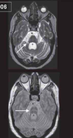
    • “Hot cross bun sign” on T2-weighted imaging: hyperintense Hyperintense Magnetic Resonance Imaging (MRI) shape of a cross within the atrophied pons Pons The front part of the hindbrain (rhombencephalon) that lies between the medulla and the midbrain (mesencephalon) ventral to the cerebellum. It is composed of two parts, the dorsal and the ventral. The pons serves as a relay station for neural pathways between the cerebellum to the cerebrum. Brain Stem: Anatomy
Mri of a brain showing multiple system atrophy (msa)

MRI of a brain showing multiple system atrophy (MSA):
Note the brain stem and cerebellar atrophy (top, arrow), and the “hot cross bun” sign (bottom, arrow).

Image: “MRI of a patient with multiple system atrophy (MSA)” by J. Vijayan, S. Sinha, S. Ravishankar,1 and A. B. Taly. License: CC BY 4.0

Management

  • No definitive treatment 
  • Medical therapy:
    • Botulinum toxin Botulinum toxin Toxic proteins produced from the species Clostridium botulinum. The toxins are synthesized as a single peptide chain which is processed into a mature protein consisting of a heavy chain and light chain joined via a disulfide bond. The botulinum toxin light chain is a zinc-dependent protease which is released from the heavy chain upon endocytosis into presynaptic nerve endings. Once inside the cell the botulinum toxin light chain cleaves specific snare proteins which are essential for secretion of acetylcholine by synaptic vesicles. This inhibition of acetylcholine release results in muscular paralysis. Botulism injections can be used for focal dystonia Focal Dystonia Dystonia.
    • Orthostatic hypotension Orthostatic hypotension A significant drop in blood pressure after assuming a standing position. Orthostatic hypotension is a finding, and defined as a 20-mm hg decrease in systolic pressure or a 10-mm hg decrease in diastolic pressure 3 minutes after the person has risen from supine to standing. Symptoms generally include dizziness, blurred vision, and syncope. Hypotension: fludrocortisone Fludrocortisone A synthetic mineralocorticoid with anti-inflammatory activity. Mineralocorticoids acetate (1st-line therapy), midodrine Midodrine An ethanolamine derivative that is an adrenergic alpha-1 agonist. It is used as a vasoconstrictor agent in the treatment of hypotension. Sympathomimetic Drugs (2nd-line therapy)
    • Urogenital symptoms: oxybutynin Oxybutynin Anticholinergic Drugs or tolterodine Tolterodine An antimuscarinic agent selective for the muscarinic receptors of the bladder that is used in the treatment of urinary incontinence and urinary urge incontinence. Anticholinergic Drugs

Prognosis Prognosis A prediction of the probable outcome of a disease based on a individual’s condition and the usual course of the disease as seen in similar situations. Non-Hodgkin Lymphomas

  • Rapid progression
  • Death as soon as 6 years after diagnosis

Corticobasal Degeneration

Definition

Corticobasal degeneration (CBD) is a progressive, asymmetric dementia Dementia Major neurocognitive disorders (NCD), also known as dementia, are a group of diseases characterized by decline in a person’s memory and executive function. These disorders are progressive and persistent diseases that are the leading cause of disability among elderly people worldwide. Major Neurocognitive Disorders–movement disorder with sporadic Sporadic Selective IgA Deficiency neurodegenerative 4-repeat tauopathy.

Epidemiology and etiology

  • Prevalence Prevalence The total number of cases of a given disease in a specified population at a designated time. It is differentiated from incidence, which refers to the number of new cases in the population at a given time. Measures of Disease Frequency: 5–7 per 100,000
  • Incidence Incidence The number of new cases of a given disease during a given period in a specified population. It also is used for the rate at which new events occur in a defined population. It is differentiated from prevalence, which refers to all cases in the population at a given time. Measures of Disease Frequency: 0.6–0.9 per 100,000
  • 5% of cases of parkinsonism Parkinsonism West Nile Virus
  • 6th to 8th decades of life
  • May be related to abnormalities in the microtubule-associated protein tau (MAPT) gene Gene A category of nucleic acid sequences that function as units of heredity and which code for the basic instructions for the development, reproduction, and maintenance of organisms. Basic Terms of Genetics

Pathophysiology

  • Hyperphosphorylation of tau protein leads to less affinity for microtubules Microtubules Slender, cylindrical filaments found in the cytoskeleton of plant and animal cells. They are composed of the protein tubulin and are influenced by tubulin modulators. The Cell: Cytosol and Cytoskeleton → hyperphosphorylated tau protein resists proteolysis and forms aggregates → tau protein is accumulated and neurofibrillary tangles Neurofibrillary Tangles Abnormal structures located in various parts of the brain and composed of dense arrays of paired helical filaments (neurofilaments and microtubules). These double helical stacks of transverse subunits are twisted into left-handed ribbon-like filaments that likely incorporate the following proteins: (1) the intermediate filaments: medium- and high-molecular-weight neurofilaments; (2) the microtubule-associated proteins map-2 and tau; (3) actin; and (4) ubiquitins. As one of the hallmarks of alzheimer disease, the neurofibrillary tangles eventually occupy the whole of the cytoplasm in certain classes of cell in the neocortex, hippocampus, brain stem, and diencephalon. The number of these tangles, as seen in post mortem histology, correlates with the degree of dementia during life. Some studies suggest that tangle antigens leak into the systemic circulation both in the course of normal aging and in cases of alzheimer disease. Alzheimer Disease are formed
  • Classified as a 4-repeat tauopathy (a different interval than other tau-related syndromes)
  • Ballooned, achromatic, tau-positive neurons Neurons The basic cellular units of nervous tissue. Each neuron consists of a body, an axon, and dendrites. Their purpose is to receive, conduct, and transmit impulses in the nervous system. Nervous System: Histology, astrocytic plaques, and other dystrophic glial tau findings, similar to those seen in PSP
  • Extensive neuronal loss

Clinical presentation

History:

  • Asymmetrical affectation
  • Frequent falls
  • Behavioral changes consistent with dementia Dementia Major neurocognitive disorders (NCD), also known as dementia, are a group of diseases characterized by decline in a person’s memory and executive function. These disorders are progressive and persistent diseases that are the leading cause of disability among elderly people worldwide. Major Neurocognitive Disorders

Physical examination:

  • May display PSP features
  • Bradykinesia Bradykinesia Parkinson’s Disease/akinesia
  • Limb rigidity Rigidity Continuous involuntary sustained muscle contraction which is often a manifestation of basal ganglia diseases. When an affected muscle is passively stretched, the degree of resistance remains constant regardless of the rate at which the muscle is stretched. This feature helps to distinguish rigidity from muscle spasticity. Megacolon and dystonia Dystonia Dystonia is a hyperkinetic movement disorder characterized by the involuntary contraction of muscles, resulting in abnormal postures or twisting and repetitive movements. Dystonia can present in various ways as may affect many different skeletal muscle groups. Dystonia
  • Speech disturbances
  • Action tremor Tremor Cyclical movement of a body part that can represent either a physiologic process or a manifestation of disease. Intention or action tremor, a common manifestation of cerebellar diseases, is aggravated by movement. In contrast, resting tremor is maximal when there is no attempt at voluntary movement, and occurs as a relatively frequent manifestation of parkinson disease. Myotonic Dystrophies, myoclonus Myoclonus Involuntary shock-like contractions, irregular in rhythm and amplitude, followed by relaxation, of a muscle or a group of muscles. This condition may be a feature of some central nervous system diseases; (e.g., epilepsy-myoclonic). Nocturnal myoclonus is the principal feature of the nocturnal myoclonus syndrome. Neurological Examination, and hyperreflexia
  • “Alien-limb” phenomena: unintended motor Motor Neurons which send impulses peripherally to activate muscles or secretory cells. Nervous System: Histology actions such as grasping, groping, drifting, or undoing Undoing Defense Mechanisms
  • Slow gait Gait Manner or style of walking. Neurological Examination
  • Dementia Dementia Major neurocognitive disorders (NCD), also known as dementia, are a group of diseases characterized by decline in a person’s memory and executive function. These disorders are progressive and persistent diseases that are the leading cause of disability among elderly people worldwide. Major Neurocognitive Disorders 
  • Slow and saccadic eye movements Saccadic Eye Movements Internuclear Ophthalmoplegia

Diagnosis

Diagnosis is made clinically, and definitive diagnosis is by postmortem pathology examination. 
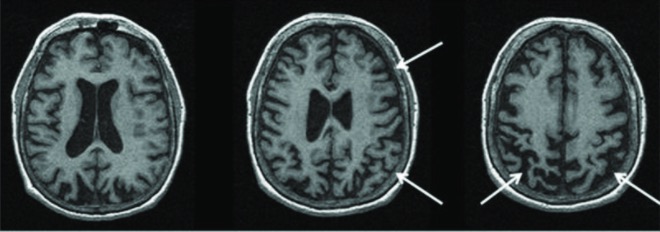
Magnetic resonance imaging of a patient with a pathology-proven diagnosis of corticobasal degeneration

MRI of a brain showing a pathologic diagnosis of corticobasal degeneration:
MRI shows the right greater than left parietofrontal atrophy typical of corticobasal syndrome.

Image: “Magnetic resonance imaging of a patient with a pathology-proven diagnosis of corticobasal degeneration” by Saeed et al. License: CC BY 4.0

Medical therapy

  • Levodopa Levodopa The naturally occurring form of dihydroxyphenylalanine and the immediate precursor of dopamine. Unlike dopamine itself, it can be taken orally and crosses the blood-brain barrier. It is rapidly taken up by dopaminergic neurons and converted to dopamine. It is used for the treatment of parkinsonian disorders and is usually given with agents that inhibit its conversion to dopamine outside of the central nervous system. Parkinson’s Disease Drugs trial: does not reduce symptoms, unlike in Parkinson’s disease
  • No definitive treatment
  • Propranolol Propranolol A widely used non-cardioselective beta-adrenergic antagonist. Propranolol has been used for myocardial infarction; arrhythmia; angina pectoris; hypertension; hyperthyroidism; migraine; pheochromocytoma; and anxiety but adverse effects instigate replacement by newer drugs. Antiadrenergic Drugs or clonazepam Clonazepam An anticonvulsant used for several types of seizures, including myotonic or atonic seizures, photosensitive epilepsy, and absence seizures, although tolerance may develop. It is seldom effective in generalized tonic-clonic or partial seizures. The mechanism of action appears to involve the enhancement of gamma-aminobutyric acid receptor responses. Benzodiazepines can be used to treat tremor Tremor Cyclical movement of a body part that can represent either a physiologic process or a manifestation of disease. Intention or action tremor, a common manifestation of cerebellar diseases, is aggravated by movement. In contrast, resting tremor is maximal when there is no attempt at voluntary movement, and occurs as a relatively frequent manifestation of parkinson disease. Myotonic Dystrophies and myoclonus Myoclonus Involuntary shock-like contractions, irregular in rhythm and amplitude, followed by relaxation, of a muscle or a group of muscles. This condition may be a feature of some central nervous system diseases; (e.g., epilepsy-myoclonic). Nocturnal myoclonus is the principal feature of the nocturnal myoclonus syndrome. Neurological Examination.
  • Cholinesterase Cholinesterase Liver Function Tests inhibitors (e.g., donepezil Donepezil An indan and piperidine derivative that acts as a selective and reversible inhibitor of acetylcholinesterase. Donepezil is highly selective for the central nervous system and is used in the management of mild to moderate dementia in alzheimer disease. Cholinomimetic Drugs, rivastigmine Rivastigmine A carbamate-derived reversible cholinesterase inhibitor that is selective for the central nervous system and is used for the treatment of dementia in alzheimer disease and parkinson disease. Cholinomimetic Drugs) can be used to treat cognitive dysfunction.

Prognosis Prognosis A prediction of the probable outcome of a disease based on a individual’s condition and the usual course of the disease as seen in similar situations. Non-Hodgkin Lymphomas

  • Poor
  • Death as soon as 5 years after diagnosis

Differential Diagnosis

  • Parkinson’s disease: degenerative disease of the basal ganglia Basal Ganglia Basal ganglia are a group of subcortical nuclear agglomerations involved in movement, and are located deep to the cerebral hemispheres. Basal ganglia include the striatum (caudate nucleus and putamen), globus pallidus, substantia nigra, and subthalamic nucleus. Basal Ganglia: Anatomy. Parkinson’s disease is characterized by diminished facial expression, stooped posture, slowing of voluntary movement, festinating gait Gait Manner or style of walking. Neurological Examination (progressively shortened and accelerated steps), cogwheel rigidity Cogwheel Rigidity Parkinson’s Disease, and a “pill rolling Rolling Movement of tethered, spherical leukocytes along the endothelial surface of the microvasculature. The tethering and rolling involves interaction with selectins and other adhesion molecules in both the endothelium and leukocyte. The rolling leukocyte then becomes activated by chemokines, flattens out, and firmly adheres to the endothelial surface in preparation for transmigration through the interendothelial cell junction. Inflammation tremor Tremor Cyclical movement of a body part that can represent either a physiologic process or a manifestation of disease. Intention or action tremor, a common manifestation of cerebellar diseases, is aggravated by movement. In contrast, resting tremor is maximal when there is no attempt at voluntary movement, and occurs as a relatively frequent manifestation of parkinson disease. Myotonic Dystrophies at rest. 
  • Frontotemporal dementia Dementia Major neurocognitive disorders (NCD), also known as dementia, are a group of diseases characterized by decline in a person’s memory and executive function. These disorders are progressive and persistent diseases that are the leading cause of disability among elderly people worldwide. Major Neurocognitive Disorders: neurodegenerative disorder characterized by loss of cognitive functions ( memory Memory Complex mental function having four distinct phases: (1) memorizing or learning, (2) retention, (3) recall, and (4) recognition. Clinically, it is usually subdivided into immediate, recent, and remote memory. Psychiatric Assessment, abstract thinking, reasoning Reasoning Decision-making Capacity and Legal Competence, and executive function), which severely impairs functioning and quality Quality Activities and programs intended to assure or improve the quality of care in either a defined medical setting or a program. The concept includes the assessment or evaluation of the quality of care; identification of problems or shortcomings in the delivery of care; designing activities to overcome these deficiencies; and follow-up monitoring to ensure effectiveness of corrective steps. Quality Measurement and Improvement of life. Clinical presentation includes behavioral changes, such as apathy Apathy Lack of emotion or emotional expression; a disorder of motivation that persists over time. Wernicke Encephalopathy and Korsakoff Syndrome.
  • Wilson disease Wilson disease Wilson disease (hepatolenticular degeneration) is an autosomal recessive disorder caused by various mutations in the ATP7B gene, which regulates copper transport within hepatocytes. Dysfunction of this transport mechanism leads to abnormal copper accumulations in the liver, brain, eyes, and other organs, with consequent major and variably expressed hepatic, neurologic, and psychiatric disturbances. Wilson Disease: autosomal recessive Autosomal recessive Autosomal inheritance, both dominant and recessive, refers to the transmission of genes from the 22 autosomal chromosomes. Autosomal recessive diseases are only expressed when 2 copies of the recessive allele are inherited. Autosomal Recessive and Autosomal Dominant Inheritance disorder associated with mutations in the ATP7B gene Gene A category of nucleic acid sequences that function as units of heredity and which code for the basic instructions for the development, reproduction, and maintenance of organisms. Basic Terms of Genetics, which regulates copper Copper A heavy metal trace element with the atomic symbol cu, atomic number 29, and atomic weight 63. 55. Trace Elements transport within hepatocytes Hepatocytes The main structural component of the liver. They are specialized epithelial cells that are organized into interconnected plates called lobules. Liver: Anatomy. This mutation Mutation Genetic mutations are errors in DNA that can cause protein misfolding and dysfunction. There are various types of mutations, including chromosomal, point, frameshift, and expansion mutations. Types of Mutations causes copper Copper A heavy metal trace element with the atomic symbol cu, atomic number 29, and atomic weight 63. 55. Trace Elements to accumulate in the liver Liver The liver is the largest gland in the human body. The liver is found in the superior right quadrant of the abdomen and weighs approximately 1.5 kilograms. Its main functions are detoxification, metabolism, nutrient storage (e.g., iron and vitamins), synthesis of coagulation factors, formation of bile, filtration, and storage of blood. Liver: Anatomy, brain Brain The part of central nervous system that is contained within the skull (cranium). Arising from the neural tube, the embryonic brain is comprised of three major parts including prosencephalon (the forebrain); mesencephalon (the midbrain); and rhombencephalon (the hindbrain). The developed brain consists of cerebrum; cerebellum; and other structures in the brain stem. Nervous System: Anatomy, Structure, and Classification, and cornea Cornea The transparent anterior portion of the fibrous coat of the eye consisting of five layers: stratified squamous corneal epithelium; bowman membrane; corneal stroma; descemet membrane; and mesenchymal corneal endothelium. It serves as the first refracting medium of the eye. Eye: Anatomy. Wilson disease Wilson disease Wilson disease (hepatolenticular degeneration) is an autosomal recessive disorder caused by various mutations in the ATP7B gene, which regulates copper transport within hepatocytes. Dysfunction of this transport mechanism leads to abnormal copper accumulations in the liver, brain, eyes, and other organs, with consequent major and variably expressed hepatic, neurologic, and psychiatric disturbances. Wilson Disease often presents in young adulthood with signs of liver Liver The liver is the largest gland in the human body. The liver is found in the superior right quadrant of the abdomen and weighs approximately 1.5 kilograms. Its main functions are detoxification, metabolism, nutrient storage (e.g., iron and vitamins), synthesis of coagulation factors, formation of bile, filtration, and storage of blood. Liver: Anatomy disease and neurologic and psychiatric symptoms (often resembling parkinsonism Parkinsonism West Nile Virus).
  • Niemann-Pick disease Niemann-Pick disease Niemann-Pick disease (NPD) is a rare, inherited, lysosomal storage disorder. The disease is classified on the basis of the genetic mutation. Type A and type B result from mutations in the SMPD-1 gene, resulting in acid sphingomyelinase enzyme deficiency. Type C results from NPC1 or NPC2 gene mutations, which are needed for intracellular transport of lipids. Niemann-Pick Disease: rare inherited lysosomal storage disorder. Niemann-Pick disease Niemann-Pick disease Niemann-Pick disease (NPD) is a rare, inherited, lysosomal storage disorder. The disease is classified on the basis of the genetic mutation. Type A and type B result from mutations in the SMPD-1 gene, resulting in acid sphingomyelinase enzyme deficiency. Type C results from NPC1 or NPC2 gene mutations, which are needed for intracellular transport of lipids. Niemann-Pick Disease is classified on the basis of the genetic mutation Mutation Genetic mutations are errors in DNA that can cause protein misfolding and dysfunction. There are various types of mutations, including chromosomal, point, frameshift, and expansion mutations. Types of Mutations. Type A and type B result from mutations in the SMPD-1 gene Gene A category of nucleic acid sequences that function as units of heredity and which code for the basic instructions for the development, reproduction, and maintenance of organisms. Basic Terms of Genetics, resulting in acid sphingomyelinase Acid sphingomyelinase Niemann-Pick Disease enzyme deficiency. Type C results from NPC1 or NPC2 gene Gene A category of nucleic acid sequences that function as units of heredity and which code for the basic instructions for the development, reproduction, and maintenance of organisms. Basic Terms of Genetics mutations; these genes Genes A category of nucleic acid sequences that function as units of heredity and which code for the basic instructions for the development, reproduction, and maintenance of organisms. DNA Types and Structure are needed for intracellular transport of lipids Lipids Lipids are a diverse group of hydrophobic organic molecules, which include fats, oils, sterols, and waxes. Fatty Acids and Lipids.
  • Myasthenia gravis Myasthenia Gravis Myasthenia gravis (MG) is an autoimmune neuromuscular disorder characterized by weakness and fatigability of skeletal muscles caused by dysfunction/destruction of acetylcholine receptors at the neuromuscular junction. MG presents with fatigue, ptosis, diplopia, dysphagia, respiratory difficulties, and progressive weakness in the limbs, leading to difficulty in movement. Myasthenia Gravis: autoimmune neuromuscular disorder characterized by weakness and fatigability of skeletal muscles Skeletal muscles A subtype of striated muscle, attached by tendons to the skeleton. Skeletal muscles are innervated and their movement can be consciously controlled. They are also called voluntary muscles. Muscle Tissue: Histology caused by dysfunction/destruction of acetylcholine Acetylcholine A neurotransmitter found at neuromuscular junctions, autonomic ganglia, parasympathetic effector junctions, a subset of sympathetic effector junctions, and at many sites in the central nervous system. Receptors and Neurotransmitters of the CNS receptors Receptors Receptors are proteins located either on the surface of or within a cell that can bind to signaling molecules known as ligands (e.g., hormones) and cause some type of response within the cell. Receptors at the neuromuscular junction Neuromuscular junction The synapse between a neuron and a muscle. Skeletal Muscle Contraction. Myasthenia gravis Myasthenia Gravis Myasthenia gravis (MG) is an autoimmune neuromuscular disorder characterized by weakness and fatigability of skeletal muscles caused by dysfunction/destruction of acetylcholine receptors at the neuromuscular junction. MG presents with fatigue, ptosis, diplopia, dysphagia, respiratory difficulties, and progressive weakness in the limbs, leading to difficulty in movement. Myasthenia Gravis presents with fatigue Fatigue The state of weariness following a period of exertion, mental or physical, characterized by a decreased capacity for work and reduced efficiency to respond to stimuli. Fibromyalgia, ptosis Ptosis Cranial Nerve Palsies, diplopia Diplopia A visual symptom in which a single object is perceived by the visual cortex as two objects rather than one. Disorders associated with this condition include refractive errors; strabismus; oculomotor nerve diseases; trochlear nerve diseases; abducens nerve diseases; and diseases of the brain stem and occipital lobe. Myasthenia Gravis, dysphagia Dysphagia Dysphagia is the subjective sensation of difficulty swallowing. Symptoms can range from a complete inability to swallow, to the sensation of solids or liquids becoming “stuck.” Dysphagia is classified as either oropharyngeal or esophageal, with esophageal dysphagia having 2 sub-types: functional and mechanical. Dysphagia, respiratory difficulties, and progressive weakness in the limbs leading to difficulty in movement.

References

  1. Agarwal S, Gilbert R. (2021). Progressive supranuclear palsy. StatPearls. Retrieved August 14, 2021, from http://www.ncbi.nlm.nih.gov/books/NBK526098/ 
  2. Seeley WW, Miller BL. (2018). Frontotemporal dementia. Jameson J, Fauci AS, Kasper DL, Hauser SL, Longo DL, Loscalzo J (Eds.), Harrison’s Principles of Internal Medicine, 20th ed. McGraw-Hill. 
  3. Jankovic J. (2022). Parkinson Disease and Other Movement Disorders. In Jankovic, J., et al. (Eds.). Bradley and Daroff’s Neurology in Clinical Practice. pp. 1498–1534.e5. Elsevier.
  4. Höglinger GU, et al. (2017). Clinical diagnosis of progressive supranuclear palsy: the movement disorder society criteria. Movement Disorders 32:853–864. https://doi.org/10.1002/mds.26987 
  5. Zibetti M. (2015). Parkinson disease: epidemiology, pathology, and clinical diagnosis. DeckerMed Medicine. Retrieved July 31, 2021, from
  6. Factor, S. A., Doss Esper, C. (2021). Multiple system atrophy: Clinical features and diagnosis. UpToDate. Retrieved July 31, from https://www.uptodate.com/contents/multiple-system-atrophy-clinical-features-and-diagnosis
  7. Factor, S. A., Doss Esper, C. (2021). Multiple system atrophy: Prognosis and treatment. UpToDate. Retrieved July 31, from https://www.uptodate.com/contents/multiple-system-atrophy-prognosis-and-treatment
  8. Gibbons CH, Engstrom JW. (2018). Disorders of the autonomic nervous system. Jameson J, Fauci AS, Kasper DL, Hauser SL, Longo DL, Loscalzo J (Eds.), Harrison’s Principles of Internal Medicine, 20th ed. McGraw-Hill. 
  9. Factor, S.A., Tripathi, R. (2021). Corticobasal degeneration. UpToDate. Retrieved July 31, from https://www.uptodate.com/contents/corticobasal-degeneration
  10. Eggenberger, E. (2018). Progressive supranuclear palsy. Emedicine. Retrieved August 9, 2021, from https://emedicine.medscape.com/article/1151430-overview?ecd=ppc_google_rlsa-traf_mscp_emed_md-ldlm-cohort_us#a6 
  11. Diedrich A. (2018). Multiple system atrophy. Emedicine. Retrieved August 9, 2021, from https://emedicine.medscape.com/article/1154583-overview?ecd=ppc_google_rlsa-traf_mscp_emed_md-ldlm-cohort_us#a5
  12. Pantelyat A. (2019). Corticobasal syndrome and corticobasal degeneration. Emedicine. Retrieved August 9, 2021, from https://emedicine.medscape.com/article/1150039-overview#a5

Create your free account or log in to continue reading!

Sign up now and get free access to Lecturio with concept pages, medical videos, and questions for your medical education.

User Reviews

Details