Advertisement

Advertisement

Advertisement

Advertisement

Shoulder Joint: Anatomy

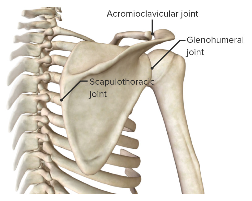
The shoulder complex comprises the glenohumeral joint, sternoclavicular joint, acromioclavicular joint, and the scapulothoracic articulation, and connects the upper limb to the trunk. This group of joints consists of the clavicle Clavicle A bone on the ventral side of the shoulder girdle, which in humans is commonly called the collar bone. Clavicle Fracture, scapula, and humerus Humerus Bone in humans and primates extending from the shoulder joint to the elbow joint. Arm: Anatomy bones, multiple muscles and supporting ligaments, cartilage Cartilage Cartilage is a type of connective tissue derived from embryonic mesenchyme that is responsible for structural support, resilience, and the smoothness of physical actions. Perichondrium (connective tissue membrane surrounding cartilage) compensates for the absence of vasculature in cartilage by providing nutrition and support. Cartilage: Histology, and bursae. The muscles ensure the mobility and stability of the shoulder and upper limb and are divided into 3 groups: anterior axioappendicular, posterior axioappendicular, and scapulohumeral muscles.

Last updated: Oct 30, 2024

Editorial responsibility: Stanley Oiseth, Lindsay Jones, Evelin Maza

Advertisement

Advertisement

Advertisement

Advertisement

Advertisement

Advertisement

Bones

Clavicle Clavicle A bone on the ventral side of the shoulder girdle, which in humans is commonly called the collar bone. Clavicle Fracture

  • The only horizontal long bone Bone Bone is a compact type of hardened connective tissue composed of bone cells, membranes, an extracellular mineralized matrix, and central bone marrow. The 2 primary types of bone are compact and spongy. Bones: Structure and Types in the body
  • S-shaped, connects the upper limb with the axial Axial Computed Tomography (CT) skeleton
  • Along with the scapula, forms the pectoral girdle
  • Landmarks and articulations:
    • Medially articulates with the sternum Sternum A long, narrow, and flat bone commonly known as breastbone occurring in the midsection of the anterior thoracic segment or chest region, which stabilizes the rib cage and serves as the point of origin for several muscles that move the arms, head, and neck. Chest Wall: Anatomy to form the sternoclavicular joint Sternoclavicular Joint Examination of the Upper Limbs
    • Laterally articulates with the scapula to form the acromioclavicular joint Acromioclavicular joint The gliding joint formed by the outer extremity of the clavicle and the inner margin of the acromion process of the scapula. Examination of the Upper Limbs
Inferior view of the right clavicle

Inferior view of the right clavicle

Image by BioDigital, edited by Lecturio

Scapula

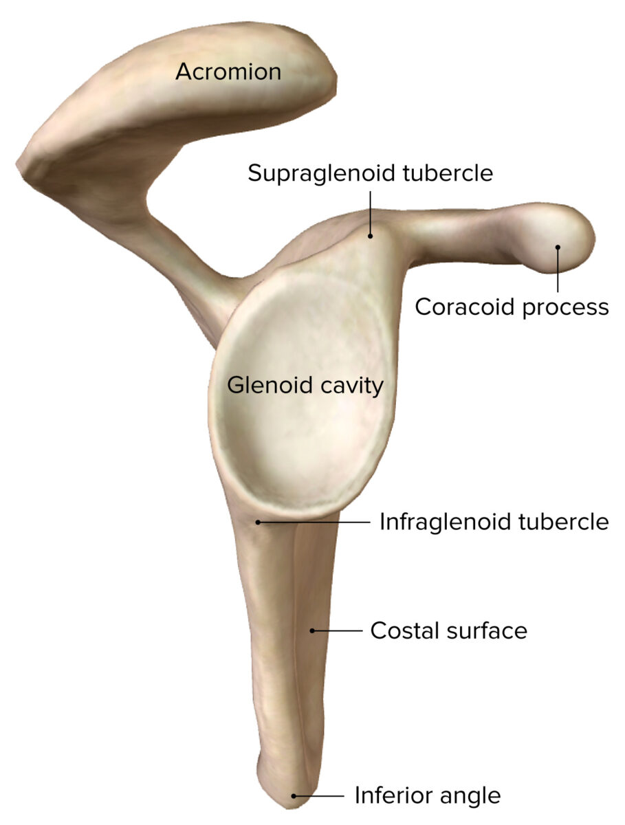
  • Flat, triangular-shaped, with 3 angles, 3 borders, and an anterior and posterior surface
  • Along with the clavicle Clavicle A bone on the ventral side of the shoulder girdle, which in humans is commonly called the collar bone. Clavicle Fracture, it forms the shoulder or pectoral girdle, connecting the upper limb to the trunk.
  • Landmarks and articulations:
    • Glenoid cavity: lateral, articulates with the head of the humerus Head of The Humerus The upper rounded extremity of the humerus fitting into the glenoid cavity of the scapula. Arm: Anatomy to form the glenohumeral joint
    • Acromion: continuation of the scapular spine Spine The human spine, or vertebral column, is the most important anatomical and functional axis of the human body. It consists of 7 cervical vertebrae, 12 thoracic vertebrae, and 5 lumbar vertebrae and is limited cranially by the skull and caudally by the sacrum. Vertebral Column: Anatomy, articulates with the lateral end of the clavicle Clavicle A bone on the ventral side of the shoulder girdle, which in humans is commonly called the collar bone. Clavicle Fracture to form the acromioclavicular joint Acromioclavicular joint The gliding joint formed by the outer extremity of the clavicle and the inner margin of the acromion process of the scapula. Examination of the Upper Limbs
    • Coracoid process: anterior, hook-shaped, place of attachment for multiple muscles and stabilizing ligaments

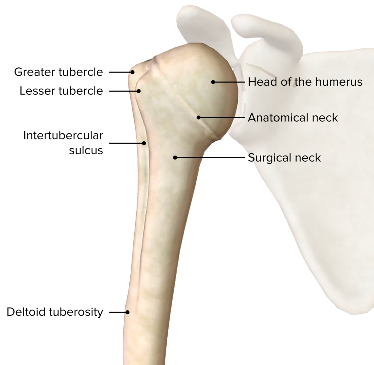
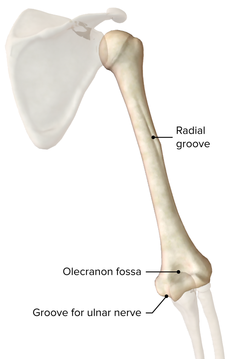
Humerus Humerus Bone in humans and primates extending from the shoulder joint to the elbow joint. Arm: Anatomy

  • Bone Bone Bone is a compact type of hardened connective tissue composed of bone cells, membranes, an extracellular mineralized matrix, and central bone marrow. The 2 primary types of bone are compact and spongy. Bones: Structure and Types of the upper arm Upper Arm The arm, or “upper arm” in common usage, is the region of the upper limb that extends from the shoulder to the elbow joint and connects inferiorly to the forearm through the cubital fossa. It is divided into 2 fascial compartments (anterior and posterior). Arm: Anatomy
  • The proximal end comprises the head, the neck Neck The part of a human or animal body connecting the head to the rest of the body. Peritonsillar Abscess, the greater and lesser tubercles, and the shaft.
  • Landmarks and articulations:
    • Head of the humerus Head of The Humerus The upper rounded extremity of the humerus fitting into the glenoid cavity of the scapula. Arm: Anatomy: spherical, articulates with the glenoid cavity
    • Tubercles: greater is lateral, lesser is anterior. The supraspinatus, infraspinatus, and teres minor rotator cuff muscles attach to the greater tubercle, while the subscapularis attaches to the lesser tubercle.

Joints

Acromioclavicular (AC) joint

  • Type of joint: plane synovial joint Synovial joint Jaw and Temporomandibular Joint: Anatomy between the acromion and the clavicle Clavicle A bone on the ventral side of the shoulder girdle, which in humans is commonly called the collar bone. Clavicle Fracture
  • Function: allows for motion between the clavicle Clavicle A bone on the ventral side of the shoulder girdle, which in humans is commonly called the collar bone. Clavicle Fracture and scapula during arm Arm The arm, or “upper arm” in common usage, is the region of the upper limb that extends from the shoulder to the elbow joint and connects inferiorly to the forearm through the cubital fossa. It is divided into 2 fascial compartments (anterior and posterior). Arm: Anatomy movement
  • Ligaments: named according to their anatomical insertions
    • Acromioclavicular ligament: reinforces the superior part of the joint capsule Capsule An envelope of loose gel surrounding a bacterial cell which is associated with the virulence of pathogenic bacteria. Some capsules have a well-defined border, whereas others form a slime layer that trails off into the medium. Most capsules consist of relatively simple polysaccharides but there are some bacteria whose capsules are made of polypeptides. Bacteroides and resists anteroposterior displacement Displacement The process by which an emotional or behavioral response that is appropriate for one situation appears in another situation for which it is inappropriate. Defense Mechanisms at the AC joint
    • Coracoclavicular ligament: consists of the trapezoid and conoid ligaments and resists vertical and rotational displacement Displacement The process by which an emotional or behavioral response that is appropriate for one situation appears in another situation for which it is inappropriate. Defense Mechanisms of the clavicle Clavicle A bone on the ventral side of the shoulder girdle, which in humans is commonly called the collar bone. Clavicle Fracture relative to the scapula

Glenohumeral joint

  • Type of joint: synovial ball-and-socket joint between the head of the humerus Head of The Humerus The upper rounded extremity of the humerus fitting into the glenoid cavity of the scapula. Arm: Anatomy and the glenoid cavity of the scapula
  • Function: most mobile joint in the body with multiple degrees of motion: flexion Flexion Examination of the Upper Limbs and extension Extension Examination of the Upper Limbs, abduction Abduction Examination of the Upper Limbs and adduction Adduction Examination of the Upper Limbs, medial and lateral rotation Rotation Motion of an object in which either one or more points on a line are fixed. It is also the motion of a particle about a fixed point. X-rays, and circumduction of the shoulder
  • Ligaments and supporting structures:
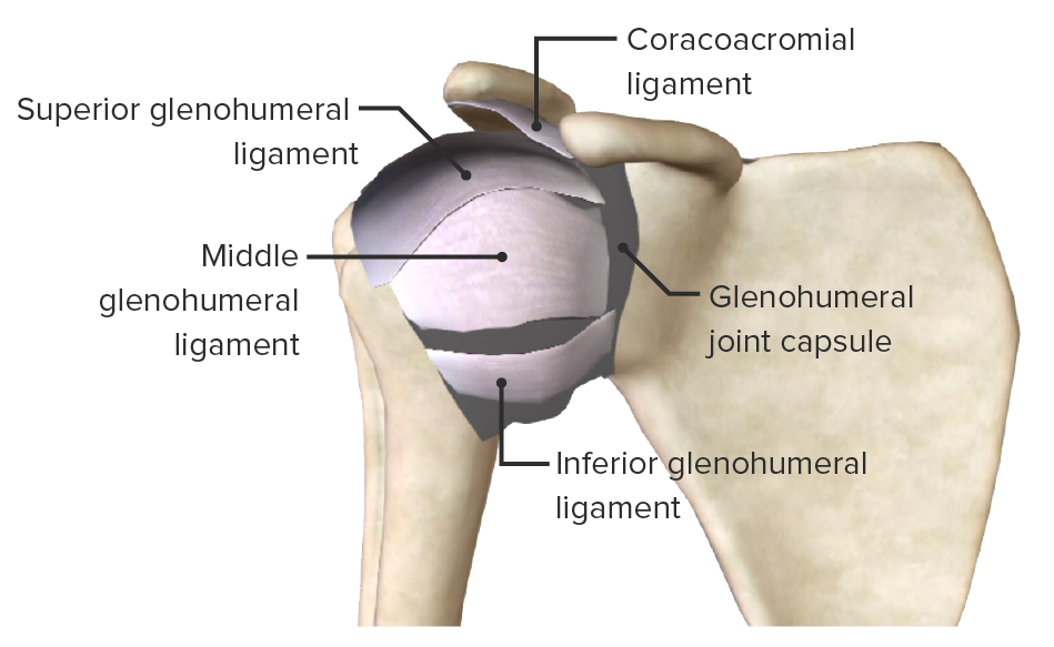
    • Fibrous capsule Fibrous capsule Hip Joint: Anatomy: extends from the anatomical neck Neck The part of a human or animal body connecting the head to the rest of the body. Peritonsillar Abscess of the humerus Humerus Bone in humans and primates extending from the shoulder joint to the elbow joint. Arm: Anatomy to the rim of the glenoid 
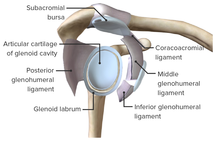
    • Glenoid labrum: fibrocartilaginous, meniscus-like structure that deepens the glenoid cavity
    • Ligaments: named according to their anatomical insertions
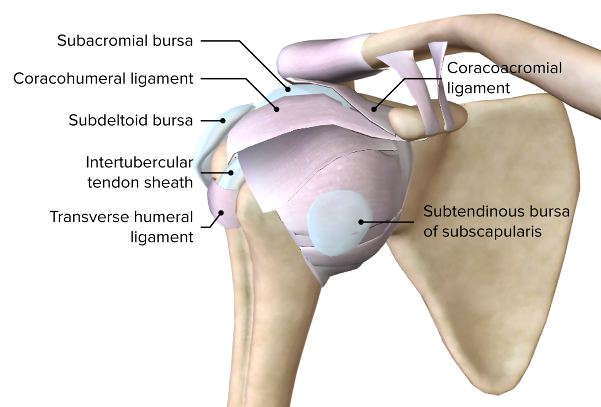
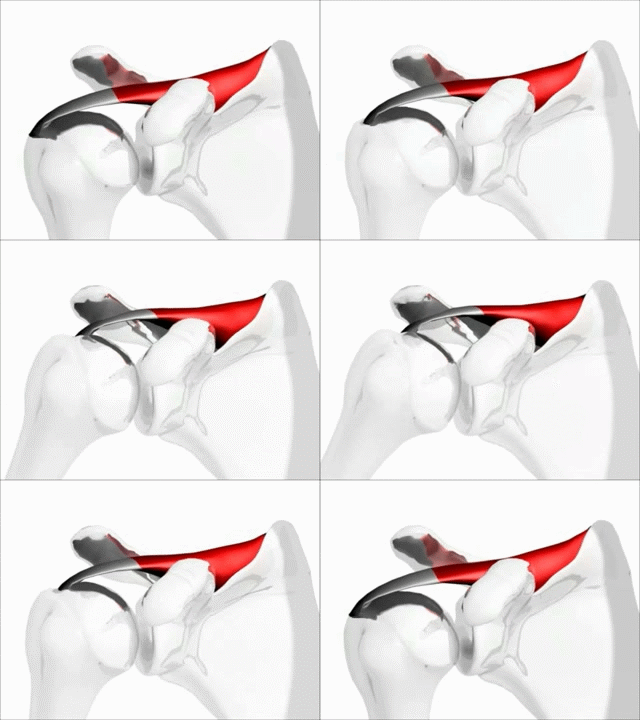
      • Coracoacromial ligament: resists superior displacement Displacement The process by which an emotional or behavioral response that is appropriate for one situation appears in another situation for which it is inappropriate. Defense Mechanisms of the humeral head Humeral head The upper rounded extremity of the humerus fitting into the glenoid cavity of the scapula. Arm: Anatomy from the glenoid cavity 
      • Coracohumeral ligament: resists superior and inferior displacement Displacement The process by which an emotional or behavioral response that is appropriate for one situation appears in another situation for which it is inappropriate. Defense Mechanisms and reinforces the anterior portion of the joint capsule Capsule An envelope of loose gel surrounding a bacterial cell which is associated with the virulence of pathogenic bacteria. Some capsules have a well-defined border, whereas others form a slime layer that trails off into the medium. Most capsules consist of relatively simple polysaccharides but there are some bacteria whose capsules are made of polypeptides. Bacteroides
      • Glenohumeral ligaments: consists of superior, middle, and inferior bands and stabilizes the joint during adduction Adduction Examination of the Upper Limbs, abduction Abduction Examination of the Upper Limbs, and external rotation External Rotation Examination of the Upper Limbs, depending on the position of the shoulder
      • Transverse humeral ligament: maintains the long head of the biceps Biceps Arm: Anatomy tendon within the bicipital groove
    • Subacromial bursa:
      • Synovial cavity located inferior to the acromion and coracoid and superior to the supraspinatus/infraspinatus tendons
      • The function of the bursa is to decrease friction and facilitate movement between these structures during movement of the shoulder.
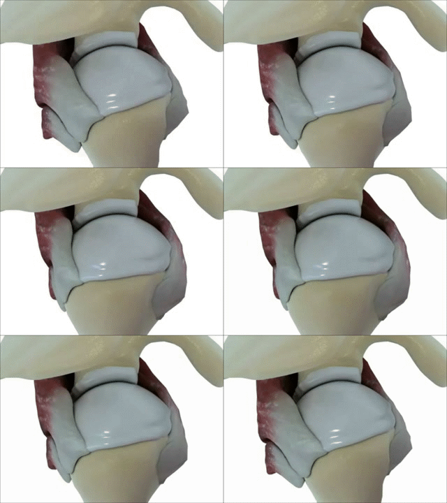
Superficial ligaments and bursa glenohumeral joint

Anterior view of the superficial layer of ligaments and bursa of the right glenohumeral joint

Image by BioDigital, edited by Lecturio
Sternoclavicular (SC) joint

  • Type of joint: synovial saddle joint between the sternum Sternum A long, narrow, and flat bone commonly known as breastbone occurring in the midsection of the anterior thoracic segment or chest region, which stabilizes the rib cage and serves as the point of origin for several muscles that move the arms, head, and neck. Chest Wall: Anatomy and medial clavicle Clavicle A bone on the ventral side of the shoulder girdle, which in humans is commonly called the collar bone. Clavicle Fracture
  • Primary skeletal attachment between the axial Axial Computed Tomography (CT) skeleton and upper limb
  • Function: allows for motion of the clavicle Clavicle A bone on the ventral side of the shoulder girdle, which in humans is commonly called the collar bone. Clavicle Fracture in multiple planes
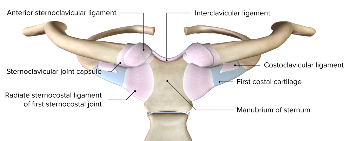
  • Ligaments:
    • Sternoclavicular ligament: primarily provides anterior/posterior stabilization and resists superior displacement Displacement The process by which an emotional or behavioral response that is appropriate for one situation appears in another situation for which it is inappropriate. Defense Mechanisms
    • Costoclavicular ligament: anchors clavicle Clavicle A bone on the ventral side of the shoulder girdle, which in humans is commonly called the collar bone. Clavicle Fracture to the first rib and resists clavicular elevation
    • Interclavicular ligament: strengthens the capsule Capsule An envelope of loose gel surrounding a bacterial cell which is associated with the virulence of pathogenic bacteria. Some capsules have a well-defined border, whereas others form a slime layer that trails off into the medium. Most capsules consist of relatively simple polysaccharides but there are some bacteria whose capsules are made of polypeptides. Bacteroides and resists downward displacement Displacement The process by which an emotional or behavioral response that is appropriate for one situation appears in another situation for which it is inappropriate. Defense Mechanisms of the clavicle Clavicle A bone on the ventral side of the shoulder girdle, which in humans is commonly called the collar bone. Clavicle Fracture
Anterior view of the sternoclavicular joint

Anterior view of the sternoclavicular joint and the first sternocostal joint

Image by BioDigital, edited by Lecturio
Scapulothoracic articulation

  • Not a true synovial joint Synovial joint Jaw and Temporomandibular Joint: Anatomy
  • Function:
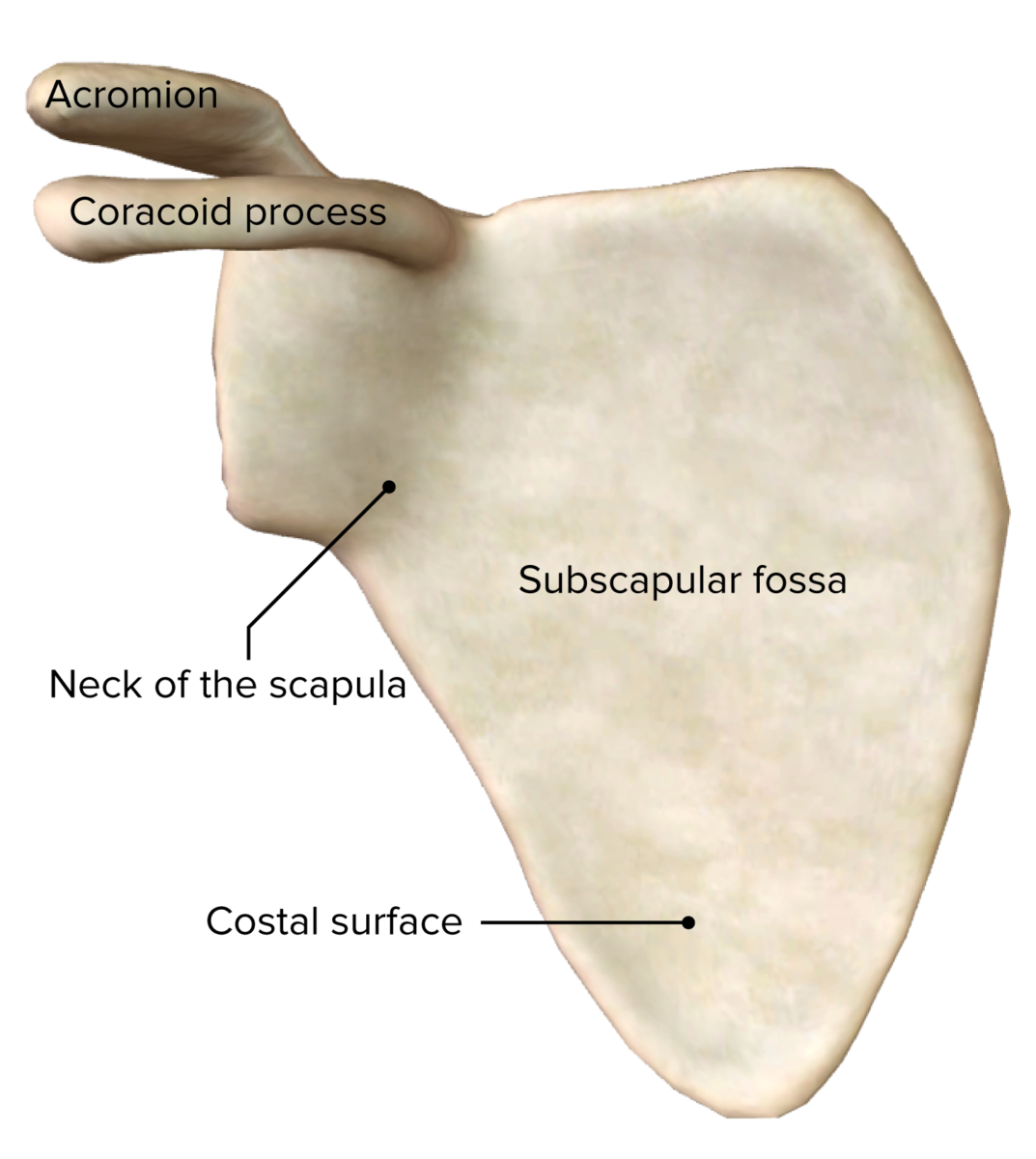
    • Articulation between the concave anterior scapula and convex surface of the posterior thorax
    • Movement of the scapula on the thorax at this articulation requires movement at the AC joint and SC joint.
Scapulothoracic pseudojoint

Posterior view of the scapular region, highlighting the scapulothoracic pseudo-joint

Image by BioDigital, edited by Lecturio
Anterior Axioappendicular Muscles

The anterior axioappendicular muscles function to move the pectoral girdle, stabilize the clavicle Clavicle A bone on the ventral side of the shoulder girdle, which in humans is commonly called the collar bone. Clavicle Fracture, and move the upper arm Upper Arm The arm, or “upper arm” in common usage, is the region of the upper limb that extends from the shoulder to the elbow joint and connects inferiorly to the forearm through the cubital fossa. It is divided into 2 fascial compartments (anterior and posterior). Arm: Anatomy.

Table: Origin, insertion, innervation, and function of the anterior axioappendicular muscles
Muscle Origin Insertion Nerve supply Function
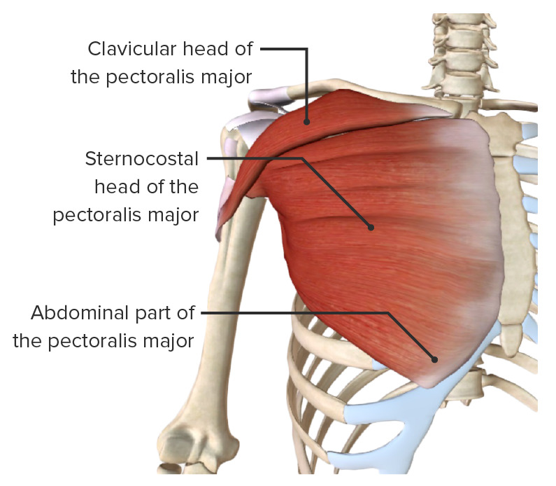
Pectoralis major
  • Clavicular head: anterior surface of medial clavicle Clavicle A bone on the ventral side of the shoulder girdle, which in humans is commonly called the collar bone. Clavicle Fracture
  • Sternocostal head: sternum Sternum A long, narrow, and flat bone commonly known as breastbone occurring in the midsection of the anterior thoracic segment or chest region, which stabilizes the rib cage and serves as the point of origin for several muscles that move the arms, head, and neck. Chest Wall: Anatomy and superior 6 costal cartilages
Lateral lip of intertubercular sulcus Intertubercular sulcus Arm: Anatomy Lateral and medial pectoral nerves (C5, C6: clavicular head; C7, C8: sternocostal head)
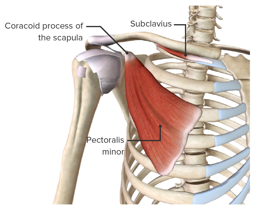
Pectoralis minor Ribs Ribs A set of twelve curved bones which connect to the vertebral column posteriorly, and terminate anteriorly as costal cartilage. Together, they form a protective cage around the internal thoracic organs. Chest Wall: Anatomy 3–5 Coracoid process of scapula Medial pectoral nerves (C8, T1) Stabilizes scapula and pulls anteriorly and inferiorly
Subclavius Subclavius Muscles of the Neck: Anatomy Rib 1 and sternum Sternum A long, narrow, and flat bone commonly known as breastbone occurring in the midsection of the anterior thoracic segment or chest region, which stabilizes the rib cage and serves as the point of origin for several muscles that move the arms, head, and neck. Chest Wall: Anatomy junction Middle 3rd of clavicle Clavicle A bone on the ventral side of the shoulder girdle, which in humans is commonly called the collar bone. Clavicle Fracture Subclavian nerve (C5) Stabilizes and depresses clavicle Clavicle A bone on the ventral side of the shoulder girdle, which in humans is commonly called the collar bone. Clavicle Fracture
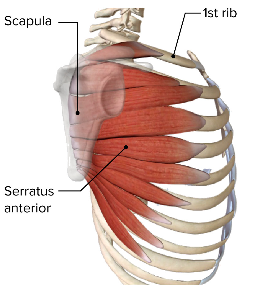
Serratus anterior External surface of ribs Ribs A set of twelve curved bones which connect to the vertebral column posteriorly, and terminate anteriorly as costal cartilage. Together, they form a protective cage around the internal thoracic organs. Chest Wall: Anatomy 1–8 Medial border of scapula Long thoracic nerve Long thoracic nerve Axilla and Brachial Plexus: Anatomy (C6, C7) Protracts scapula; holds scapula against posterior thoracic wall

Posterior Axioappendicular Muscles

The posterior axioappendicular muscles stabilize and move the scapula and consist of a superficial and a deep layer.

Superficial or extrinsic layer

Table: Origin, insertion, innervation, and function of the superficial or extrinsic layer of the posterior axioappendicular muscles
Muscle Origin Insertion Nerve supply Function
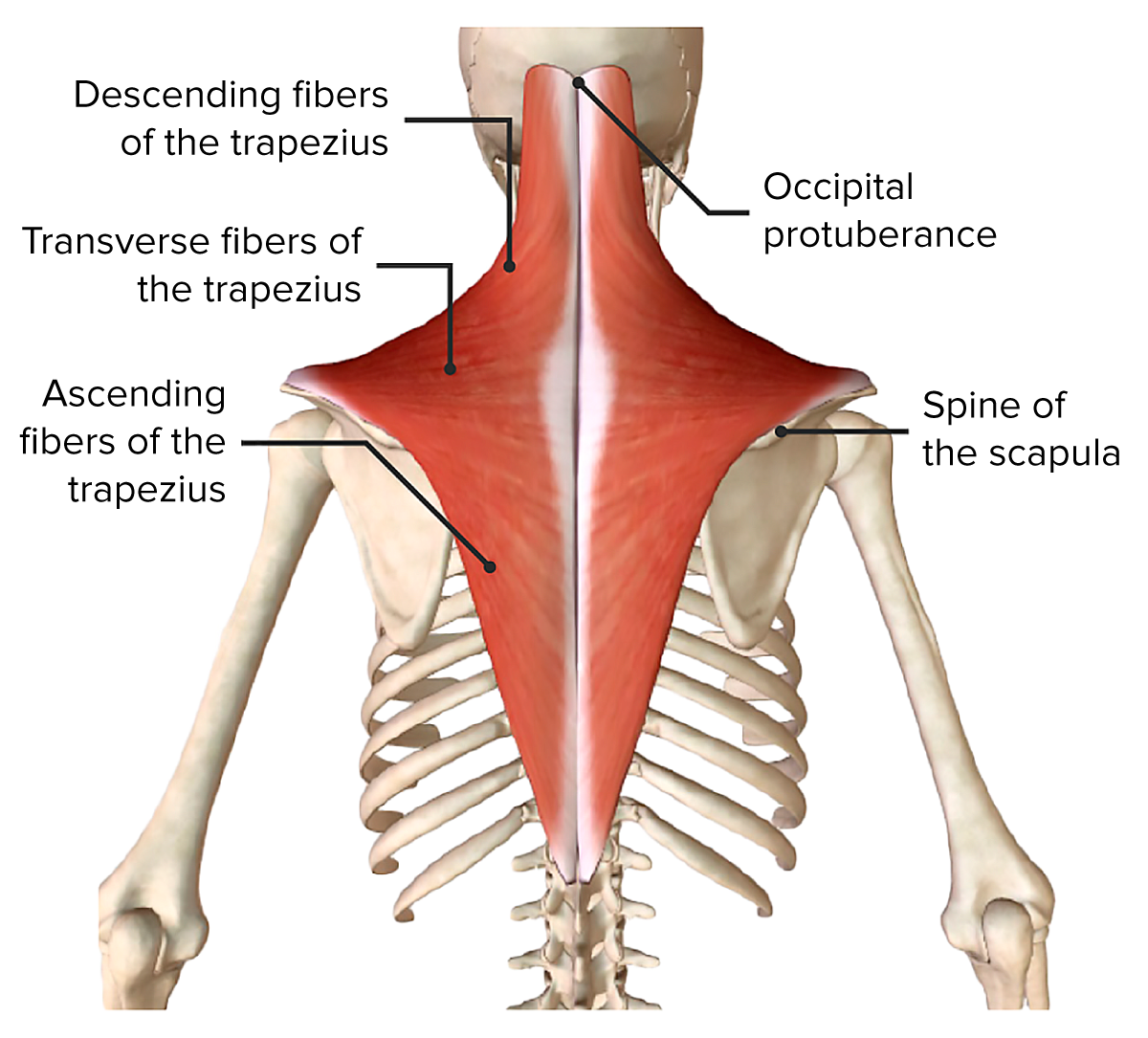
Trapezius
  • Descending part: superior nuchal line and external occipital Occipital Part of the back and base of the cranium that encloses the foramen magnum. Skull: Anatomy protuberance
  • Transverse part: nuchal ligament
  • Ascending part: C7–T12 spinous processes
Lateral 3rd of clavicle Clavicle A bone on the ventral side of the shoulder girdle, which in humans is commonly called the collar bone. Clavicle Fracture and spine Spine The human spine, or vertebral column, is the most important anatomical and functional axis of the human body. It consists of 7 cervical vertebrae, 12 thoracic vertebrae, and 5 lumbar vertebrae and is limited cranially by the skull and caudally by the sacrum. Vertebral Column: Anatomy of scapula Spinal accessory nerve Spinal accessory nerve The 11th cranial nerve which originates from neurons in the medulla and in the cervical spinal cord. It has a cranial root, which joins the vagus nerve (10th cranial) and sends motor fibers to the muscles of the larynx, and a spinal root, which sends motor fibers to the trapezius and the sternocleidomastoid muscles. The 12 Cranial Nerves: Overview and Functions (CN XI) and C3, C4 spinal nerves Spinal nerves The 31 paired peripheral nerves formed by the union of the dorsal and ventral spinal roots from each spinal cord segment. The spinal nerve plexuses and the spinal roots are also included. Spinal Cord: Anatomy for proprioception Proprioception Sensory functions that transduce stimuli received by proprioceptive receptors in joints, tendons, muscles, and the inner ear into neural impulses to be transmitted to the central nervous system. Proprioception provides sense of stationary positions and movements of one’s body parts, and is important in maintaining kinesthesia and postural balance. Neurological Examination
  • Descending part: elevates scapula
  • Transverse part: retracts scapula
  • Ascending part: depresses scapula
  • Descending and ascending: rotates scapula superiorly
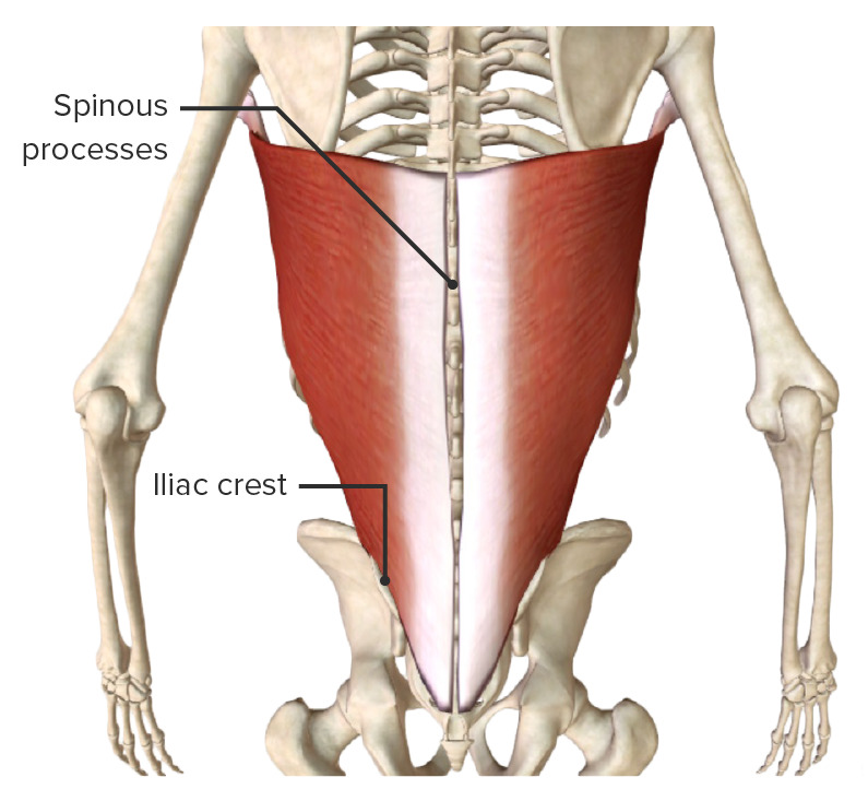
Latissimus dorsi T6–12 spinous processes, thoracolumbar fascia Thoracolumbar fascia Posterior Abdominal Wall: Anatomy and iliac crest Floor of intertubercular groove Thoracodorsal nerve Thoracodorsal nerve Axilla and Brachial Plexus: Anatomy (C6, C7) Extends, adducts, and medially rotates shoulder joint Shoulder joint The articulation between the head of the humerus and the glenoid cavity of the scapula. Examination of the Upper Limbs
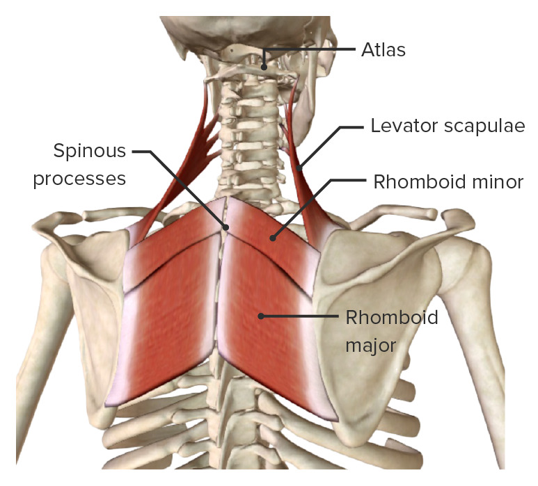
Rhomboid
  • Minor: nuchal ligament and C7–T1 spinous processes
  • Major: T2–T5 spinous processes
  • Minor: medial border of scapula (superior)
  • Major: medial border of scapula (inferior)
Dorsal scapula (C5)
  • Retracts scapula
  • Rotates scapula inferiorly (depressing glenoid cavity)
Levator scapulae Transverse process of C1–C4 Superior angle of scapula Dorsal scapula (C5)
  • Elevates scapula
  • Rotates scapula inferiorly (depressing glenoid cavity)
Deep or intrinsic posterior axioappendicular muscles

Rhomboid major, rhomboid minor, and levator scapulae muscles, the deep or intrinsic posterior axioappendicular muscles

Image by BioDigital, edited by Lecturio

Deep or intrinsic layer

The deep or intrinsic layer consists of 6 muscles:

  • Deltoid
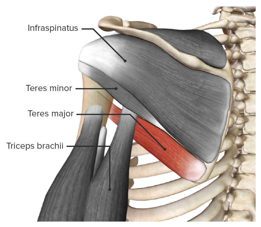
  • Teres major
  • Supraspinatus
  • Infraspinatus
  • Teres minor
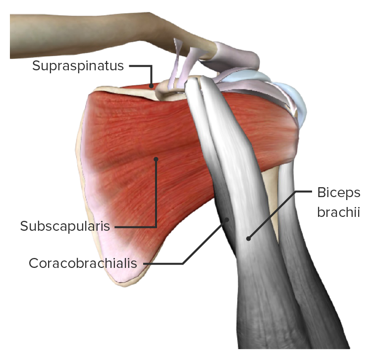
  • Subscapularis

Scapulohumeral Muscles

The scapulohumeral muscles stabilize the glenohumeral joint by connecting the humerus Humerus Bone in humans and primates extending from the shoulder joint to the elbow joint. Arm: Anatomy to the scapula, including the rotator cuff muscles.

Table: Origin, insertion, innervation, and function of the scapulohumeral muscles
Muscle Origin Insertion Nerve supply Function
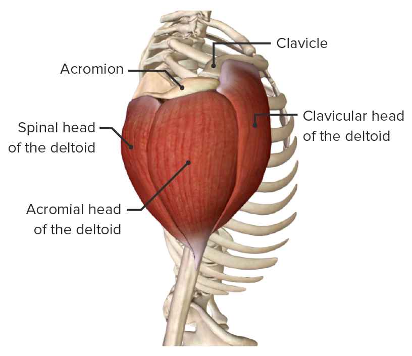
Deltoid
  • Clavicular head: lateral 3rd of clavicle Clavicle A bone on the ventral side of the shoulder girdle, which in humans is commonly called the collar bone. Clavicle Fracture
  • Acromial head: acromion
  • Spinal head: spine Spine The human spine, or vertebral column, is the most important anatomical and functional axis of the human body. It consists of 7 cervical vertebrae, 12 thoracic vertebrae, and 5 lumbar vertebrae and is limited cranially by the skull and caudally by the sacrum. Vertebral Column: Anatomy of scapula
Deltoid tuberosity Deltoid tuberosity Arm: Anatomy of humerus Humerus Bone in humans and primates extending from the shoulder joint to the elbow joint. Arm: Anatomy Axillary nerve Axillary nerve Axilla and Brachial Plexus: Anatomy (C5)
Teres major Lateral border of scapula, inferior portion Medial lip of intertubercular groove Lower subscapular nerve Lower subscapular nerve Axilla and Brachial Plexus: Anatomy (C6) Adducts and medially rotates shoulder joint Shoulder joint The articulation between the head of the humerus and the glenoid cavity of the scapula. Examination of the Upper Limbs
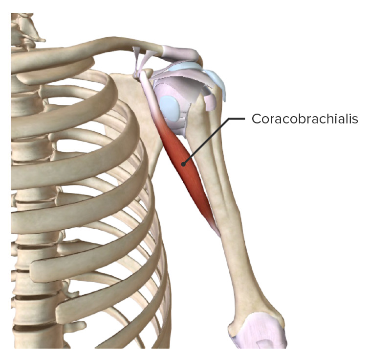
Coracobrachialis Coracobrachialis Arm: Anatomy Coracoid process of scapula Shaft of humerus Humerus Bone in humans and primates extending from the shoulder joint to the elbow joint. Arm: Anatomy, anteromedial Musculocutaneous nerve Musculocutaneous Nerve A major nerve of the upper extremity. The fibers of the musculocutaneous nerve originate in the lower cervical spinal cord (usually C5 to C7), travel via the lateral cord of the brachial plexus, and supply sensory and motor innervation to the upper arm, elbow, and forearm. Axilla and Brachial Plexus: Anatomy (C5, C6, C7) Flexes, adducts, and medially rotates the shoulder joint Shoulder joint The articulation between the head of the humerus and the glenoid cavity of the scapula. Examination of the Upper Limbs

Rotator cuff muscles

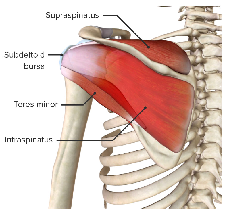
Rotator cuff muscles stabilize the shoulder joint Shoulder joint The articulation between the head of the humerus and the glenoid cavity of the scapula. Examination of the Upper Limbs and prevent detachment of the head of the humerus Head of The Humerus The upper rounded extremity of the humerus fitting into the glenoid cavity of the scapula. Arm: Anatomy from the glenoid cavity via “concavity compression Compression Blunt Chest Trauma,” especially during abduction Abduction Examination of the Upper Limbs of the arm Arm The arm, or “upper arm” in common usage, is the region of the upper limb that extends from the shoulder to the elbow joint and connects inferiorly to the forearm through the cubital fossa. It is divided into 2 fascial compartments (anterior and posterior). Arm: Anatomy.

Mnemonic

To recall the rotator cuff muscles, remember “SITS”:

S: Supraspinatus

I: Infraspinatus

T: Teres minor

S: Subscapularis

Table: Origin, insertion, innervation, and function of the rotator cuff muscles
Muscle Origin Insertion Nerve supply Function
Supraspinatus Supraspinous fossa Superior facet of greater tubercle of humerus Humerus Bone in humans and primates extending from the shoulder joint to the elbow joint. Arm: Anatomy Suprascapular nerve Suprascapular nerve Axilla and Brachial Plexus: Anatomy (C5) Initiates and assists deltoid in abduction Abduction Examination of the Upper Limbs
Infraspinatus Infraspinous fossa Middle facet of greater tubercle of humerus Humerus Bone in humans and primates extending from the shoulder joint to the elbow joint. Arm: Anatomy Suprascapular nerve Suprascapular nerve Axilla and Brachial Plexus: Anatomy (C5) Laterally rotates the shoulder joint Shoulder joint The articulation between the head of the humerus and the glenoid cavity of the scapula. Examination of the Upper Limbs, holds head of humerus Humerus Bone in humans and primates extending from the shoulder joint to the elbow joint. Arm: Anatomy in glenoid cavity
Teres minor Lateral border of scapula, middle portion Inferior facet of greater tubercle of humerus Humerus Bone in humans and primates extending from the shoulder joint to the elbow joint. Arm: Anatomy Axillary nerve Axillary nerve Axilla and Brachial Plexus: Anatomy (C6) Laterally rotates the shoulder joint Shoulder joint The articulation between the head of the humerus and the glenoid cavity of the scapula. Examination of the Upper Limbs, holds head of humerus Humerus Bone in humans and primates extending from the shoulder joint to the elbow joint. Arm: Anatomy in glenoid cavity
Subscapularis Subscapular fossa Lesser tubercle of humerus Humerus Bone in humans and primates extending from the shoulder joint to the elbow joint. Arm: Anatomy Upper and lower subscapular nerves (C6) Medially rotates and adducts shoulder joint Shoulder joint The articulation between the head of the humerus and the glenoid cavity of the scapula. Examination of the Upper Limbs, holds head of humerus Humerus Bone in humans and primates extending from the shoulder joint to the elbow joint. Arm: Anatomy in glenoid cavity
Rotator cuff muscles

Posterior view of the scapular region and rotator cuff muscles (subscapularis missing)

Image by BioDigital, edited by Lecturio
Clinical Relevance

The following conditions are common conditions associated with the shoulder:

  • Shoulder dislocation: the separation of the head of the humerus Head of The Humerus The upper rounded extremity of the humerus fitting into the glenoid cavity of the scapula. Arm: Anatomy from the glenoid cavity. The glenohumeral joint is the most commonly dislocated joint. The vast majority of these dislocations are anterior-inferior (95%). Presents as severe shoulder pain Pain An unpleasant sensation induced by noxious stimuli which are detected by nerve endings of nociceptive neurons. Pain: Types and Pathways and restricted range of motion Range of motion The distance and direction to which a bone joint can be extended. Range of motion is a function of the condition of the joints, muscles, and connective tissues involved. Joint flexibility can be improved through appropriate muscle strength exercises. Examination of the Upper Limbs.
  • Rotator cuff pathology: consists of a tear of one or more of the tendons of the 4 rotator cuff muscles. The supraspinatus is the most commonly torn rotator cuff tendon. May be associated with trauma or repeated activities. Presents with severe pain Pain An unpleasant sensation induced by noxious stimuli which are detected by nerve endings of nociceptive neurons. Pain: Types and Pathways or discomfort, loss of strength, and restricted range of motion Range of motion The distance and direction to which a bone joint can be extended. Range of motion is a function of the condition of the joints, muscles, and connective tissues involved. Joint flexibility can be improved through appropriate muscle strength exercises. Examination of the Upper Limbs. These injuries may require surgical repair.
  • SLAP SLAP Chronic Shoulder Pain (Superior Labral tear from Anterior to Posterior) lesion: consists of injury to the glenoid labrum. Presents as dull pain Pain An unpleasant sensation induced by noxious stimuli which are detected by nerve endings of nociceptive neurons. Pain: Types and Pathways, discomfort, decreased range of motion Range of motion The distance and direction to which a bone joint can be extended. Range of motion is a function of the condition of the joints, muscles, and connective tissues involved. Joint flexibility can be improved through appropriate muscle strength exercises. Examination of the Upper Limbs and strength, joint instability, and change in velocity in overhead athletes.
  • Subacromial bursitis: inflammation Inflammation Inflammation is a complex set of responses to infection and injury involving leukocytes as the principal cellular mediators in the body’s defense against pathogenic organisms. Inflammation is also seen as a response to tissue injury in the process of wound healing. The 5 cardinal signs of inflammation are pain, heat, redness, swelling, and loss of function. Inflammation of the bursa located between the acromion and the deltoid muscle superficially and the supraspinatus muscle. Often caused by repetitive overhead motion. Presents as pain Pain An unpleasant sensation induced by noxious stimuli which are detected by nerve endings of nociceptive neurons. Pain: Types and Pathways, swelling Swelling Inflammation, loss of strength, and stiffness of the shoulder. Can progress to impingement syndrome.
  • Impingement syndrome: a spectrum of clinical manifestations caused by the compression Compression Blunt Chest Trauma of tissues as they pass through the subacromial space. Impingement syndrome can progress to a rotator cuff tear. Caused by many etiologies, including repetitive overhead motions or any condition that further narrows the subacromial space. Impingement syndrome presents as shoulder pain Pain An unpleasant sensation induced by noxious stimuli which are detected by nerve endings of nociceptive neurons. Pain: Types and Pathways with overhead motions (“painful arc”), restriction of range of motion Range of motion The distance and direction to which a bone joint can be extended. Range of motion is a function of the condition of the joints, muscles, and connective tissues involved. Joint flexibility can be improved through appropriate muscle strength exercises. Examination of the Upper Limbs, loss of strength, and pain Pain An unpleasant sensation induced by noxious stimuli which are detected by nerve endings of nociceptive neurons. Pain: Types and Pathways when sleeping on the shoulder.
  • Adhesive capsulitis Adhesive Capsulitis Chronic Shoulder Pain: also known as “ frozen shoulder Frozen Shoulder Chronic Shoulder Pain,” the painful loss of motion of the shoulder due to inflammation Inflammation Inflammation is a complex set of responses to infection and injury involving leukocytes as the principal cellular mediators in the body’s defense against pathogenic organisms. Inflammation is also seen as a response to tissue injury in the process of wound healing. The 5 cardinal signs of inflammation are pain, heat, redness, swelling, and loss of function. Inflammation and fibrosis Fibrosis Any pathological condition where fibrous connective tissue invades any organ, usually as a consequence of inflammation or other injury. Bronchiolitis Obliterans of the fibrous capsule Fibrous capsule Hip Joint: Anatomy; may be associated with diabetes Diabetes Diabetes mellitus (DM) is a metabolic disease characterized by hyperglycemia and dysfunction of the regulation of glucose metabolism by insulin. Type 1 DM is diagnosed mostly in children and young adults as the result of autoimmune destruction of β cells in the pancreas and the resulting lack of insulin. Type 2 DM has a significant association with obesity and is characterized by insulin resistance. Diabetes Mellitus, thyroid Thyroid The thyroid gland is one of the largest endocrine glands in the human body. The thyroid gland is a highly vascular, brownish-red gland located in the visceral compartment of the anterior region of the neck. Thyroid Gland: Anatomy disease, trauma, or prolonged immobilization Immobilization Delirium.

References

  1. Drake, R. L., Vogl, W., & Mitchell, A. W. M. (2019). Gray’s anatomy for students (4th ed.). Elsevier.
  2. Gilroy, A. M., MacPherson, B. R., & Ross, L. M. (2020). Atlas Atlas The first cervical vertebra. Vertebral Column: Anatomy of anatomy (4th ed.). Thieme.
  3. Moore, K. L., Dalley, A. F., & Agur, A. M. R. (2022). Clinically oriented anatomy (9th ed.). Wolters Kluwer.
  4. Netter, F. H. (2019). Atlas Atlas The first cervical vertebra. Vertebral Column: Anatomy of human anatomy (7th ed.). Elsevier.
  5. Paulsen, F., & Waschke, J. (Eds.). (2022). Sobotta atlas Atlas The first cervical vertebra. Vertebral Column: Anatomy of human anatomy (16th ed.). Elsevier.
  6. Standring, S. (Ed.). (2021). Gray’s anatomy: The anatomical basis of clinical practice (42nd ed.). Elsevier.
  7. Tank Tank ACES and RUSH: Resuscitation Ultrasound Protocols, P. W., & Grant, J. C. B. (2021). Grant’s dissector (17th ed.). Wolters Kluwer.

Create your free account or log in to continue reading!

Sign up now and get free access to Lecturio with concept pages, medical videos, and questions for your medical education.

User Reviews

Details