Las respuestas inmunes contra patógenos se dividen en sistemas de respuesta inmune innata y adaptativa. La respuesta inmune adaptativa, también denominada sistema inmune adquirido, consta de 2 mecanismos principales: las respuestas inmunes humoral y celular. La inmunidad humoral está mediada por las células B (que producen anticuerpos), mientras que la inmunidad mediada por células involucra a las células T. Como 2da línea de defensa, el sistema inmune adaptativo es más lento y responde durante un período de tiempo más largo, pero el efecto generalmente conduce a una memoria inmunológica específica. Las 2 características importantes de la respuesta adaptativa son la especificidad (con reconocimiento de antígeno) y la memoria (respuesta inmune inducida por reinfección).
Last updated: Dec 15, 2025
El sistema inmune proporciona defensa (inmunidad) contra patógenos invasores que van desde virus Virus Viruses are infectious, obligate intracellular parasites composed of a nucleic acid core surrounded by a protein capsid. Viruses can be either naked (non-enveloped) or enveloped. The classification of viruses is complex and based on many factors, including type and structure of the nucleoid and capsid, the presence of an envelope, the replication cycle, and the host range. Virology hasta parásitos, y sus componentes están interconectados por la circulación sanguínea y linfática.
2 líneas de defensa (que se superponen):
| Inmunidad innata | Inmunidad adaptativa | |
|---|---|---|
| Genética | Línea germinal codificada | Reordenamientos genéticos implicados en EN Erythema nodosum is an immune-mediated panniculitis (inflammation of the subcutaneous fat) caused by a type IV (delayed-type) hypersensitivity reaction. It commonly manifests in young women as tender, erythematous nodules on the shins. Erythema Nodosum el desarrollo de linfocitos |
| Respuesta inmune | Inespecífica | Altamente específica |
| Tiempo de respuesta | Inmediato (minutos a horas) | Se desarrolla durante un período de tiempo más prolongado |
| Respuesta de memoria | Ninguna | Con respuesta de memoria, que responde rápidamente al AL Amyloidosis reconocimiento del antígeno |
| Reconocimiento del patógeno | Los LOS Neisseria receptores de reconocimiento de patrones, como los LOS Neisseria TLR, reconocen patrones moleculares asociados a patógenos |
|
| Componentes |
|
|
La respuesta inmune adaptativa se divide en EN Erythema nodosum is an immune-mediated panniculitis (inflammation of the subcutaneous fat) caused by a type IV (delayed-type) hypersensitivity reaction. It commonly manifests in young women as tender, erythematous nodules on the shins. Erythema Nodosum los LOS Neisseria sistemas inmunes mediados por respuesta humoral y celular.

Inmunidad mediada por células:
La activación de los linfocitos T auxiliares da lugar a la liberación de citoquinas, activando así los linfocitos T citotóxicos y los fagocitos (como los macrófagos).
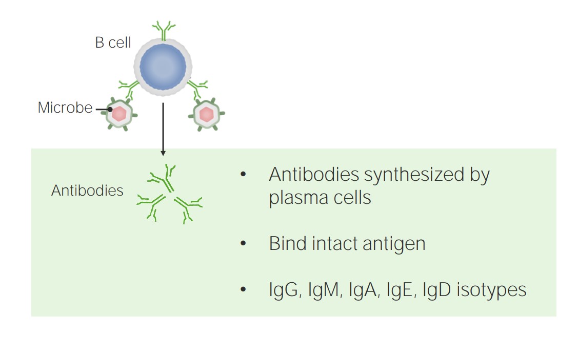
La inmunidad humoral está mediada por las células B y los anticuerpos.
Imagen por Lecturio.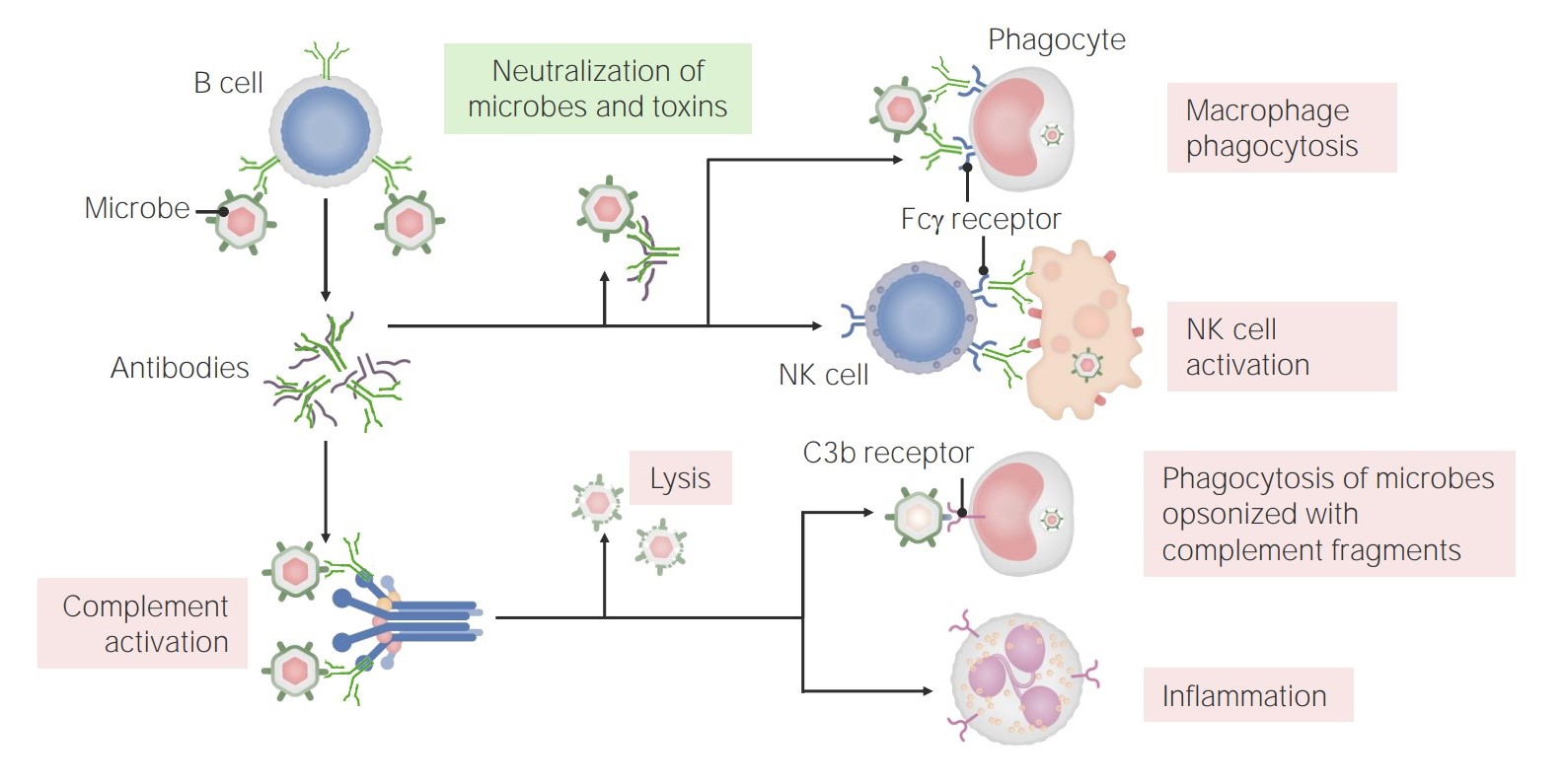
Las funciones de los anticuerpos:
Los anticuerpos tienen múltiples funciones en la inmunidad, incluyendo la neutralización (de microbios y toxinas), la promoción de la fagocitosis y la activación de las células NK. Además, los anticuerpos tienen un papel en la activación del complemento, que puede conducir a la lisis directa de los microbios, la opsonización y la fagocitosis, y el reclutamiento/activación de los neutrófilos.
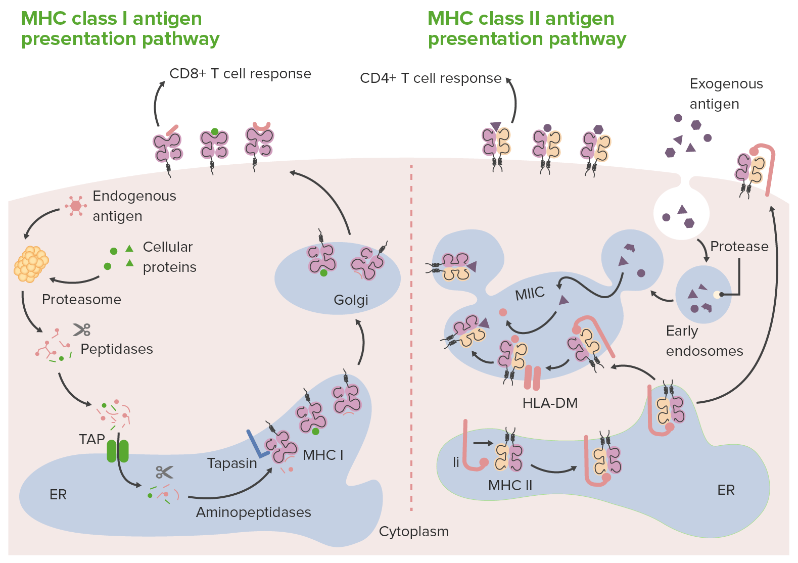
Vías de presentación de antígenos por moléculas del complejo mayor de histocompatibilidad (MHC, por sus siglas en inglés, de clase I y II:
En la presentación de antígenos de clase I (izquierda), los proteosomas degradan antígenos o proteínas endógenas (dentro de la célula) en péptidos. Los fragmentos de péptidos se transportan (a través del transportador asociado con el procesamiento de antígenos (TAP, por sus siglas en inglés)) al reticulo endoplasmico (ER, por sus siglas en inglés), donde las aminopeptidasas los recortan aún más y los acoplan en la molécula MHC de clase I. Los complejos Antígeno-MHC de clase I van al aparato de Golgi para la modificación postraduccional. Luego, los complejos se transportan a la superficie celular, donde se presentan a las células T CD8+. En la presentación de antígenos de clase II (derecha), las células presentadoras de antígenos captan los antígenos extracelulares/exógenos dentro de los fagosomas. Los fagosomas luego se fusionan con lisosomas llenos de enzimas proteolíticas. Esto da como resultado la descomposición de las proteínas fagocitadas en pequeños péptidos. Mientras tanto, en el ER se sintetizan nuevas moléculas MHC de clase II. Estas moléculas tienen la cadena invariable (estructura rosa en la imagen de la derecha, marcada como “Ii”), que se une a la hendidura de unión a antígeno. Con la hendidura ocluida (por la cadena invariable), los péptidos residentes en el ER no pueden unirse. La cadena invariable dirige el complejo MHC II al endosoma acidificado (donde están los péptidos del antígeno) a medida que sale del ER. Cuando los complejos MHC II se entregan al endosoma, la cadena invariable se libera, lo que permite el acople de péptidos del antígeno (acompañados por una proteína, HLA-DM) en las moléculas MHC de clase II. Una vez acoplados, los complejos péptido del antígeno-MHC de clase II formados se llevan a la superficie celular, listos para presentar el antígeno a las células T CD4+.
Ii: cadena invariable asociada a MHC de clase II
MIIC: compartimento MHC clase II
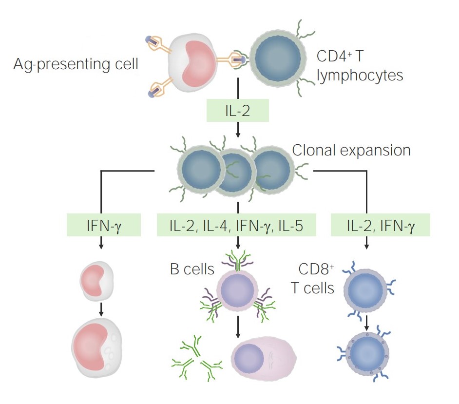
Tras interactuar con las células presentadoras de Ag, se produce la estimulación y expansión clonal de las células T CD4+:
Estas células liberan citoquinas que pueden tener diversos efectos, como la activación de macrófagos (izquierda), células B (centro) y células T CD8+.
| Células T CD4+ | Estimulado por | Citoquinas producidas | Funciones | Papel en EN Erythema nodosum is an immune-mediated panniculitis (inflammation of the subcutaneous fat) caused by a type IV (delayed-type) hypersensitivity reaction. It commonly manifests in young women as tender, erythematous nodules on the shins. Erythema Nodosum la enfermedad |
|---|---|---|---|---|
| Th1 Th1 A subset of helper-inducer T-lymphocytes which synthesize and secrete interleukin-2; interferon-gamma; and interleukin-12. Due to their ability to kill antigen-presenting cells and their lymphokine-mediated effector activity, th1 cells are associated with vigorous delayed-type hypersensitivity reactions. T cells: Types and Functions | IL-12, IFN-γ | IFN-γ, TNF TNF Tumor necrosis factor (TNF) is a major cytokine, released primarily by macrophages in response to stimuli. The presence of microbial products and dead cells and injury are among the stimulating factors. This protein belongs to the TNF superfamily, a group of ligands and receptors performing functions in inflammatory response, morphogenesis, and cell proliferation. Tumor Necrosis Factor (TNF), IL-2 |
|
|
| Th2 Th2 A subset of helper-inducer T-lymphocytes which synthesize and secrete the interleukins il-4; il-5; il-6; and il-10. These cytokines influence b-cell development and antibody production as well as augmenting humoral responses. T cells: Types and Functions | IL-2, IL-4 | IL-4, IL-5, IL-6, IL-9, IL-10, IL-13 |
|
|
| Th17 Th17 A subset of helper-effector T-lymphocytes which synthesize and secrete interleukins il-17; il-17f; and il-22. These cytokines are involved in host defenses and tissue inflammation in autoimmune diseases. T cells: Types and Functions | IL-1, IL-6, IL-23, TGF-β | IL-17, IL-21, IL-22 | Promover la inflamación neutrofílica |
|
| Tfh | IL-6 | IL-4, IL-21 | Facilitar la activación y maduración de las células B | Producción de anticuerpos |
| Treg | TGF-β, IL-2 | TGF-β, IL-10, IL-35 |
|
↓ Autoinmunidad, alergia, inflamación |
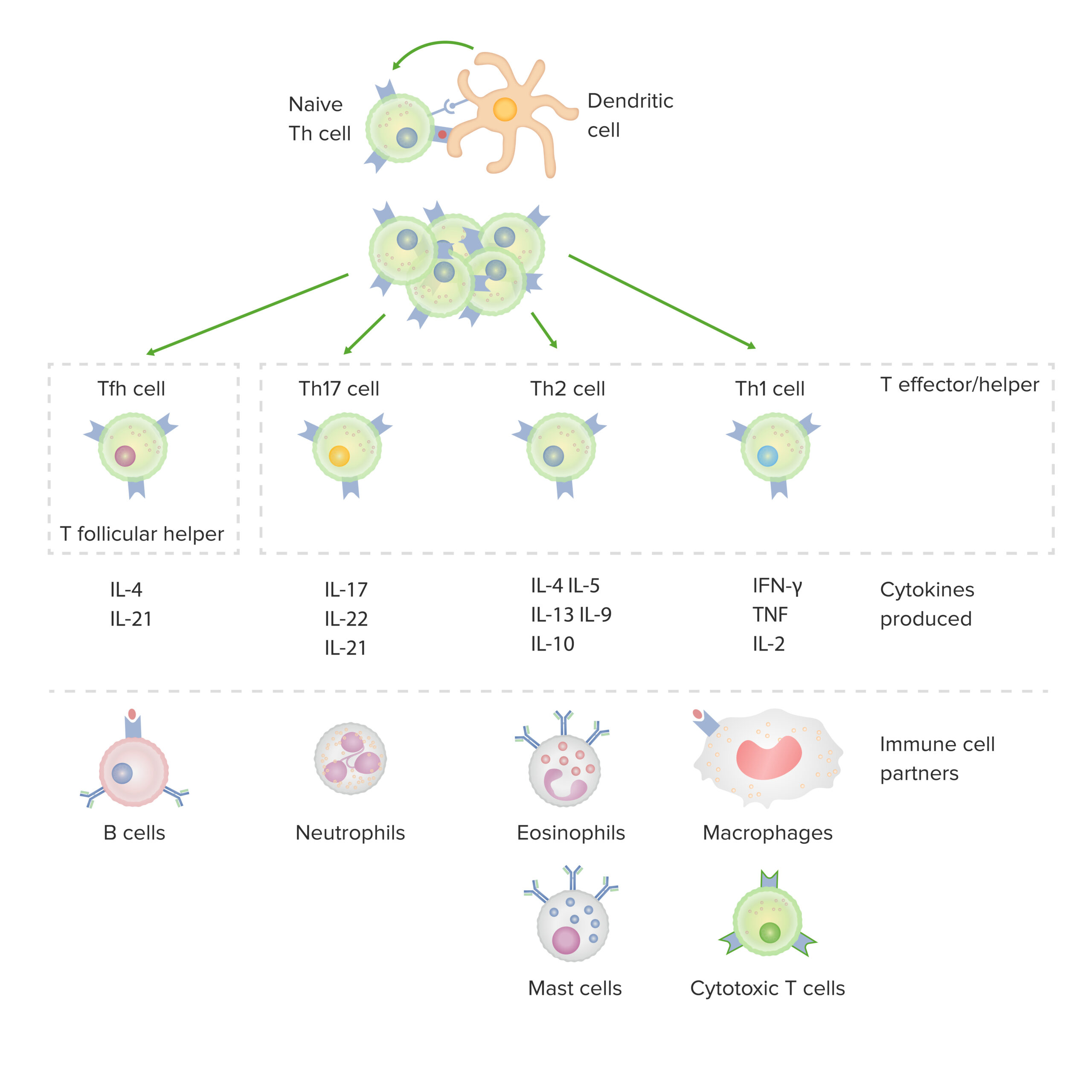
Subconjuntos de células T CD4+, incluidas las citocinas producidas y los socios celulares inmunitarios asociados
IFN: interferón
TNF: factor de necrosis tumoral
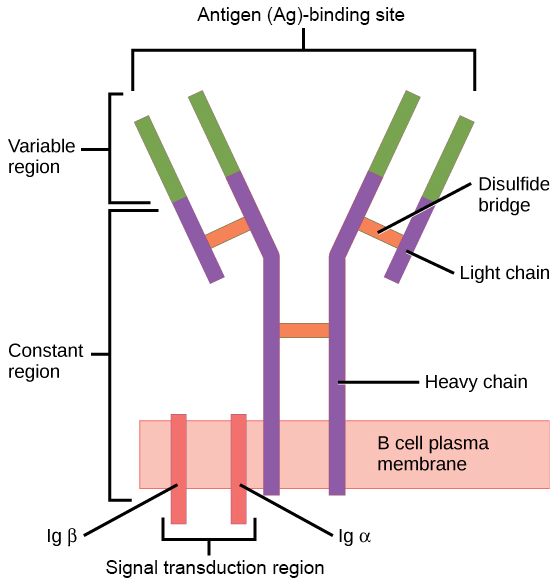
Receptor de células B:
Consiste en la molécula de inmunoglobulina y la molécula de señalización.
La inmunoglobulina unida a la membrana está anclada a la superficie celular. La Inmunoglobulina contiene 2 cadenas pesadas idénticas y 2 cadenas ligeras idénticas, unidas por un puente disulfuro.

Activación de las células B en la respuesta inmune humoral que produce los anticuerpos:
1) Una célula B se activa cuando encuentra su antígeno correspondiente.
2) El linfocito B engulle el antígeno y lo digiere.
3) A continuación, muestra fragmentos del antígeno unidos a su única molécula del complejo mayor de histocompatibilidad.
4) Esta combinación de antígeno y complejo mayor de histocompatibilidad atrae la ayuda de una célula T madura compatible.
5) Las citoquinas secretadas por la célula T ayudan a la célula B a multiplicarse y madurar en células plasmáticas productoras de anticuerpos.
6) Liberados en la sangre, los anticuerpos se fijan al antígeno correspondiente. Los complejos antígeno-anticuerpo son eliminados por la cascada del complemento o por el hígado y el bazo.
Una célula de memoria es un linfocito B o T específico para un antígeno que produce una fuerte respuesta inmune después de una nueva exposición al AL Amyloidosis mismo patógeno. Tanto las células B como las T de memoria expresan el marcador de superficie CD27.
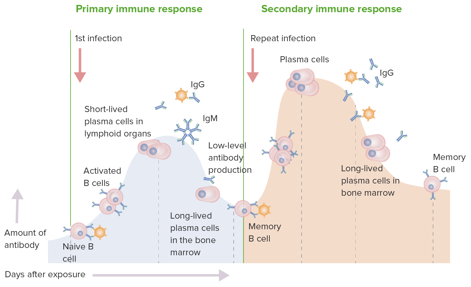
Respuestas inmunes primarias y secundarias:
En una respuesta inmune primaria, las células B vírgenes son estimuladas por el antígeno. Se produce la activación de las células B y luego la diferenciación en células secretoras de anticuerpos. Los anticuerpos son específicos para el antígeno desencadenante. Luego, la producción de IgM es seguida por IgG. Si bien hay una respuesta inmune, la producción es de bajo nivel. En la respuesta inmune secundaria, el mismo antígeno estimula las células B de memoria, lo que conduce a la producción de mayores cantidades de anticuerpos específicos que se producen en la respuesta primaria. La producción y liberación de IgG también ocurre antes.