La reacción de hipersensibilidad tipo I es una respuesta inmunitaria anormal provocada por la exposición a antígenos específicos conocidos como alérgenos. En EN Erythema nodosum is an immune-mediated panniculitis (inflammation of the subcutaneous fat) caused by a type IV (delayed-type) hypersensitivity reaction. It commonly manifests in young women as tender, erythematous nodules on the shins. Erythema Nodosum este tipo de reacción de hipersensibilidad, la presentación del antígeno a las células T colaboradoras inicia una cascada de eventos inmunológicos que conducen a la producción de anticuerpos inmunoglobulina E ( IgE IgE An immunoglobulin associated with mast cells. Overexpression has been associated with allergic hypersensitivity. Immunoglobulins: Types and Functions) específicos de antígeno. La reexposición al AL Amyloidosis antígeno promueve la degranulación de los LOS Neisseria mastocitos y basófilos unidos a IgE IgE An immunoglobulin associated with mast cells. Overexpression has been associated with allergic hypersensitivity. Immunoglobulins: Types and Functions, liberando mediadores químicos que causan varios síntomas de alergia. Las manifestaciones pueden ser locales, dependiendo de la vía de entrada del antígeno. En EN Erythema nodosum is an immune-mediated panniculitis (inflammation of the subcutaneous fat) caused by a type IV (delayed-type) hypersensitivity reaction. It commonly manifests in young women as tender, erythematous nodules on the shins. Erythema Nodosum casos severos, la reacción sistémica conduce a un shock Shock Shock is a life-threatening condition associated with impaired circulation that results in tissue hypoxia. The different types of shock are based on the underlying cause: distributive (↑ cardiac output (CO), ↓ systemic vascular resistance (SVR)), cardiogenic (↓ CO, ↑ SVR), hypovolemic (↓ CO, ↑ SVR), obstructive (↓ CO), and mixed. Types of Shock anafiláctico. Para determinar la etiología alérgica, se dispone de pruebas cutáneas y pruebas in vitro. El tratamiento incluye evitar los LOS Neisseria desencadenantes para reducir la exacerbación. Las opciones de tratamiento comunes son los LOS Neisseria antihistamínicos y los LOS Neisseria glucocorticoides para controlar la respuesta inflamatoria. La anafilaxia, sin embargo, es una emergencia médica que requiere acceso inmediato a las vías respiratorias con administración de epinefrina y reanimación con líquidos.
Last updated: Apr 14, 2025
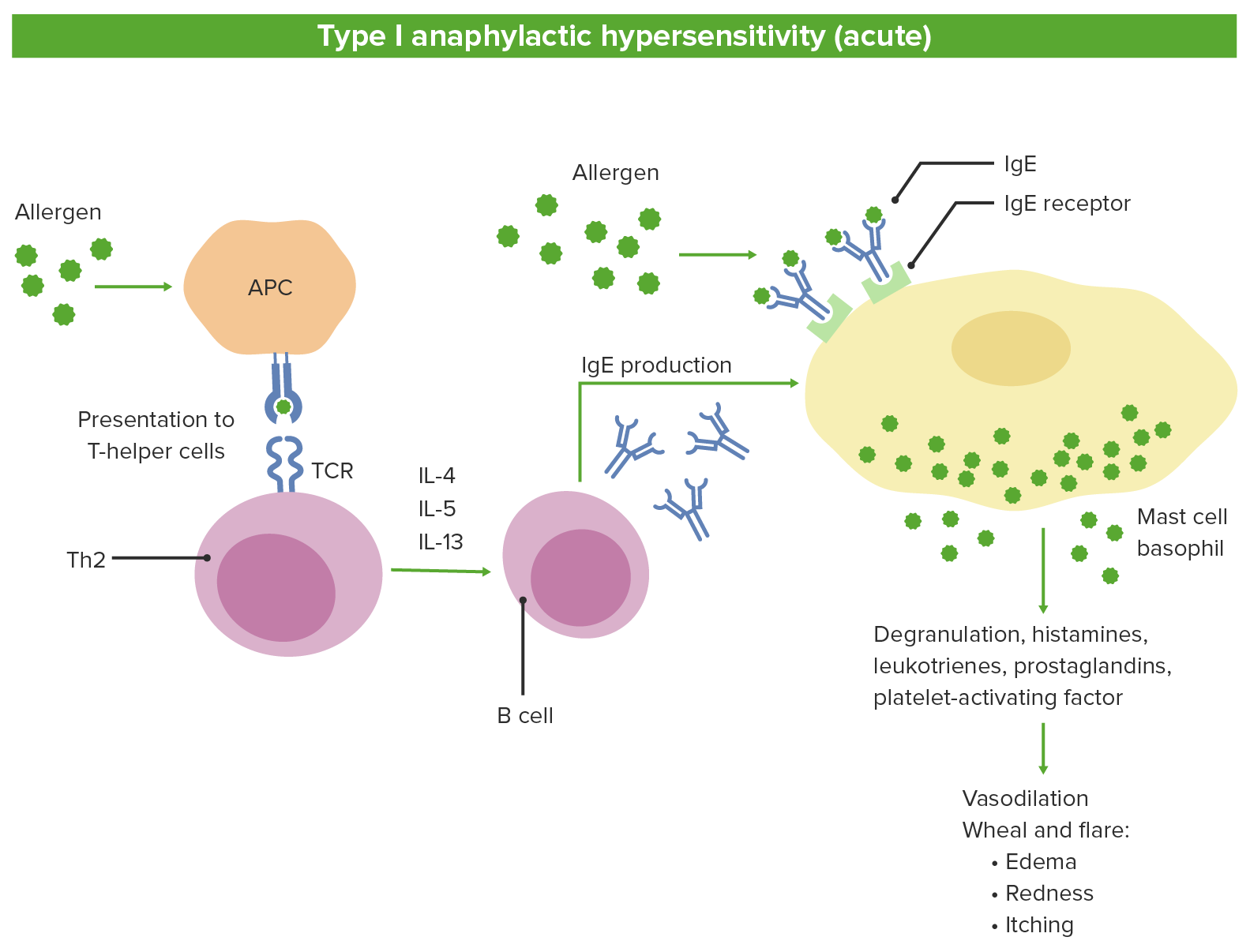
Fisiopatología de la hipersensibilidad tipo 1:
1. Las células presentadoras de antígenos (APC) reconocen el alérgeno y lo presentan a las células T vírgenes
2. Las células T se diferencian en Th2 que liberan las interleuquinas.
3. Las interleuquinas estimulan las células B para producir IgE.
4. La IgE específica de antígeno se une a los mastocitos y basófilos.
5. La exposición posterior al mismo antígeno conduce a la degranulación y liberación de mediadores.
TCR: receptor de células T
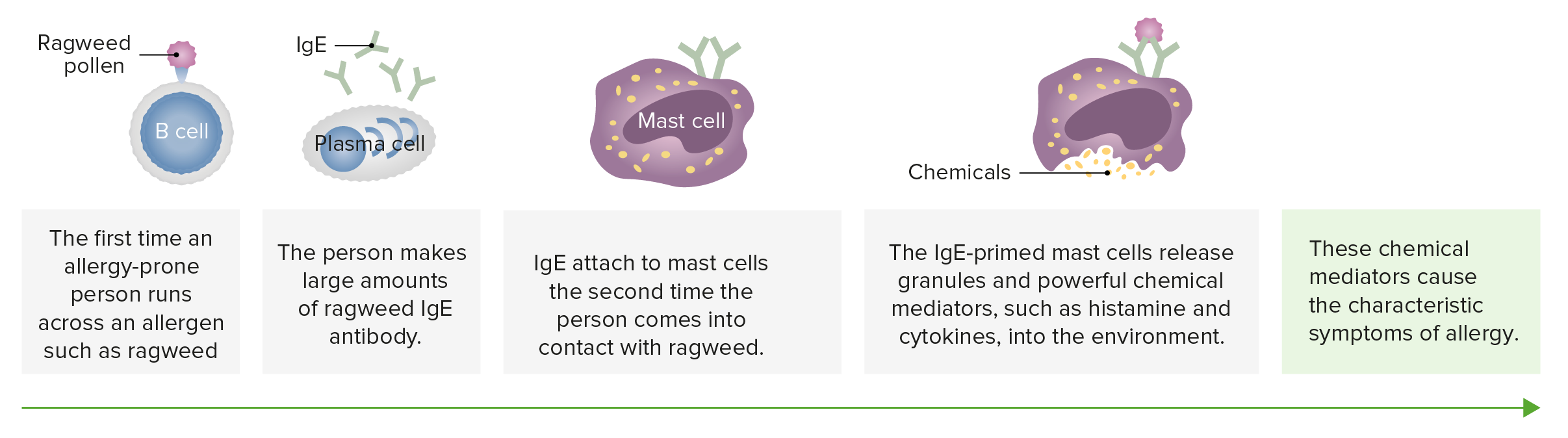
Los mastocitos están implicados en la alergia. Las alergias como la alergia al polen están relacionadas con el anticuerpo conocido como IgE. Como otros anticuerpos, cada anticuerpo IgE es específico; uno actúa contra el polen del roble, otro contra la ambrosía.
Imagen por Lecturio.| Tipo I | Tipo II | Tipo III | Tipo IV |
|---|---|---|---|
| Hipersensibilidad mediada por IgE IgE An immunoglobulin associated with mast cells. Overexpression has been associated with allergic hypersensitivity. Immunoglobulins: Types and Functions | Hipersensibilidad citotóxica mediada por inmunoglobulina G ( IgG IgG The major immunoglobulin isotype class in normal human serum. There are several isotype subclasses of igg, for example, igg1, igg2a, and igg2b. Hypersensitivity Pneumonitis) | Hipersensibilidad mediada por inmunocomplejos | Hipersensibilidad mediada por células |
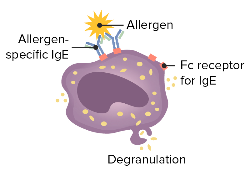
| La IgE IgE An immunoglobulin associated with mast cells. Overexpression has been associated with allergic hypersensitivity. Immunoglobulins: Types and Functions se une a los LOS Neisseria mastocitos a través de su fragmento cristalizable ( Fc Fc Crystallizable fragments composed of the carboxy-terminal halves of both immunoglobulin heavy chains linked to each other by disulfide bonds. Fc fragments contain the carboxy-terminal parts of the heavy chain constant regions that are responsible for the effector functions of an immunoglobulin (complement fixation, binding to the cell membrane via fc receptors, and placental transport). This fragment can be obtained by digestion of immunoglobulins with the proteolytic enzyme papain. Immunoglobulins: Types and Functions). Cuando un alérgeno se une a estos anticuerpos, el entrecruzamiento de IgE IgE An immunoglobulin associated with mast cells. Overexpression has been associated with allergic hypersensitivity. Immunoglobulins: Types and Functions induce la degranulación. | Las células son destruidas por el anticuerpo unido, ya sea por la activación del complemento o por una célula NK con un receptor Receptor Receptors are proteins located either on the surface of or within a cell that can bind to signaling molecules known as ligands (e.g., hormones) and cause some type of response within the cell. Receptors Fc Fc Crystallizable fragments composed of the carboxy-terminal halves of both immunoglobulin heavy chains linked to each other by disulfide bonds. Fc fragments contain the carboxy-terminal parts of the heavy chain constant regions that are responsible for the effector functions of an immunoglobulin (complement fixation, binding to the cell membrane via fc receptors, and placental transport). This fragment can be obtained by digestion of immunoglobulins with the proteolytic enzyme papain. Immunoglobulins: Types and Functions para el anticuerpo (citotoxicidad celular dependiente de anticuerpos) | Los LOS Neisseria complejos antígeno-anticuerpo se depositan en EN Erythema nodosum is an immune-mediated panniculitis (inflammation of the subcutaneous fat) caused by a type IV (delayed-type) hypersensitivity reaction. It commonly manifests in young women as tender, erythematous nodules on the shins. Erythema Nodosum los LOS Neisseria tejidos, provocando la activación del complemento, que atrae a los LOS Neisseria neutrófilos al AL Amyloidosis sitio. | Las células Th1 Th1 A subset of helper-inducer T-lymphocytes which synthesize and secrete interleukin-2; interferon-gamma; and interleukin-12. Due to their ability to kill antigen-presenting cells and their lymphokine-mediated effector activity, th1 cells are associated with vigorous delayed-type hypersensitivity reactions. T cells: Types and Functions secretan citoquinas, que activan los LOS Neisseria macrófagos y las células T citotóxicas y pueden provocar la acumulación de macrófagos en EN Erythema nodosum is an immune-mediated panniculitis (inflammation of the subcutaneous fat) caused by a type IV (delayed-type) hypersensitivity reaction. It commonly manifests in young women as tender, erythematous nodules on the shins. Erythema Nodosum el sitio. |
| Causa anafilaxia localizada y sistémica; alergias estacionales, incluida la fiebre del heno; alergias alimentarias, como las de los LOS Neisseria mariscos y los LOS Neisseria cacahuetes; urticaria Urticaria Urticaria is raised, well-circumscribed areas (wheals) of edema (swelling) and erythema (redness) involving the dermis and epidermis with associated pruritus (itch). Urticaria is not a single disease but rather is a reaction pattern representing cutaneous mast cell degranulation. Urticaria (Hives); y eccema. | Los LOS Neisseria eritrocitos son destruidos por el complemento y los LOS Neisseria anticuerpos durante una transfusión de tipo incompatible o durante la eritroblastosis fetal. | Las formas más comunes de enfermedad por complejos inmunes incluyen glomerulonefritis, artritis reumatoide y lupus eritematoso sistémico. | Las formas más comunes son la dermatitis Dermatitis Any inflammation of the skin. Atopic Dermatitis (Eczema) de contacto, reacción a la tuberculina, diabetes Diabetes Diabetes mellitus (DM) is a metabolic disease characterized by hyperglycemia and dysfunction of the regulation of glucose metabolism by insulin. Type 1 DM is diagnosed mostly in children and young adults as the result of autoimmune destruction of β cells in the pancreas and the resulting lack of insulin. Type 2 DM has a significant association with obesity and is characterized by insulin resistance. Diabetes Mellitus mellitus tipo I, esclerosis múltiple y artritis reumatoide. |
Hipersensibilidad tipo 1
Imagen: “Figure 1” por Phil Schatz. Licencia: CC BY 4.0, editado por Lecturio.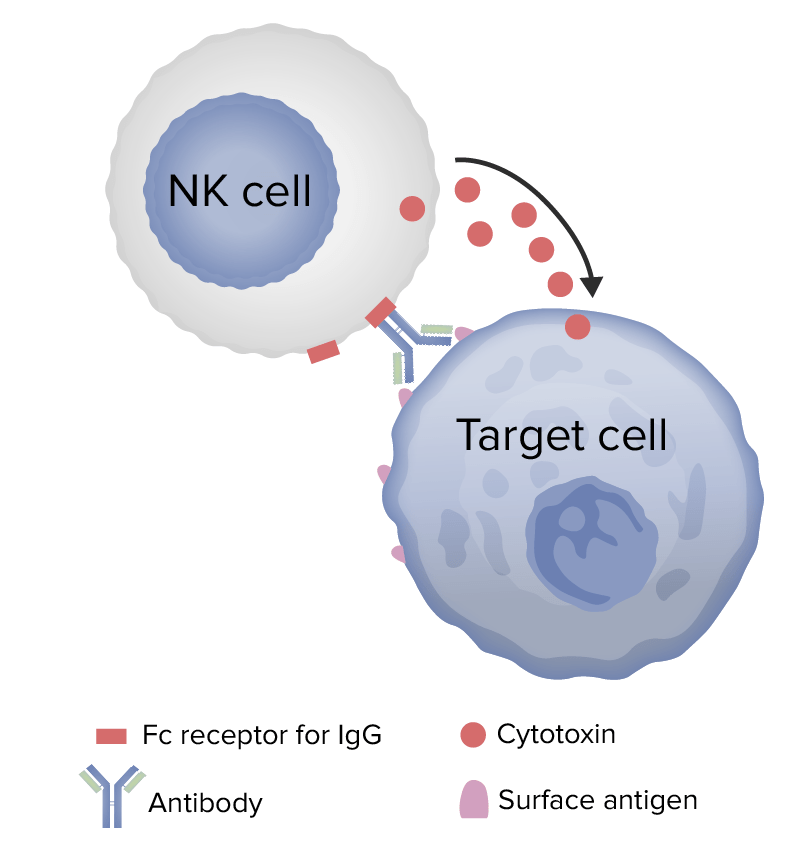
Mecanismo de hipersensibilidad de tipo 2
Image: “Figure 1” by Phil Schatz. License: CC BY 4.0, edited by Lecturio.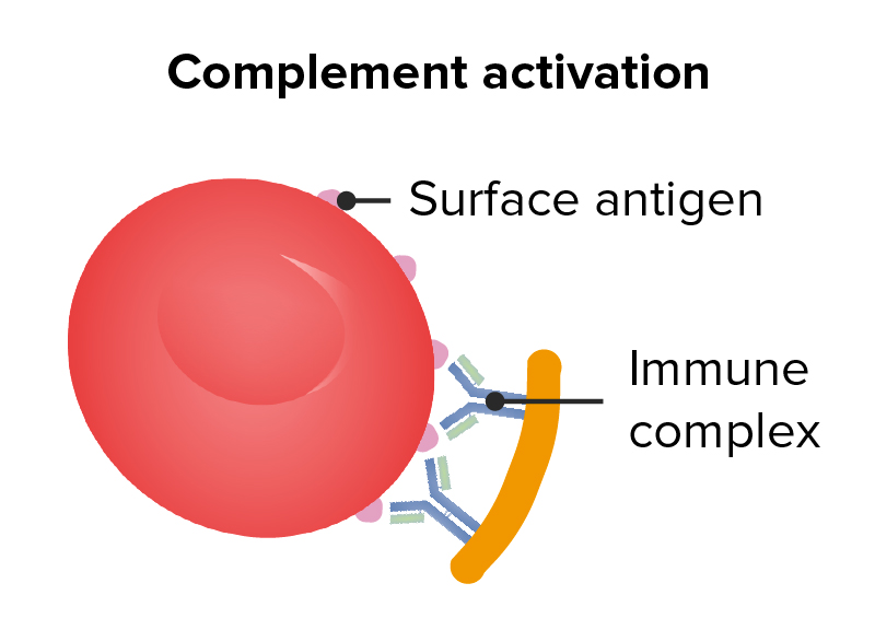
Hipersensibilidad tipo 2
Imagen: “Figure 1” por Phil Schatz. Licencia: CC BY 4.0, editado por Lecturio.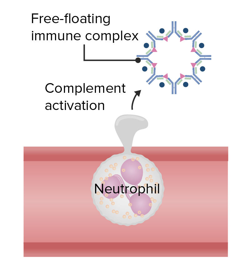
Hipersensibilidad tipo 3
Imagen: “Figure 1” por Phil Schatz. Licencia: CC BY 4.0, editado por Lecturio.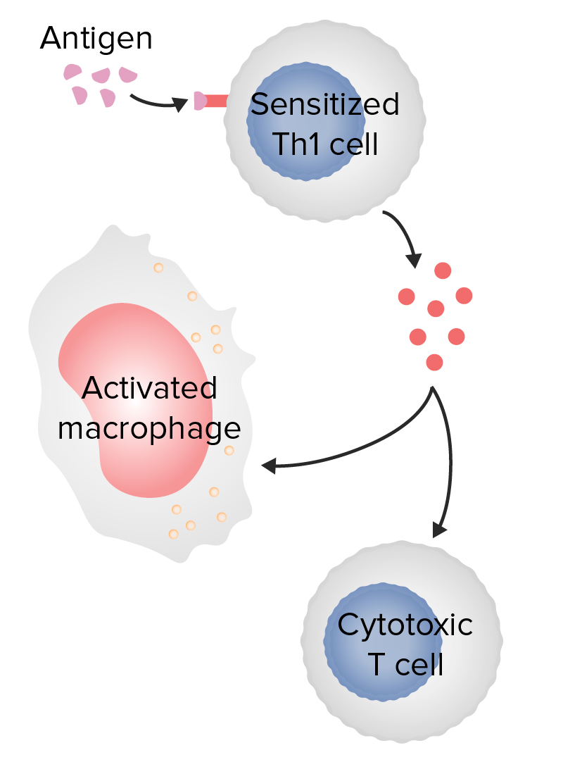
Hipersensibilidad tipo 4
Imagen: “Figure 1” por Phil Schatz. Licencia: CC BY 4.0, editado por Lecturio.| Síntomas anafilácticos | Efectos de la histamina |
|---|---|
| Rinitis, conjuntivitis | Vasodilatación periférica, aumento de la permeabilidad vascular, aumento de la secreción de moco |
| Eritema | Acumulación de sangre en EN Erythema nodosum is an immune-mediated panniculitis (inflammation of the subcutaneous fat) caused by a type IV (delayed-type) hypersensitivity reaction. It commonly manifests in young women as tender, erythematous nodules on the shins. Erythema Nodosum el lecho capilar por vasodilatación |
| Edema Edema Edema is a condition in which excess serous fluid accumulates in the body cavity or interstitial space of connective tissues. Edema is a symptom observed in several medical conditions. It can be categorized into 2 types, namely, peripheral (in the extremities) and internal (in an organ or body cavity). Edema pulmonar, angioedema Angioedema Angioedema is a localized, self-limited (but potentially life-threatening), nonpitting, asymmetrical edema occurring in the deep layers of the skin and mucosal tissue. The common underlying pathophysiology involves inflammatory mediators triggering significant vasodilation and increased capillary permeability. Angioedema, hipotensión | Cambio de líquido hacia el estroma debido al AL Amyloidosis aumento de la permeabilidad de los LOS Neisseria vasos, vasodilatación |
| Prurito o urticaria Urticaria Urticaria is raised, well-circumscribed areas (wheals) of edema (swelling) and erythema (redness) involving the dermis and epidermis with associated pruritus (itch). Urticaria is not a single disease but rather is a reaction pattern representing cutaneous mast cell degranulation. Urticaria (Hives) | Extravasación de líquido en EN Erythema nodosum is an immune-mediated panniculitis (inflammation of the subcutaneous fat) caused by a type IV (delayed-type) hypersensitivity reaction. It commonly manifests in young women as tender, erythematous nodules on the shins. Erythema Nodosum la dermis Dermis A layer of vascularized connective tissue underneath the epidermis. The surface of the dermis contains innervated papillae. Embedded in or beneath the dermis are sweat glands; hair follicles; and sebaceous glands. Skin: Structure and Functions; aumenta el desencadenante de los LOS Neisseria nervios sensoriales de la piel (prurito) |
| Broncoespasmo, broncoconstricción | Contracción del músculo liso bronquial, aumento de la secreción de moco |