LosLOSNeisseria rayos X son partículas de radiación electromagnética de alta energía que se utilizan enENErythema nodosum is an immune-mediated panniculitis (inflammation of the subcutaneous fat) caused by a type IV (delayed-type) hypersensitivity reaction. It commonly manifests in young women as tender, erythematous nodules on the shins.Erythema Nodosum el campo médico para la generación de imágenes anatómicas. LosLOSNeisseria rayos X se proyectan a través del cuerpo de un paciente y sobre una película, y esta técnica se denomina radiografía convencional o de proyección. Como la radiación por rayos X puede causar efectos secundarios dependiendo de la dosis absorbida, es necesario tomar medidas de protección para reducir el daño. La radiografía digital utiliza el formato de datos digitales y permite la manipulación digital de imágenes. LosLOSNeisseria usos comunes incluyen la evaluación de afecciones del tórax, el mediastino, la columna vertebral y losLOSNeisseria huesos/articulaciones. Si bien la radiografía todavía se usa para visualizar las estructuras de la cabeza y el abdomen, ahora se prefieren modalidades más avanzadas (TC y RM). La radiografía sigue siendo un componente esencial de las pruebas iniciales enENErythema nodosum is an immune-mediated panniculitis (inflammation of the subcutaneous fat) caused by a type IV (delayed-type) hypersensitivity reaction. It commonly manifests in young women as tender, erythematous nodules on the shins.Erythema Nodosum muchas enfermedades, dada su amplia disponibilidad, bajo costo y facilidad de operación.
Un rayo X es una partícula discreta de radiación electromagnética (fotón) de alta energía que se propaga a través del espacio a la velocidad de la luz.
Producción de rayos X
LosLOSNeisseria rayos X se producen a través de diferentes procesos:
Radiación característica de losLOSNeisseria rayos X:
Resultado del movimiento o transición de electrones desde una capa externa (órbita) a vacantes enENErythema nodosum is an immune-mediated panniculitis (inflammation of the subcutaneous fat) caused by a type IV (delayed-type) hypersensitivity reaction. It commonly manifests in young women as tender, erythematous nodules on the shins.Erythema Nodosum la capa interna
La emisión de fotones de rayos X depende del material.
LosLOSNeisseria electrones se mueven rápidamente hacia el ánodo (electrodo cargado positivamente) y desaceleran cuando colisionan.
Durante la desaceleración, el 99% de la energía se disipa como calorCalorInflammation y el 1% se libera como fotones de rayos X.
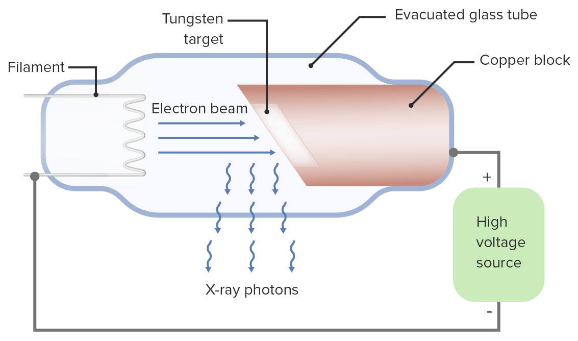
La imagenología con rayos X utilizan un tubo de rayos X que consta de:
Un filamento caliente que emite electrones
Un objetivo/ánodo de tungsteno donde chocan losLOSNeisseria electrones, produciendo rayos X
LosLOSNeisseria rayos X penetran la materia e interactúan con losLOSNeisseria electrones atómicos del material. Durante este proceso, losLOSNeisseria rayos X pueden absorberse o dispersarse.
No todos losLOSNeisseria rayos X pueden penetrar alALAmyloidosis paciente. La mayoría de losLOSNeisseria rayos X son dispersados y no contribuyen a la creación de imágenes.
Diagrama de un tubo de rayos X: En el tubo, los electrones son acelerados hacia un objetivo de tungsteno (ánodo), que luego desaceleran después de golpear el objetivo, liberando calor y fotones de rayos X.
Imagen por Lecturio.
Efectos de la radiación de rayos X
El daño biológico de losLOSNeisseria rayos X se atribuye a la radiación ionizante que se produce cuando losLOSNeisseria rayos X interactúan con la materia.
La dosis absorbida es la energía (de la interacción) depositada enENErythema nodosum is an immune-mediated panniculitis (inflammation of the subcutaneous fat) caused by a type IV (delayed-type) hypersensitivity reaction. It commonly manifests in young women as tender, erythematous nodules on the shins.Erythema Nodosum la materia.
Radiación absorbida: medida enENErythema nodosum is an immune-mediated panniculitis (inflammation of the subcutaneous fat) caused by a type IV (delayed-type) hypersensitivity reaction. It commonly manifests in young women as tender, erythematous nodules on the shins.Erythema Nodosum unidades conocidas como Gray (Gy) o rad (100 rad equivalen a 1 Gy)
Tipos de efectos de la radiación:
Efecto determinista:
El daño ocurre cuando se cruza un umbral de radiación, de modo que la capacidad de una célula para repararse a sí misma se veVEVentilation: Mechanics of Breathing abrumada.
Es el resultado de dosis muy altas de radiación, que causan eritema cutáneo, cataratas y esterilidad
Efecto estocástico: el daño es aditivo y la probabilidad del efecto aumenta con una mayor exposición.
El daño se produce a nivel genético durante la división celular y puede provocar carcinogénesis.
La probabilidad de efectos aumenta con la dosis de radiación.
EnENErythema nodosum is an immune-mediated panniculitis (inflammation of the subcutaneous fat) caused by a type IV (delayed-type) hypersensitivity reaction. It commonly manifests in young women as tender, erythematous nodules on the shins.Erythema Nodosum última instancia, el daño resultante incluye:
Formación de radicales libres
Interrupción de la función metabólica y la mitosisMitosisA type of cell nucleus division by means of which the two daughter nuclei normally receive identical complements of the number of chromosomes of the somatic cells of the species.Cell Cycle
Inducción de cáncer:
LosLOSNeisseria órganos con las células que se dividen más rápidamente son losLOSNeisseria más susceptibles, lo que también explica por qué losLOSNeisseria niños, enENErythema nodosum is an immune-mediated panniculitis (inflammation of the subcutaneous fat) caused by a type IV (delayed-type) hypersensitivity reaction. It commonly manifests in young women as tender, erythematous nodules on the shins.Erythema Nodosum general, son más susceptibles.
Órganos más susceptibles:
Médula ósea
ColonColonThe large intestines constitute the last portion of the digestive system. The large intestine consists of the cecum, appendix, colon (with ascending, transverse, descending, and sigmoid segments), rectum, and anal canal. The primary function of the colon is to remove water and compact the stool prior to expulsion from the body via the rectum and anal canal. Colon, Cecum, and Appendix: Anatomy
Pulmones
Estómago
Órganos moderadamente susceptibles:
Vejiga
Mamas
Hígado
Esófago
Tiroides
Riesgo fetal de radiación
Tabla: Riesgo fetal de radiación
Semanas después de la concepción
Efectos de una exposición importante
2
10–50 rad: riesgo de falla enENErythema nodosum is an immune-mediated panniculitis (inflammation of the subcutaneous fat) caused by a type IV (delayed-type) hypersensitivity reaction. It commonly manifests in young women as tender, erythematous nodules on the shins.Erythema Nodosum la implantación
> 50 rad: alta probabilidad de implantación fallida
3–5
10–50 rad: posible restricción del crecimiento
> 50 rad: anomalías congénitas, restricción del crecimiento, riesgo de aborto espontáneo
6–13
10–50 rad: posible restricción del crecimiento
> 50 rad: restricción del crecimiento, riesgo de aborto espontáneo
14–23
10–50 rad: es poco probable que se produzcan efectos no cancerosos enENErythema nodosum is an immune-mediated panniculitis (inflammation of the subcutaneous fat) caused by a type IV (delayed-type) hypersensitivity reaction. It commonly manifests in young women as tender, erythematous nodules on the shins.Erythema Nodosum la salud
> 50 rad: restricción del crecimiento, riesgo de aborto espontáneo, posibles anomalías congénitas
24 semanas hasta el término del embarazo
10–50 rad: es poco probable que se produzcan efectos no cancerosos enENErythema nodosum is an immune-mediated panniculitis (inflammation of the subcutaneous fat) caused by a type IV (delayed-type) hypersensitivity reaction. It commonly manifests in young women as tender, erythematous nodules on the shins.Erythema Nodosum la salud
> 50 rad: aborto espontáneo, muerte neonatal (según la dosis)
Protección contra la radiación
Minimice la dosis de radiación siempre que sea posible, lo más como sea posible.
Medidas:
El personal expuesto debe ser monitorizado usando un dosímetro de película.
Blindaje de plomo y aumento de la distancia desde la fuente
Blindaje dentro de las habitaciones
Aumentar el kilovoltaje del haz de rayos X, aumenta su penetración
Radiografía de proyección: generación de una imagen radiográfica proyectando un haz de partículas de rayos X a través de un sujeto y hacia una película:
La imagen de rayos X es una imagen de sombra obtenida utilizando una sola fuente de “luz”.
Fluoroscopia: el uso de radiografía de proyección para observar estructuras internas enENErythema nodosum is an immune-mediated panniculitis (inflammation of the subcutaneous fat) caused by a type IV (delayed-type) hypersensitivity reaction. It commonly manifests in young women as tender, erythematous nodules on the shins.Erythema Nodosum tiempo real (e.g., imagenología gastrointestinal)
TC: generación de una imagen de varias capas mediante un haz proyectado por un tubo de rayos X rotatorio, hacia detectores de radiación
Generación de imágenes por rayos X
Orden de producción de una imagen con rayos X:
Tubo de rayos X: rayos X generados después de que losLOSNeisseria electrones colisionan con el ánodo.
Paciente: el haz de rayos X atraviesa alALAmyloidosis paciente y se atenúa enENErythema nodosum is an immune-mediated panniculitis (inflammation of the subcutaneous fat) caused by a type IV (delayed-type) hypersensitivity reaction. It commonly manifests in young women as tender, erythematous nodules on the shins.Erythema Nodosum función de losLOSNeisseria tejidos a su paso.
Rejilla antidispersión: tiras de plomo que mejoran el contraste de la imagen alALAmyloidosis reducir losLOSNeisseria fotones dispersos
La captura de imágenes se realiza mediante el uso de una placa de imágenes enENErythema nodosum is an immune-mediated panniculitis (inflammation of the subcutaneous fat) caused by a type IV (delayed-type) hypersensitivity reaction. It commonly manifests in young women as tender, erythematous nodules on the shins.Erythema Nodosum un casete.
Tecnologías que producen imágenes radiográficas:
Radiografía convencional:
Se utiliza una placa y se revela la película.
Alta sensibilidad, bajo costo y fácil manejo
Radiografía digital (utiliza un formato de datos digitales, lo que permite la manipulación digital de las imágenes):
Radiografía computarizada: se inserta un casete enENErythema nodosum is an immune-mediated panniculitis (inflammation of the subcutaneous fat) caused by a type IV (delayed-type) hypersensitivity reaction. It commonly manifests in young women as tender, erythematous nodules on the shins.Erythema Nodosum un escáner y la imagen se muestra enENErythema nodosum is an immune-mediated panniculitis (inflammation of the subcutaneous fat) caused by a type IV (delayed-type) hypersensitivity reaction. It commonly manifests in young women as tender, erythematous nodules on the shins.Erythema Nodosum un monitor.
Radiografía directa: no se utiliza casete. LosLOSNeisseria rayos X se convierten enENErythema nodosum is an immune-mediated panniculitis (inflammation of the subcutaneous fat) caused by a type IV (delayed-type) hypersensitivity reaction. It commonly manifests in young women as tender, erythematous nodules on the shins.Erythema Nodosum cargas eléctricas mediante un fotoconductor.
Imagen de una radiografía temprana: radiografía de una mano izquierda tomada en una conferencia pública por Wilhelm Röntgen
Imagen: “An early X-ray” por Wilhelm Röntgen; versión actual creada por Old Moonraker. Licencia: Dominio Público
Suma de sombras: las imágenes aparecen más radiopacas debido a densidades superpuestas.
Signo de la silueta:
LosLOSNeisseria bordes de un objeto son indistinguibles cuando las densidades son adyacentes entre sí.
Piense enENErythema nodosum is an immune-mediated panniculitis (inflammation of the subcutaneous fat) caused by a type IV (delayed-type) hypersensitivity reaction. It commonly manifests in young women as tender, erythematous nodules on the shins.Erythema Nodosum una neumonía enENErythema nodosum is an immune-mediated panniculitis (inflammation of the subcutaneous fat) caused by a type IV (delayed-type) hypersensitivity reaction. It commonly manifests in young women as tender, erythematous nodules on the shins.Erythema Nodosum el lóbulo medio derecho, que oculta el borde derecho del corazón.
Imagenología ortogonal: tomar 2 proyecciones de la misma estructura para documentar mejor su tridimensionalidad
Elementos que reducen el rendimiento diagnóstico de una radiografía
Penetración excesiva o insuficiente
Rotación del paciente
Magnificación de la imagen
Movimiento del paciente
Artefactos, como partículas de polvo
Tórax
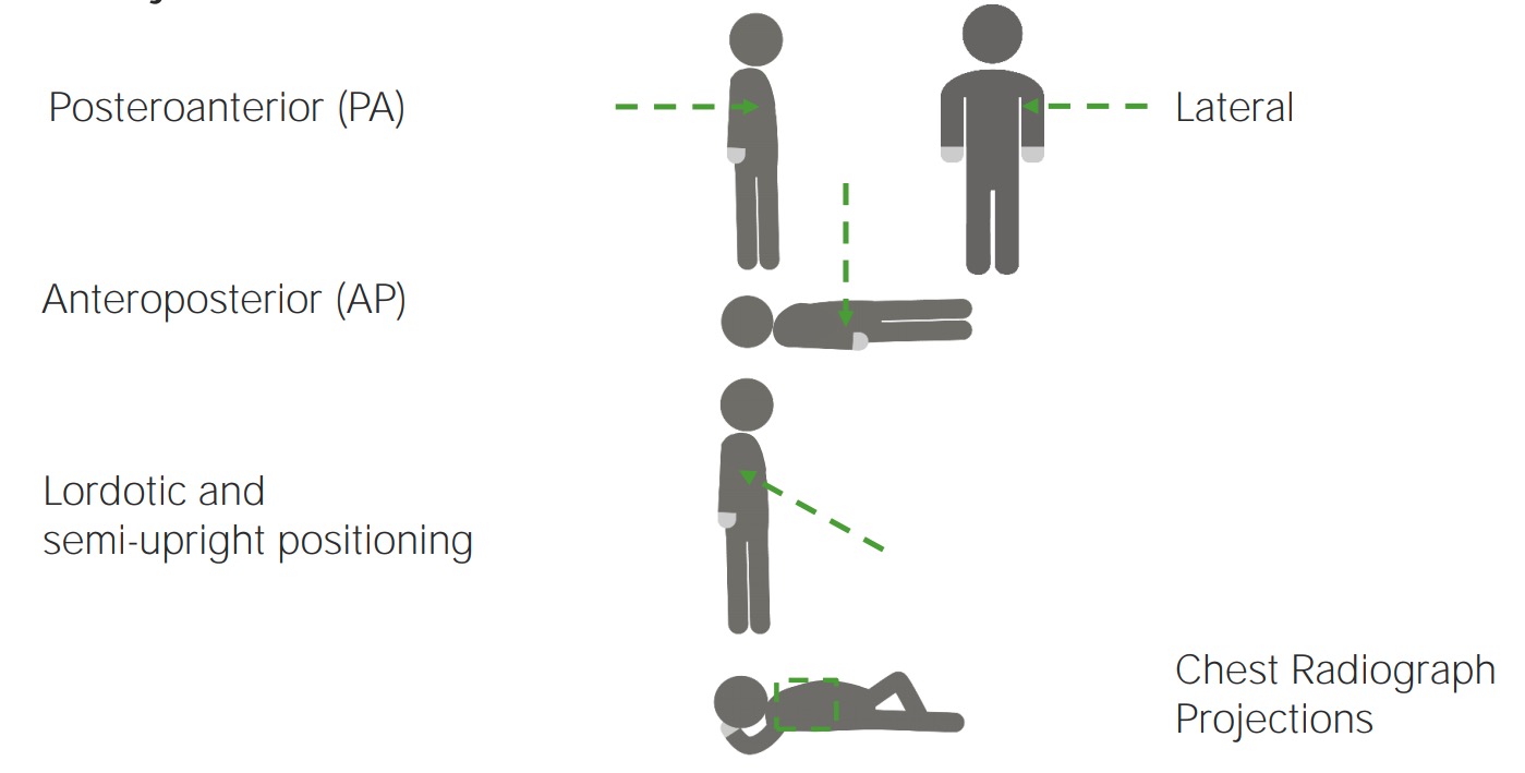
Proyecciones
Las imágenes de rayos X del tórax se pueden producir enENErythema nodosum is an immune-mediated panniculitis (inflammation of the subcutaneous fat) caused by a type IV (delayed-type) hypersensitivity reaction. It commonly manifests in young women as tender, erythematous nodules on the shins.Erythema Nodosum las siguientes proyecciones:
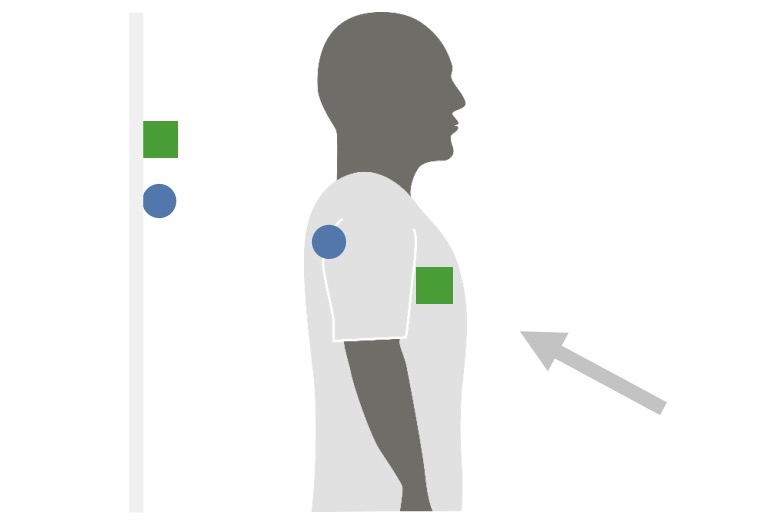
Posteroanterior:
El haz de rayos X penetra inicialmente enENErythema nodosum is an immune-mediated panniculitis (inflammation of the subcutaneous fat) caused by a type IV (delayed-type) hypersensitivity reaction. It commonly manifests in young women as tender, erythematous nodules on the shins.Erythema Nodosum la cara posterior del cuerpo, mientras que el casete se coloca enENErythema nodosum is an immune-mediated panniculitis (inflammation of the subcutaneous fat) caused by a type IV (delayed-type) hypersensitivity reaction. It commonly manifests in young women as tender, erythematous nodules on the shins.Erythema Nodosum contacto directo con la cara anterior.
Método preferido para evaluar el tamaño de la silueta cardíaca
Anteroposterior:
El haz de rayos X penetra inicialmente enENErythema nodosum is an immune-mediated panniculitis (inflammation of the subcutaneous fat) caused by a type IV (delayed-type) hypersensitivity reaction. It commonly manifests in young women as tender, erythematous nodules on the shins.Erythema Nodosum la cara anterior del cuerpo, mientras que el casete se coloca enENErythema nodosum is an immune-mediated panniculitis (inflammation of the subcutaneous fat) caused by a type IV (delayed-type) hypersensitivity reaction. It commonly manifests in young women as tender, erythematous nodules on the shins.Erythema Nodosum contacto directo con la cara posterior.
Utilizada enENErythema nodosum is an immune-mediated panniculitis (inflammation of the subcutaneous fat) caused by a type IV (delayed-type) hypersensitivity reaction. It commonly manifests in young women as tender, erythematous nodules on the shins.Erythema Nodosum radiografía portátil (muy común enENErythema nodosum is an immune-mediated panniculitis (inflammation of the subcutaneous fat) caused by a type IV (delayed-type) hypersensitivity reaction. It commonly manifests in young women as tender, erythematous nodules on the shins.Erythema Nodosum pacientes hospitalizados que no pueden moverse)
Las estructuras más alejadas del casete se ven aumentadas, creando falsos positivos de cardiomegalia.
Lateral:
El haz de rayos X incide enENErythema nodosum is an immune-mediated panniculitis (inflammation of the subcutaneous fat) caused by a type IV (delayed-type) hypersensitivity reaction. It commonly manifests in young women as tender, erythematous nodules on the shins.Erythema Nodosum una cara lateral del cuerpo y el casete se coloca enENErythema nodosum is an immune-mediated panniculitis (inflammation of the subcutaneous fat) caused by a type IV (delayed-type) hypersensitivity reaction. It commonly manifests in young women as tender, erythematous nodules on the shins.Erythema Nodosum contacto con la otra cara lateral.
Posicionamiento lordótico y semi-erguido:
El haz de rayos X penetra enENErythema nodosum is an immune-mediated panniculitis (inflammation of the subcutaneous fat) caused by a type IV (delayed-type) hypersensitivity reaction. It commonly manifests in young women as tender, erythematous nodules on the shins.Erythema Nodosum el paciente enENErythema nodosum is an immune-mediated panniculitis (inflammation of the subcutaneous fat) caused by a type IV (delayed-type) hypersensitivity reaction. It commonly manifests in young women as tender, erythematous nodules on the shins.Erythema Nodosum un ángulo para mostrar 2 estructuras diferentes enENErythema nodosum is an immune-mediated panniculitis (inflammation of the subcutaneous fat) caused by a type IV (delayed-type) hypersensitivity reaction. It commonly manifests in young women as tender, erythematous nodules on the shins.Erythema Nodosum diferentes niveles.
Decúbito:
El paciente se acuesta sobre su lado derecho o izquierdo.
Reemplazo de la proyección lateral que se usa para pacientes que no pueden ponerse de pie
Proyecciones de la radiografía de tórax
Imagen por Lecturio.
Radiografía de proyección posteroanterior: El haz de rayos X penetra al paciente desde la cara posterior y la cara anterior se coloca en contacto directo con el receptor.
Imagen por Lecturio.
Radiografía de proyección anteroposterior: El haz de rayos X penetra al paciente desde la cara anterior y la cara posterior se coloca en contacto directo con el receptor.
Imagen por Lecturio.
Radiografía de proyección lordótica apical: El haz de rayos X penetra en el paciente en un ángulo para mostrar 2 elementos diferentes en diferentes niveles.
Imagen por Lecturio.
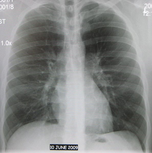
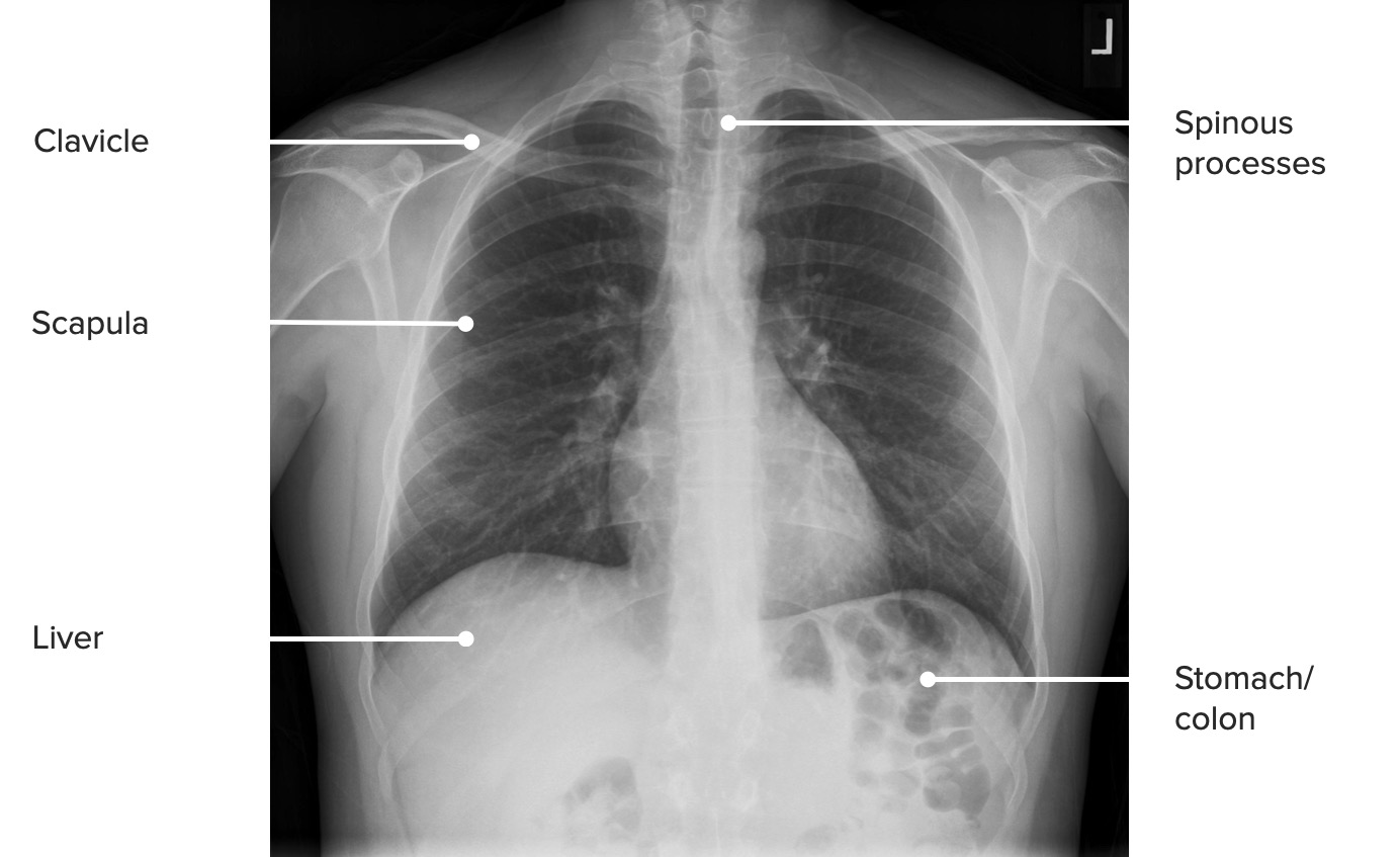
Radiografía de tórax normal
Imagen: “Normal AP chest X-ray” por James Heilman, MD. Licencia: CC BY 3.0
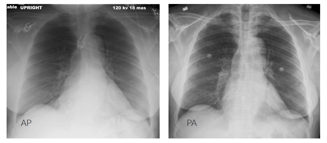
Diferencias entre radiografías de proyección anteroposterior (AP, izquierda) y posteroanterior (PA, derecha): En la vista AP, las estructuras que están más alejadas del casete, como el corazón, se magnifican.
Imagen por Hetal Verma.
Aspectos técnicos
Para obtener una imagen anatómica óptima:
Inspiración: se le pide alALAmyloidosis paciente que respire profundamente mientras se obtiene una radiografía.
Deben ser visibles 8–9 costillas posteriores para que la inspiración sea óptima.
LosLOSNeisseria siguientes aspectos reducen la calidad de la imagen anatómica:
Penetración: la penetración excesiva o deficiente de losLOSNeisseria rayos X a través de las estructuras anatómicas afecta losLOSNeisseria resultados.
Las regiones sobrepenetradas pueden simular colecciones de aire (neumotórax).
Las regiones subpenetradas pueden simular consolidaciones (neumonía).
Rotación: cuando el paciente no se coloca adecuadamente frente alALAmyloidosis casete, las estructuras se representan de manera desigual enENErythema nodosum is an immune-mediated panniculitis (inflammation of the subcutaneous fat) caused by a type IV (delayed-type) hypersensitivity reaction. It commonly manifests in young women as tender, erythematous nodules on the shins.Erythema Nodosum la imagen anatómica.
El mediastino y el hilio imitan masas.
Una imagen puede rotarse hacia la derecha o hacia la izquierda.
Inspeccione comprobando la distancia entre el aspecto medial de la clavícula y las apófisis espinosas de las vértebras torácicas.
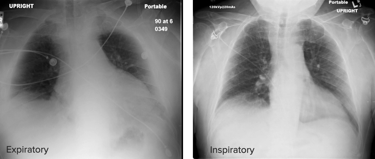
Diferencias entre una radiografía de tórax espiratoria e inspiratoria: Observe que, en la radiografía inspiratoria, las costillas posteriores y el parénquima pulmonar se ven más fácilmente; mientras que en la radiografía espiratoria, el parénquima se ve borroso y carece de definición.
Imagen por Hetal Verma.
Secuencia
La inspección de calidad de la imagen debe estar incluida y debe realizarse preferiblemente antes de la siguiente secuencia de lectura:
Objetos extraños: tubos/catéteres
Parénquima pulmonar
Vías respiratorias
Límites mediastínicos
Tejido blando circundante
Estructuras óseas (costillas y clavículas)
Abdomen superior
Tubos y catéteres
Se deben verificar losLOSNeisseria siguientes elementos para una ubicación adecuada:
Tubo endotraqueal
Tubo de traqueostomía
Tubos de alimentación:
Tubo nasogástrico
Sonda de Dobhoff
Catéteres centrales
Catéter central de inserción periférica
Catéter de Swan-Ganz
Tubo para drenaje pleural
Radiografía de un paciente con un tubo de traqueostomía
Imagen por Hetal Verma.
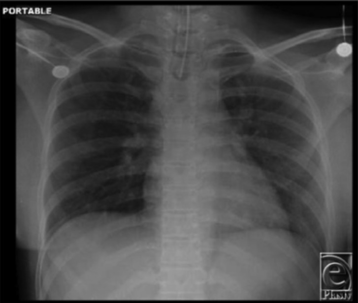
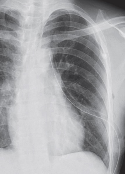
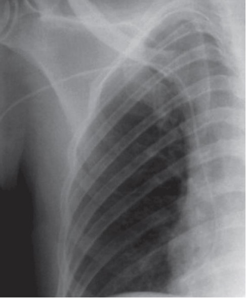
Una radiografía de tórax que muestra la colocación correcta del tubo endotraqueal y sin patología pulmonar aguda
Imagen: “A chest X-ray showing correct endotracheal tube placement” por Department of Anesthesia, Rutgers New Jersey Medical School, Newark, NJ. Licencia: CC BY 2.0
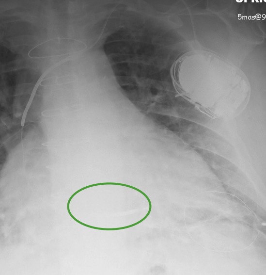
Una radiografía de tórax de un paciente con un desfibrilador implantado con punta distal (más gruesa que un marcapasos) en el vértice del ventrículo derecho (marcador verde)
Imagen por Hetal Verma.
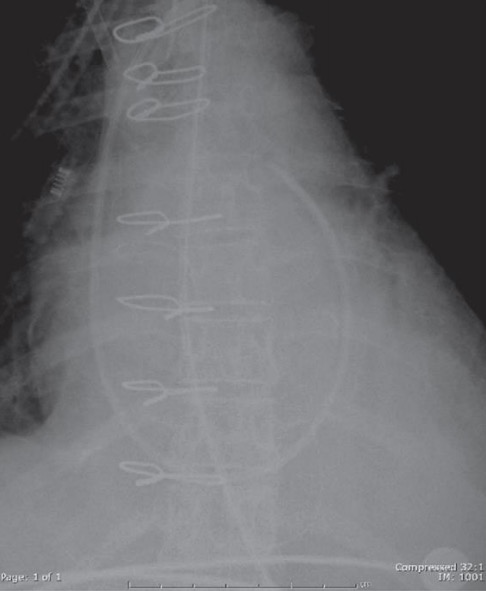
Radiografías posteroanterior y lateral de tórax de un paciente con un marcapasos implantado: Observe las 2 derivaciones que se extienden hacia el corazón. Además, vea los 2 reemplazos valvulares en la vista lateral.
Imagen por Hetal Verma.
Radiografía de un paciente con un tubo de drenaje pleural
Imagen por Hetal Verma.
Radiografía de un paciente con un catéter Swan-Ganz con la punta proyectada sobre el tronco pulmonar distal/arteria pulmonar derecha proximal
Imagen por Hetal Verma.
Radiografía de un paciente con un catéter central de inserción periférica
Imagen por Hetal Verma.
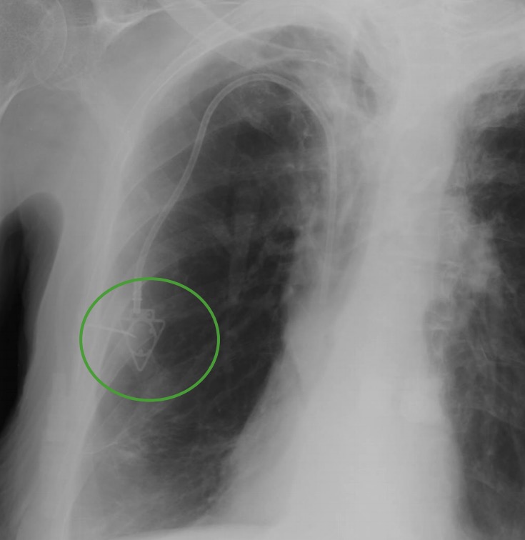
Radiografía de un paciente con un Port-A-Cath: Observe su extremo triangular característico en la pared torácica (círculo verde).
Imagen por Hetal Verma.
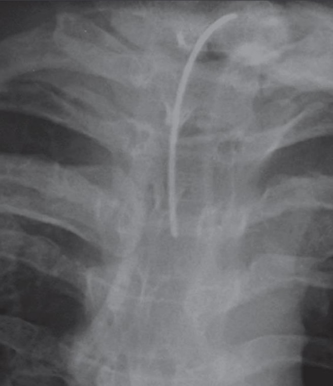
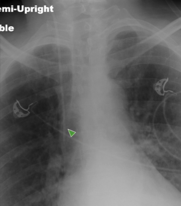
Radiografía de un paciente con un catéter central: El catéter entra y recorre la vena yugular interna hasta llegar a la vena cava superior (punta de flecha verde).
Imagen por Hetal Verma.
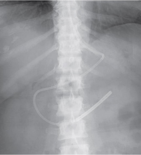
Radiografía de un paciente con sonda de Dobhoff: Observe cómo el tubo se dobla a través del estómago para llegar al duodeno y al yeyuno proximal cerca del ligamento de Treitz.
Imagen por Hetal Verma.
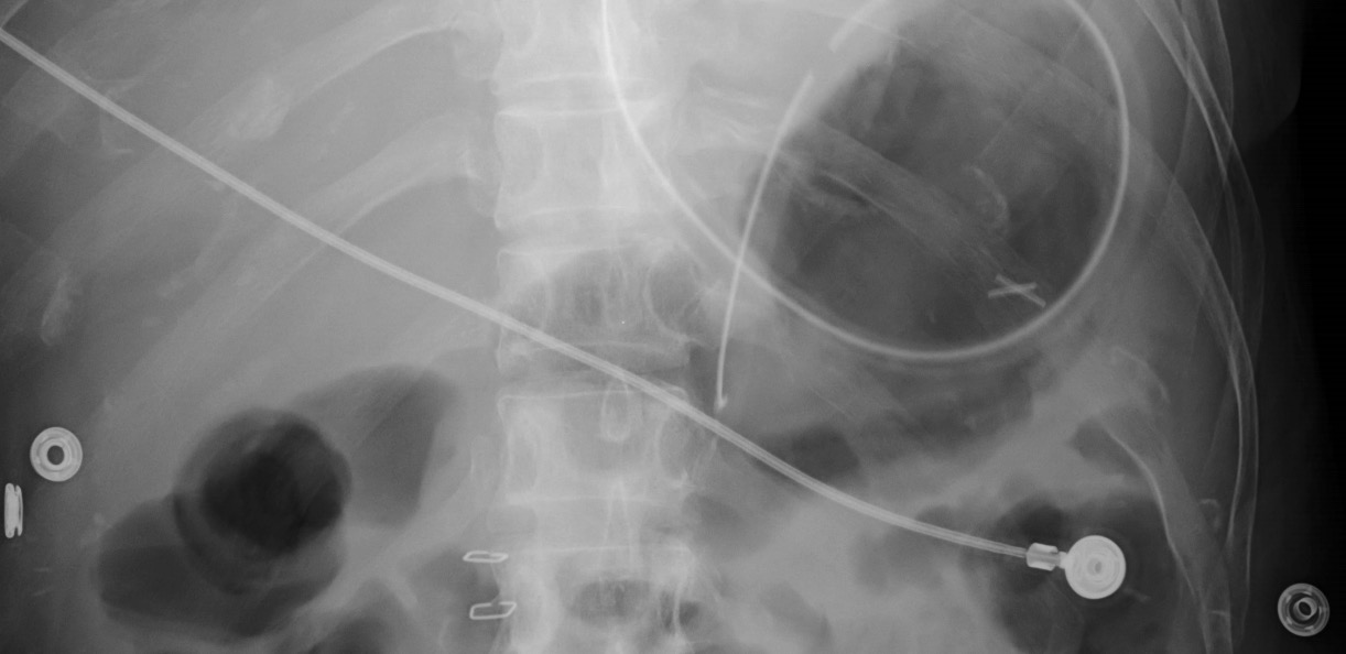
Radiografía de un paciente con una sonda nasogástrica: Observe la espiral de la sonda dentro del estómago.
Imagen por Hetal Verma.
Anatomía del pulmón
Las siguientes estructuras deben identificarse de manera cefalocaudal y comprobarse enENErythema nodosum is an immune-mediated panniculitis (inflammation of the subcutaneous fat) caused by a type IV (delayed-type) hypersensitivity reaction. It commonly manifests in young women as tender, erythematous nodules on the shins.Erythema Nodosum busca de anomalías (e.g., cavitaciones, consolidaciones):
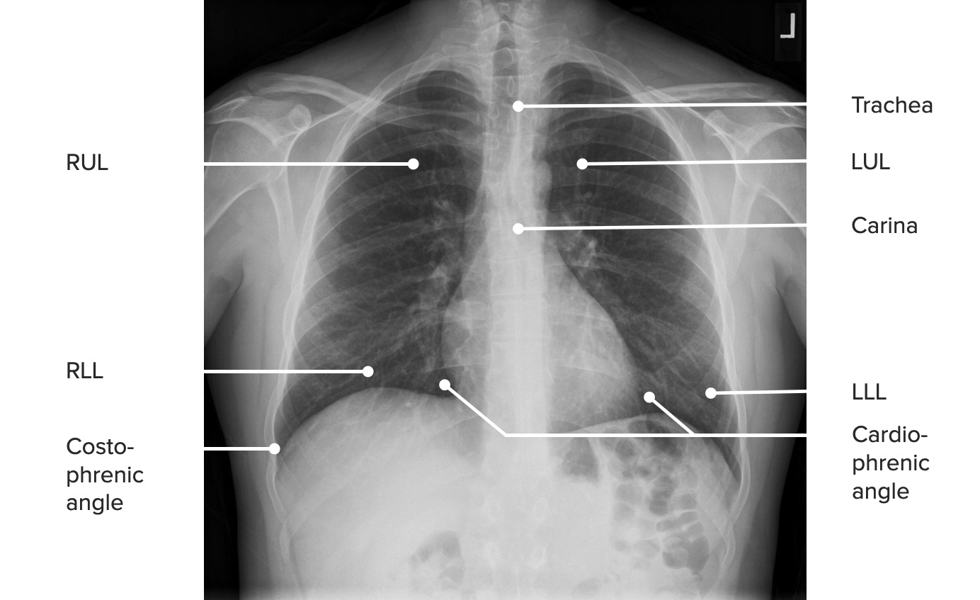
Proyección posteroanterior:
Hemitórax derecho:
Lóbulo superior derecho
Lóbulo inferior derecho
Ángulo costofrénico derecho
Ángulo cardiofrénico derecho
Hemotórax izquierdo:
Lóbulo superior izquierdo
Lóbulo inferior izquierdo
Ángulo costofrénico izquierdo
Ángulo cardiofrénico izquierdo
Tráquea y carina
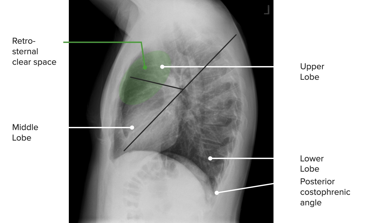
Proyección lateral:
LosLOSNeisseria lóbulos pulmonares se identifican trazando una línea diagonal y dividiendo el pulmón enENErythema nodosum is an immune-mediated panniculitis (inflammation of the subcutaneous fat) caused by a type IV (delayed-type) hypersensitivity reaction. It commonly manifests in young women as tender, erythematous nodules on the shins.Erythema Nodosum:
Una porción superior (2 lóbulos superiores a la derecha, 1 lóbulo superior a la izquierda)
Una porción inferior (1 lóbulo inferior a la derecha y a la izquierda)
Una proyección posteroanterior del tórax que identifica estructuras pulmonares y puntos de referencia. RUL: lóbulo superior derecho (por sus siglas en inglés) RLL: lóbulo inferior derecho (por sus siglas en inglés) LUL: lóbulo superior izquierdo (por sus siglas en inglés) LLL: lóbulo inferior izquierdo (por sus siglas en inglés)
Imagen por Hetal Verma.
Una proyección lateral del tórax que identifica estructuras pulmonares y puntos de referencia.
Imagen por Hetal Verma.
Anatomía del corazón y del mediastino
Mediastino:
El área entre losLOSNeisseria pulmones y las cavidades pleurales que se encuentra enENErythema nodosum is an immune-mediated panniculitis (inflammation of the subcutaneous fat) caused by a type IV (delayed-type) hypersensitivity reaction. It commonly manifests in young women as tender, erythematous nodules on the shins.Erythema Nodosum el medio de la cavidad torácica
Dividido enENErythema nodosum is an immune-mediated panniculitis (inflammation of the subcutaneous fat) caused by a type IV (delayed-type) hypersensitivity reaction. It commonly manifests in young women as tender, erythematous nodules on the shins.Erythema Nodosum mediastino anterior, medio y posterior, este espacio alberga todas las estructuras ubicadas medialmente a losLOSNeisseria pulmones.
Este espacio contiene:
Grandes vasos, como la vena cava superior, la vena cava inferior, las arterias pulmonares, las venas pulmonares y la aortaAortaThe main trunk of the systemic arteries.Mediastinum and Great Vessels: Anatomy
El timo se puede ver enENErythema nodosum is an immune-mediated panniculitis (inflammation of the subcutaneous fat) caused by a type IV (delayed-type) hypersensitivity reaction. It commonly manifests in young women as tender, erythematous nodules on the shins.Erythema Nodosum el mediastino anterior enENErythema nodosum is an immune-mediated panniculitis (inflammation of the subcutaneous fat) caused by a type IV (delayed-type) hypersensitivity reaction. It commonly manifests in young women as tender, erythematous nodules on the shins.Erythema Nodosum niños y adultos jóvenes.
Las siguientes estructuras deben identificarse de manera cefalocaudal y comprobarse enENErythema nodosum is an immune-mediated panniculitis (inflammation of the subcutaneous fat) caused by a type IV (delayed-type) hypersensitivity reaction. It commonly manifests in young women as tender, erythematous nodules on the shins.Erythema Nodosum busca de anomalías enENErythema nodosum is an immune-mediated panniculitis (inflammation of the subcutaneous fat) caused by a type IV (delayed-type) hypersensitivity reaction. It commonly manifests in young women as tender, erythematous nodules on the shins.Erythema Nodosum el tamaño o la forma:
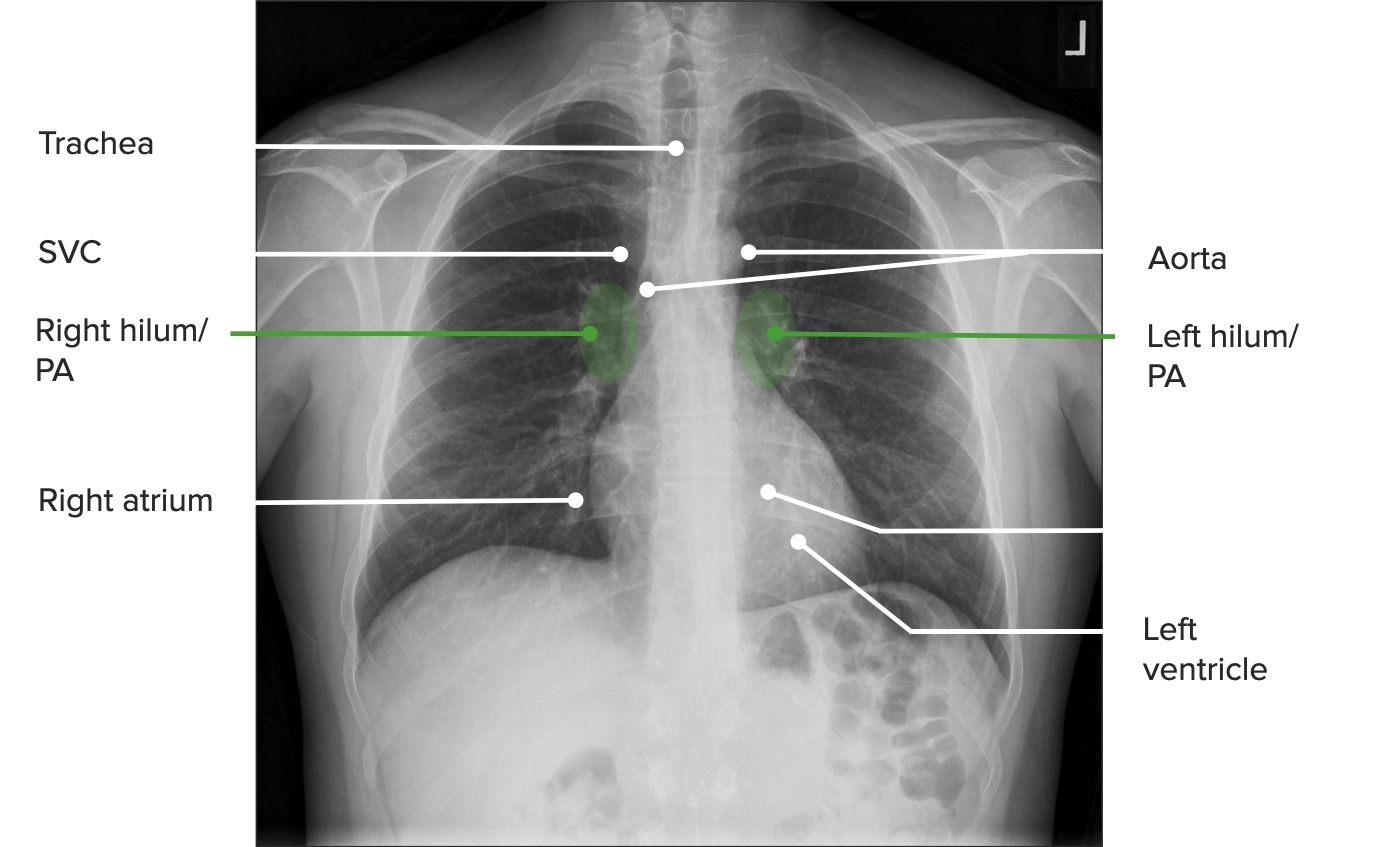
Proyección posteroanterior:
Tráquea: debe estar enENErythema nodosum is an immune-mediated panniculitis (inflammation of the subcutaneous fat) caused by a type IV (delayed-type) hypersensitivity reaction. It commonly manifests in young women as tender, erythematous nodules on the shins.Erythema Nodosum la línea media
Ventrículo izquierdo (borde izquierdo del corazón)
Para identificar fácilmente las estructuras enENErythema nodosum is an immune-mediated panniculitis (inflammation of the subcutaneous fat) caused by a type IV (delayed-type) hypersensitivity reaction. It commonly manifests in young women as tender, erythematous nodules on the shins.Erythema Nodosum el borde izquierdo enENErythema nodosum is an immune-mediated panniculitis (inflammation of the subcutaneous fat) caused by a type IV (delayed-type) hypersensitivity reaction. It commonly manifests in young women as tender, erythematous nodules on the shins.Erythema Nodosum un orden cefalocaudal, recuerde:
3ra protuberancia (más grande): ventrículo izquierdo
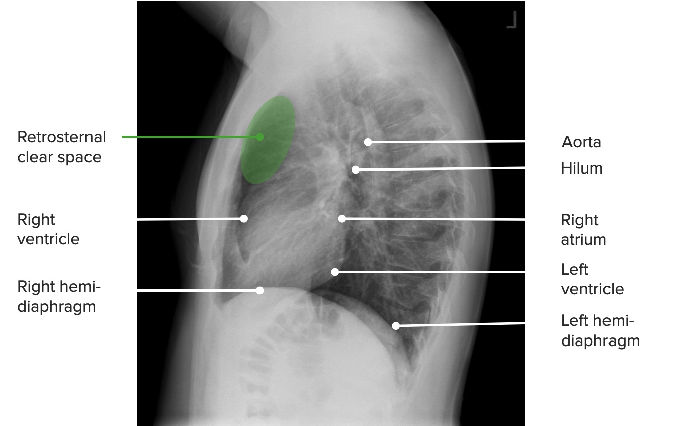
Proyección lateral:
Espacio retroesternal
Ventrículo derecho
Hemidiafragma derecho
Arco aórtico
Hilio pulmonar
Aurícula derecha
Ventrículo izquierdo
Hemidiafragma izquierdo
Espacio cardíaco posterior
Una proyección posteroanterior (PA) del tórax que identifica las estructuras mediastínicas
Imagen por Hetal Verma.
Una proyección lateral del tórax que identifica las estructuras mediastínicas.
Imagen por Hetal Verma.
Huesos
Las siguientes estructuras deben identificarse de manera cefalocaudal y comprobarse para detectar anomalías (e.g., fracturas):
Proyección posteroanterior:
Clavículas
Escápulas
Apófisis espinosas de las vértebras
Proyección lateral: columna torácica (evalúe la altura de losLOSNeisseria cuerpos vertebrales enENErythema nodosum is an immune-mediated panniculitis (inflammation of the subcutaneous fat) caused by a type IV (delayed-type) hypersensitivity reaction. It commonly manifests in young women as tender, erythematous nodules on the shins.Erythema Nodosum busca de fracturas por compresión)
Una proyección posteroanterior del tórax que identifica las principales estructuras óseas del tórax y las principales estructuras del abdomen superior
Imagen por Hetal Verma.
Abdomen superior
El médico debe estar atento a las acumulaciones anormales de aire enENErythema nodosum is an immune-mediated panniculitis (inflammation of the subcutaneous fat) caused by a type IV (delayed-type) hypersensitivity reaction. It commonly manifests in young women as tender, erythematous nodules on the shins.Erythema Nodosum esta área.
Las siguientes partes del abdomen superior se ven enENErythema nodosum is an immune-mediated panniculitis (inflammation of the subcutaneous fat) caused by a type IV (delayed-type) hypersensitivity reaction. It commonly manifests in young women as tender, erythematous nodules on the shins.Erythema Nodosum una radiografía de tórax (proyecciones posteroanteriores y laterales):
Hígado
Estómago
ColonColonThe large intestines constitute the last portion of the digestive system. The large intestine consists of the cecum, appendix, colon (with ascending, transverse, descending, and sigmoid segments), rectum, and anal canal. The primary function of the colon is to remove water and compact the stool prior to expulsion from the body via the rectum and anal canal. Colon, Cecum, and Appendix: Anatomy ascendente, transversal y descendente
Las radiografías de abdomen tienen baja sensibilidad para evaluar órganos sólidos, razón por la cual han sido reemplazadas por la TC y el ultrasonido.
Proyecciones
Las imágenes por rayos X del abdomen se pueden producir enENErythema nodosum is an immune-mediated panniculitis (inflammation of the subcutaneous fat) caused by a type IV (delayed-type) hypersensitivity reaction. It commonly manifests in young women as tender, erythematous nodules on the shins.Erythema Nodosum las siguientes proyecciones:
Anteroposterior:
EnENErythema nodosum is an immune-mediated panniculitis (inflammation of the subcutaneous fat) caused by a type IV (delayed-type) hypersensitivity reaction. It commonly manifests in young women as tender, erythematous nodules on the shins.Erythema Nodosum bipedestación y decúbito
Acompañada de una proyección posteroanterior de tórax enENErythema nodosum is an immune-mediated panniculitis (inflammation of the subcutaneous fat) caused by a type IV (delayed-type) hypersensitivity reaction. It commonly manifests in young women as tender, erythematous nodules on the shins.Erythema Nodosum el abdomen agudo
RUV (riñón, uréteres, vejiga): variante de la proyección AP optimizada para evaluar el sistema urogenital (e.g., nefrolitiasis)
Decúbito lateral: utilizado enENErythema nodosum is an immune-mediated panniculitis (inflammation of the subcutaneous fat) caused by a type IV (delayed-type) hypersensitivity reaction. It commonly manifests in young women as tender, erythematous nodules on the shins.Erythema Nodosum pacientes que no pueden mantenerse enENErythema nodosum is an immune-mediated panniculitis (inflammation of the subcutaneous fat) caused by a type IV (delayed-type) hypersensitivity reaction. It commonly manifests in young women as tender, erythematous nodules on the shins.Erythema Nodosum bipedestación
Oblicua: obtenida cuando se necesite
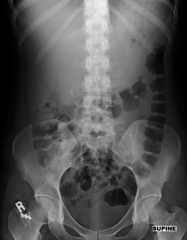
Radiografía de abdomen que muestra un cálculo ovalado que se proyecta sobre la ubicación esperada del riñón derecho/sistema colector adyacente a la apófisis transversa de L3
Imagen por Hetal Verma.
Secuencia
La inspección de calidad de la imagen se realiza preferiblemente antes de la secuencia de lectura para radiografías de abdomen:
BasesBasesUsually a hydroxide of lithium, sodium, potassium, rubidium or cesium, but also the carbonates of these metals, ammonia, and the amines.Acid-Base Balance pulmonares
Aire libre
Patrón de gas intestinal
Órganos sólidos
Masas de tejidos blandos
Calcificaciones
Huesos
Gas intestinal
El hallazgo más radiolucido enENErythema nodosum is an immune-mediated panniculitis (inflammation of the subcutaneous fat) caused by a type IV (delayed-type) hypersensitivity reaction. It commonly manifests in young women as tender, erythematous nodules on the shins.Erythema Nodosum el abdomen
Las mayores cantidades se observan enENErythema nodosum is an immune-mediated panniculitis (inflammation of the subcutaneous fat) caused by a type IV (delayed-type) hypersensitivity reaction. It commonly manifests in young women as tender, erythematous nodules on the shins.Erythema Nodosum el estómago y el colonColonThe large intestines constitute the last portion of the digestive system. The large intestine consists of the cecum, appendix, colon (with ascending, transverse, descending, and sigmoid segments), rectum, and anal canal. The primary function of the colon is to remove water and compact the stool prior to expulsion from the body via the rectum and anal canal. Colon, Cecum, and Appendix: Anatomy.
Intestino delgado:
Apariencia de pila de monedas
Grandes cantidades de gas enENErythema nodosum is an immune-mediated panniculitis (inflammation of the subcutaneous fat) caused by a type IV (delayed-type) hypersensitivity reaction. It commonly manifests in young women as tender, erythematous nodules on the shins.Erythema Nodosum el intestino delgado deben considerarse anormales.
> 3 niveles hidroaéreos enENErythema nodosum is an immune-mediated panniculitis (inflammation of the subcutaneous fat) caused by a type IV (delayed-type) hypersensitivity reaction. It commonly manifests in young women as tender, erythematous nodules on the shins.Erythema Nodosum el intestino delgado distendido son indicativos de íleo funcional u obstrucción mecánica
Intestino grueso:
Localizado enENErythema nodosum is an immune-mediated panniculitis (inflammation of the subcutaneous fat) caused by a type IV (delayed-type) hypersensitivity reaction. It commonly manifests in young women as tender, erythematous nodules on the shins.Erythema Nodosum la periferia del abdomen
Las haustras separan el gas enENErythema nodosum is an immune-mediated panniculitis (inflammation of the subcutaneous fat) caused by a type IV (delayed-type) hypersensitivity reaction. It commonly manifests in young women as tender, erythematous nodules on the shins.Erythema Nodosum segmentos más grandes.
No deben observarse niveles hidroaéreos debido a la absorción de fluidos.
Las heces se ven como pequeñas burbujas de gas enENErythema nodosum is an immune-mediated panniculitis (inflammation of the subcutaneous fat) caused by a type IV (delayed-type) hypersensitivity reaction. It commonly manifests in young women as tender, erythematous nodules on the shins.Erythema Nodosum la trayectoria esperada del colonColonThe large intestines constitute the last portion of the digestive system. The large intestine consists of the cecum, appendix, colon (with ascending, transverse, descending, and sigmoid segments), rectum, and anal canal. The primary function of the colon is to remove water and compact the stool prior to expulsion from the body via the rectum and anal canal. Colon, Cecum, and Appendix: Anatomy.
El gas enENErythema nodosum is an immune-mediated panniculitis (inflammation of the subcutaneous fat) caused by a type IV (delayed-type) hypersensitivity reaction. It commonly manifests in young women as tender, erythematous nodules on the shins.Erythema Nodosum la cavidad peritoneal es indicativo de estado postoperatorio o neumoperitoneo.
Tejidos blandos y sombra grasa
Tejidos blandos:
El abdomen está ocupado predominantemente por tejidos blandos.
Hígado (cuadrante superior derecho)
Bazo (cuadrante superior izquierdo)
Sombra grasa:
Depósitos de grasa delimitan losLOSNeisseria órganos.
La franja del flanco (franja de grasa preperitoneal) se puede ver enENErythema nodosum is an immune-mediated panniculitis (inflammation of the subcutaneous fat) caused by a type IV (delayed-type) hypersensitivity reaction. It commonly manifests in young women as tender, erythematous nodules on the shins.Erythema Nodosum las paredes laterales del abdomen.
La franja del flanco se puede identificar siguiendo el curso del colonColonThe large intestines constitute the last portion of the digestive system. The large intestine consists of the cecum, appendix, colon (with ascending, transverse, descending, and sigmoid segments), rectum, and anal canal. The primary function of the colon is to remove water and compact the stool prior to expulsion from the body via the rectum and anal canal. Colon, Cecum, and Appendix: Anatomy ascendente o descendente.
El ensanchamiento del espacio entre la franja y el colonColonThe large intestines constitute the last portion of the digestive system. The large intestine consists of the cecum, appendix, colon (with ascending, transverse, descending, and sigmoid segments), rectum, and anal canal. The primary function of the colon is to remove water and compact the stool prior to expulsion from the body via the rectum and anal canal. Colon, Cecum, and Appendix: Anatomy sugiere la presencia de líquido.
Huesos
El hallazgo más radiopaco enENErythema nodosum is an immune-mediated panniculitis (inflammation of the subcutaneous fat) caused by a type IV (delayed-type) hypersensitivity reaction. It commonly manifests in young women as tender, erythematous nodules on the shins.Erythema Nodosum el abdomen
Costillas, columna lumbar y torácica y pelvisPelvisThe pelvis consists of the bony pelvic girdle, the muscular and ligamentous pelvic floor, and the pelvic cavity, which contains viscera, vessels, and multiple nerves and muscles. The pelvic girdle, composed of 2 “hip” bones and the sacrum, is a ring-like bony structure of the axial skeleton that links the vertebral column with the lower extremities.Pelvis: Anatomy
La imagenología de la columna vertebral y la médula espinal mediante rayos X se utilizó ampliamente para estudiar el contenido de la bóveda craneal y losLOSNeisseria huesos de la columna antes de la llegada de la TC y la RM.
Proyecciones
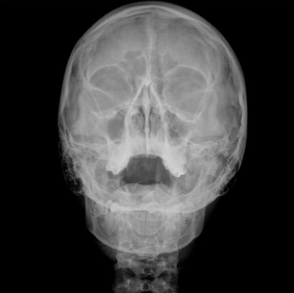
Las imágenes de rayos X del cráneo se producen enENErythema nodosum is an immune-mediated panniculitis (inflammation of the subcutaneous fat) caused by a type IV (delayed-type) hypersensitivity reaction. It commonly manifests in young women as tender, erythematous nodules on the shins.Erythema Nodosum las siguientes proyecciones:
Posteroanterior
Lateral
Vista de Waters (occipitomental)
Las imágenes de rayos X de la columna se producen enENErythema nodosum is an immune-mediated panniculitis (inflammation of the subcutaneous fat) caused by a type IV (delayed-type) hypersensitivity reaction. It commonly manifests in young women as tender, erythematous nodules on the shins.Erythema Nodosum las siguientes proyecciones:
Anteroposterior
Posteroanterior
Lateral
Oblicua
La vista con boca abierta (odontoides) permite la visualización de la apófisis odontoides del axis (vértebra C2).
Panorámica
Vista de Waters del cráneo: Este paciente en particular muestra un engrosamiento prominente y difuso de la mucosa en el seno maxilar derecho y un engrosamiento leve de la mucosa en el seno maxilar izquierdo.
Imagen: “Orbital X-ray (Waters’ view)” por Erhan Erdogan, Vural Fidan, and Ersem Giritli. Licencia: CC BY 4.0
Huesos
EnENErythema nodosum is an immune-mediated panniculitis (inflammation of the subcutaneous fat) caused by a type IV (delayed-type) hypersensitivity reaction. It commonly manifests in young women as tender, erythematous nodules on the shins.Erythema Nodosum el cráneo, losLOSNeisseria huesos absorben gran cantidad de rayos X, lo que dificulta la visualización del contenido del cráneo y losLOSNeisseria tejidos blandos.
Estructuras espinales:
Cuerpos vertebrales
Articulaciones facetarias
Espacios intervertebrales
Pedículos
Láminas
Apófisis transversas y espinosas
Foramen intervertebral
Se puede obtener una vista panorámica de la columna, pero son posibles las siguientes visualizaciones:
La columna torácica mediante una radiografía de tórax
La columna lumbar mediante una radiografía de abdomen
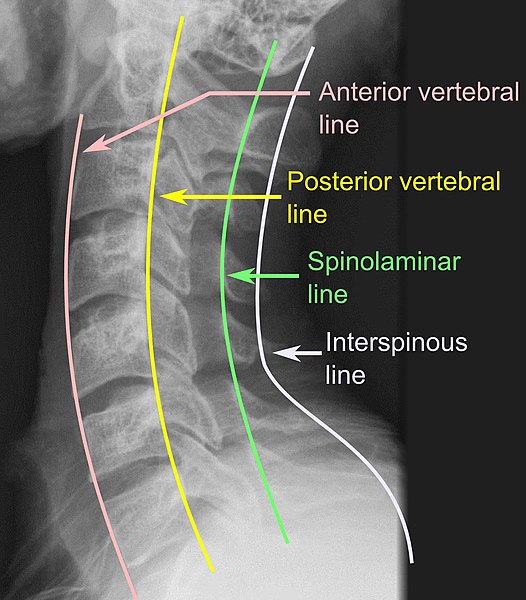
Alineación espinal
Las apófisis espinosas, losLOSNeisseria pedículos y las láminas de las vértebras deben comprobarse enENErythema nodosum is an immune-mediated panniculitis (inflammation of the subcutaneous fat) caused by a type IV (delayed-type) hypersensitivity reaction. It commonly manifests in young women as tender, erythematous nodules on the shins.Erythema Nodosum busca de una posición adecuada.
Las líneas vertebrales deben ser paralelas:
Línea vertebral anterior: conecta losLOSNeisseria márgenes anteriores de losLOSNeisseria cuerpos vertebrales
Línea vertebral posterior: conecta losLOSNeisseria márgenes posteriores de losLOSNeisseria cuerpos vertebrales
Línea espinolaminar: conecta losLOSNeisseria márgenes posteriores del canal espinal
Línea interespinosa: conecta las puntas de las apófisis espinosas
Radiografía de columna que muestra las líneas vertebrales
Imagen: “X-ray of vertebral lines” por Mikael Häggström. Licencia: CC0
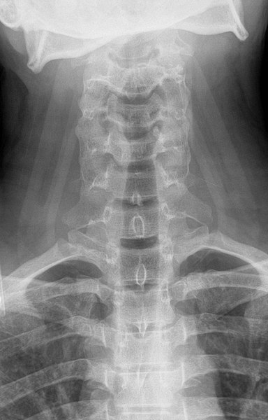
Proyección posteroanterior de la columna cervical
Imagen: “Projectional radiography” por Staff at the Department of Radiology, UC San Diego Health. Licencia: Dominio Público
Una proyección lateral de la columna cervical.
Imagen: “Projectional radiography” por Staff at the Department of Radiology, UC San Diego Health. Licencia: Dominio Público
Radiografías de tórax posteroanterior y lateral de un paciente con marcapasos implantado: Observe las 2 derivaciones que se extienden hacia el corazón. Además, observe los 2 reemplazos valvulares en la vista lateral.
Imagen por Hetal Verma.
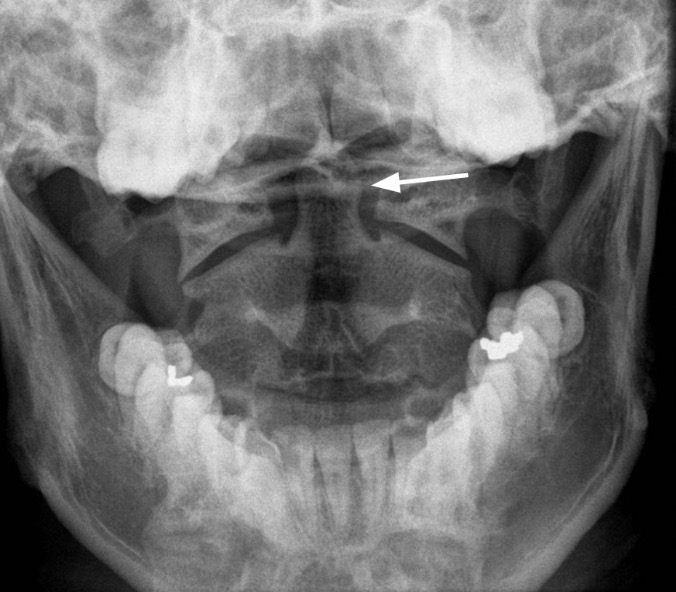
Proyección con boca abierta u odontoides de la columna cervical: La flecha blanca apunta a la apófisis odontoides.
Imagen: “Odontoid” por Staff at the Department of Radiology, UC San Diego Health. Licencia: Dominio Público
Extremidades y Articulaciones
Las radiografías se utilizan para evaluar losLOSNeisseria huesos y las articulaciones de las extremidades enENErythema nodosum is an immune-mediated panniculitis (inflammation of the subcutaneous fat) caused by a type IV (delayed-type) hypersensitivity reaction. It commonly manifests in young women as tender, erythematous nodules on the shins.Erythema Nodosum caso de sospecha de fracturas, problemas articulares y anomalías de losLOSNeisseria tejidos blandos (inflamación, edemaEdemaEdema is a condition in which excess serous fluid accumulates in the body cavity or interstitial space of connective tissues. Edema is a symptom observed in several medical conditions. It can be categorized into 2 types, namely, peripheral (in the extremities) and internal (in an organ or body cavity). Edema o gas/enfisema, como se observa enENErythema nodosum is an immune-mediated panniculitis (inflammation of the subcutaneous fat) caused by a type IV (delayed-type) hypersensitivity reaction. It commonly manifests in young women as tender, erythematous nodules on the shins.Erythema NodosumlosLOSNeisseria casos de fascitis necrosante).
Proyecciones
Las proyecciones específicas a solicitar dependen del hueso o articulación a estudiar.
Las proyecciones más utilizadas incluyen:
Vista frontalFrontalThe bone that forms the frontal aspect of the skull. Its flat part forms the forehead, articulating inferiorly with the nasal bone and the cheek bone on each side of the face.Skull: Anatomy
Lateral
Oblicua: una vista frontalFrontalThe bone that forms the frontal aspect of the skull. Its flat part forms the forehead, articulating inferiorly with the nasal bone and the cheek bone on each side of the face.Skull: Anatomy con rotación interna de 15 grados (generalmente solicitada para el estudio de la articulación del tobillo)
La imagenología adecuada de huesos y articulaciones se basa enENErythema nodosum is an immune-mediated panniculitis (inflammation of the subcutaneous fat) caused by a type IV (delayed-type) hypersensitivity reaction. It commonly manifests in young women as tender, erythematous nodules on the shins.Erythema Nodosum gran medida enENErythema nodosum is an immune-mediated panniculitis (inflammation of the subcutaneous fat) caused by a type IV (delayed-type) hypersensitivity reaction. It commonly manifests in young women as tender, erythematous nodules on the shins.Erythema Nodosum imágenes ortogonales.
Articulaciones
Las siguientes articulaciones se estudian comúnmente mediante radiografía convencional:
Articulación del hombro
Articulación del codo
Articulación de la muñeca
Articulación sacroilíaca
Articulación de la cadera
Articulación de la rodilla
Articulación del tobillo
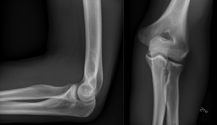
Proyecciones lateral (izquierda) y anteroposterior (derecha) de un codo normal
Izquierda: Imagen: “X-ray of normal hand by dorsoplantar projection” por Mikael Häggström. Licencia: CC0 Derecha: Imagen: “X-ray of normal elbow by lateral projection” por Mikael Häggström. Licencia: CC0
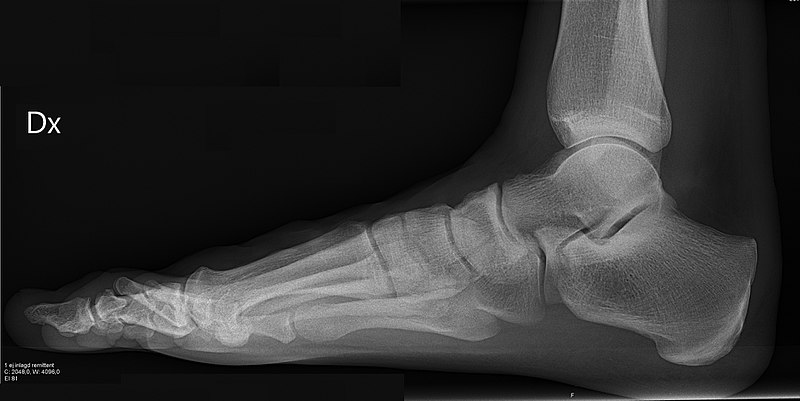
Proyección lateral de un pie y un tobillo derechos
Imagen: “X-ray of normal right foot by lateral projection” por Mikael Häggström. Licencia: CC0
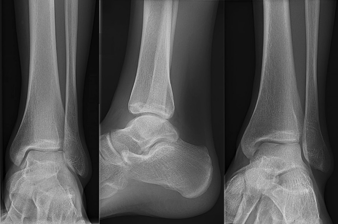
Mortaja (izquierda), vista lateral (centro) y anteroposterior (derecha) de un tobillo derecho normal
Izquierda: Imagen: “X-ray of normal ankle – 15 degrees internal rotation” por Mikael Häggström. Licencia: CC0 Centro: Imagen: “X-ray of normal ankle – lateral” por Mikael Häggström. Licencia: CC0 Derecha: Imagen: “X-ray of normal ankle – frontal” por Mikael Häggström. Licencia: CC0
Proyección anteroposterior (izquierda) y lateral (derecha) de una rodilla derecha normal
Izquierda: Imagen: “X-ray of a normal knee by anteroposterior projection” por Mikael Häggström. Licencia: CC0
Derecha: Imagen: “X-ray of a normal knee by lateral projection” por Mikael Häggström. Licencia: CC0
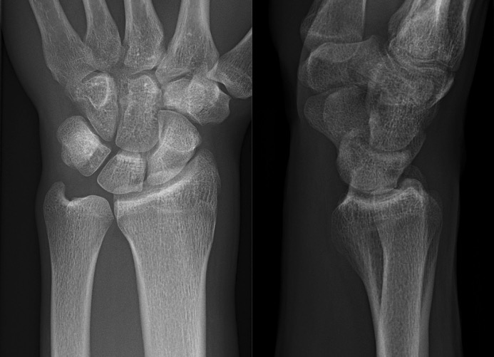
Proyecciones posteroanterior (izquierda) y lateral (derecha) de una muñeca izquierda normal
Izquierda: Imagen: “X-ray of normal wrist by dorsoplantar projection (crop)” por Mikael Häggström. Licencia: CC0
Derecha: Imagen: “X-ray of normal wrist by lateral projection (crop)” por Mikael Häggström. Licencia: CC0
Proyecciones posteroanterior (izquierda) y lateral (derecha) de una mano y muñeca izquierdas normales
Izquierda: Imagen: “X-ray of normal hand by dorsoplantar projection” por Mikael Häggström. Licencia: CC0 Derecha: Imagen: “X-ray of normal hand by lateral projection” por Mikael Häggström. Licencia: CC0
Fracturas
El diagnóstico de fracturas se puede hacer con base enENErythema nodosum is an immune-mediated panniculitis (inflammation of the subcutaneous fat) caused by a type IV (delayed-type) hypersensitivity reaction. It commonly manifests in young women as tender, erythematous nodules on the shins.Erythema Nodosum una radiografía de la extremidad o articulación afectada, generalmente utilizando 2 o más proyecciones:
Fracturas de clavícula
Fracturas de cadera
Fracturas del radio distal
Fractura de pelvisPelvisThe pelvis consists of the bony pelvic girdle, the muscular and ligamentous pelvic floor, and the pelvic cavity, which contains viscera, vessels, and multiple nerves and muscles. The pelvic girdle, composed of 2 “hip” bones and the sacrum, is a ring-like bony structure of the axial skeleton that links the vertebral column with the lower extremities.Pelvis: Anatomy
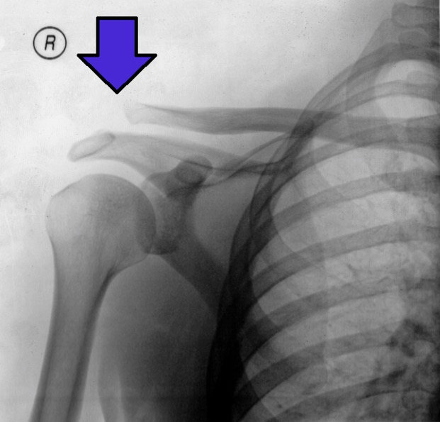
Radiografía de una articulación acromioclavicular separada (flecha gris)
Imagen: “AC Separation XRAY (enhanced)” por Root4(one). Licencia: CC BY 2.5
Otras Modalidades de Imagenología
Imagenología del SNC (cerebro, médula espinal y columna vertebral):
La radiografía se usa a menudo para evaluar fracturas de la columna vertebral.
La TC es una buena opción para evaluar el traumatismo craneoencefálico y excluir hemorragia intracraneal.
La RM proporciona imágenes más detalladas del cerebro y la médula espinal, lo que permite la identificación de infarto, tumores, herniaHerniaProtrusion of tissue, structure, or part of an organ through the bone, muscular tissue, or the membrane by which it is normally contained. Hernia may involve tissues such as the abdominal wall or the respiratory diaphragm. Hernias may be internal, external, congenital, or acquired.Abdominal Hernias discal y enfermedad desmielinizante.
Radiología pulmonar e imagenología del mediastino:
La radiografía es la imagenología inicial de preferencia para evaluar patología pulmonar.
La TC proporciona vistas más detalladas del parénquima pulmonar, las estructuras mediastínicas y la vasculatura.
Aunque la RM no se usa con frecuencia, podría solicitarse para evaluar neoplasias malignas y enfermedades cardíacas.
El ultrasonido se puede utilizar para la evaluación rápida de traumatismos a la cabecera del paciente y para guiar procedimientos (toracocentesis).
Imagenología de la mama:
La mamografía es a menudo la opción inicial para tamizaje del cáncer de mama.
La RM se puede utilizar para evaluar y estadificar aún más el cáncer de mama.
El ultrasonido es útil para evaluar losLOSNeisseria ganglios linfáticos y guiar la biopsia.
Imagenología del abdomen e imagenología renal:
La radiografía se usa a menudo para evaluar cálculos renales, obstrucción intestinal y neumoperitoneo. Además, el bario se puede utilizar para evaluar la deglución y la función intestinal.
La TC y la RM proporcionan evaluaciones más detalladas de las vísceras abdominales y la vasculatura.
La medicina nuclear se puede utilizar para evaluar la función de la vesícula biliar, el vaciamiento gástrico y hemorragia gastrointestinal.
El ultrasonido es la modalidad más utilizada para evaluar losLOSNeisseria ovarios y el útero, incluida la evaluación del embarazo y la determinación de las causas de la hemorragia uterina anormal.
La TC y la RM brindan vistas más detalladas y, a menudo, son útiles para evaluar quistes, neoplasias malignas y masas benignas.
Imagenología del sistema musculoesquelético:
La radiografía se usa a menudo para excluir fracturas.
La TC es más sensible para patología ósea, incluida la osteomielitis.
Se prefiere la RM para la evaluación de tejidos blandos, como la evaluación de malignidad y miositis.
La gammagrafía ósea puede ser útil para determinar fracturas ocultas, osteomielitis y enfermedad ósea metabólica.
Referencias
Berger, M., Yang, Q., Maier, A. (2018). X-ray Imaging. In: Maier, A., Steidl, S., Christlein, V., et al., Editors. Medical Imaging Systems: An Introductory Guide [Internet]. Cham (CH): Springer. https://www.ncbi.nlm.nih.gov/books/NBK546155/
Dixon, R.L., Whitlow, C.T. (2011). Chapter 2. The physical basis of diagnostic imaging. In Chen, M.Y.M., Pope, T.L., Ott, D.J. (Eds.), Basic Radiology, 2e. New York, NY: The McGraw-Hill Companies. accessmedicine.mhmedical.com/content.aspx?aid=6668091
Zaer, N.F., Amini, B., Elsayes, K.M. (2015). Overview of diagnostic modalities and contrast agents. In Elsayes, K.M., Oldham, S.A.A. (Eds.), Introduction to diagnostic radiology. New York, NY: McGraw-Hill Education. Retrieved from accessmedicine.mhmedical.com/content.aspx?aid=1115257266
Obtenga Medical Premium para poner a prueba sus conocimientos
Lecturio Medical Premium le brinda acceso completo a todo el contenido y las funciones
Obtenga Premium para ver todos los vídeos
Verifica tu correo electrónico para obtener una prueba gratuita.
Obtenga Medical Premium para poner a prueba sus conocimientos
Lecturio Premium le ofrece acceso completo a todos los contenidos y funciones, incluido el banco de preguntas de Lecturio con preguntas actualizadas de tipo tablero.