La piel, también conocida como sistema tegumentario, es el órgano más grande del cuerpo. La piel está compuesta principalmente por la epidermis Epidermis The external, nonvascular layer of the skin. It is made up, from within outward, of five layers of epithelium: (1) basal layer (stratum basale epidermidis); (2) spinous layer (stratum spinosum epidermidis); (3) granular layer (stratum granulosum epidermidis); (4) clear layer (stratum lucidum epidermidis); and (5) horny layer (stratum corneum epidermidis). Skin: Structure and Functions (capa externa) y la dermis Dermis A layer of vascularized connective tissue underneath the epidermis. The surface of the dermis contains innervated papillae. Embedded in or beneath the dermis are sweat glands; hair follicles; and sebaceous glands. Skin: Structure and Functions (capa profunda). La epidermis Epidermis The external, nonvascular layer of the skin. It is made up, from within outward, of five layers of epithelium: (1) basal layer (stratum basale epidermidis); (2) spinous layer (stratum spinosum epidermidis); (3) granular layer (stratum granulosum epidermidis); (4) clear layer (stratum lucidum epidermidis); and (5) horny layer (stratum corneum epidermidis). Skin: Structure and Functions se compone principalmente de queratinocitos que experimentan un rápido recambio, mientras que la dermis Dermis A layer of vascularized connective tissue underneath the epidermis. The surface of the dermis contains innervated papillae. Embedded in or beneath the dermis are sweat glands; hair follicles; and sebaceous glands. Skin: Structure and Functions contiene densas capas de tejido conectivo. La piel está compuesta por epitelio superficial, componentes exocrinos, tejido conectivo, músculos y nervios. La tarea principal de la piel es servir como barrera protectora entre el cuerpo interno y el ambiente externo; también protege al AL Amyloidosis cuerpo de la pérdida excesiva de líquidos.
Last updated: Dec 15, 2025
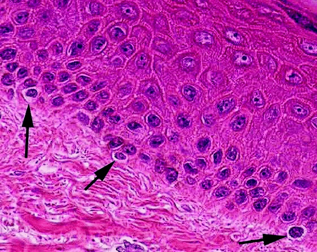
Micrografía de melanocitos en la epidermis
Imagen: “Micrograph of melanocytes in the epidermis” por Setijanti, H.B., et al. Licencia: CC BY 4.0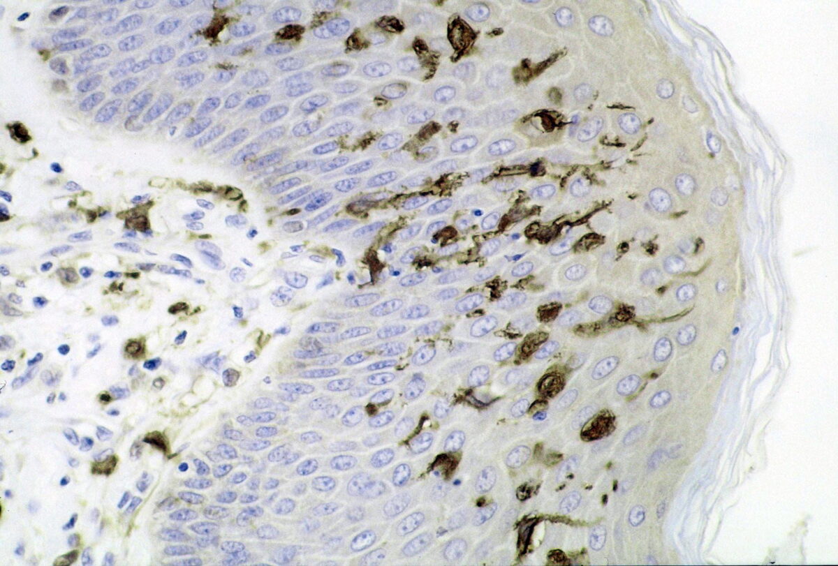
Sección de piel que muestra un gran número de células dendríticas (Langerhans) en la epidermis:
Infección por Mycobacterium ulcerans, tinción de inmunoperoxidasa S100
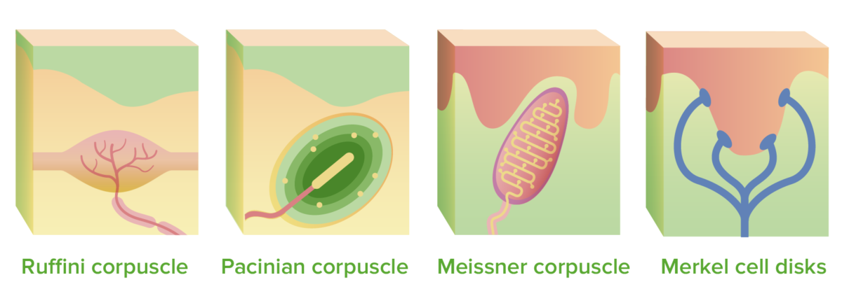
Mecanorreceptores:
Los corpúsculos de Ruffini y de Pacini se encuentran en la dermis profunda, los corpúsculos de Meissner se extienden hacia las papilas dérmicas y los discos de células de Merkel se conectan a la epidermis.
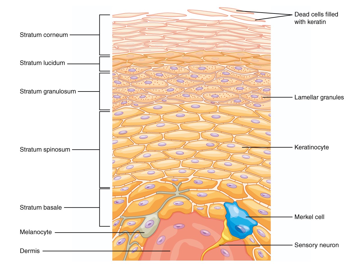
Hay 5 capas de la epidermis:
Partiendo de las células ubicadas en el estrato basal, se diferencian y proliferan hacia la superficie de la piel y se integran en las otras capas, siendo la última capa el estrato córneo, que está compuesto por células queratinizadas muertas.

La dermis está formada por 2 capas:
La capa papilar está formada por fibras de colágeno y elastina y contiene fagocitos, fibroblastos y adipocitos. La capa reticular le da a la piel su elasticidad y ayuda a endurecer la piel. En esta capa se encuentran las glándulas sudoríparas y los folículos pilosos. Esta capa también está bien vascularizada.
Cada capa de la piel tiene una función única.
Epidermis Epidermis The external, nonvascular layer of the skin. It is made up, from within outward, of five layers of epithelium: (1) basal layer (stratum basale epidermidis); (2) spinous layer (stratum spinosum epidermidis); (3) granular layer (stratum granulosum epidermidis); (4) clear layer (stratum lucidum epidermidis); and (5) horny layer (stratum corneum epidermidis). Skin: Structure and Functions y dermis Dermis A layer of vascularized connective tissue underneath the epidermis. The surface of the dermis contains innervated papillae. Embedded in or beneath the dermis are sweat glands; hair follicles; and sebaceous glands. Skin: Structure and Functions:
Hipodermis:

Estructura de la unidad folicular:
El bulbo piloso se encuentra en la dermis/hipodermis profunda y el tallo del pelo se extiende a través de la dermis antes de salir de la epidermis. El músculo arrector pili se encuentra en la dermis y es responsable de la piloerección.
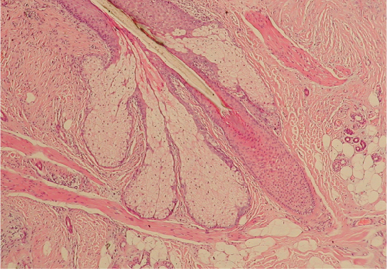
Corte transversal de un folículo piloso:
El pelo está compuesto por células queratinizadas que emergen y migran hacia arriba dentro del folículo piloso. Cuando el tallo del pelo emerge de la superficie de la piel, lo hace en un ángulo ligeramente inclinado. Por cada folículo piloso, hay una glándula sebácea adyacente.
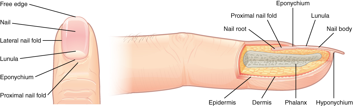
Estructura de la uña:
El extremo blanquecino en forma de media luna de la uña (hacia el cuerpo) se conoce como lúnula (“luna pequeña”). Entre la lúnula y la piel, hay una capa protectora, llamada cutícula, que evita que los patógenos entren debajo de la piel. Detrás de la cutícula (hacia el cuerpo), está la raíz de la uña, que forma células queratinizadas que empujan la uña hacia adelante.
Las uñas de las manos crecen más rápido que las de los pies. La tasa de crecimiento típica de una uña de las manos es de 1 mm por semana; una uña de los pies crece 0,5 mm por semana.