Los LOS Neisseria antibióticos betalactámicos contienen un anillo betalactámico como parte de su estructura química. Los LOS Neisseria medicamentos de esta clase incluyen las penicilinas G y V, las penicilinas sensibles y resistentes a la penicilinasa, las cefalosporinas, los LOS Neisseria carbapenémicos y el aztreonam Aztreonam The carbapenems and aztreonam are both members of the bactericidal beta-lactam family of antibiotics (similar to penicillins). They work by preventing bacteria from producing their cell wall, ultimately leading to bacterial cell death. Carbapenems and Aztreonam. Los LOS Neisseria antibióticos betalactámicos bloquean la transpeptidasa bacteriana (proteína fijadora de penicilina) y, por lo tanto, inactivan el entrecruzamiento de peptidoglucano en EN Erythema nodosum is an immune-mediated panniculitis (inflammation of the subcutaneous fat) caused by a type IV (delayed-type) hypersensitivity reaction. It commonly manifests in young women as tender, erythematous nodules on the shins. Erythema Nodosum la pared celular. Todos los LOS Neisseria antibióticos betalactámicos son bactericidas. Los LOS Neisseria mecanismos comunes de resistencia incluyen la producción de betalactamasas o la mutación en EN Erythema nodosum is an immune-mediated panniculitis (inflammation of the subcutaneous fat) caused by a type IV (delayed-type) hypersensitivity reaction. It commonly manifests in young women as tender, erythematous nodules on the shins. Erythema Nodosum el gen de la proteína de unión a la penicilina. Los LOS Neisseria efectos secundarios comunes incluyen reacciones de hipersensibilidad, malestar gastrointestinal y anemia Anemia Anemia is a condition in which individuals have low Hb levels, which can arise from various causes. Anemia is accompanied by a reduced number of RBCs and may manifest with fatigue, shortness of breath, pallor, and weakness. Subtypes are classified by the size of RBCs, chronicity, and etiology. Anemia: Overview and Types hemolítica.
Last updated: Dec 15, 2025
Las penicilinas son miembros de la familia de medicamentos betalactámicos y consisten en EN Erythema nodosum is an immune-mediated panniculitis (inflammation of the subcutaneous fat) caused by a type IV (delayed-type) hypersensitivity reaction. It commonly manifests in young women as tender, erythematous nodules on the shins. Erythema Nodosum:
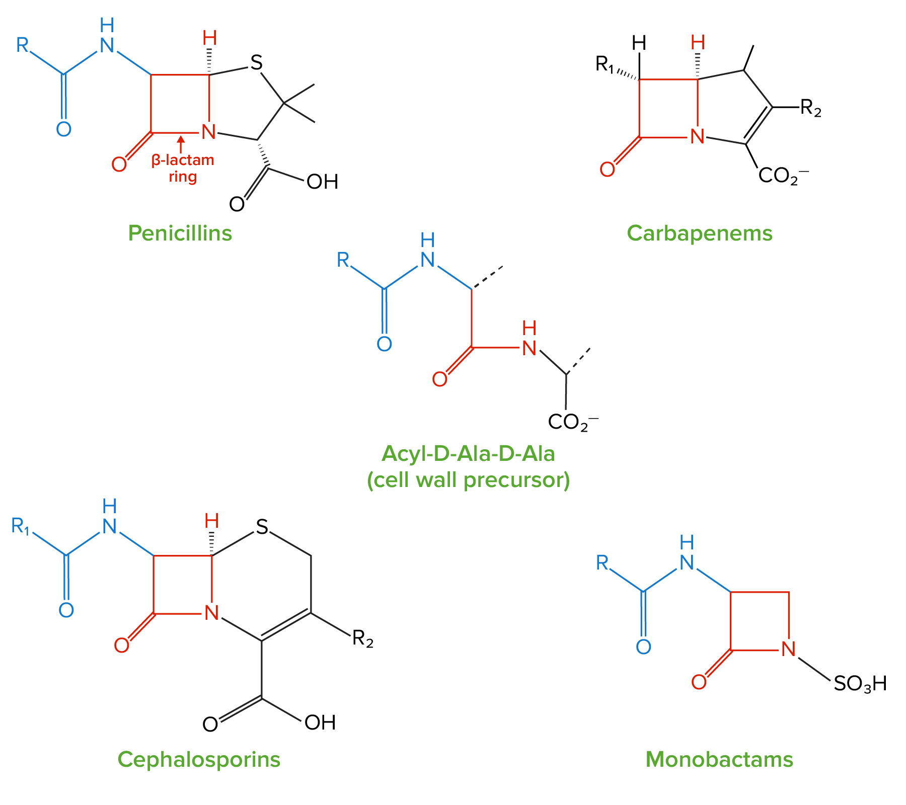
Estructura de los betalactámicos:
Todos los antibióticos betalactámicos contienen el mismo anillo central “betalactámico” de 4 miembros (resaltado en rojo). Este anillo es responsable de las propiedades antibacterianas del medicamento porque es la región que se une e inhibe las proteínas de unión a penicilina. Las proteínas de unión a penicilina catalizan la formación de la pared celular al generar enlaces cruzados entre las cadenas peptídicas en las moléculas de peptidoglicano; Las proteínas de unión a penicilina forman estos enlaces cruzados entre los péptidos acil-D-Ala-D-Ala, que tienen una estructura similar al anillo betalactámico.
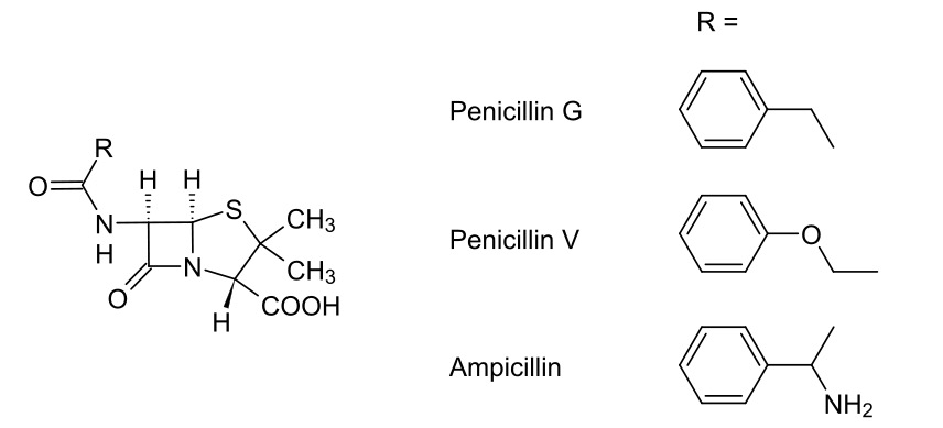
Penicilinas
Imagen: “Strukturen verschiedener Penicillinen” por Roland Mattern. Licencia: Dominio PúblicoTodos los LOS Neisseria betalactámicos, incluidas las penicilinas, ejercen sus efectos al AL Amyloidosis inhibir la síntesis de la pared celular bacteriana.
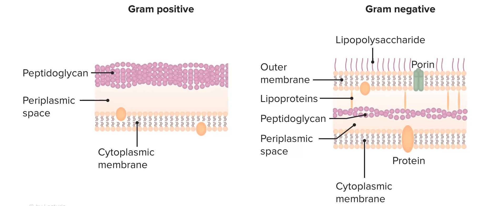
Estructura de las paredes celulares bacterianas
Imagen por Lecturio. Licencia: CC BY-NC-SA 4.0Todos los LOS Neisseria betalactámicos actúan inhibiendo irreversiblemente las proteínas de unión a penicilina → los LOS Neisseria antibióticos betalactámicos inhiben la síntesis de la pared celular
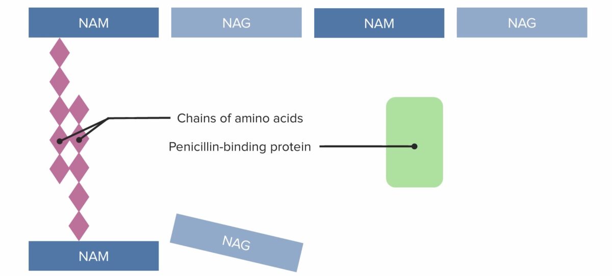
(1) Proteína de unión a penicilina que forma puentes entrecruzados entre cadenas de peptidoglicano adyacentes
NAM: ácido N-acetilmurámico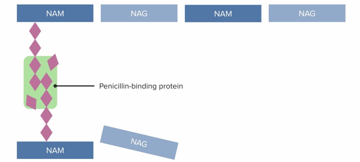
(2) Proteína de unión a penicilina que forma puentes entrecruzados entre cadenas de peptidoglicano adyacentes
NAM: ácido N-acetilmurámico
(3) Proteína de unión a penicilina que forma puentes entrecruzados entre cadenas de peptidoglicano adyacentes
NAM: ácido N-acetilmurámico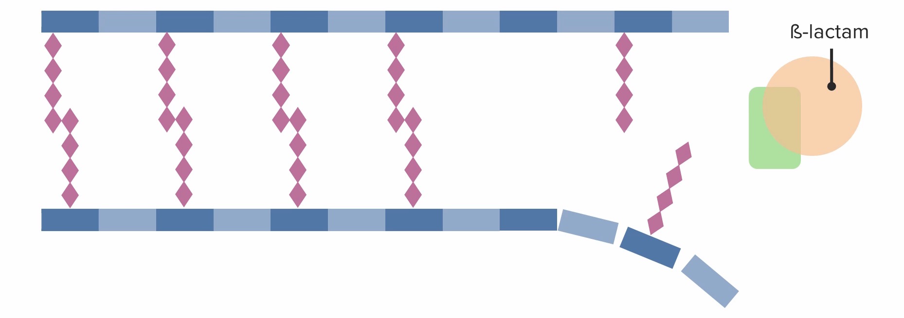
Presencia de un antibiótico betalactámico, que se une de forma irreversible e inhibe la proteína de unión a penicilina, evitando que forme nuevos enlaces cruzados:
El antibiótico betalactámico inhibe eficazmente la síntesis de la pared celular y, en última instancia, conduce a la muerte celular.
Los LOS Neisseria betalactámicos, incluidas las penicilinas, ejercen un efecto bactericida ( en EN Erythema nodosum is an immune-mediated panniculitis (inflammation of the subcutaneous fat) caused by a type IV (delayed-type) hypersensitivity reaction. It commonly manifests in young women as tender, erythematous nodules on the shins. Erythema Nodosum lugar de bacteriostático).
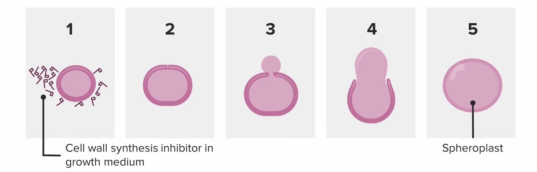
Bacteria que intenta dividirse en presencia de penicilina:
La bacteria se despoja de su pared y se convierte en un esferoplasto. El esferoplasto no puede sobrevivir y se autocataliza (muere).
Las bacterias utilizan 3 mecanismos principales para resistir las penicilinas:
Las penicilinas se pueden clasificar en EN Erythema nodosum is an immune-mediated panniculitis (inflammation of the subcutaneous fat) caused by a type IV (delayed-type) hypersensitivity reaction. It commonly manifests in young women as tender, erythematous nodules on the shins. Erythema Nodosum penicilinas naturales, penicilinas antiestafilocócicas y penicilinas de amplio espectro. Las penicilinas también se pueden clasificar como compuestos sensibles a la penicilinasa o resistentes a la penicilinasa.
Las penicilinas resistentes a la penicilinasa tienen un gran grupo R junto al AL Amyloidosis anillo betalactámico, lo que impide la degradación de los LOS Neisseria medicamentos por la penicilinasa. Las penicilinas resistentes a la penicilinasa son eficaces contra los LOS Neisseria estafilococos sensibles a la meticilina; por lo tanto, se les conoce comúnmente como penicilinas antiestafilocócicas.
| Medicamento (vía de administración) | Espectro de actividad | Usos clínicos |
|---|---|---|
| Penicilina G (intravenosa/intramuscular) y penicilina V (oral) | Estrecho:
|
|
| Cloxacilina y dicloxacilina | Estrecho: Cocos grampositivos:
|
|
| Ampicilina (intravenosa/oral) y amoxicilina (oral) | Más amplio:
|
|
| Piperacilina (solo disponible como piperacilina/ tazobactam Tazobactam A penicillanic acid and sulfone derivative and potent beta-lactamase inhibitor that enhances the activity of other anti-bacterial agents against beta-lactamase producing bacteria. Cephalosporins en EN Erythema nodosum is an immune-mediated panniculitis (inflammation of the subcutaneous fat) caused by a type IV (delayed-type) hypersensitivity reaction. It commonly manifests in young women as tender, erythematous nodules on the shins. Erythema Nodosum los LOS Neisseria EE. UU.) | Más ancho:
|
|
| Mezlocilina | Amplio: buena cobertura de gramnegativos | Infecciones del tracto biliar (e.g., colangitis biliar) |
Los LOS Neisseria efectos más comunes están relacionados con reacciones alérgicas.
Los LOS Neisseria antibióticos se pueden clasificar de varias maneras. Una forma es clasificarlos según su mecanismo de acción:
| Mecanismo | Clases de antibióticos |
|---|---|
| Inhibidores de la síntesis de la pared celular bacteriana |
|
| Inhibidores de la síntesis de proteínas bacterianas |
|
| Agentes que actúan contra el ADN y/o el folato |
|
| Agentes antimicobacterianos |
|
Diferentes antibióticos tienen diferentes grados de actividad contra diferentes bacterias. La siguiente tabla describe los LOS Neisseria antibióticos que son activos contra 3 clases importantes de bacterias, que incluyen cocos grampositivos, bacilos gramnegativos y anaerobios.
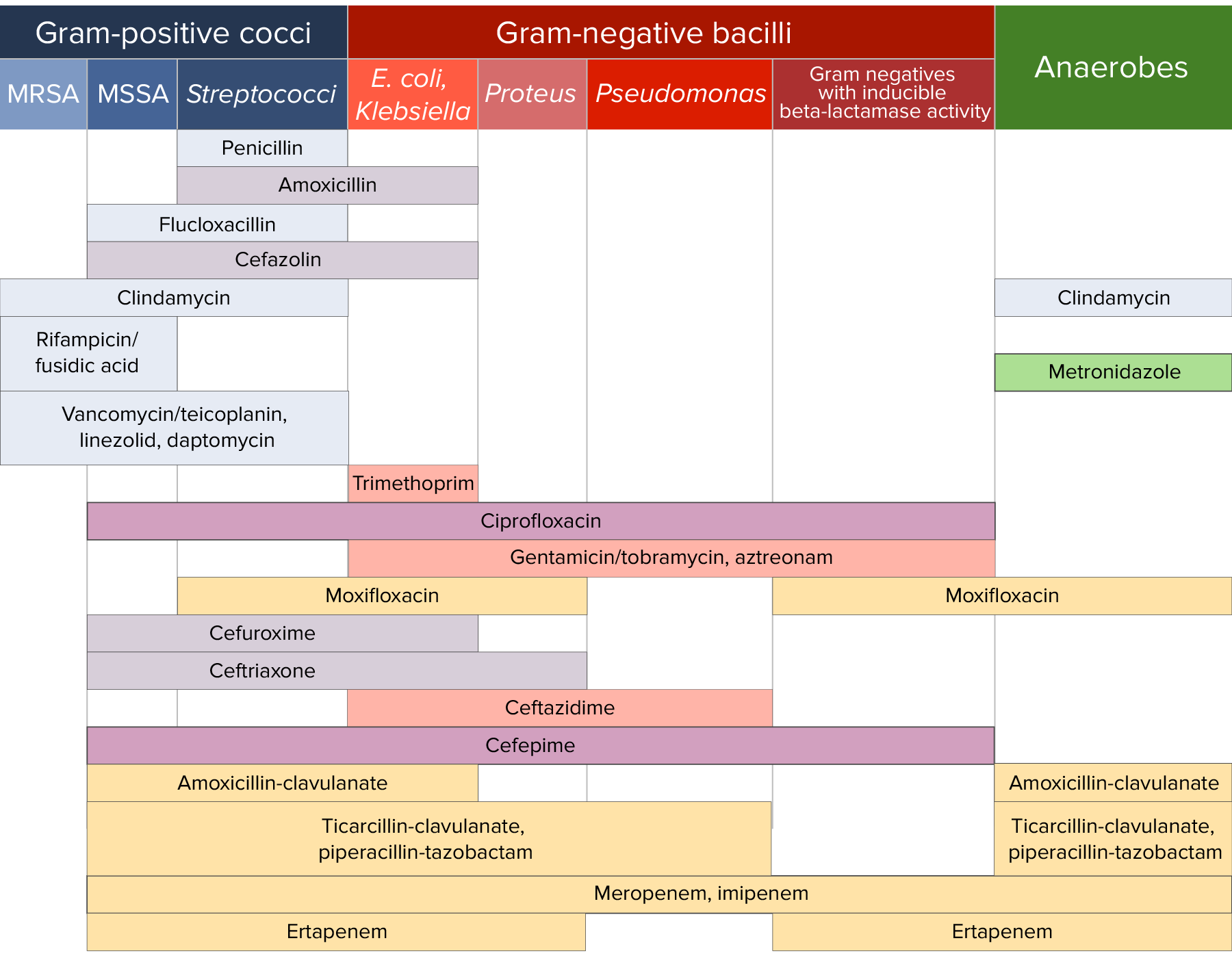
Sensibilidad a los antibióticos:
Gráfico que compara la cobertura microbiana de diferentes antibióticos para cocos grampositivos, bacilos gramnegativos y anaerobios.