Domina Conceptos Médicos

Estudia para la escuela de medicina y tus examenes con Lecturio.

Oligodendroglioma

Los oligodendrogliomas son tumores malignos del SNC que surgen de los precursores neuronales de las células gliales. Los oligodendrogliomas suelen surgir en los lóbulos frontales del cerebro y tienen un pronóstico generalmente favorable en comparación con otros gliomas. Los oligodendrogliomas son el 3er tumor más frecuente del SNC. El síntoma de presentación más frecuente es una convulsión; otros síntomas incluyen cefaleas, pérdida visual y déficits neurológicos focales. El diagnóstico se establece por medio de una biopsia cerebral que demuestra una apariencia clásica de "huevo frito" (núcleos redondos con citoplasma claro). Los LOS Neisseria oligodendrogliomas son de crecimiento lento, pero como son mortales, se tratan con una combinación de resección quirúrgica, radiación y quimioterapia.

Last updated: Dec 15, 2025

Editorial responsibility: Stanley Oiseth, Lindsay Jones, Evelin Maza

Descripción General

Definición

Los LOS Neisseria oligodendrogliomas son tumores neuroepiteliales malignos que surgen de los LOS Neisseria precursores neuronales de las células gliales. Estas células tumorales parecen histológicamente similares a los LOS Neisseria oligodendrocitos, pero carecen de la capacidad mielinizante de los LOS Neisseria oligodendrocitos. Los LOS Neisseria oligodendrogliomas son un tipo de glioma:

  • Los LOS Neisseria gliomas surgen de las células gliales, que son tejidos de soporte dentro del cerebro y de la médula espinal.
  • Los LOS Neisseria oligodendrocitos son células gliales del SNC que producen mielina.
  • Los LOS Neisseria astrocitomas y los LOS Neisseria ependimomas son los LOS Neisseria otros tumores de células gliales más comunes.

Clasificación de los LOS Neisseria tumores del sistema nervioso

Tabla: Clasificación de los LOS Neisseria tumores del sistema nervioso
Categorías Tumores específicos
Tumores neuroepiteliales en EN Erythema nodosum is an immune-mediated panniculitis (inflammation of the subcutaneous fat) caused by a type IV (delayed-type) hypersensitivity reaction. It commonly manifests in young women as tender, erythematous nodules on the shins. Erythema Nodosum el SNC
  • Astrocitomas, incluido el glioblastoma multiforme Glioblastoma multiforme Glioblastoma multiforme is a high-grade astrocytoma, an aggressive brain tumor arising from astrocytes, with an unknown cause and a poorly understood link to risk factors. There are two main types: primary, a more aggressive form seen more commonly in older patients, and secondary, developing from lower-grade astrocytomas and seen more commonly in younger patients. Glioblastoma Multiforme
  • Oligodendroglioma Oligodendroglioma Oligodendrogliomas are malignant CNS tumors arising from neural glial cell precursors. Oligodendrogliomas often arise in the frontal lobes of the brain and have a generally favorable prognosis when compared to other gliomas. Oligodendrogliomas are the 3rd most common CNS tumor. The most frequent presenting symptom is a seizure. Oligodendroglioma
  • Ependimoma y tumores del plexo coroideo
  • Meduloblastomas (tumores embrionarios)
Tumores meníngeos
  • Meningiomas
  • Hemangioblastomas Hemangioblastomas A benign tumor of the nervous system that may occur sporadically or in association with von Hippel-Lindau disease. It accounts for approximately 2% of intracranial tumors, arising most frequently in the cerebellar hemispheres and vermis. Histologically, the tumors are composed of multiple capillary and sinusoidal channels lined with endothelial cells and clusters of lipid-laden pseudoxanthoma cells. Usually solitary, these tumors can be multiple and may also occur in the brain stem, spinal cord, retina, and supratentorial compartment. Cerebellar hemangioblastomas usually present in the third decade with intracranial hypertension, and ataxia. Von Hippel-Lindau Disease
Tumores de la región selar
  • Craneofaringioma
  • Adenoma hipofisario
  • Pinealoma/pinealoblastoma
Linfoma primario del SNC Linfoma primario del SNC
Metástasis al AL Amyloidosis cerebro (5 veces más frecuente que los LOS Neisseria tumores cerebrales primarios) Más comúnmente surgen de:
  • Carcinomas de pulmón, mama y células renales
  • Melanoma Melanoma Melanoma is a malignant tumor arising from melanocytes, the melanin-producing cells of the epidermis. These tumors are most common in fair-skinned individuals with a history of excessive sun exposure and sunburns. Melanoma
Tumores periféricos
  • Schwannomas, incluido el neurinoma del acústico
  • Neuroblastoma Neuroblastoma Neuroblastoma is a malignancy that arises from the neural crest cell derivatives along the sympathetic chain (neuroblasts) and is most commonly located in the adrenal medulla. The tumor often presents in childhood with a flank mass that crosses the midline. Neuroblastoma

Epidemiología

  • Incidencia:
    • 3ra neoplasia cerebral primaria más frecuente (después del glioblastoma multiforme Glioblastoma multiforme Glioblastoma multiforme is a high-grade astrocytoma, an aggressive brain tumor arising from astrocytes, with an unknown cause and a poorly understood link to risk factors. There are two main types: primary, a more aggressive form seen more commonly in older patients, and secondary, developing from lower-grade astrocytomas and seen more commonly in younger patients. Glioblastoma Multiforme y del astrocitoma difuso de bajo grado)
    • Representa aproximadamente el 5% de los LOS Neisseria tumores primarios del SNC
    • 0,2 por cada 100 000 habitantes
    • Cada año se diagnostican aproximadamente 1 000 pacientes con oligodendrogliomas en EN Erythema nodosum is an immune-mediated panniculitis (inflammation of the subcutaneous fat) caused by a type IV (delayed-type) hypersensitivity reaction. It commonly manifests in young women as tender, erythematous nodules on the shins. Erythema Nodosum Estados Unidos.
  • Mediana de edad al AL Amyloidosis momento del diagnóstico: 25–45 años
  • Ligeramente más común en EN Erythema nodosum is an immune-mediated panniculitis (inflammation of the subcutaneous fat) caused by a type IV (delayed-type) hypersensitivity reaction. It commonly manifests in young women as tender, erythematous nodules on the shins. Erythema Nodosum los LOS Neisseria hombres que en EN Erythema nodosum is an immune-mediated panniculitis (inflammation of the subcutaneous fat) caused by a type IV (delayed-type) hypersensitivity reaction. It commonly manifests in young women as tender, erythematous nodules on the shins. Erythema Nodosum las mujeres

Fisiopatología

Fisiopatología

  • Localización:
    • Suelen surgir en EN Erythema nodosum is an immune-mediated panniculitis (inflammation of the subcutaneous fat) caused by a type IV (delayed-type) hypersensitivity reaction. It commonly manifests in young women as tender, erythematous nodules on the shins. Erythema Nodosum los LOS Neisseria hemisferios cerebrales (cortical o subcortical)
    • Raramente ocurren dentro del cerebelo, los LOS Neisseria ventrículos o la médula espinal
  • Los LOS Neisseria efectos regionales en EN Erythema nodosum is an immune-mediated panniculitis (inflammation of the subcutaneous fat) caused by a type IV (delayed-type) hypersensitivity reaction. It commonly manifests in young women as tender, erythematous nodules on the shins. Erythema Nodosum el parénquima cerebral incluyen:
    • Compresión
    • Invasión
    • Destrucción
  • ↑ Presión intracraneal (PIC) probablemente debido a:
    • Efecto de masa del tumor Tumor Inflammation
    • Edema Edema Edema is a condition in which excess serous fluid accumulates in the body cavity or interstitial space of connective tissues. Edema is a symptom observed in several medical conditions. It can be categorized into 2 types, namely, peripheral (in the extremities) and internal (in an organ or body cavity). Edema en EN Erythema nodosum is an immune-mediated panniculitis (inflammation of the subcutaneous fat) caused by a type IV (delayed-type) hypersensitivity reaction. It commonly manifests in young women as tender, erythematous nodules on the shins. Erythema Nodosum el tejido cerebral circundante
    • ↑ Volumen sanguíneo
    • ↑ Volumen de LCR/hidrocefalia
  • Las alteraciones de las funciones normales del parénquima se deben a:
    • Hipoxia
    • Competencia por los LOS Neisseria nutrientes
    • Liberación de productos finales del metabolismo:
      • Radicales libres
      • Alteración de los LOS Neisseria electrolitos
      • Neurotransmisores
    • Liberación y reclutamiento de mediadores celulares (e.g., citoquinas)

Patogénesis

Los LOS Neisseria oligodendrogliomas son tumores definidos histológicamente por mutaciones de la isocitrato deshidrogenasa (IDH) 1 y 2 como codeleciones de los LOS Neisseria brazos cromosómicos 1p y 19q.

  • Mutaciones en IDH1 (que se encuentra en el citosol) y/o IDH2 (que se encuentra en la mitocondria):
    • IDH:
      • Cataliza la descarboxilación oxidativa reversible de isocitrato → α-cetoglutarato (α-KG, por sus siglas en inglés) en el ciclo del ácido tricarboxílico.
      • Productor primario de NADPH en la mayoría de los tejidos, especialmente en el cerebro
      • También participa en la mitigación del daño oxidativo
    • Las mutaciones conducen a la producción y acumulación de 2-hidroxiglutarato (2-HG):
      • La 2-HG inhibe la función enzimática de la α-KG dependiente de dioxigenasas, que participan en la desmetilación del ADN.
      • ↑ El 2-HG provoca una desregulación epigenética → puede conducir al desarrollo de tumores
  • Codeleciones de los brazos cromosómicos 1p y 19q:
    • Conduce a cambios epigenéticos y a la hipermetilación del genoma
    • Típicamente suele producirse por una translocación desequilibrada entre los cromosomas 1 y 19
  • Otras mutaciones:
    • Mutaciones activadoras del promotor de la transcriptasa inversa de la telomerasa (TERT, por sus siglas en inglés):
      • TERT es una subunidad catalítica de la telomerasa → las mutaciones activadoras permiten que las células tumorales se vuelvan “inmortales”
      • Un marcador de diagnóstico primario para el glioblastoma multiforme Glioblastoma multiforme Glioblastoma multiforme is a high-grade astrocytoma, an aggressive brain tumor arising from astrocytes, with an unknown cause and a poorly understood link to risk factors. There are two main types: primary, a more aggressive form seen more commonly in older patients, and secondary, developing from lower-grade astrocytomas and seen more commonly in younger patients. Glioblastoma Multiforme, aunque también puede estar presente en EN Erythema nodosum is an immune-mediated panniculitis (inflammation of the subcutaneous fat) caused by a type IV (delayed-type) hypersensitivity reaction. It commonly manifests in young women as tender, erythematous nodules on the shins. Erythema Nodosum los LOS Neisseria oligodendrogliomas
    • Metilación (i.e., silenciamiento) del promotor de la O-6-metilguanina-ADN metiltransferasa (MGMT): La MGMT es una enzima que participa en EN Erythema nodosum is an immune-mediated panniculitis (inflammation of the subcutaneous fat) caused by a type IV (delayed-type) hypersensitivity reaction. It commonly manifests in young women as tender, erythematous nodules on the shins. Erythema Nodosum la reparación del ADN.

Clasificación

Los LOS Neisseria oligodendrogliomas son clasificados por la OMS según su histología y los LOS Neisseria tipos de mutaciones que albergan. ( Los LOS Neisseria tumores de grado I se consideran benignos, por lo que, a diferencia de los LOS Neisseria astrocitomas, no existen oligodendrogliomas de grado I).

  • Grado II de acuerdo a la OMS:
    • Oligodendroglioma Oligodendroglioma Oligodendrogliomas are malignant CNS tumors arising from neural glial cell precursors. Oligodendrogliomas often arise in the frontal lobes of the brain and have a generally favorable prognosis when compared to other gliomas. Oligodendrogliomas are the 3rd most common CNS tumor. The most frequent presenting symptom is a seizure. Oligodendroglioma, con mutación en EN Erythema nodosum is an immune-mediated panniculitis (inflammation of the subcutaneous fat) caused by a type IV (delayed-type) hypersensitivity reaction. It commonly manifests in young women as tender, erythematous nodules on the shins. Erythema Nodosum IDH y codeleción en EN Erythema nodosum is an immune-mediated panniculitis (inflammation of the subcutaneous fat) caused by a type IV (delayed-type) hypersensitivity reaction. It commonly manifests in young women as tender, erythematous nodules on the shins. Erythema Nodosum 1p/19q
    • Oligodendroglioma Oligodendroglioma Oligodendrogliomas are malignant CNS tumors arising from neural glial cell precursors. Oligodendrogliomas often arise in the frontal lobes of the brain and have a generally favorable prognosis when compared to other gliomas. Oligodendrogliomas are the 3rd most common CNS tumor. The most frequent presenting symptom is a seizure. Oligodendroglioma, no especificado
  • Grado III de acuerdo a la OMS:
    • Oligodendroglioma Oligodendroglioma Oligodendrogliomas are malignant CNS tumors arising from neural glial cell precursors. Oligodendrogliomas often arise in the frontal lobes of the brain and have a generally favorable prognosis when compared to other gliomas. Oligodendrogliomas are the 3rd most common CNS tumor. The most frequent presenting symptom is a seizure. Oligodendroglioma anaplásico, con mutación en EN Erythema nodosum is an immune-mediated panniculitis (inflammation of the subcutaneous fat) caused by a type IV (delayed-type) hypersensitivity reaction. It commonly manifests in young women as tender, erythematous nodules on the shins. Erythema Nodosum IDH y codeleción 1p/19q
    • Oligodendroglioma Oligodendroglioma Oligodendrogliomas are malignant CNS tumors arising from neural glial cell precursors. Oligodendrogliomas often arise in the frontal lobes of the brain and have a generally favorable prognosis when compared to other gliomas. Oligodendrogliomas are the 3rd most common CNS tumor. The most frequent presenting symptom is a seizure. Oligodendroglioma anaplásico, no especificado
  • Los LOS Neisseria tumores no especificados se sometieron a pruebas genéticas que no fueron concluyentes o no se realizaron pruebas genéticas.
  • Ya no se prefiere el diagnóstico de oligoastrocitoma, ya que los LOS Neisseria tumores con histología mixta deben clasificarse según su impronta molecular como astrocitomas u oligodendrogliomas. Si la codeleción en EN Erythema nodosum is an immune-mediated panniculitis (inflammation of the subcutaneous fat) caused by a type IV (delayed-type) hypersensitivity reaction. It commonly manifests in young women as tender, erythematous nodules on the shins. Erythema Nodosum 1p/19q está ausente, el tumor Tumor Inflammation suele clasificarse como astrocitoma.

Presentación Clínica

La mayoría de los LOS Neisseria signos y síntomas dependen de la localización exacta del tumor Tumor Inflammation. Los LOS Neisseria oligodendrogliomas son de crecimiento lento y suelen ser asintomáticos durante años antes del diagnóstico. El síntoma de presentación clínica más frecuente es una convulsión.

  • Convulsiones: pueden ser parciales o generalizadas
  • Síntomas generales: síntomas que pueden aparecer con tumores en EN Erythema nodosum is an immune-mediated panniculitis (inflammation of the subcutaneous fat) caused by a type IV (delayed-type) hypersensitivity reaction. It commonly manifests in young women as tender, erythematous nodules on the shins. Erythema Nodosum cualquier localización
    • Cefalea (normalmente por la mañana)
    • Náuseas y/o vómitos
    • Dificultades cognitivas:
      • Problemas de memoria
      • Cambios de estado de ánimo o de personalidad (sobre todo en EN Erythema nodosum is an immune-mediated panniculitis (inflammation of the subcutaneous fat) caused by a type IV (delayed-type) hypersensitivity reaction. It commonly manifests in young women as tender, erythematous nodules on the shins. Erythema Nodosum los LOS Neisseria tumores del lóbulo frontal Frontal The bone that forms the frontal aspect of the skull. Its flat part forms the forehead, articulating inferiorly with the nasal bone and the cheek bone on each side of the face. Skull: Anatomy)
    • Papiledema
  • Déficits neurológicos focales: depende de la localización concreta del tumor Tumor Inflammation
    • Debilidad muscular (daño en EN Erythema nodosum is an immune-mediated panniculitis (inflammation of the subcutaneous fat) caused by a type IV (delayed-type) hypersensitivity reaction. It commonly manifests in young women as tender, erythematous nodules on the shins. Erythema Nodosum la corteza motora contralateral)
    • Anomalías sensoriales (daño en EN Erythema nodosum is an immune-mediated panniculitis (inflammation of the subcutaneous fat) caused by a type IV (delayed-type) hypersensitivity reaction. It commonly manifests in young women as tender, erythematous nodules on the shins. Erythema Nodosum la corteza somatosensorial contralateral)
    • Apraxia Apraxia A group of cognitive disorders characterized by the inability to perform previously learned skills that cannot be attributed to deficits of motor or sensory function. The two major subtypes of this condition are ideomotor and ideational apraxia, which refers to loss of the ability to mentally formulate the processes involved with performing an action. For example, dressing apraxia may result from an inability to mentally formulate the act of placing clothes on the body. Apraxias are generally associated with lesions of the dominant parietal lobe and supramarginal gyrus. Cranial Nerve Palsies
    • Afasia
    • Déficit en EN Erythema nodosum is an immune-mediated panniculitis (inflammation of the subcutaneous fat) caused by a type IV (delayed-type) hypersensitivity reaction. It commonly manifests in young women as tender, erythematous nodules on the shins. Erythema Nodosum el campo visual
    • Hemiparesia

Diagnóstico

Imagenología

La RM de cabeza es el método de imagen de elección para evaluar los LOS Neisseria tumores cerebrales.
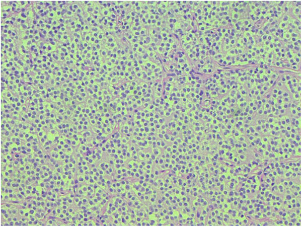
  • RM del cerebro:
    • Imágenes ponderadas en EN Erythema nodosum is an immune-mediated panniculitis (inflammation of the subcutaneous fat) caused by a type IV (delayed-type) hypersensitivity reaction. It commonly manifests in young women as tender, erythematous nodules on the shins. Erythema Nodosum T2: lesiones hiperintensas o de intensidad de señal mixta
    • Imágenes ponderadas en EN Erythema nodosum is an immune-mediated panniculitis (inflammation of the subcutaneous fat) caused by a type IV (delayed-type) hypersensitivity reaction. It commonly manifests in young women as tender, erythematous nodules on the shins. Erythema Nodosum T1: lesiones hipointensas o de intensidad de señal mixta
    • Es más probable que tenga bordes indistintos y señales no uniformes/heterogéneas en EN Erythema nodosum is an immune-mediated panniculitis (inflammation of the subcutaneous fat) caused by a type IV (delayed-type) hypersensitivity reaction. It commonly manifests in young women as tender, erythematous nodules on the shins. Erythema Nodosum comparación con los LOS Neisseria astrocitomas
    • También se suelen observar calcificaciones.
    • Realce con contraste:
      • Las lesiones de grado II no suelen realzarse
      • Las lesiones anaplásicas suelen mostrar cierto realce.
  • TC de la cabeza:
    • Se realiza cuando hay contraindicaciones para realizar la RM o en EN Erythema nodosum is an immune-mediated panniculitis (inflammation of the subcutaneous fat) caused by a type IV (delayed-type) hypersensitivity reaction. It commonly manifests in young women as tender, erythematous nodules on the shins. Erythema Nodosum situaciones de cuidado agudo
    • Los LOS Neisseria hallazgos incluyen:
      • Lesiones hipodensas con edema Edema Edema is a condition in which excess serous fluid accumulates in the body cavity or interstitial space of connective tissues. Edema is a symptom observed in several medical conditions. It can be categorized into 2 types, namely, peripheral (in the extremities) and internal (in an organ or body cavity). Edema circundante
      • Pueden verse calcificaciones y hemorragias.
      • Normalmente, solo las lesiones anaplásicas tienen realce.

Biopsia

  • Aspecto macroscópico:
    • Masa gris, gelatinosa, bien circunscrita
    • Frecuentemente con áreas de quistes, hemorragias y calcificaciones
  • Aspecto microscópico:
    • Compuesto por células bien diferenciadas que se asemejan a los LOS Neisseria oligodendrocitos
    • Láminas de células en EN Erythema nodosum is an immune-mediated panniculitis (inflammation of the subcutaneous fat) caused by a type IV (delayed-type) hypersensitivity reaction. It commonly manifests in young women as tender, erythematous nodules on the shins. Erythema Nodosum huevo frito (células grandes con citoplasma claro y núcleo redondo hipercromático)
    • Capilares ramificados: redes de vasos sanguíneos finos en EN Erythema nodosum is an immune-mediated panniculitis (inflammation of the subcutaneous fat) caused by a type IV (delayed-type) hypersensitivity reaction. It commonly manifests in young women as tender, erythematous nodules on the shins. Erythema Nodosum todo el tumor Tumor Inflammation debido a una angiogénesis importante
    • Los LOS Neisseria tumores anaplásicos presentan:
      • Mayor densidad celular
      • Atipia nuclear
      • Actividad mitótica
      • Proliferación microvascular
      • Necrosis Necrosis The death of cells in an organ or tissue due to disease, injury or failure of the blood supply. Ischemic Cell Damage
  • Pruebas moleculares:
    • Presencia de mutaciones en EN Erythema nodosum is an immune-mediated panniculitis (inflammation of the subcutaneous fat) caused by a type IV (delayed-type) hypersensitivity reaction. It commonly manifests in young women as tender, erythematous nodules on the shins. Erythema Nodosum IDH1/2 y codeleción en EN Erythema nodosum is an immune-mediated panniculitis (inflammation of the subcutaneous fat) caused by a type IV (delayed-type) hypersensitivity reaction. It commonly manifests in young women as tender, erythematous nodules on the shins. Erythema Nodosum 1p/19q:
      • Tinción inmunohistoquímica para mutaciones en EN Erythema nodosum is an immune-mediated panniculitis (inflammation of the subcutaneous fat) caused by a type IV (delayed-type) hypersensitivity reaction. It commonly manifests in young women as tender, erythematous nodules on the shins. Erythema Nodosum IDH1/2
      • Estudios FISH FISH A type of in situ hybridization in which target sequences are stained with fluorescent dye so their location and size can be determined using fluorescence microscopy. This staining is sufficiently distinct that the hybridization signal can be seen both in metaphase spreads and in interphase nuclei. Chromosome Testing para codeleciones 1p/19q
    • Carecen de la proteína básica de la mielina (MBP, por sus siglas en EN Erythema nodosum is an immune-mediated panniculitis (inflammation of the subcutaneous fat) caused by a type IV (delayed-type) hypersensitivity reaction. It commonly manifests in young women as tender, erythematous nodules on the shins. Erythema Nodosum inglés) que se encuentra en EN Erythema nodosum is an immune-mediated panniculitis (inflammation of the subcutaneous fat) caused by a type IV (delayed-type) hypersensitivity reaction. It commonly manifests in young women as tender, erythematous nodules on the shins. Erythema Nodosum los LOS Neisseria oligodendrocitos productores de mielina normales y en EN Erythema nodosum is an immune-mediated panniculitis (inflammation of the subcutaneous fat) caused by a type IV (delayed-type) hypersensitivity reaction. It commonly manifests in young women as tender, erythematous nodules on the shins. Erythema Nodosum las células de Schwann
    • Puede tener una tinción positiva para el factor de transcripción oligodendrocitario 2 (Olig2), un marcador de las células progenitoras oligodendrocitarias que participa en EN Erythema nodosum is an immune-mediated panniculitis (inflammation of the subcutaneous fat) caused by a type IV (delayed-type) hypersensitivity reaction. It commonly manifests in young women as tender, erythematous nodules on the shins. Erythema Nodosum la diferenciación celular
    • Puede tener una tinción positiva para la proteína ácida fibrilar glial (GFAP, por sus siglas en EN Erythema nodosum is an immune-mediated panniculitis (inflammation of the subcutaneous fat) caused by a type IV (delayed-type) hypersensitivity reaction. It commonly manifests in young women as tender, erythematous nodules on the shins. Erythema Nodosum inglés), un marcador común de los LOS Neisseria astrocitos
Oligodendroglioma

Oligodendroglioma:
Observe las células en forma de huevo frito con núcleos oscuros y redondos y los capilares ramificados en toda la muestra de tejido.

Imagen:“Morphologic spectrum of glial tumors: an increased trend towards oligodendroglial tumors in Pakistan” por Hashmi AA, Faridi N, Malik B, Edhi MM, Khurshid A, Khan M. Licencia: CC BY 4.0

Tratamiento y Pronóstico

Tratamiento

Los LOS Neisseria oligodendrogliomas son tumores infiltrativos que suelen considerarse incurables debido al AL Amyloidosis alto riesgo de recaída/recurrencia. El tratamiento óptimo suele consistir en EN Erythema nodosum is an immune-mediated panniculitis (inflammation of the subcutaneous fat) caused by a type IV (delayed-type) hypersensitivity reaction. It commonly manifests in young women as tender, erythematous nodules on the shins. Erythema Nodosum cirugía, radiación y quimioterapia.

  • Cirugía: funciones diagnósticas y terapéuticas
    • Confirma el diagnóstico, que puede guiar las terapias adyuvantes
    • Para obtener los LOS Neisseria mejores resultados, el objetivo de la cirugía es una resección total; sin embargo, esto no siempre es posible, ya que depende de la región del cerebro que esté afectada.
  • Radioterapia:
    • Normalmente se produce después de la cirugía
    • Se administra en EN Erythema nodosum is an immune-mediated panniculitis (inflammation of the subcutaneous fat) caused by a type IV (delayed-type) hypersensitivity reaction. It commonly manifests in young women as tender, erythematous nodules on the shins. Erythema Nodosum una zona restringida alrededor de la zona quirúrgica para maximizar el beneficio terapéutico y minimizar los LOS Neisseria efectos secundarios
    • Normalmente no se realiza en EN Erythema nodosum is an immune-mediated panniculitis (inflammation of the subcutaneous fat) caused by a type IV (delayed-type) hypersensitivity reaction. It commonly manifests in young women as tender, erythematous nodules on the shins. Erythema Nodosum niños muy pequeños debido a la grave comorbilidad en EN Erythema nodosum is an immune-mediated panniculitis (inflammation of the subcutaneous fat) caused by a type IV (delayed-type) hypersensitivity reaction. It commonly manifests in young women as tender, erythematous nodules on the shins. Erythema Nodosum ese grupo de edad
  • Quimioterapia:
    • Normalmente se administra después de la radiación
    • Mejora la supervivencia global cuando se combina con radioterapia adyuvante
    • El régimen estándar es la PCV:
      • Procarbazina
      • Lomustina (CCNU)
      • Vincristina
    • También se puede considerar la temozolomida como agente de 2da línea en EN Erythema nodosum is an immune-mediated panniculitis (inflammation of the subcutaneous fat) caused by a type IV (delayed-type) hypersensitivity reaction. It commonly manifests in young women as tender, erythematous nodules on the shins. Erythema Nodosum el oligodendroglioma Oligodendroglioma Oligodendrogliomas are malignant CNS tumors arising from neural glial cell precursors. Oligodendrogliomas often arise in the frontal lobes of the brain and have a generally favorable prognosis when compared to other gliomas. Oligodendrogliomas are the 3rd most common CNS tumor. The most frequent presenting symptom is a seizure. Oligodendroglioma (es tratamiento de 1ra línea para los LOS Neisseria astrocitomas)
  • Otras terapias:
    • Tratamiento anticonvulsivo en EN Erythema nodosum is an immune-mediated panniculitis (inflammation of the subcutaneous fat) caused by a type IV (delayed-type) hypersensitivity reaction. It commonly manifests in young women as tender, erythematous nodules on the shins. Erythema Nodosum pacientes con convulsiones
    • Corticosteroides para el edema Edema Edema is a condition in which excess serous fluid accumulates in the body cavity or interstitial space of connective tissues. Edema is a symptom observed in several medical conditions. It can be categorized into 2 types, namely, peripheral (in the extremities) and internal (in an organ or body cavity). Edema cerebral

Pronóstico

  • Supervivencia media de 5 años:
    • Oligodendroglioma Oligodendroglioma Oligodendrogliomas are malignant CNS tumors arising from neural glial cell precursors. Oligodendrogliomas often arise in the frontal lobes of the brain and have a generally favorable prognosis when compared to other gliomas. Oligodendrogliomas are the 3rd most common CNS tumor. The most frequent presenting symptom is a seizure. Oligodendroglioma de bajo grado: 5–10 años
    • Oligodendroglioma Oligodendroglioma Oligodendrogliomas are malignant CNS tumors arising from neural glial cell precursors. Oligodendrogliomas often arise in the frontal lobes of the brain and have a generally favorable prognosis when compared to other gliomas. Oligodendrogliomas are the 3rd most common CNS tumor. The most frequent presenting symptom is a seizure. Oligodendroglioma anaplásico: 3–4 años
  • Factores que afectan al AL Amyloidosis pronóstico:
    • Edad del paciente en EN Erythema nodosum is an immune-mediated panniculitis (inflammation of the subcutaneous fat) caused by a type IV (delayed-type) hypersensitivity reaction. It commonly manifests in young women as tender, erythematous nodules on the shins. Erythema Nodosum el momento del diagnóstico ( los LOS Neisseria pacientes más jóvenes tienen mejor pronóstico)
    • Localización del tumor Tumor Inflammation
    • Extensión de la resección quirúrgica
    • Grado histológico del tumor Tumor Inflammation
    • Estado del paciente en EN Erythema nodosum is an immune-mediated panniculitis (inflammation of the subcutaneous fat) caused by a type IV (delayed-type) hypersensitivity reaction. It commonly manifests in young women as tender, erythematous nodules on the shins. Erythema Nodosum el postoperatorio

Related videos

Diagnóstico Diferencial

  • Astrocitoma: Tumor Tumor Inflammation del SNC que surge de los LOS Neisseria astrocitos. Los LOS Neisseria astrocitomas agresivos de grado IV se denominan glioblastoma multiforme Glioblastoma multiforme Glioblastoma multiforme is a high-grade astrocytoma, an aggressive brain tumor arising from astrocytes, with an unknown cause and a poorly understood link to risk factors. There are two main types: primary, a more aggressive form seen more commonly in older patients, and secondary, developing from lower-grade astrocytomas and seen more commonly in younger patients. Glioblastoma Multiforme, mientras que los LOS Neisseria astrocitomas de grado II se denominan astrocitomas difusos de bajo grado. Los LOS Neisseria astrocitomas pueden presentarse de forma similar a los LOS Neisseria oligodendrogliomas y se diagnostican mediante RM y biopsia. Para diagnosticar un oligodendroglioma Oligodendroglioma Oligodendrogliomas are malignant CNS tumors arising from neural glial cell precursors. Oligodendrogliomas often arise in the frontal lobes of the brain and have a generally favorable prognosis when compared to other gliomas. Oligodendrogliomas are the 3rd most common CNS tumor. The most frequent presenting symptom is a seizure. Oligodendroglioma se requieren mutaciones en EN Erythema nodosum is an immune-mediated panniculitis (inflammation of the subcutaneous fat) caused by a type IV (delayed-type) hypersensitivity reaction. It commonly manifests in young women as tender, erythematous nodules on the shins. Erythema Nodosum IDH y la codeleción de 1p/19q; si solo hay mutaciones en EN Erythema nodosum is an immune-mediated panniculitis (inflammation of the subcutaneous fat) caused by a type IV (delayed-type) hypersensitivity reaction. It commonly manifests in young women as tender, erythematous nodules on the shins. Erythema Nodosum IDH, probablemente se trate de un astrocitoma de bajo grado. Al AL Amyloidosis igual que los LOS Neisseria oligodendrogliomas, el tratamiento de los LOS Neisseria astrocitomas incluye escisión quirúrgica, radiación y quimioterapia.
  • Meningioma Meningioma Meningiomas are slow-growing tumors that arise from the meninges of the brain and spinal cord. The vast majority are benign. These tumors commonly occur in individuals with a history of high doses of skull radiation, head trauma, and neurofibromatosis 2. Meningioma: tumor Tumor Inflammation benigno que surge de las meninges Meninges The brain and the spinal cord are enveloped by 3 overlapping layers of connective tissue called the meninges. The layers are, from the most external layer to the most internal layer, the dura mater, arachnoid mater, and pia mater. Between these layers are 3 potential spaces called the epidural, subdural, and subarachnoid spaces. Meninges: Anatomy del cerebro o de la médula espinal. Los LOS Neisseria meningiomas se presentan con cefalea, alteraciones de la visión y posibles convulsiones. El diagnóstico se confirma mediante resonancia magnética, biopsia y los LOS Neisseria meningiomas se tratan con escisión quirúrgica. A diferencia de los LOS Neisseria meningiomas, los LOS Neisseria oligodendrogliomas suelen ser más sintomáticos y no tienen asociación con la duramadre.
  • Tumor Tumor Inflammation metastásico: células neoplásicas que se han extendido al AL Amyloidosis cerebro a partir de tumores primarios en EN Erythema nodosum is an immune-mediated panniculitis (inflammation of the subcutaneous fat) caused by a type IV (delayed-type) hypersensitivity reaction. It commonly manifests in young women as tender, erythematous nodules on the shins. Erythema Nodosum otra parte del cuerpo. Las neoplasias metastásicas son las neoplasias más frecuentes en EN Erythema nodosum is an immune-mediated panniculitis (inflammation of the subcutaneous fat) caused by a type IV (delayed-type) hypersensitivity reaction. It commonly manifests in young women as tender, erythematous nodules on the shins. Erythema Nodosum el cerebro. La neuroimagen suele mostrar múltiples focos del carcinoma, lo que sugiere un origen fuera del cerebro. La presentación clínica depende del tumor Tumor Inflammation primario y de la localización y extensión de la metástasis cerebral. El tratamiento se dirige a la neoplasia subyacente y puede incluir resección quirúrgica, radioterapia y quimioterapia.
  • Toxoplasmosis Toxoplasmosis Toxoplasmosis is an infectious disease caused by Toxoplasma gondii, an obligate intracellular protozoan parasite. Felines are the definitive host, but transmission to humans can occur through contact with cat feces or the consumption of contaminated foods. The clinical presentation and complications depend on the host’s immune status. Toxoplasma/Toxoplasmosis cerebral: infección por Toxoplasma Toxoplasma Toxoplasmosis is an infectious disease caused by Toxoplasma gondii, an obligate intracellular protozoan parasite. Felines are the definitive host, but transmission to humans can occur through contact with cat feces or the consumption of contaminated foods. The clinical presentation and complications depend on the host’s immune status. Toxoplasma/Toxoplasmosis gondii en EN Erythema nodosum is an immune-mediated panniculitis (inflammation of the subcutaneous fat) caused by a type IV (delayed-type) hypersensitivity reaction. It commonly manifests in young women as tender, erythematous nodules on the shins. Erythema Nodosum el SNC en EN Erythema nodosum is an immune-mediated panniculitis (inflammation of the subcutaneous fat) caused by a type IV (delayed-type) hypersensitivity reaction. It commonly manifests in young women as tender, erythematous nodules on the shins. Erythema Nodosum pacientes inmunodeprimidos (e.g., pacientes con VIH). Esta enfermedad suele ser sintomática solo en EN Erythema nodosum is an immune-mediated panniculitis (inflammation of the subcutaneous fat) caused by a type IV (delayed-type) hypersensitivity reaction. It commonly manifests in young women as tender, erythematous nodules on the shins. Erythema Nodosum pacientes con recuentos de CD4 < 100 células/µL y se presenta con fiebre, cefalea, déficits neurológicos focales y/o convulsiones. La toxoplasmosis Toxoplasmosis Toxoplasmosis is an infectious disease caused by Toxoplasma gondii, an obligate intracellular protozoan parasite. Felines are the definitive host, but transmission to humans can occur through contact with cat feces or the consumption of contaminated foods. The clinical presentation and complications depend on the host’s immune status. Toxoplasma/Toxoplasmosis cerebral se diagnostica con estudios serológicos y con una resonancia magnética cerebral que muestra lesiones con realce en EN Erythema nodosum is an immune-mediated panniculitis (inflammation of the subcutaneous fat) caused by a type IV (delayed-type) hypersensitivity reaction. It commonly manifests in young women as tender, erythematous nodules on the shins. Erythema Nodosum forma de anillo (lo que es diferente a los LOS Neisseria oligodendrogliomas). La toxoplasmosis Toxoplasmosis Toxoplasmosis is an infectious disease caused by Toxoplasma gondii, an obligate intracellular protozoan parasite. Felines are the definitive host, but transmission to humans can occur through contact with cat feces or the consumption of contaminated foods. The clinical presentation and complications depend on the host’s immune status. Toxoplasma/Toxoplasmosis cerebral se trata con pirimetamina, sulfadiazina y leucovorina, junto con una terapia antirretroviral.

Referencias

  1. Van den Bent, M. (2021). Clinical features, diagnosis, and pathology of IDH-mutant, 1p/19q-codeleted oligodendrogliomas. UpToDate. Retrieved May 21, 2021, from https://www.uptodate.com/contents/clinical-features-diagnosis-and-pathology-of-idh-mutant-1p-19q-codeleted-oligodendrogliomas
  2. Van den Bent, M. (2021). Treatment and prognosis of IDH-mutant, 1p/19q-codeleted (grade II and III) oligodendrogliomas. UpToDate. Retrieved May 21, 2021, from https://www.uptodate.com/contents/treatment-and-prognosis-of-idh-mutant-1p-19q-codeleted-grade-ii-and-iii-oligodendrogliomas
  3. Uddin, S. (2020). Oligodendroglioma. Medscape. Retrieved May 21, 2021, from https://emedicine.medscape.com/article/1156699-overview
  4. Tork, C.A., Atkinson, C. (2020). Oligodendroglioma. StatPearls. Retrieved May 21, 2021, from https://www.ncbi.nlm.nih.gov/books/NBK559184/

¡Crea tu cuenta gratis o inicia una sesión para seguir leyendo!

Regístrate ahora y obtén acceso gratuito a Lecturio con páginas de concepto, videos médicos y cuestionarios para tu educación médica.

User Reviews

Que tengas una sesión de estudio alegre y navideña 🎁 Ahorra 50% en todos los planes >>

Details