Domina Conceptos Médicos

Estudia para la escuela de medicina y tus examenes con Lecturio.

Linfocitos: Histología

Los LOS Neisseria linfocitos son leucocitos heterogéneos que participan en EN Erythema nodosum is an immune-mediated panniculitis (inflammation of the subcutaneous fat) caused by a type IV (delayed-type) hypersensitivity reaction. It commonly manifests in young women as tender, erythematous nodules on the shins. Erythema Nodosum la respuesta inmunitaria. Los LOS Neisseria linfocitos se desarrollan a partir de la médula ósea, empezando por las células madre hematopoyéticas y progresando hacia los LOS Neisseria progenitores linfoides comunes. De este linaje surgen los LOS Neisseria linfocitos B y T y las células asesinas naturales. Los LOS Neisseria linfocitos B y T desempeñan un papel en EN Erythema nodosum is an immune-mediated panniculitis (inflammation of the subcutaneous fat) caused by a type IV (delayed-type) hypersensitivity reaction. It commonly manifests in young women as tender, erythematous nodules on the shins. Erythema Nodosum la inmunidad adaptativa, y las células asesinas naturales proporcionan la defensa del huésped contra las proteínas atípicas, como las células tumorales. Aunque todas las etapas de desarrollo comienzan en EN Erythema nodosum is an immune-mediated panniculitis (inflammation of the subcutaneous fat) caused by a type IV (delayed-type) hypersensitivity reaction. It commonly manifests in young women as tender, erythematous nodules on the shins. Erythema Nodosum la médula ósea, la maduración de los LOS Neisseria linfocitos es diferente. Los LOS Neisseria linfocitos B y las células asesinas naturales se diferencian en EN Erythema nodosum is an immune-mediated panniculitis (inflammation of the subcutaneous fat) caused by a type IV (delayed-type) hypersensitivity reaction. It commonly manifests in young women as tender, erythematous nodules on the shins. Erythema Nodosum la médula ósea antes de migrar a los LOS Neisseria órganos linfoides secundarios (como los LOS Neisseria ganglios linfáticos). Sin embargo, los LOS Neisseria linfocitos T pasan al AL Amyloidosis timo para seguir madurando.

Last updated: Dec 18, 2025

Editorial responsibility: Stanley Oiseth, Lindsay Jones, Evelin Maza

Descripción General

Definiciones y descripción

Los LOS Neisseria linfocitos son células sanguíneas que participan en EN Erythema nodosum is an immune-mediated panniculitis (inflammation of the subcutaneous fat) caused by a type IV (delayed-type) hypersensitivity reaction. It commonly manifests in young women as tender, erythematous nodules on the shins. Erythema Nodosum la respuesta inmunitaria y que surgen del progenitor linfoide común.

  • Descripción:
    • 30% de los LOS Neisseria leucocitos circulantes
    • Células esféricas y/u ovoides
    • Diámetro: 6–15 μm
    • Vida útil: de semanas a años
  • Pertenecen a un grupo heterogéneo de células llamadas leucocitos, que se dividen de la siguiente manera
    • Granulocitos: derivados del progenitor mieloide
    • Agranulocitos: incluye linfocitos (del progenitor linfoide) y monocitos (del progenitor mieloide)
  • Tipos de linfocitos y función:
    • Linfocitos B, o células B (derivadas de la bursa): inmunidad adaptativa humoral
    • Linfocitos T, o células T (derivadas del timo): inmunidad adaptativa celular
    • Células asesinas naturales: inmunidad innata con cierta respuesta inmunitaria adaptativa:
      • Un papel importante en EN Erythema nodosum is an immune-mediated panniculitis (inflammation of the subcutaneous fat) caused by a type IV (delayed-type) hypersensitivity reaction. It commonly manifests in young women as tender, erythematous nodules on the shins. Erythema Nodosum la defensa del huésped de las células tumorales, las células infectadas por virus Virus Viruses are infectious, obligate intracellular parasites composed of a nucleic acid core surrounded by a protein capsid. Viruses can be either naked (non-enveloped) or enveloped. The classification of viruses is complex and based on many factors, including type and structure of the nucleoid and capsid, the presence of an envelope, the replication cycle, and the host range. Virology y otras proteínas atípicas, “no propias”
      • 5%–20% de los LOS Neisseria linfocitos en EN Erythema nodosum is an immune-mediated panniculitis (inflammation of the subcutaneous fat) caused by a type IV (delayed-type) hypersensitivity reaction. It commonly manifests in young women as tender, erythematous nodules on the shins. Erythema Nodosum la sangre periférica
  • Tejidos linfoides implicados:
    • Primario: participa en EN Erythema nodosum is an immune-mediated panniculitis (inflammation of the subcutaneous fat) caused by a type IV (delayed-type) hypersensitivity reaction. It commonly manifests in young women as tender, erythematous nodules on the shins. Erythema Nodosum la generación inicial de linfocitos B y T:
      • Médula ósea
      • Timo
    • Secundario:
      • Ganglios linfáticos
      • Bazo
      • Amígdalas
      • Agregados de tejido linfoide en EN Erythema nodosum is an immune-mediated panniculitis (inflammation of the subcutaneous fat) caused by a type IV (delayed-type) hypersensitivity reaction. It commonly manifests in young women as tender, erythematous nodules on the shins. Erythema Nodosum el tracto gastrointestinal y respiratorio

Estructura

  • Dificultad para distinguir los LOS Neisseria tipos de linfocitos en EN Erythema nodosum is an immune-mediated panniculitis (inflammation of the subcutaneous fat) caused by a type IV (delayed-type) hypersensitivity reaction. It commonly manifests in young women as tender, erythematous nodules on the shins. Erythema Nodosum la microscopía
  • Núcleo:
    • Forma ovoide o de riñón con cromatina nuclear densamente empaquetada
    • Ocupa aproximadamente el 90% de la célula (alta proporción entre el núcleo y el citoplasma)
  • En EN Erythema nodosum is an immune-mediated panniculitis (inflammation of the subcutaneous fat) caused by a type IV (delayed-type) hypersensitivity reaction. It commonly manifests in young women as tender, erythematous nodules on the shins. Erythema Nodosum el citoplasma azul pálido:
    • Ribosomas libres
    • Lisosomas
    • Retículo endoplásmico de superficie rugosa
    • Golgi
    • Las mitocondrias y los LOS Neisseria centríolos son adyacentes a la membrana.
  • Las proteínas del citoesqueleto incluyen la tubulina, miosina y actina (entre otras) organizadas en EN Erythema nodosum is an immune-mediated panniculitis (inflammation of the subcutaneous fat) caused by a type IV (delayed-type) hypersensitivity reaction. It commonly manifests in young women as tender, erythematous nodules on the shins. Erythema Nodosum microtúbulos y microfilamentos.
  • Los LOS Neisseria linfocitos T citotóxicos y las células asesinas naturales tienen abundantes gránulos citoplasmáticos:
    • Perforina: enzima proteolítica formadora de poros
    • Granzimas: proteinasas de serina que facilitan la apoptosis Apoptosis A regulated cell death mechanism characterized by distinctive morphologic changes in the nucleus and cytoplasm, including the endonucleolytic cleavage of genomic DNA, at regularly spaced, internucleosomal sites, I.e., DNA fragmentation. It is genetically-programmed and serves as a balance to mitosis in regulating the size of animal tissues and in mediating pathologic processes associated with tumor growth. Ischemic Cell Damage (almacenadas como proenzimas inactivas)
    • Serpinas (inhibidores de la serina-proteínasa): impiden la autolisis de los LOS Neisseria gránulos
Glóbulos blancos

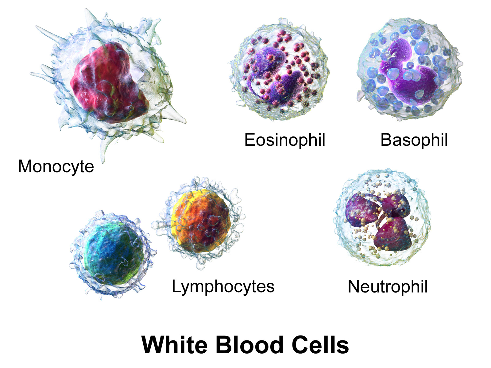
Los glóbulos blancos (WBC), o leucocitos, en la sangre:
Los granulocitos incluyen basófilos, eosinófilos y neutrófilos; los agranulocitos incluyen linfocitos y monocitos.

Imagen: “Blausen 0909 WhiteBloodCells” por Blausen. Licencia: CC BY 3.0

Linfopoyesis

Producción de linfocitos

  • Hematopoyesis:
    • Del 1er al AL Amyloidosis 2do mes en EN Erythema nodosum is an immune-mediated panniculitis (inflammation of the subcutaneous fat) caused by a type IV (delayed-type) hypersensitivity reaction. It commonly manifests in young women as tender, erythematous nodules on the shins. Erythema Nodosum el útero: mesodermo del saco vitelino
    • Al AL Amyloidosis 2do mes: se desplaza al AL Amyloidosis hígado (y al AL Amyloidosis bazo)
    • Al AL Amyloidosis 5to mes: se produce en EN Erythema nodosum is an immune-mediated panniculitis (inflammation of the subcutaneous fat) caused by a type IV (delayed-type) hypersensitivity reaction. It commonly manifests in young women as tender, erythematous nodules on the shins. Erythema Nodosum la médula ósea, convirtiéndose en EN Erythema nodosum is an immune-mediated panniculitis (inflammation of the subcutaneous fat) caused by a type IV (delayed-type) hypersensitivity reaction. It commonly manifests in young women as tender, erythematous nodules on the shins. Erythema Nodosum la fuente predominante de células sanguíneas
  • La linfopoyesis comienza con las células madre hematopoyéticas multipotenciales de la médula ósea.
  • Células madre hematopoyéticas → células progenitoras multipotenciales → progenitores linfoides comunes (se convierten en EN Erythema nodosum is an immune-mediated panniculitis (inflammation of the subcutaneous fat) caused by a type IV (delayed-type) hypersensitivity reaction. It commonly manifests in young women as tender, erythematous nodules on the shins. Erythema Nodosum células T, células B y células asesinas naturales):
    • Linfocitos B → órganos linfoides periféricos
    • Los LOS Neisseria linfocitos migran al AL Amyloidosis timo → linfocitos T → órganos linfoides periféricos:
      • El timo mantiene la producción de células T hasta la pubertad.
      • En EN Erythema nodosum is an immune-mediated panniculitis (inflammation of the subcutaneous fat) caused by a type IV (delayed-type) hypersensitivity reaction. It commonly manifests in young women as tender, erythematous nodules on the shins. Erythema Nodosum la pubertad, el timo sufre una involución → ↓ masa de tejido linfoide y ↓ producción de células T
    • Células asesinas naturales → órganos linfoides periféricos
Hematopoyesis de la médula ósea

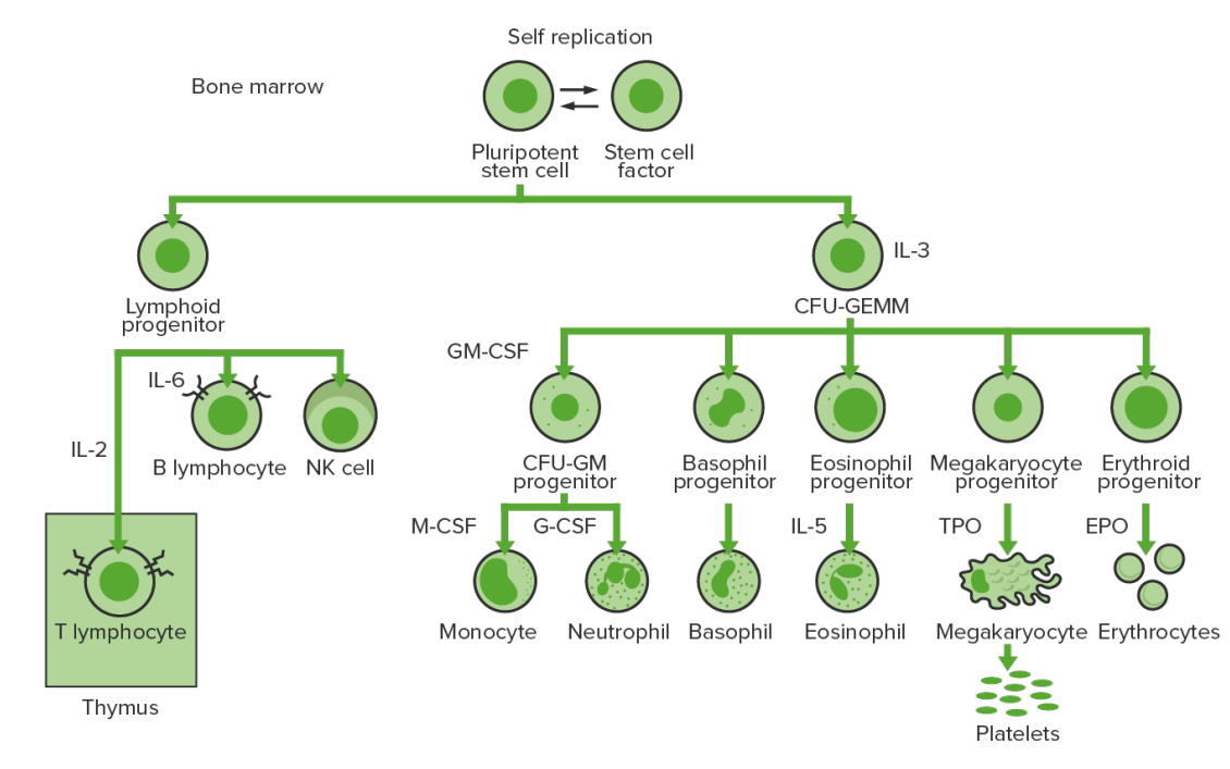
Hematopoyesis de la médula ósea: proliferación y diferenciación de los elementos formados de la sangre.
IL-3: interleuquina-3
CFU-GEMM: unidad formadora de colonias de granulocitos, eritrocitos, monocitos y megacariocitos
IL-2: interleuquina-2
IL-6: interleuquina-6
CFU-GM: unidad formadora de colonias-granulocitos-macrófagos
GM-CSF: factor estimulante de colonias de granulocitos y macrófagos
M-CSF: factor estimulante de colonias de macrófagos
G-CSF: factor estimulante de colonias de granulocitos
IL-5: interleuquina-5
NK: asesina natural
TPO: trombopoyetina
EPO: eritropoyetina

Imagen por Lecturio. Licencia: CC BY-NC-SA 4.0

Regulación

Tabla: Interleuquinas
Citoquinas Actividades Fuente
Factor de células madre Estimula todas las células progenitoras hematopoyéticas Células estromales de la médula ósea
Interleuquina-2 (IL-2)
  • Mitógeno para las células T y B activadas
  • Diferenciación de las células asesinas naturales
Células T colaboradoras
Interleuquina-4 (IL-4)
  • Desarrollo de basófilos y mastocitos
  • Activación de los LOS Neisseria linfocitos B
Células T colaboradoras
Interleuquina-6 (IL-6)
  • Mitógeno para los LOS Neisseria leucocitos
  • Activación de células B y células T reguladoras
  • Macrófagos
  • Neutrófilos
  • Células endoteliales
Interleuquina-7 ( IL-7 IL-7 A proinflammatory cytokine produced primarily by T-lymphocytes or their precursors. Several subtypes of interleukin-17 have been identified, each of which is a product of a unique gene. Severe Combined Immunodeficiency (SCID)) Estimulación de todas las células madre linfoides Células estromales de la médula ósea

Células B

Desarrollo

  • Comienza en EN Erythema nodosum is an immune-mediated panniculitis (inflammation of the subcutaneous fat) caused by a type IV (delayed-type) hypersensitivity reaction. It commonly manifests in young women as tender, erythematous nodules on the shins. Erythema Nodosum la médula ósea: células madre hematopoyéticas → progenitores linfoides comunes
  • Para producir una célula B funcional y madura a partir del progenitor linfoide común:
    • Expresión de la molécula inmunoglobulina de la superficie celular (parte del receptor Receptor Receptors are proteins located either on the surface of or within a cell that can bind to signaling molecules known as ligands (e.g., hormones) and cause some type of response within the cell. Receptors de células B)
    • El ácido desoxirribonucleico (ADN) de la línea germinal no tiene los LOS Neisseria genes Genes A category of nucleic acid sequences that function as units of heredity and which code for the basic instructions for the development, reproduction, and maintenance of organisms. DNA Types and Structure completos para codificar una molécula de inmunoglobulina completa.
    • Los LOS Neisseria reordenamientos genéticos (unión de diferentes segmentos de genes Genes A category of nucleic acid sequences that function as units of heredity and which code for the basic instructions for the development, reproduction, and maintenance of organisms. DNA Types and Structure) dentro de las células B son necesarios para ensamblar la molécula de inmunoglobulina.
    • El proceso también produce un repertorio de células B diversas, creando una protección contra diferentes tipos de infecciones.
  • Molécula de superficie celular (inmunoglobulina):
    • Tiene cadenas pesadas (μ, δ, γ, α, o ε) unidas por disulfuro a las cadenas ligeras (κ o λ)
    • Los LOS Neisseria genes Genes A category of nucleic acid sequences that function as units of heredity and which code for the basic instructions for the development, reproduction, and maintenance of organisms. DNA Types and Structure de la cadena pesada (que se encuentran en EN Erythema nodosum is an immune-mediated panniculitis (inflammation of the subcutaneous fat) caused by a type IV (delayed-type) hypersensitivity reaction. It commonly manifests in young women as tender, erythematous nodules on the shins. Erythema Nodosum un único locus Locus Specific regions that are mapped within a genome. Genetic loci are usually identified with a shorthand notation that indicates the chromosome number and the position of a specific band along the P or Q arm of the chromosome where they are found. For example the locus 6p21 is found within band 21 of the P-arm of chromosome 6. Many well known genetic loci are also known by common names that are associated with a genetic function or hereditary disease. Basic Terms of Genetics genético, el IGH), se ensamblan a partir de 4 segmentos genéticos:
      • Región variable Variable Variables represent information about something that can change. The design of the measurement scales, or of the methods for obtaining information, will determine the data gathered and the characteristics of that data. As a result, a variable can be qualitative or quantitative, and may be further classified into subgroups. Types of Variables (V)
      • Segmento de diversidad (D)
      • Región de unión (J)
      • Región constante (C)
    • Los LOS Neisseria genes Genes A category of nucleic acid sequences that function as units of heredity and which code for the basic instructions for the development, reproduction, and maintenance of organisms. DNA Types and Structure de la cadena ligera (que se encuentran como dos loci génicos separados -el locus Locus Specific regions that are mapped within a genome. Genetic loci are usually identified with a shorthand notation that indicates the chromosome number and the position of a specific band along the P or Q arm of the chromosome where they are found. For example the locus 6p21 is found within band 21 of the P-arm of chromosome 6. Many well known genetic loci are also known by common names that are associated with a genetic function or hereditary disease. Basic Terms of Genetics κ [IGK] y el locus Locus Specific regions that are mapped within a genome. Genetic loci are usually identified with a shorthand notation that indicates the chromosome number and the position of a specific band along the P or Q arm of the chromosome where they are found. For example the locus 6p21 is found within band 21 of the P-arm of chromosome 6. Many well known genetic loci are also known by common names that are associated with a genetic function or hereditary disease. Basic Terms of Genetics λ [IGL]) provienen de 3 segmentos génicos:
      • Región variable Variable Variables represent information about something that can change. The design of the measurement scales, or of the methods for obtaining information, will determine the data gathered and the characteristics of that data. As a result, a variable can be qualitative or quantitative, and may be further classified into subgroups. Types of Variables (V)
      • Región de unión (J)
      • Región constante (C)
Receptor de células B (BCR)

El receptor de células B está formado por la molécula de inmunoglobulina (Ig) y la molécula de señalización. La inmunoglobina contiene 2 cadenas pesadas idénticas y 2 cadenas ligeras idénticas unidas por un puente disulfuro; la inmunoglobulina unida a la membrana está anclada a la superficie celular.

Imagen: “Figure 42 02 06” por OpenStax. Licencia: CC BY 4.0

Etapas

Para alcanzar la funcionalidad, la célula B pasa por etapas en EN Erythema nodosum is an immune-mediated panniculitis (inflammation of the subcutaneous fat) caused by a type IV (delayed-type) hypersensitivity reaction. It commonly manifests in young women as tender, erythematous nodules on the shins. Erythema Nodosum la médula ósea y en EN Erythema nodosum is an immune-mediated panniculitis (inflammation of the subcutaneous fat) caused by a type IV (delayed-type) hypersensitivity reaction. It commonly manifests in young women as tender, erythematous nodules on the shins. Erythema Nodosum los LOS Neisseria órganos linfoides secundarios.

  • En EN Erythema nodosum is an immune-mediated panniculitis (inflammation of the subcutaneous fat) caused by a type IV (delayed-type) hypersensitivity reaction. It commonly manifests in young women as tender, erythematous nodules on the shins. Erythema Nodosum las etapas iniciales que se producen en EN Erythema nodosum is an immune-mediated panniculitis (inflammation of the subcutaneous fat) caused by a type IV (delayed-type) hypersensitivity reaction. It commonly manifests in young women as tender, erythematous nodules on the shins. Erythema Nodosum la médula ósea, el objetivo es construir el receptor Receptor Receptors are proteins located either on the surface of or within a cell that can bind to signaling molecules known as ligands (e.g., hormones) and cause some type of response within the cell. Receptors (que no requiere ningún antígeno).
  • Cuando se libera en EN Erythema nodosum is an immune-mediated panniculitis (inflammation of the subcutaneous fat) caused by a type IV (delayed-type) hypersensitivity reaction. It commonly manifests in young women as tender, erythematous nodules on the shins. Erythema Nodosum los LOS Neisseria órganos linfoides secundarios, un antígeno (con o sin ayuda de las células T) activará la célula B para continuar el proceso de maduración.
Tabla: Etapas independientes del antígeno en EN Erythema nodosum is an immune-mediated panniculitis (inflammation of the subcutaneous fat) caused by a type IV (delayed-type) hypersensitivity reaction. It commonly manifests in young women as tender, erythematous nodules on the shins. Erythema Nodosum la diferenciación de las células B
Etapa de maduración Genes Genes A category of nucleic acid sequences that function as units of heredity and which code for the basic instructions for the development, reproduction, and maintenance of organisms. DNA Types and Structure Ig Ig X-linked Agammaglobulinemia BCR BCR Lymphocytes: Histology Eventos asociados
Célula pre-pro-B ADN de la línea germinal Ninguno No hay expresión de la cadena pesada o ligera
Célula pro-B IGH D-J reordenado Ninguno Comienza a expresar CD19, CD34 y HLA-DR (antígeno de histocompatibilidad de clase II)
Célula pre-B IGH V-D-J reordenado Se forma el pre-BCR:
  • La cadena pesada está presente.
  • La cadena ligera sustituta está presente.
Aparecen otros marcadores (e.g., CD79, CD10, CD20, CD40 CD40 Members of the tumor necrosis factor receptor superfamily with specificity for CD40 ligand. They are found on mature B-lymphocytes, some epithelial cells; and lymphoid dendritic cells. Evidence suggests that CD40-dependent activation of B-cells is important for generation of memory B-cells within the germinal centers. Mutations in the CD40 antigen gene result in hyper-igm immunodeficiency syndrome, type 3. Signaling of the receptor occurs through its association with tnf receptor-associated factors. Hyper-IgM Syndrome, TdT TdT Acute Lymphoblastic Leukemia)
Célula B inmadura
  • IGH V-D-J reordenado
  • Cadena ligera V-J reordenada
BCR BCR Lymphocytes: Histology maduro (molécula IgM IgM A class of immunoglobulin bearing mu chains (immunoglobulin mu-chains). Igm can fix complement. The name comes from its high molecular weight and originally being called a macroglobulin. Immunoglobulins: Types and Functions) Continúa la expresión de HLA-DR, CD19, CD20 y CD40 CD40 Members of the tumor necrosis factor receptor superfamily with specificity for CD40 ligand. They are found on mature B-lymphocytes, some epithelial cells; and lymphoid dendritic cells. Evidence suggests that CD40-dependent activation of B-cells is important for generation of memory B-cells within the germinal centers. Mutations in the CD40 antigen gene result in hyper-igm immunodeficiency syndrome, type 3. Signaling of the receptor occurs through its association with tnf receptor-associated factors. Hyper-IgM Syndrome, pero no de otros marcadores (e.g., CD10, CD34, TdT TdT Acute Lymphoblastic Leukemia)
Célula B madura (virgen)
  • IGH V-D-J reordenado
  • Cadena ligera V-J reordenada
Con BCR BCR Lymphocytes: Histology maduro ( IgM IgM A class of immunoglobulin bearing mu chains (immunoglobulin mu-chains). Igm can fix complement. The name comes from its high molecular weight and originally being called a macroglobulin. Immunoglobulins: Types and Functions) → salida de la médula ósea Expresión de CD19 y CD20 por todas
Ig: inmunoglobulina
BCR: Receptor de células B
IGH: cadena pesada de inmunoglobulina
Región variable (V)
Segmento de diversidad (D)
Región de unión (J)
TdT: desoxitransferasa terminal
Tabla: Etapas dependientes del antígeno en EN Erythema nodosum is an immune-mediated panniculitis (inflammation of the subcutaneous fat) caused by a type IV (delayed-type) hypersensitivity reaction. It commonly manifests in young women as tender, erythematous nodules on the shins. Erythema Nodosum la diferenciación de las células B
Etapa de maduración BCR BCR Lymphocytes: Histology Eventos asociados
Célula B madura ( en EN Erythema nodosum is an immune-mediated panniculitis (inflammation of the subcutaneous fat) caused by a type IV (delayed-type) hypersensitivity reaction. It commonly manifests in young women as tender, erythematous nodules on the shins. Erythema Nodosum los LOS Neisseria tejidos linfoides secundarios) Madura (expresa IgM IgM A class of immunoglobulin bearing mu chains (immunoglobulin mu-chains). Igm can fix complement. The name comes from its high molecular weight and originally being called a macroglobulin. Immunoglobulins: Types and Functions e IgD IgD An immunoglobulin which accounts for less than 1% of plasma immunoglobulin. It is found on the membrane of many circulating B lymphocytes. Immunoglobulins: Types and Functions cuando está en EN Erythema nodosum is an immune-mediated panniculitis (inflammation of the subcutaneous fat) caused by a type IV (delayed-type) hypersensitivity reaction. It commonly manifests in young women as tender, erythematous nodules on the shins. Erythema Nodosum los LOS Neisseria tejidos linfoides secundarios) Las células pueden descansar o puede producirse la activación de las células B (las células B interactúan con el antígeno exógeno y/o las células T colaboradoras).
Célula B activada Conmutación de clases Una vez activado, puede permanecer como IgM IgM A class of immunoglobulin bearing mu chains (immunoglobulin mu-chains). Igm can fix complement. The name comes from its high molecular weight and originally being called a macroglobulin. Immunoglobulins: Types and Functions o cambiar a IgE IgE An immunoglobulin associated with mast cells. Overexpression has been associated with allergic hypersensitivity. Immunoglobulins: Types and Functions, IgG IgG The major immunoglobulin isotype class in normal human serum. There are several isotype subclasses of igg, for example, igg1, igg2a, and igg2b. Hypersensitivity Pneumonitis o IgA IgA Represents 15-20% of the human serum immunoglobulins, mostly as the 4-chain polymer in humans or dimer in other mammals. Secretory iga is the main immunoglobulin in secretions. Immunoglobulins: Types and Functions
Memoria de la célula B
  • Célula B activada → algunas se convierten en EN Erythema nodosum is an immune-mediated panniculitis (inflammation of the subcutaneous fat) caused by a type IV (delayed-type) hypersensitivity reaction. It commonly manifests in young women as tender, erythematous nodules on the shins. Erythema Nodosum células B de memoria
  • Circulan, reaccionan a la estimulación de antígenos, generan células plasmáticas
Célula plasmática
  • Célula B activada → algunas se convierten en EN Erythema nodosum is an immune-mediated panniculitis (inflammation of the subcutaneous fat) caused by a type IV (delayed-type) hypersensitivity reaction. It commonly manifests in young women as tender, erythematous nodules on the shins. Erythema Nodosum células plasmáticas
  • Las células grandes segregan anticuerpos y combaten las infecciones
  • Migran a la médula ósea
BCR: Receptor de células B
Differentiation stages of the B cell

Etapas de diferenciación de la célula B:
En las etapas independientes del antígeno, la producción de células B comienza con la célula madre hematopoyética, que se convierte en un progenitor linfoide común, y luego en una célula pre-pro-B o en una célula progenitora B. Los siguientes pasos incluyen el reordenamiento de los genes para ensamblar la molécula de inmunoglobulina (Ig). Las cadenas pesadas de inmunoglobulina comienzan con el reordenamiento del segmento de diversidad y de unión para formar la célula pro-B. En el siguiente paso (célula pre-B), se completa la recombinación de la cadena pesada de Ig (variable, diversidad, unión) y se forma el receptor de la célula pre-B. Se produce un reordenamiento de la cadena ligera (kappa (κ) o lambda (λ)) que da lugar a la expresión de una molécula completa de anticuerpos IgM por parte de una célula B inmadura. A continuación se produce la formación de la célula B madura (virgen) con IgM e IgD.
Las etapas dependientes del antígeno tienen lugar en los tejidos linfoides secundarios. Una vez que la célula B madura produce IgM e IgD, puede producirse un cambio de clase para fabricar IgE, IgG e IgA. Las células B se activan y se convierten en células plasmáticas o células de memoria.

Imagen por Lecturio.

Células T

Desarrollo

  • Células madre hematopoyéticas → progenitores linfoides comunes → células progenitoras tímicas tempranas → timo
  • Las células T en EN Erythema nodosum is an immune-mediated panniculitis (inflammation of the subcutaneous fat) caused by a type IV (delayed-type) hypersensitivity reaction. It commonly manifests in young women as tender, erythematous nodules on the shins. Erythema Nodosum desarrollo en EN Erythema nodosum is an immune-mediated panniculitis (inflammation of the subcutaneous fat) caused by a type IV (delayed-type) hypersensitivity reaction. It commonly manifests in young women as tender, erythematous nodules on the shins. Erythema Nodosum el timo se denominan timocitos.
  • Los LOS Neisseria reordenamientos genéticos forman el receptor Receptor Receptors are proteins located either on the surface of or within a cell that can bind to signaling molecules known as ligands (e.g., hormones) and cause some type of response within the cell. Receptors de células T:
    • La mayoría de las células T contienen cadenas ɑ y β y los LOS Neisseria coreceptores CD4 o CD8.
    • Las células T restantes contienen cadenas ɣ y δ.
    • Receptor Receptor Receptors are proteins located either on the surface of or within a cell that can bind to signaling molecules known as ligands (e.g., hormones) and cause some type of response within the cell. Receptors de célula T + CD3 CD3 Complex of at least five membrane-bound polypeptides in mature T-lymphocytes that are non-covalently associated with one another and with the T-cell receptor. The CD3 complex includes the gamma, delta, epsilon, zeta, and eta chains (subunits). When antigen binds to the T-cell receptor, the CD3 complex transduces the activating signals to the cytoplasm of the T-cell. The CD3 gamma and delta chains (subunits) are separate from and not related to the gamma/delta chains of the T-cell receptor. T cells: Types and Functions forman el complejo TCR TCR Molecules on the surface of T-lymphocytes that recognize and combine with antigens. The receptors are non-covalently associated with a complex of several polypeptides collectively called CD3 antigens. Recognition of foreign antigen and the major histocompatibility complex is accomplished by a single heterodimeric antigen-receptor structure, composed of either alpha-beta or gamma-delta chains. Adaptive Cell-mediated Immunity.
    • CD3 CD3 Complex of at least five membrane-bound polypeptides in mature T-lymphocytes that are non-covalently associated with one another and with the T-cell receptor. The CD3 complex includes the gamma, delta, epsilon, zeta, and eta chains (subunits). When antigen binds to the T-cell receptor, the CD3 complex transduces the activating signals to the cytoplasm of the T-cell. The CD3 gamma and delta chains (subunits) are separate from and not related to the gamma/delta chains of the T-cell receptor. T cells: Types and Functions: El marcador más utilizado para identificar las células T.
Comparación del receptor de células B (BCR) y del receptor de células T (TCR)

Comparación del receptor de células B y del receptor de células T

Imagen: “Antigen receptor chem114A” por Tinastella. Licencia: Dominio Público

Etapas

Para alcanzar la funcionalidad, la célula T pasa por etapas, liberándose de la médula ósea como células progenitoras para continuar su desarrollo en EN Erythema nodosum is an immune-mediated panniculitis (inflammation of the subcutaneous fat) caused by a type IV (delayed-type) hypersensitivity reaction. It commonly manifests in young women as tender, erythematous nodules on the shins. Erythema Nodosum el timo.

  • En EN Erythema nodosum is an immune-mediated panniculitis (inflammation of the subcutaneous fat) caused by a type IV (delayed-type) hypersensitivity reaction. It commonly manifests in young women as tender, erythematous nodules on the shins. Erythema Nodosum las etapas iniciales, el objetivo es construir el receptor Receptor Receptors are proteins located either on the surface of or within a cell that can bind to signaling molecules known as ligands (e.g., hormones) and cause some type of response within the cell. Receptors (que no requiere ningún antígeno).
  • A continuación, se dan otros pasos para activar la célula T (con un antígeno) y para diferenciarse en EN Erythema nodosum is an immune-mediated panniculitis (inflammation of the subcutaneous fat) caused by a type IV (delayed-type) hypersensitivity reaction. It commonly manifests in young women as tender, erythematous nodules on the shins. Erythema Nodosum célula T colaboradora o célula T citotóxica.
Tabla: Etapas de la diferenciación de las células T
Etapa de maduración Receptor Receptor Receptors are proteins located either on the surface of or within a cell that can bind to signaling molecules known as ligands (e.g., hormones) and cause some type of response within the cell. Receptors de células T Eventos asociados
Células progenitoras Ninguno
  • Médula ósea → timo para su posterior maduración
  • Se convierten en EN Erythema nodosum is an immune-mediated panniculitis (inflammation of the subcutaneous fat) caused by a type IV (delayed-type) hypersensitivity reaction. It commonly manifests in young women as tender, erythematous nodules on the shins. Erythema Nodosum células doblemente negativas (todavía sin CD4 y CD8)
Células doblemente negativas Reordenamiento de la cadena β (pre-TCR) (la falta de reordenamiento conduce a la apoptosis Apoptosis A regulated cell death mechanism characterized by distinctive morphologic changes in the nucleus and cytoplasm, including the endonucleolytic cleavage of genomic DNA, at regularly spaced, internucleosomal sites, I.e., DNA fragmentation. It is genetically-programmed and serves as a balance to mitosis in regulating the size of animal tissues and in mediating pathologic processes associated with tumor growth. Ischemic Cell Damage)
  • Expresión de CD3 CD3 Complex of at least five membrane-bound polypeptides in mature T-lymphocytes that are non-covalently associated with one another and with the T-cell receptor. The CD3 complex includes the gamma, delta, epsilon, zeta, and eta chains (subunits). When antigen binds to the T-cell receptor, the CD3 complex transduces the activating signals to the cytoplasm of the T-cell. The CD3 gamma and delta chains (subunits) are separate from and not related to the gamma/delta chains of the T-cell receptor. T cells: Types and Functions
  • CD4-, CD8- (sin CD4 y CD8)
Células doblemente positivas Reordenamiento de la cadena ɑ → las cadenas ɑ se ensamblan con las cadenas β → complejo completo ɑ-β-TCR-CD3 (expresado en EN Erythema nodosum is an immune-mediated panniculitis (inflammation of the subcutaneous fat) caused by a type IV (delayed-type) hypersensitivity reaction. It commonly manifests in young women as tender, erythematous nodules on the shins. Erythema Nodosum la superficie)
  • CD4+, CD8+
  • Las células doblemente positivas interactúan con los LOS Neisseria autoantígenos ( en EN Erythema nodosum is an immune-mediated panniculitis (inflammation of the subcutaneous fat) caused by a type IV (delayed-type) hypersensitivity reaction. It commonly manifests in young women as tender, erythematous nodules on the shins. Erythema Nodosum el contexto de las moléculas MHC)
  • Con la presentación del MHC, algunas células sufren una selección positiva en EN Erythema nodosum is an immune-mediated panniculitis (inflammation of the subcutaneous fat) caused by a type IV (delayed-type) hypersensitivity reaction. It commonly manifests in young women as tender, erythematous nodules on the shins. Erythema Nodosum la corteza tímica:
    • Interacción intermedia o moderada entre el MHC y el TCR TCR Molecules on the surface of T-lymphocytes that recognize and combine with antigens. The receptors are non-covalently associated with a complex of several polypeptides collectively called CD3 antigens. Recognition of foreign antigen and the major histocompatibility complex is accomplished by a single heterodimeric antigen-receptor structure, composed of either alpha-beta or gamma-delta chains. Adaptive Cell-mediated Immunity
    • Producción de células funcionales
  • Algunas células se someten a una selección negativa en EN Erythema nodosum is an immune-mediated panniculitis (inflammation of the subcutaneous fat) caused by a type IV (delayed-type) hypersensitivity reaction. It commonly manifests in young women as tender, erythematous nodules on the shins. Erythema Nodosum la médula tímica:
    • Alta afinidad o fuerte interacción entre el MHC y el TCR TCR Molecules on the surface of T-lymphocytes that recognize and combine with antigens. The receptors are non-covalently associated with a complex of several polypeptides collectively called CD3 antigens. Recognition of foreign antigen and the major histocompatibility complex is accomplished by a single heterodimeric antigen-receptor structure, composed of either alpha-beta or gamma-delta chains. Adaptive Cell-mediated Immunity
    • Muerte celular ( apoptosis Apoptosis A regulated cell death mechanism characterized by distinctive morphologic changes in the nucleus and cytoplasm, including the endonucleolytic cleavage of genomic DNA, at regularly spaced, internucleosomal sites, I.e., DNA fragmentation. It is genetically-programmed and serves as a balance to mitosis in regulating the size of animal tissues and in mediating pathologic processes associated with tumor growth. Ischemic Cell Damage)
    • Evita la liberación de células T disfuncionales (puede activar la autoinmunidad)
  • Falta de interacción → apoptosis Apoptosis A regulated cell death mechanism characterized by distinctive morphologic changes in the nucleus and cytoplasm, including the endonucleolytic cleavage of genomic DNA, at regularly spaced, internucleosomal sites, I.e., DNA fragmentation. It is genetically-programmed and serves as a balance to mitosis in regulating the size of animal tissues and in mediating pathologic processes associated with tumor growth. Ischemic Cell Damage
Células T monopositivas
  • Las señales celulares desencadenan la expresión celular de CD4 o CD8 (no de ambos).
    • Th con CD4: interactúan con las células para expresar MHC clase II
    • Tc con CD8: interactúan con las células para expresar MHC clase I
  • Los LOS Neisseria Th y Tc virgenes circulan (sangre → tejidos linfáticos → linfa) y esperan a ser activados por las CPA (que llevan un complejo péptido-MHC complementario)
MHC: complejo mayor de histocompatibilidad
TCR: receptor de células T
Th: Células T colaboradoras
Tc: células T citotóxicas
CPAs: células presentadoras de antígenos
Etapas de diferenciación de las células T

Etapas de diferenciación de las células T:
Desde la médula ósea, las células progenitoras pasan al timo para su posterior maduración. Las células doblemente negativas (sin expresión de CD4/CD8 o CD4-/CD8 -) no han desarrollado el receptor de células T (TCR). Las células doblemente negativas sufren un reordenamiento del gen TCR y se convierten en células pro-T, y luego en células pre-T. A través de la serie, se expresan los CD4 y CD8, y el TCR se ensambla a través de reordenamientos genéticos (células doblemente positivas). A continuación, el timo presenta las moléculas del complejo mayor de histocompatibilidad (MHC) a las células T en desarrollo. Algunas células se someten a una selección positiva (se produce una interacción intermedia entre el MHC y el TCR) y producen células funcionales. Algunas células se someten a una selección negativa (fuerte interacción entre el MHC y el TCR), lo que provoca la muerte celular. Se evita la liberación de células T disfuncionales, que pueden activar la autoinmunidad. Algunas células T no logran interactuar, lo que conduce a la apoptosis. Las células T maduras expresan o bien CD4 (células T colaboradoras) o bien CD8 (células T citotóxicas), pero no ambas.

Imagen por Lecturio. Licencia: CC BY-NC-SA 4.0

Células Asesinas Naturales

  • Células madre hematopoyéticas en EN Erythema nodosum is an immune-mediated panniculitis (inflammation of the subcutaneous fat) caused by a type IV (delayed-type) hypersensitivity reaction. It commonly manifests in young women as tender, erythematous nodules on the shins. Erythema Nodosum la médula ósea → progenitores linfoides comunes → linfoblastos → prolinfocitos → células asesinas naturales
  • Producción estimulada por la interleuquina-15 (IL-15)
  • Se activan por la exposición a células infectadas por virus Virus Viruses are infectious, obligate intracellular parasites composed of a nucleic acid core surrounded by a protein capsid. Viruses can be either naked (non-enveloped) or enveloped. The classification of viruses is complex and based on many factors, including type and structure of the nucleoid and capsid, the presence of an envelope, the replication cycle, and the host range. Virology o células con patrones anormales de expresión de antígenos de superficie (células cancerosas)
  • También participan en EN Erythema nodosum is an immune-mediated panniculitis (inflammation of the subcutaneous fat) caused by a type IV (delayed-type) hypersensitivity reaction. It commonly manifests in young women as tender, erythematous nodules on the shins. Erythema Nodosum la citotoxicidad mediada por células dependientes de anticuerpos

Relevancia Clínica

  • Linfoma de Hodgkin: neoplasia de los LOS Neisseria linfocitos B que se originan en EN Erythema nodosum is an immune-mediated panniculitis (inflammation of the subcutaneous fat) caused by a type IV (delayed-type) hypersensitivity reaction. It commonly manifests in young women as tender, erythematous nodules on the shins. Erythema Nodosum los LOS Neisseria ganglios linfáticos. El hallazgo patognomónico es una célula de Hodgkin/Reed-Sternberg (una célula B gigante multinucleada con inclusiones eosinófilas). La enfermedad se presenta más comúnmente con linfadenopatía, sudores nocturnos, pérdida de peso y fiebre. Puede haber esplenomegalia o hepatomegalia. Las pruebas de diagnóstico incluyen el análisis histológico de los LOS Neisseria ganglios linfáticos para mostrar las células de Hodgkin/Reed-Sternberg, análisis de sangre y estudios de imagenología.
  • Linfoma no Hodgkin: un grupo diverso de neoplasias malignas que se originan en EN Erythema nodosum is an immune-mediated panniculitis (inflammation of the subcutaneous fat) caused by a type IV (delayed-type) hypersensitivity reaction. It commonly manifests in young women as tender, erythematous nodules on the shins. Erythema Nodosum las células B, en EN Erythema nodosum is an immune-mediated panniculitis (inflammation of the subcutaneous fat) caused by a type IV (delayed-type) hypersensitivity reaction. It commonly manifests in young women as tender, erythematous nodules on the shins. Erythema Nodosum las células T o (raramente) en EN Erythema nodosum is an immune-mediated panniculitis (inflammation of the subcutaneous fat) caused by a type IV (delayed-type) hypersensitivity reaction. It commonly manifests in young women as tender, erythematous nodules on the shins. Erythema Nodosum las células asesinas naturales. Dos tercios de los LOS Neisseria linfomas no Hodgkin afectan a los LOS Neisseria ganglios linfáticos; el resto son extraganglionares. El linfoma no Hodgkin afecta a todas las edades. Los LOS Neisseria linfomas no Hodgkin de células B incluyen el linfoma de Burkitt, linfoma difuso de células B grandes, linfoma de células del manto y linfoma de la zona marginal. Los LOS Neisseria linfomas no Hodgkin de células T incluyen el linfoma de células T del adulto y la micosis fungoide. Los LOS Neisseria signos y síntomas más comunes son fiebre, pérdida de peso, sudores nocturnos, linfadenopatía y hepatoesplenomegalia.
  • Leucemia linfoblástica aguda: la forma más común de cáncer que afecta a los LOS Neisseria niños. La leucemia linfoblástica aguda se caracteriza por la proliferación incontrolada de células precursoras linfoides (aumento de linfoblastos). La médula normal es sustituida por linfoblastos, que pasan a la circulación y se infiltran en EN Erythema nodosum is an immune-mediated panniculitis (inflammation of the subcutaneous fat) caused by a type IV (delayed-type) hypersensitivity reaction. It commonly manifests in young women as tender, erythematous nodules on the shins. Erythema Nodosum otros órganos. Los LOS Neisseria signos y síntomas están relacionados con anemia Anemia Anemia is a condition in which individuals have low Hb levels, which can arise from various causes. Anemia is accompanied by a reduced number of RBCs and may manifest with fatigue, shortness of breath, pallor, and weakness. Subtypes are classified by the size of RBCs, chronicity, and etiology. Anemia: Overview and Types, trombocitopenia y falta de leucocitos funcionales. El frotis de sangre periférica y la biopsia de médula ósea identifican linfoblastos. Los LOS Neisseria estudios inmunofenotípicos, histoquímicos y genéticos ayudan al AL Amyloidosis diagnóstico y al AL Amyloidosis tratamiento.
  • Leucemia linfocítica crónica: cáncer de la sangre y de la médula ósea de crecimiento lento caracterizado por una producción excesiva de linfocitos monoclonales B en EN Erythema nodosum is an immune-mediated panniculitis (inflammation of the subcutaneous fat) caused by a type IV (delayed-type) hypersensitivity reaction. It commonly manifests in young women as tender, erythematous nodules on the shins. Erythema Nodosum la sangre periférica. Cuando la afectación es principalmente ganglionar, la enfermedad se denomina linfoma linfocítico pequeño. La enfermedad suele presentarse de forma asintomática en EN Erythema nodosum is an immune-mediated panniculitis (inflammation of the subcutaneous fat) caused by a type IV (delayed-type) hypersensitivity reaction. It commonly manifests in young women as tender, erythematous nodules on the shins. Erythema Nodosum los LOS Neisseria adultos mayores. El diagnóstico se realiza a partir de una linfocitosis anormal en EN Erythema nodosum is an immune-mediated panniculitis (inflammation of the subcutaneous fat) caused by a type IV (delayed-type) hypersensitivity reaction. It commonly manifests in young women as tender, erythematous nodules on the shins. Erythema Nodosum las pruebas de laboratorio. Las células B son linfocitos funcionalmente incompetentes, lo que puede dar lugar a infecciones recurrentes.
  • Mieloma múltiple: afección maligna de las células plasmáticas (linfocitos B activados). La proliferación monoclonal de células plasmáticas da lugar a una secreción excesiva de anticuerpos IgG IgG The major immunoglobulin isotype class in normal human serum. There are several isotype subclasses of igg, for example, igg1, igg2a, and igg2b. Hypersensitivity Pneumonitis y a una actividad osteoclástica impulsada por las citoquinas (dolores óseos, fracturas patológicas y alteraciones metabólicas). La secreción excesiva de anticuerpos provoca proteinuria Proteinuria The presence of proteins in the urine, an indicator of kidney diseases. Nephrotic Syndrome in Children, daño renal y producción/deposición tisular de fibrillas amiloides. El diagnóstico se realiza mediante electroforesis de plasma Plasma The residual portion of blood that is left after removal of blood cells by centrifugation without prior blood coagulation. Transfusion Products y biopsia de médula ósea.

Referencias

  1. Abel, A., Yang, C., Thakar, M., Malarkannan, S. (2018). Natural Killer Cells: development, maturation and clinical utilization. Front. Immunol. 9, 1869. https://doi.org/10.3389/fimmu.2018.01869
  2. Aster, J. (2023). Normal B and T lymphocyte development. UpToDate. Retrieved December 17, 2025, from https://www.uptodate.com/contents/normal-b-and-t-lymphocyte-development
  3. Aster, J.C., & Scadden, D. (2016). Hematopoiesis. In Aster, J.C., & Bunn, H. (Eds.), Pathophysiology of Blood Disorders, 2e. McGraw-Hill. https://accessmedicine.mhmedical.com/content.aspx?bookid=1900&sectionid=137394642
  4. Mescher, A.L. (Ed.). (2021). Hemopoiesis. Junqueira’s Basic Histology Text and Atlas, 16e. McGraw-Hill. https://accessmedicine.mhmedical.com/content.aspx?bookid=3047&sectionid=255121548
  5. Muthusamy, N., & Caligiuri, M.A. (2021). The structure of lymphocytes and plasma cells. In Kaushansky K, et al.(Eds.), Williams Hematology, 10e. McGraw-Hill. https://accessmedicine.mhmedical.com/content.aspx?bookid=2962&sectionid=252532359
  6. Seet, C.S., & Crooks, G.M. (2021). Lymphopoiesis. In Kaushansky K., et al. (Eds.), Williams Hematology, 10e. McGraw-Hill. https://accessmedicine.mhmedical.com/content.aspx?bookid=2962&sectionid=252532458

¡Crea tu cuenta gratis o inicia una sesión para seguir leyendo!

Regístrate ahora y obtén acceso gratuito a Lecturio con páginas de concepto, videos médicos y cuestionarios para tu educación médica.

User Reviews

Que tengas una sesión de estudio alegre y navideña 🎁 Ahorra 50% en todos los planes >>

Details