La glomerulonefritis membranoproliferativa también se conoce como glomerulonefritis mesangiocapilar. La glomerulonefritis membranoproliferativa es un patrón de lesión glomerular caracterizado por hipercelularidad mesangial, proliferación endocapilar y engrosamiento de la membrana basal glomerular (formación de doble contorno). Los LOS Neisseria cambios se deben al AL Amyloidosis depósito de inmunoglobulinas, factores del complemento, o ambos, en EN Erythema nodosum is an immune-mediated panniculitis (inflammation of the subcutaneous fat) caused by a type IV (delayed-type) hypersensitivity reaction. It commonly manifests in young women as tender, erythematous nodules on the shins. Erythema Nodosum el mesangio glomerular y a lo largo de las paredes de los LOS Neisseria capilares glomerulares. Las variantes patogénicas incluyen glomerulonefritis membranoproliferativa mediada por inmunocomplejos/inmunoglobulinas monoclonales (e.g., de infecciones, enfermedades autoinmunes) y mediada por complemento. En EN Erythema nodosum is an immune-mediated panniculitis (inflammation of the subcutaneous fat) caused by a type IV (delayed-type) hypersensitivity reaction. It commonly manifests in young women as tender, erythematous nodules on the shins. Erythema Nodosum casos raros, glomerulonefritis membranoproliferativa no se asocia con inmunoglobulinas o el sistema del complemento, como en EN Erythema nodosum is an immune-mediated panniculitis (inflammation of the subcutaneous fat) caused by a type IV (delayed-type) hypersensitivity reaction. It commonly manifests in young women as tender, erythematous nodules on the shins. Erythema Nodosum el caso de lesión endotelial. Con múltiples etiologías, la presentación y el curso clínico varían. Las características de presentación pueden ser proteinuria Proteinuria The presence of proteins in the urine, an indicator of kidney diseases. Nephrotic Syndrome in Children y hematuria Hematuria Presence of blood in the urine. Renal Cell Carcinoma asintomática, síndrome nefrótico, síndrome nefrítico o insuficiencia renal crónica. El diagnóstico definitivo requiere una biopsia renal, aunque las pruebas de laboratorio y de imagenología adicionales pueden indicar la enfermedad asociada. El tratamiento se basa en EN Erythema nodosum is an immune-mediated panniculitis (inflammation of the subcutaneous fat) caused by a type IV (delayed-type) hypersensitivity reaction. It commonly manifests in young women as tender, erythematous nodules on the shins. Erythema Nodosum la causa subyacente. Los LOS Neisseria esteroides, los LOS Neisseria inmunosupresores y el trasplante de riñón se encuentran entre las modalidades de tratamiento más utilizadas.
Last updated: Dec 15, 2025
La glomerulonefritis membranoproliferativa es una lesión glomerular caracterizada por un engrosamiento de la membrana basal glomerular (“membrano-”) y un aumento de la celularidad endocapilar y mesangial (“proliferativa”).
Tradicionalmente, la glomerulonefritis membranoproliferativa se clasificaba según los LOS Neisseria hallazgos de la microscopía electrónica (clasificación vieja):
Una clasificación diferente basada en EN Erythema nodosum is an immune-mediated panniculitis (inflammation of the subcutaneous fat) caused by a type IV (delayed-type) hypersensitivity reaction. It commonly manifests in young women as tender, erythematous nodules on the shins. Erythema Nodosum el proceso patogénico ayuda a señalar la etiología o enfermedad subyacente, dirigiendo así el tratamiento.
| Glomerulonefritis membranoproliferativa mediada por inmunoglobulina monoclonal/inmunocomplejos | Glomerulonefritis membranoproliferativa mediada por complemento | Glomerulonefritis membranoproliferativa sin inmunoglobulina ni complemento | |
|---|---|---|---|
| Microscopía óptica | “Rieles de tranvía” (doble contorno) de la membrana basal | ||
| Microscopía de inmunofluorescencia |
|
Complemento positivo y sin tinción (o mínima) para inmunoglobulinas | Sin tinción de inmunoglobulinas o complemento |
| Microscopía electrónica | Depósitos subendoteliales y mesangiales ( en EN Erythema nodosum is an immune-mediated panniculitis (inflammation of the subcutaneous fat) caused by a type IV (delayed-type) hypersensitivity reaction. It commonly manifests in young women as tender, erythematous nodules on the shins. Erythema Nodosum algunas enfermedades autoinmunes, + depósitos subepiteliales) |
|
Sin depósitos electro-densos a lo largo de las paredes capilares |
| Diagnóstico diferencial |
|
|
Lesión endotelial, que puede deberse a:
|
| Etiología | Hallazgos microscópicos | |
|---|---|---|
| Glomerulonefritis membranoproliferativa mediada por inmunoglobulina monoclonal/inmunocomplejos | Hepatitis B Hepatitis B Hepatitis B virus (HBV) is a partially double-stranded DNA virus, which belongs to the Orthohepadnavirus genus and the Hepadnaviridae family. Most individuals with acute HBV infection are asymptomatic or have mild, self-limiting symptoms. Chronic infection can be asymptomatic or create hepatic inflammation, leading to liver cirrhosis and hepatocellular carcinoma (HCC). Hepatitis B Virus o C (u otras infecciones virales) |
|
| Gammapatía monoclonal | Cadenas ligeras kappa o lambda | |
| Enfermedades autoinmunes | “Patrón de casa llena”:
|
|
| Glomerulonefritis membranoproliferativa mediada por complemento | Glomerulopatía C3 |
|
| Glomerulopatía C4 |
|
|
| Glomerulonefritis membranoproliferativa no asociada con el complemento o la deposición de inmunoglobulinas | Microangiopatías (comúnmente asociadas con lesión endotelial) | No se observa depósito significativo de inmunoglobulinas o complemento |
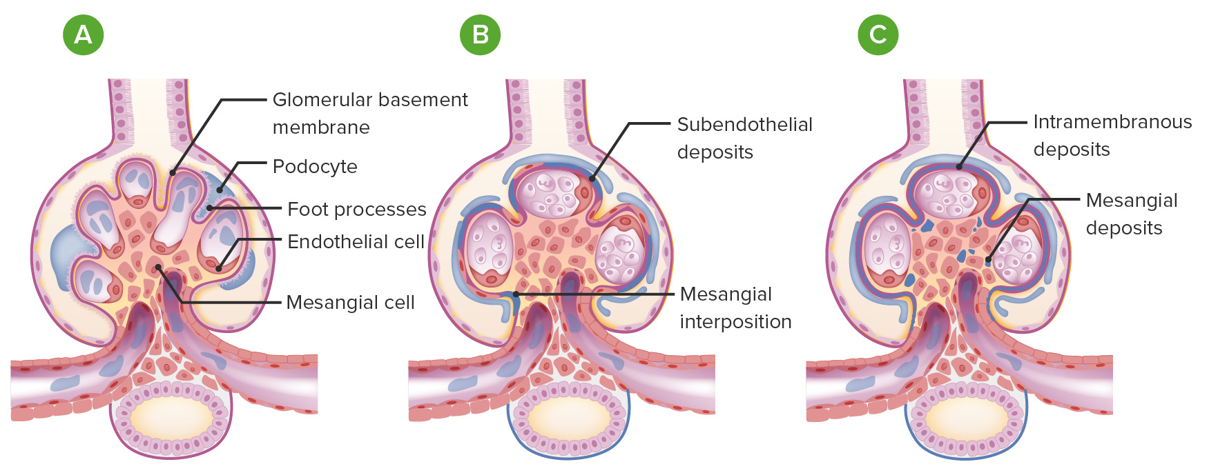
Glomerulonefritis membranoproliferativa versus glomérulos normales:
A: glomérulo normal (con asas capilares abiertas, ≤ 3 núcleos en cada área mesangial, procesos podocitarios intactos y sin depósitos ni proliferación)
B: Glomerulonefritis membranoproliferativa: Los glomérulos se lobulan con proliferación endocapilar y la membrana basal glomerular tiene una apariencia dividida (por depósitos subendoteliales e interposición mesangial).
C: Glomerulonefritis membranoproliferativa con depósitos mesangiales y depósitos intramembranosos (como se observa en la enfermedad por depósitos densos)
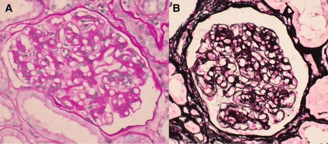
Características al microscopio óptico de la glomerulonefritis membranoproliferativa
A: El mesangio se expande y las paredes de los capilares glomerulares aparecen engrosadas (ácido periódico-Schiff).
B: Las paredes de los capilares glomerulares exhiben contornos dobles engrosados y segmentarios (metenamina de plata).
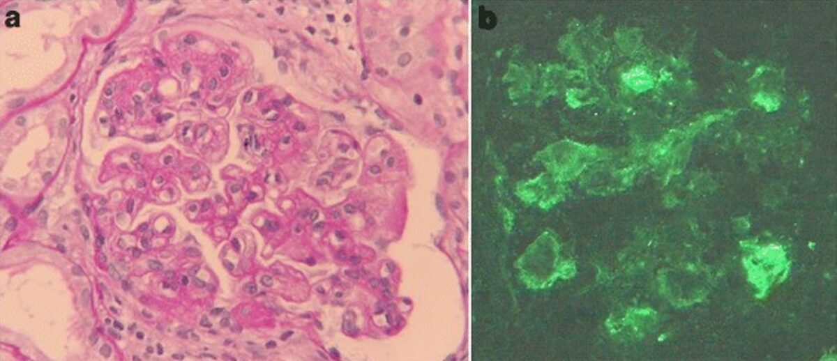
Glomerulonefritis membranoproliferativa asociada a hepatitis C
a: hallazgos de microscopía óptica de biopsia renal: la tinción de ácido periódico-Schiff revela hipercelularidad mesangial, acentuación lobulillar y doble contorno de la membrana basal (aumento original, ×400).
b: La tinción por inmunofluorescencia de IgM es positiva a lo largo del asa capilar (aumento original, ×400).
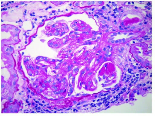
Glomerulonefritis membranoproliferativa:
Proliferación endocapilar con extensos depósitos subendoteliales a lo largo de las paredes de los capilares glomerulares. También se encuentran presentes depósitos mesangiales (microscopía electrónica).
Estudios adicionales
Se indica un estudio adicional cuando la biopsia muestra cambios consistentes con: