Domina Conceptos Médicos

Estudia para la escuela de medicina y tus examenes con Lecturio.

Filariasis Linfática (Elefantiasis)

La filariasis linfática, también conocida como elefantiasis, es una infección crónica transmitida por mosquitos y causada por Wuchereria bancrofti Wuchereria bancrofti A white threadlike worm which causes elephantiasis, lymphangitis, and chyluria by interfering with the lymphatic circulation. The microfilaria are found in the circulating blood and are carried by mosquitoes. Lymphatic Filariasis (Elephantiasis), Brugia malayi Brugia malayi A species of parasitic nematode causing malayan filariasis and having a distribution centering roughly on the malay peninsula. The life cycle of B. malayi is similar to that of Wuchereria bancrofti, except that in most areas the principal mosquito vectors belong to the genus Mansonia. Lymphatic Filariasis (Elephantiasis) y B. timori B. timori Lymphatic Filariasis (Elephantiasis). La mayoría de las causas se deben a W. bancrofti. Los LOS Neisseria mosquitos son los LOS Neisseria vectores y el ser humano es el principal reservorio. Los LOS Neisseria pacientes con infección aguda pueden presentar fiebre, adenolinfangitis, dermatolinfangioadenitis y eosinofilia pulmonar tropical. Los LOS Neisseria pacientes con infección crónica presentan linfedema, que suele afectar las extremidades inferiores (pero puede causar edema Edema Edema is a condition in which excess serous fluid accumulates in the body cavity or interstitial space of connective tissues. Edema is a symptom observed in several medical conditions. It can be categorized into 2 types, namely, peripheral (in the extremities) and internal (in an organ or body cavity). Edema testicular o hidrocele). Los LOS Neisseria efectos a largo plazo también incluyen manifestaciones renales. Los LOS Neisseria frotis de sangre periférica y por gota gruesa son los LOS Neisseria pilares del diagnóstico. La filariasis linfática sin coinfección suele tratarse con dietilcarbamazina. El pronóstico es bueno cuando se realiza un diagnóstico y una intervención temprana. La elefantiasis, o linfedema en EN Erythema nodosum is an immune-mediated panniculitis (inflammation of the subcutaneous fat) caused by a type IV (delayed-type) hypersensitivity reaction. It commonly manifests in young women as tender, erythematous nodules on the shins. Erythema Nodosum estadio avanzado, se asocia a una discapacidad importante y se requiere de diferentes métodos (incluida la cirugía) para reducir el edema Edema Edema is a condition in which excess serous fluid accumulates in the body cavity or interstitial space of connective tissues. Edema is a symptom observed in several medical conditions. It can be categorized into 2 types, namely, peripheral (in the extremities) and internal (in an organ or body cavity). Edema y las complicaciones.

Last updated: Dec 15, 2025

Editorial responsibility: Stanley Oiseth, Lindsay Jones, Evelin Maza

Epidemiología y Etiología

Epidemiología

  • 120 millones de pacientes infectados a nivel mundial a partir del 2019
  • Distribución geográfica:
    • Wuchereria bancrofti Wuchereria bancrofti A white threadlike worm which causes elephantiasis, lymphangitis, and chyluria by interfering with the lymphatic circulation. The microfilaria are found in the circulating blood and are carried by mosquitoes. Lymphatic Filariasis (Elephantiasis):
      • Etiología del 90% de las filariasis linfáticas
      • Es más frecuente en EN Erythema nodosum is an immune-mediated panniculitis (inflammation of the subcutaneous fat) caused by a type IV (delayed-type) hypersensitivity reaction. It commonly manifests in young women as tender, erythematous nodules on the shins. Erythema Nodosum el África Subsahariana, Sudeste Asiático, Subcontinente Indio, Islas del Pacífico y los LOS Neisseria climas tropicales y subtropicales del Caribe y Sudamérica
    • Brugia malayi Brugia malayi A species of parasitic nematode causing malayan filariasis and having a distribution centering roughly on the malay peninsula. The life cycle of B. malayi is similar to that of Wuchereria bancrofti, except that in most areas the principal mosquito vectors belong to the genus Mansonia. Lymphatic Filariasis (Elephantiasis): Sudeste asiático, China, India e Islas del Pacífico
    • B. timori B. timori Lymphatic Filariasis (Elephantiasis): isla de Timor en EN Erythema nodosum is an immune-mediated panniculitis (inflammation of the subcutaneous fat) caused by a type IV (delayed-type) hypersensitivity reaction. It commonly manifests in young women as tender, erythematous nodules on the shins. Erythema Nodosum Indonesia
  • La mayoría de los LOS Neisseria pacientes de las zonas endémicas estuvieron expuestos a sus 30 o 40 años.
  • La filariasis linfática tiene una predilección de 10:1 por los LOS Neisseria hombres sobre las mujeres.

Etiología

La filariasis linfática es causada por nemátodos.

Especies causantes:

Características generales:

  • Nemátodo (helminto redondo)
  • Eucariota
  • En EN Erythema nodosum is an immune-mediated panniculitis (inflammation of the subcutaneous fat) caused by a type IV (delayed-type) hypersensitivity reaction. It commonly manifests in young women as tender, erythematous nodules on the shins. Erythema Nodosum forma de hilo, los LOS Neisseria núcleos no aparecen al AL Amyloidosis final de la cola
  • Anaerobio
  • Se reproducen mediante reproducción sexual

Estadios de vida:

  • Microfilarias:
    • Se encuentran en EN Erythema nodosum is an immune-mediated panniculitis (inflammation of the subcutaneous fat) caused by a type IV (delayed-type) hypersensitivity reaction. It commonly manifests in young women as tender, erythematous nodules on the shins. Erythema Nodosum la sangre periférica del huésped humano
    • Producidos por nemátodos adultos
    • Ingeridos por un mosquito mientras se alimenta de la sangre
  • Larvas:
    • Microfilarias que pierden sus envolturas
    • En EN Erythema nodosum is an immune-mediated panniculitis (inflammation of the subcutaneous fat) caused by a type IV (delayed-type) hypersensitivity reaction. It commonly manifests in young women as tender, erythematous nodules on the shins. Erythema Nodosum el mosquito, las microfilarias maduran hasta convertirse en EN Erythema nodosum is an immune-mediated panniculitis (inflammation of the subcutaneous fat) caused by a type IV (delayed-type) hypersensitivity reaction. It commonly manifests in young women as tender, erythematous nodules on the shins. Erythema Nodosum larvas dentro del mosquito: larvas L1 (1er estadio) a larvas L3 (3er estadio)
    • Las larvas L3 se desplazan hasta la probóscide del mosquito y la infección al AL Amyloidosis humano ocurre mientras el mosquito se alimenta de la sangre.
    • En EN Erythema nodosum is an immune-mediated panniculitis (inflammation of the subcutaneous fat) caused by a type IV (delayed-type) hypersensitivity reaction. It commonly manifests in young women as tender, erythematous nodules on the shins. Erythema Nodosum el ser humano infectado, las larvas migran al AL Amyloidosis torrente sanguíneo y a los LOS Neisseria vasos linfáticos.
    • La madurez se alcanza en EN Erythema nodosum is an immune-mediated panniculitis (inflammation of the subcutaneous fat) caused by a type IV (delayed-type) hypersensitivity reaction. It commonly manifests in young women as tender, erythematous nodules on the shins. Erythema Nodosum 6–9 meses
  • Adultos:
    • Maduración de larvas L3 en EN Erythema nodosum is an immune-mediated panniculitis (inflammation of the subcutaneous fat) caused by a type IV (delayed-type) hypersensitivity reaction. It commonly manifests in young women as tender, erythematous nodules on the shins. Erythema Nodosum los LOS Neisseria vasos linfáticos regionales
    • Helmintos Wuchereria adultos, hembras: 80–100 mm de longitud y 0,24–0,30 mm de diámetro
    • Helmintos Wuchereria adultos, machos: alrededor de 40 mm por 0,1 mm
    • Helmintos Brugia adultos, hembras: 43–55 mm de longitud
    • Helmintos Brugia adultos, machos: 13–23 mm de longitud

Se transmiten a los LOS Neisseria mosquitos:

Wuchereria Bancrofti

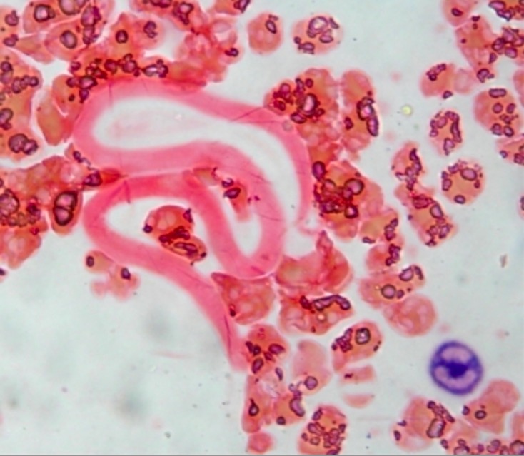
Microfilaria Wuchereria bancrofti

Imagen: “Neutrophil Alkaline Phosphatase stained peripheral smear” por Department of Hematology, All India Institute of Medical Sciences, New Delhi. Licencia: CC BY 2.0

Fisiopatología

Transmisión

  • El principal reservorio de la filariasis es el ser humano y los LOS Neisseria vectores son los LOS Neisseria mosquitos.
  • La transmisión de humano a humano se produce a través de picaduras de mosquitos.

Ciclo de vida

  1. Los LOS Neisseria mosquitos depositan larvas L3 en EN Erythema nodosum is an immune-mediated panniculitis (inflammation of the subcutaneous fat) caused by a type IV (delayed-type) hypersensitivity reaction. It commonly manifests in young women as tender, erythematous nodules on the shins. Erythema Nodosum la piel del ser humano, que excavan a través de la herida de la picadura hasta llegar al AL Amyloidosis torrente sanguíneo.
  2. Las larvas L3 migran a los LOS Neisseria ganglios linfáticos y a los LOS Neisseria vasos linfáticos regionales, donde maduran hasta convertirse en EN Erythema nodosum is an immune-mediated panniculitis (inflammation of the subcutaneous fat) caused by a type IV (delayed-type) hypersensitivity reaction. It commonly manifests in young women as tender, erythematous nodules on the shins. Erythema Nodosum adultos; tienen predilección por los LOS Neisseria ganglios linfáticos inguinales.
  3. Los LOS Neisseria helmintos adultos se reproducen sexualmente y las hembras paren microfilarias que migran activamente a través de la linfa y la sangre.
  4. El mosquito ingiere las microfilarias mientras se alimenta de sangre infectada.
  5. Tras la ingesta, las microfilarias se abren paso a través de la pared del intestino medio del mosquito y llegan a los LOS Neisseria músculos torácicos.
  6. Dentro del mosquito, las microfilarias se convierten en EN Erythema nodosum is an immune-mediated panniculitis (inflammation of the subcutaneous fat) caused by a type IV (delayed-type) hypersensitivity reaction. It commonly manifests in young women as tender, erythematous nodules on the shins. Erythema Nodosum larvas L1.
  7. Las larvas L1 se convierten posteriormente en EN Erythema nodosum is an immune-mediated panniculitis (inflammation of the subcutaneous fat) caused by a type IV (delayed-type) hypersensitivity reaction. It commonly manifests in young women as tender, erythematous nodules on the shins. Erythema Nodosum larvas infecciosas L2 y L3.
  8. Las larvas L3 migran a la probóscide del mosquito, donde pueden infectar a un humano durante su siguiente ingesta de sangre como alimento.
Ciclo de vida de las filarias

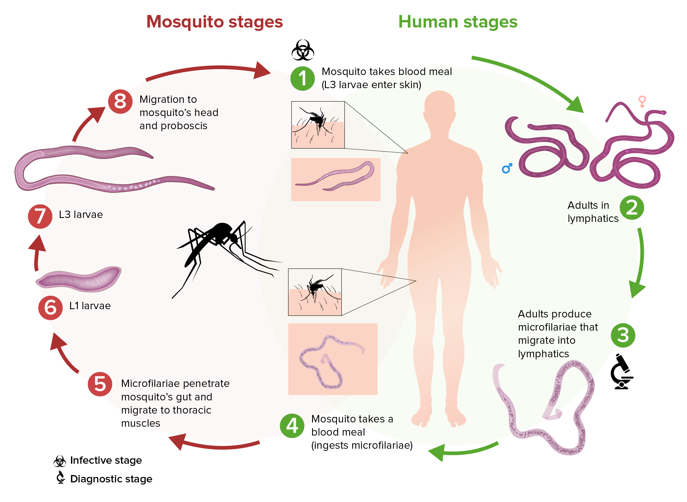
Esquema del ciclo de vida de la filaria Wuchereria bancrofti:

(1) Mientras el mosquito infectado se alimenta de la sangre, introduce las larvas L3 en la piel del huésped humano.
(2) Estas larvas se convierten en adultos que suelen residir en los vasos linfáticos.
(3) Los adultos producen microfilarias que migran activamente a través de la linfa y la sangre.
(4) Un mosquito ingiere las microfilarias mientras se alimenta de la sangre.
(5) Tras la ingesta, las microfilarias se abren paso a través de la pared del intestino medio del mosquito y llegan a los músculos torácicos.
(6) En los músculos torácicos, las microfilarias se convierten en larvas L1.
(7) Las larvas L1 se convierten posteriormente en larvas infecciosas L3.
(8) Las larvas L3 migran a la probóscide del mosquito.
(9) Las larvas L3 pueden infectar a otro ser humano cuando el mosquito realiza una nueva ingesta de sangre como alimento.

Imagen por Lecturio.

Proceso de la enfermedad

  • Tras el apareamiento, la hembra adulta pone miles de microfilarias diariamente:
    • En EN Erythema nodosum is an immune-mediated panniculitis (inflammation of the subcutaneous fat) caused by a type IV (delayed-type) hypersensitivity reaction. It commonly manifests in young women as tender, erythematous nodules on the shins. Erythema Nodosum la mayoría de las situaciones, las microfilarias de W. bancrofti están en EN Erythema nodosum is an immune-mediated panniculitis (inflammation of the subcutaneous fat) caused by a type IV (delayed-type) hypersensitivity reaction. It commonly manifests in young women as tender, erythematous nodules on the shins. Erythema Nodosum la circulación (se encuentran en EN Erythema nodosum is an immune-mediated panniculitis (inflammation of the subcutaneous fat) caused by a type IV (delayed-type) hypersensitivity reaction. It commonly manifests in young women as tender, erythematous nodules on the shins. Erythema Nodosum el torrente sanguíneo) durante las horas nocturnas.
    • En EN Erythema nodosum is an immune-mediated panniculitis (inflammation of the subcutaneous fat) caused by a type IV (delayed-type) hypersensitivity reaction. It commonly manifests in young women as tender, erythematous nodules on the shins. Erythema Nodosum el Pacífico Sur, las microfilarias se encuentran en EN Erythema nodosum is an immune-mediated panniculitis (inflammation of the subcutaneous fat) caused by a type IV (delayed-type) hypersensitivity reaction. It commonly manifests in young women as tender, erythematous nodules on the shins. Erythema Nodosum la circulación todo el día.
  • Infección aguda:
    • Los LOS Neisseria antígenos de la filaria desencadenan un aumento de citoquinas e inmunoglobulinas ( IgE IgE An immunoglobulin associated with mast cells. Overexpression has been associated with allergic hypersensitivity. Immunoglobulins: Types and Functions e IgG4).
    • Los LOS Neisseria helmintos adultos que mudan, mueren o están muertos producen cambios patológicos:
      • Dilatación de los LOS Neisseria linfáticos
      • Hiperplasia del endotelio vascular
      • Linfangitis (infiltración de eosinófilos y linfocitos en EN Erythema nodosum is an immune-mediated panniculitis (inflammation of the subcutaneous fat) caused by a type IV (delayed-type) hypersensitivity reaction. It commonly manifests in young women as tender, erythematous nodules on the shins. Erythema Nodosum los LOS Neisseria vasos linfáticos y formación de trombos)
  • Infecciones crónicas y recurrentes conducen a:
    • Formación de granulomas Granulomas A relatively small nodular inflammatory lesion containing grouped mononuclear phagocytes, caused by infectious and noninfectious agents. Sarcoidosis
    • Fibrosis Fibrosis Any pathological condition where fibrous connective tissue invades any organ, usually as a consequence of inflammation or other injury. Bronchiolitis Obliterans de los LOS Neisseria vasos linfáticos y del tejido conectivo circundante
  • Efectos:
    • Disfunción contráctil y oclusión linfática, causando linfedema y prohibiendo el drenaje linfático
    • Susceptibilidad del huésped humano a las infecciones bacterianas y fúngicas, que contribuyen aún más al AL Amyloidosis daño tisular
  • La filaria adulta puede sobrevivir en EN Erythema nodosum is an immune-mediated panniculitis (inflammation of the subcutaneous fat) caused by a type IV (delayed-type) hypersensitivity reaction. It commonly manifests in young women as tender, erythematous nodules on the shins. Erythema Nodosum el interior de los LOS Neisseria seres humanos hasta por 9 años.
  • Los LOS Neisseria helmintos adultos también llevan Wolbachia Wolbachia A genus of bacteria which comprises a heterogenous group of gram-negative small rods and coccoid forms associated with arthropods. Lymphatic Filariasis (Elephantiasis) (bacterias endosimbióticas) en EN Erythema nodosum is an immune-mediated panniculitis (inflammation of the subcutaneous fat) caused by a type IV (delayed-type) hypersensitivity reaction. It commonly manifests in young women as tender, erythematous nodules on the shins. Erythema Nodosum su intestino, que parecen beneficiar a los LOS Neisseria helmintos.

Presentación Clínica

Los LOS Neisseria síntomas pueden tardar entre 9 meses y 1 año en EN Erythema nodosum is an immune-mediated panniculitis (inflammation of the subcutaneous fat) caused by a type IV (delayed-type) hypersensitivity reaction. It commonly manifests in young women as tender, erythematous nodules on the shins. Erythema Nodosum manifestarse tras la infección inicial. Los LOS Neisseria niños o individuos de las zonas endémicas suelen permanecer asintomáticos (infección subclínica), mientras que otros muestran signos y síntomas agudos y/o crónicos.

Manifestaciones agudas

  • Fiebre filárica:
    • Fiebre típicamente de bajo grado, autolimitada
    • Puede tener mialgias
    • +/– Linfadenopatía
  • Adenolinfangitis aguda:
    • Fiebre y linfadenopatía
    • Linfangitis (inflamación que se extiende distalmente al AL Amyloidosis ganglio linfático), con vasos linfáticos en EN Erythema nodosum is an immune-mediated panniculitis (inflammation of the subcutaneous fat) caused by a type IV (delayed-type) hypersensitivity reaction. It commonly manifests in young women as tender, erythematous nodules on the shins. Erythema Nodosum la(s) pierna(s) que presenta(n) aumento de temperatura, aumento de volumen, eritema y sensibilidad
    • Se observa comúnmente en EN Erythema nodosum is an immune-mediated panniculitis (inflammation of the subcutaneous fat) caused by a type IV (delayed-type) hypersensitivity reaction. It commonly manifests in young women as tender, erythematous nodules on the shins. Erythema Nodosum los LOS Neisseria ganglios linfáticos inguinales, pero también puede afectar a los LOS Neisseria genitales (epididimitis en EN Erythema nodosum is an immune-mediated panniculitis (inflammation of the subcutaneous fat) caused by a type IV (delayed-type) hypersensitivity reaction. It commonly manifests in young women as tender, erythematous nodules on the shins. Erythema Nodosum los LOS Neisseria hombres)
    • Dura unos días y luego se resuelve, pero se repite periódicamente
  • Dermatolinfangioadenitis aguda:
    • Fiebre, escalofríos
    • Placas edematosas (comúnmente interdigitales) que se cree que son lesiones de entrada y están causadas por bacterias
    • Linfedema (empeora con episodios recurrentes)
    • Se observa en EN Erythema nodosum is an immune-mediated panniculitis (inflammation of the subcutaneous fat) caused by a type IV (delayed-type) hypersensitivity reaction. It commonly manifests in young women as tender, erythematous nodules on the shins. Erythema Nodosum zonas endémicas
  • Eosinofilia pulmonar tropical:
    • Reacción inmunitaria a las microfilarias atrapadas en EN Erythema nodosum is an immune-mediated panniculitis (inflammation of the subcutaneous fat) caused by a type IV (delayed-type) hypersensitivity reaction. It commonly manifests in young women as tender, erythematous nodules on the shins. Erythema Nodosum los LOS Neisseria pulmones
    • Enfermedad pulmonar restrictiva, que puede progresar a fibrosis Fibrosis Any pathological condition where fibrous connective tissue invades any organ, usually as a consequence of inflammation or other injury. Bronchiolitis Obliterans pulmonar intersticial
    • Sibilancias
    • Disnea
    • Esputo sanguinolento
    • Eosinofilia

Manifestaciones crónicas

  • Linfedema:
    • Edema Edema Edema is a condition in which excess serous fluid accumulates in the body cavity or interstitial space of connective tissues. Edema is a symptom observed in several medical conditions. It can be categorized into 2 types, namely, peripheral (in the extremities) and internal (in an organ or body cavity). Edema crónico de la extremidad por inflamación crónica de los LOS Neisseria vasos linfáticos
    • Se clasifica en EN Erythema nodosum is an immune-mediated panniculitis (inflammation of the subcutaneous fat) caused by a type IV (delayed-type) hypersensitivity reaction. It commonly manifests in young women as tender, erythematous nodules on the shins. Erythema Nodosum función de la extensión y la progresión de los LOS Neisseria síntomas:
      • Grado 0: subclínica
      • Grado I: edema Edema Edema is a condition in which excess serous fluid accumulates in the body cavity or interstitial space of connective tissues. Edema is a symptom observed in several medical conditions. It can be categorized into 2 types, namely, peripheral (in the extremities) and internal (in an organ or body cavity). Edema con fóvea de las extremidades, reversible (↓ con la elevación de la extremidad)
      • Grado II: edema Edema Edema is a condition in which excess serous fluid accumulates in the body cavity or interstitial space of connective tissues. Edema is a symptom observed in several medical conditions. It can be categorized into 2 types, namely, peripheral (in the extremities) and internal (in an organ or body cavity). Edema con o sin fóvea de las extremidades, no reversible
      • Grado III: edema Edema Edema is a condition in which excess serous fluid accumulates in the body cavity or interstitial space of connective tissues. Edema is a symptom observed in several medical conditions. It can be categorized into 2 types, namely, peripheral (in the extremities) and internal (in an organ or body cavity). Edema sin fóvea con engrosamiento de la piel e hipertrofia, irreversible (elefantiasis)
  • Hidrocele:
    • Enfermedad linfática que afecta al AL Amyloidosis escroto
    • Unilateral o bilateral
  • Afectación renal:
    • Quiluria (orina lechosa):
      • Normalmente, los LOS Neisseria vasos linfáticos no tienen comunicación con las vías urinarias.
      • En EN Erythema nodosum is an immune-mediated panniculitis (inflammation of the subcutaneous fat) caused by a type IV (delayed-type) hypersensitivity reaction. It commonly manifests in young women as tender, erythematous nodules on the shins. Erythema Nodosum la filariasis, el líquido con la linfa intestinal y los LOS Neisseria quilomicrones pasan a la orina de forma intermitente.
      • Conduce a la pérdida de proteínas
    • También se observa hematuria Hematuria Presence of blood in the urine. Renal Cell Carcinoma y proteinuria Proteinuria The presence of proteins in the urine, an indicator of kidney diseases. Nephrotic Syndrome in Children (pero mediante un mecanismo poco conocido)
Elefantiasis

Filariasis:
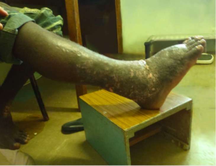
Un paciente con linfedema crónico de la extremidad inferior derecha que se manifiesta como elefantiasis

Imagen: “Elephantiasis” por Humpress Harrington et al. Licencia: CC BY 2.0

Diagnóstico

Pruebas de laboratorio

  • Sangre:
    • Frotis de sangre periférica y por gota gruesa
    • Venopunción o punción en EN Erythema nodosum is an immune-mediated panniculitis (inflammation of the subcutaneous fat) caused by a type IV (delayed-type) hypersensitivity reaction. It commonly manifests in young women as tender, erythematous nodules on the shins. Erythema Nodosum el dedo/talón
    • Se debe realizar entre las 10 p.m. y las 2 a.m. (la microfilaria tiene periodicidad nocturna)
    • Tinciones de Giemsa o Wright
  • Antígenos filáricos circulantes (W. bancrofti):
    • Detecta los LOS Neisseria antígenos de los LOS Neisseria helmintos adultos de la filaria
    • Puede ser positivo incluso en EN Erythema nodosum is an immune-mediated panniculitis (inflammation of the subcutaneous fat) caused by a type IV (delayed-type) hypersensitivity reaction. It commonly manifests in young women as tender, erythematous nodules on the shins. Erythema Nodosum los LOS Neisseria que no tienen microfilarias
  • Pruebas para anticuerpos antifiláricos:
    • Niveles elevados de IgG4 antifilárica en EN Erythema nodosum is an immune-mediated panniculitis (inflammation of the subcutaneous fat) caused by a type IV (delayed-type) hypersensitivity reaction. It commonly manifests in young women as tender, erythematous nodules on the shins. Erythema Nodosum la sangre
    • Se utiliza sobre todo para los LOS Neisseria viajeros (que no son de zonas endémicas)
  • PCR PCR Polymerase chain reaction (PCR) is a technique that amplifies DNA fragments exponentially for analysis. The process is highly specific, allowing for the targeting of specific genomic sequences, even with minuscule sample amounts. The PCR cycles multiple times through 3 phases: denaturation of the template DNA, annealing of a specific primer to the individual DNA strands, and synthesis/elongation of new DNA molecules. Polymerase Chain Reaction (PCR) para la detección de antígenos
    • Utilizado en EN Erythema nodosum is an immune-mediated panniculitis (inflammation of the subcutaneous fat) caused by a type IV (delayed-type) hypersensitivity reaction. It commonly manifests in young women as tender, erythematous nodules on the shins. Erythema Nodosum la investigación científica
    • Ensayo no aprobado para uso comercial
  • Biopsia: tejido de lesiones cutáneas
  • Pruebas de laboratorio adicionales:
    • En EN Erythema nodosum is an immune-mediated panniculitis (inflammation of the subcutaneous fat) caused by a type IV (delayed-type) hypersensitivity reaction. It commonly manifests in young women as tender, erythematous nodules on the shins. Erythema Nodosum zonas endémicas de oncocercosis (Onchocerca volvulus Volvulus A volvulus is the twisting or axial rotation of a portion of the bowel around its mesentery. The most common site of volvulus in adults is the colon; most frequently the sigmoid volvulus. Patients typically present with symptoms of bowel obstruction such as abdominal pain, distension, vomiting, and constipation/obstipation. Volvulus) o loasis (Loa loa), hay que determinar la coinfección, ya que el tratamiento será diferente.
    • Hemograma que muestra eosinofilia e IgE IgE An immunoglobulin associated with mast cells. Overexpression has been associated with allergic hypersensitivity. Immunoglobulins: Types and Functions elevada observada en EN Erythema nodosum is an immune-mediated panniculitis (inflammation of the subcutaneous fat) caused by a type IV (delayed-type) hypersensitivity reaction. It commonly manifests in young women as tender, erythematous nodules on the shins. Erythema Nodosum la eosinofilia pulmonar tropical

Imagenología

  • Ultrasonido:
    • Revela helmintos adultos moviéndose en EN Erythema nodosum is an immune-mediated panniculitis (inflammation of the subcutaneous fat) caused by a type IV (delayed-type) hypersensitivity reaction. It commonly manifests in young women as tender, erythematous nodules on the shins. Erythema Nodosum los LOS Neisseria vasos linfáticos
    • El “signo de la danza de la filaria” —un patrón de movimientos irregulares de los LOS Neisseria helmintos— pueden detectarse en EN Erythema nodosum is an immune-mediated panniculitis (inflammation of the subcutaneous fat) caused by a type IV (delayed-type) hypersensitivity reaction. It commonly manifests in young women as tender, erythematous nodules on the shins. Erythema Nodosum el Doppler Doppler Ultrasonography applying the doppler effect, with frequency-shifted ultrasound reflections produced by moving targets (usually red blood cells) in the bloodstream along the ultrasound axis in direct proportion to the velocity of movement of the targets, to determine both direction and velocity of blood flow. Ultrasound (Sonography).
  • Linfogammagrafía:
    • Evalúa el drenaje linfático
    • Puede detectar el linfedema preclínico
  • Radiografía de tórax:
    • Para evaluar por eosinofilia pulmonar tropical
    • Lesiones intersticiales difusas y marcas broncoalveolares pronunciadas

Tratamiento

Tratamiento

Filariasis sin coinfección:

  • Dietilcarbamazina:
    • Tratamiento de 1era línea
    • Dosis única
    • 2–3 semanas de tratamiento para la eosinofilia pulmonar tropical debido a W. bancrofti
  • Doxiciclina:
    • Con dietilcarbamazina o como una terapia alternativa
    • Para adultas no embarazadas y niños > 8 años de edad
    • Eficaz contra Wolbachia Wolbachia A genus of bacteria which comprises a heterogenous group of gram-negative small rods and coccoid forms associated with arthropods. Lymphatic Filariasis (Elephantiasis)

Filariasis con loasis:

  • La ingesta de dietilcarbamazina aumenta el riesgo de encefalopatía potencialmente mortal en EN Erythema nodosum is an immune-mediated panniculitis (inflammation of the subcutaneous fat) caused by a type IV (delayed-type) hypersensitivity reaction. It commonly manifests in young women as tender, erythematous nodules on the shins. Erythema Nodosum la coinfección a medida que aumenta la carga microfilárica.
  • El tratamiento varía en EN Erythema nodosum is an immune-mediated panniculitis (inflammation of the subcutaneous fat) caused by a type IV (delayed-type) hypersensitivity reaction. It commonly manifests in young women as tender, erythematous nodules on the shins. Erythema Nodosum función de la carga:
    • < 2500 microfilarias L. loa/mL: dietilcarbamazina
    • > 2500–8000 microfilarias/mL: ivermectina para disminuir la carga microfilárica, posteriormente dietilcarbamazina
    • > 8000 microfilarias/mL: doxiciclina durante 4–6 semanas

Filariasis con oncocercosis:

  • La ingesta de dietilcarbamazina empeora la enfermedad ocular (↑ riesgo de ceguera).
  • Opciones de tratamiento:
    • Tratar la oncocercosis 1ero con ivermectina y posteriormente con dietilcarbamazina.
    • Para los LOS Neisseria que tienen enfermedades oculares, se debe administrar doxiciclina seguida de ivermectina.

Tratamiento quirúrgico:

  • Citorreducción de la piel y anastomosis linfovenosa para mejorar el drenaje
  • Escisión quirúrgica del hidrocele

Tratamiento a largo plazo para reducir la progresión del linfedema:

  • Higiene de la piel
  • Portar un calzado cómodo
  • Vendas compresivas
  • Elevación de las extremidades
  • Terapia de frío y calor Calor Inflammation
  • Antibióticos y antifúngicos para prevenir brotes

Prevención

  • Evitar las picaduras de mosquitos:
    • Dormir en EN Erythema nodosum is an immune-mediated panniculitis (inflammation of the subcutaneous fat) caused by a type IV (delayed-type) hypersensitivity reaction. It commonly manifests in young women as tender, erythematous nodules on the shins. Erythema Nodosum una habitación con aire acondicionado o bajo mosquiteras.
    • Utilizar repelente de mosquitos.
    • Usar mangas largas y pantalones.
  • Control de los LOS Neisseria mosquitos con insecticidas
  • Programa Mundial para Eliminar la Filariasis Linfática:
    • Introducido para eliminar la propagación y reducir la morbilidad
    • Consiste en EN Erythema nodosum is an immune-mediated panniculitis (inflammation of the subcutaneous fat) caused by a type IV (delayed-type) hypersensitivity reaction. It commonly manifests in young women as tender, erythematous nodules on the shins. Erythema Nodosum una administración masiva anual de medicamentos (durante al AL Amyloidosis menos 5 años) aplicada mediante varios regímenes con:
      • Dietilcarbamazina
      • Ivermectina
      • Albendazol

Complicaciones y pronóstico

La filariasis oculta es una infección filárica que se extiende a los LOS Neisseria tejidos, sin ninguna evidencia en EN Erythema nodosum is an immune-mediated panniculitis (inflammation of the subcutaneous fat) caused by a type IV (delayed-type) hypersensitivity reaction. It commonly manifests in young women as tender, erythematous nodules on the shins. Erythema Nodosum la sangre. Esto conduce a complicaciones crónicas, como:

  • Eosinofilia pulmonar tropical
  • Artritis filática
  • Glomerulonefritis por inmunocomplejos asociada a filarias
  • Abscesos mamarios filáricos

Pronóstico:

  • Cuando las enfermedades se diagnostican y tratan a tiempo, el pronóstico es bueno.
  • Como los LOS Neisseria síntomas pueden presentarse más tarde en EN Erythema nodosum is an immune-mediated panniculitis (inflammation of the subcutaneous fat) caused by a type IV (delayed-type) hypersensitivity reaction. It commonly manifests in young women as tender, erythematous nodules on the shins. Erythema Nodosum la adultez, el linfedema y, posteriormente, la elefantiasis se asocian con discapacidad y morbilidad.

Diagnóstico Diferencial

  • Oncocercosis: infección causada por el nemátodo filárico Onchocerca volvulus Volvulus A volvulus is the twisting or axial rotation of a portion of the bowel around its mesentery. The most common site of volvulus in adults is the colon; most frequently the sigmoid volvulus. Patients typically present with symptoms of bowel obstruction such as abdominal pain, distension, vomiting, and constipation/obstipation. Volvulus, también llamada ceguera de los LOS Neisseria ríos: El vector de O. volvulus Volvulus A volvulus is the twisting or axial rotation of a portion of the bowel around its mesentery. The most common site of volvulus in adults is the colon; most frequently the sigmoid volvulus. Patients typically present with symptoms of bowel obstruction such as abdominal pain, distension, vomiting, and constipation/obstipation. Volvulus es la mosca negra del género Simulium. Las manifestaciones afectan al AL Amyloidosis ojo (queratitis, uveítis, coriorretinitis, atrofia óptica), la piel (nódulos subcutáneos, dermatitis Dermatitis Any inflammation of the skin. Atopic Dermatitis (Eczema)) y al AL Amyloidosis sistema nervioso (convulsiones). El diagnóstico se realiza mediante una biopsia de piel. El tratamiento es con ivermectina.
  • Loasis: infección causada por el nemátodo filárico Loa loa. La loasis también se conoce como el gusano del ojo africano. La transmisión se produce a través de la picadura de la mosca Chrysops. Los LOS Neisseria individuos infectados pueden ser asintomáticos, pero la enfermedad puede manifestarse con edema Edema Edema is a condition in which excess serous fluid accumulates in the body cavity or interstitial space of connective tissues. Edema is a symptom observed in several medical conditions. It can be categorized into 2 types, namely, peripheral (in the extremities) and internal (in an organ or body cavity). Edema subcutáneo ( edema Edema Edema is a condition in which excess serous fluid accumulates in the body cavity or interstitial space of connective tissues. Edema is a symptom observed in several medical conditions. It can be categorized into 2 types, namely, peripheral (in the extremities) and internal (in an organ or body cavity). Edema de Calabar) y la migración subconjuntival del helminto adulto. El diagnóstico se realiza a través de un frotis de sangre periférica (se observan microfilarias) o la identificación de un helminto migratorio en EN Erythema nodosum is an immune-mediated panniculitis (inflammation of the subcutaneous fat) caused by a type IV (delayed-type) hypersensitivity reaction. It commonly manifests in young women as tender, erythematous nodules on the shins. Erythema Nodosum el ojo o en EN Erythema nodosum is an immune-mediated panniculitis (inflammation of the subcutaneous fat) caused by a type IV (delayed-type) hypersensitivity reaction. It commonly manifests in young women as tender, erythematous nodules on the shins. Erythema Nodosum el tejido subcutáneo. El tratamiento es con dietilcarbamazina si la carga microfilárica es baja. Con niveles elevados de microfilarias, es necesario reducir la carga antes del tratamiento con dietilcarbamazina.
  • Otras condiciones testiculares: otras infecciones testiculares pueden ser el varicocele o la dilatación del plexo venoso pampiniforme, que está conectado a la vena espermática interna o gonadal. Los varicoceles se presentan con una apariencia de “bolsa de gusanos” indolora al AL Amyloidosis examen físico. El hidrocele o el exceso de líquido en EN Erythema nodosum is an immune-mediated panniculitis (inflammation of the subcutaneous fat) caused by a type IV (delayed-type) hypersensitivity reaction. It commonly manifests in young women as tender, erythematous nodules on the shins. Erythema Nodosum la túnica vaginal que da lugar a un escroto aumentado de volumen, puede ser congénito o puede deberse a un traumatismo. El espermatocele, o quiste epididimario, suele surgir de la cabeza del epidídimo y suele presentarse como una masa escrotal indolora e incidental en EN Erythema nodosum is an immune-mediated panniculitis (inflammation of the subcutaneous fat) caused by a type IV (delayed-type) hypersensitivity reaction. It commonly manifests in young women as tender, erythematous nodules on the shins. Erythema Nodosum el examen.
  • Otra etiología del linfedema: acumulación anormal de líquido intersticial por obstrucción linfática. El linfedema primario se debe a una alteración en EN Erythema nodosum is an immune-mediated panniculitis (inflammation of the subcutaneous fat) caused by a type IV (delayed-type) hypersensitivity reaction. It commonly manifests in young women as tender, erythematous nodules on the shins. Erythema Nodosum la función linfática o a un desarrollo anómalo debido a un trastorno genético. El linfedema secundario tiene una enfermedad subyacente o un tratamiento que ha HA Hemolytic anemia (HA) is the term given to a large group of anemias that are caused by the premature destruction/hemolysis of circulating red blood cells (RBCs). Hemolysis can occur within (intravascular hemolysis) or outside the blood vessels (extravascular hemolysis). Hemolytic Anemia provocado el edema Edema Edema is a condition in which excess serous fluid accumulates in the body cavity or interstitial space of connective tissues. Edema is a symptom observed in several medical conditions. It can be categorized into 2 types, namely, peripheral (in the extremities) and internal (in an organ or body cavity). Edema. Entre las causas se encuentran los LOS Neisseria tumores malignos (y sus tratamientos), las infecciones, los LOS Neisseria traumatismos o las enfermedades inflamatorias. El enfoque diagnóstico implica los LOS Neisseria antecedentes clínicos y un examen exhaustivo, los LOS Neisseria estudios dependen de la sospecha clínica.

Referencias

  1. Bjerum, C.M., Ouattara, A.F., Aboulaye, M., Kouadio, O., Marius, V.K., Andersen, B.J., Weil, G.J., Koudou, B.G., King, C.L. (2020). Efficacy and safety of a single dose of ivermectin, diethylcarbamazine, and albendazole for treatment of lymphatic filariasis in Côte d’Ivoire: an open-label randomized controlled trial. Clin Infect Dis 71:e68–e75.
  2. Centers for Disease Control and Prevention. (2024). About lymphatic filariasis. Filarial Worms. Retrieved October 17, 2025, from https://www.cdc.gov/filarial-worms/about/lymphatic-filariasis.html
  3. Chandy, A., Thakur, A.S., Singh, M.P., Manigauha, A. (2011). A review of neglected tropical diseases: filariasis. Asian Pac J Trop Med 4:581–586. 
  4. Kalyanasundaram, R., Khatri, V., Chauhan, N. (2020). Advances in vaccine development for human lymphatic filariasis. Trends Parasitol 36:195–205. 
  5. Klion, A.D. (2025). Lymphatic filariasis: epidemiology, clinical manifestations, and diagnosis. UpToDate. Retrieved October 17, 2025, from https://www.uptodate.com/contents/lymphatic-filariasis-epidemiology-clinical-manifestations-and-diagnosis
  6. Klion, A.D. (2025). Lymphatic filariasis: treatment and prevention. UpToDate. Retrieved October 17, 2025, from https://www.uptodate.com/contents/lymphatic-filariasis-treatment-and-prevention
  7. Lich, B. (2023). Filariasis. Medscape. Retrieved October 17, 2025, from https://emedicine.medscape.com/article/217776-overview
  8. Mehrara, B., Chang, E. I., & Ashinoff, R. L. (2025). Management of peripheral lymphedema. UpToDate. Retrieved October 17, 2025, from https://www.uptodate.com/contents/management-of-peripheral-lymphedema
  9. Newman, T.E., Juergens, A.L. (2025). Filariasis. StatPearls. Retrieved October 17, 2025, from https://www.ncbi.nlm.nih.gov/books/NBK556012/
  10. Rebollo, M.P., Bockarie, M.J. (2017). Can lymphatic filariasis be eliminated by 2020? Trends Parasitol 33(2):83–92.
  11. Ryan K.J. (Ed.), (2017). Tissue nematodes. Chapter 55 of Sherris Medical Microbiology, 7th ed. McGraw-Hill. https://accessmedicine.mhmedical.com/content.aspx?bookid=2268&sectionid=176089905
  12. Shenoy, R.K. (2008). Clinical and pathological aspects of filarial lymphedema and its management. Korean J Parasitol 46(3):119–125.
  13. Shukla, S.K., Kusum, A., Sharma, S., Kandari, D. (2019). Filariasis presenting as a solitary testicular mass. Trop Parasitol 9(2):124–126.

¡Crea tu cuenta gratis o inicia una sesión para seguir leyendo!

Regístrate ahora y obtén acceso gratuito a Lecturio con páginas de concepto, videos médicos y cuestionarios para tu educación médica.

User Reviews

Que tengas una sesión de estudio alegre y navideña 🎁 Ahorra 50% en todos los planes >>

Details