El hueso es un tipo compacto de tejido conectivo endurecido compuesto por células óseas, membranas, una matriz mineralizada extracelular y médula ósea central. LosLOSNeisseria dos tipos principales de hueso son el compacto y el esponjoso. Como la matriz está mineralizada (enENErythema nodosum is an immune-mediated panniculitis (inflammation of the subcutaneous fat) caused by a type IV (delayed-type) hypersensitivity reaction. It commonly manifests in young women as tender, erythematous nodules on the shins.Erythema Nodosum lugar de acuosa), losLOSNeisseria nutrientes y losLOSNeisseria residuos no pueden difundirse a través de la matriz. El hueso haHAHemolytic anemia (HA) is the term given to a large group of anemias that are caused by the premature destruction/hemolysis of circulating red blood cells (RBCs). Hemolysis can occur within (intravascular hemolysis) or outside the blood vessels (extravascular hemolysis).Hemolytic Anemia desarrollado una estructura única para permitir que se produzcan estas funciones. La estructura del hueso permite que este sea duro, pero no demasiado frágil, y le da la fuerza necesaria para resistir las fuerzas de compresión y flexión. Por ello, el hueso es ideal para las funciones de soporte, protección de losLOSNeisseria órganos vitales y movimiento. Además, el hueso produce células sanguíneas enENErythema nodosum is an immune-mediated panniculitis (inflammation of the subcutaneous fat) caused by a type IV (delayed-type) hypersensitivity reaction. It commonly manifests in young women as tender, erythematous nodules on the shins.Erythema Nodosum la médula y es el principal lugar de almacenamiento de calcio del organismo.
Protección (e.g., el cráneo protege el cerebro, las costillas protegen el corazón y losLOSNeisseria pulmones)
Soporte
Movimiento
Formación de células sanguíneas
Almacenamiento:
Minerales
Fosfato
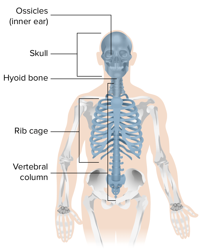
Clasificación de losLOSNeisseria huesos según su localización
Huesos axiales:
Cráneo
Columna vertebral y sacro
Caja torácica: costillas y esternón
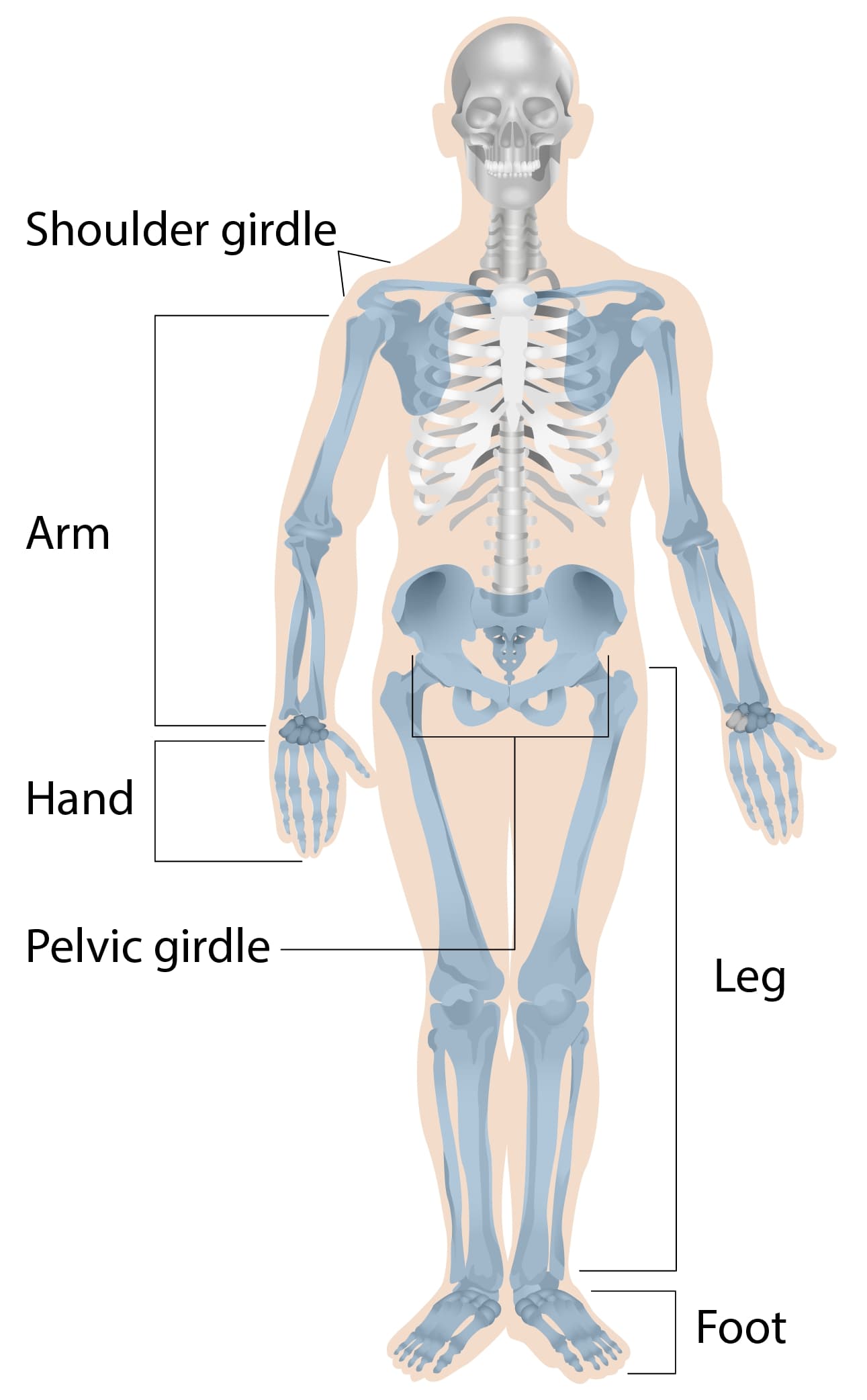
Huesos apendiculares:
Clavícula
Escápula
Brazos y manos
Huesos de la pelvisPelvisThe pelvis consists of the bony pelvic girdle, the muscular and ligamentous pelvic floor, and the pelvic cavity, which contains viscera, vessels, and multiple nerves and muscles. The pelvic girdle, composed of 2 “hip” bones and the sacrum, is a ring-like bony structure of the axial skeleton that links the vertebral column with the lower extremities.Pelvis: Anatomy
Piernas y pies
Ilustración que representa los huesos que forman el esqueleto axial
Imagen por Lecturio.
Ilustración que representa los huesos que forman el esqueleto apendicular
Imagen por Lecturio.
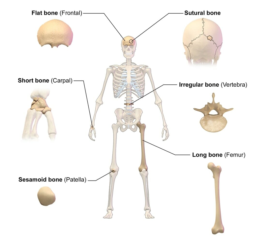
Clasificación de losLOSNeisseria huesos por su forma
Huesos largos: longitud mayor que la anchura:
Extremidad superior: húmero, radio, cúbito, metacarpianos y falanges
Extremidad inferior: fémur, tibiaTibiaThe second longest bone of the skeleton. It is located on the medial side of the lower leg, articulating with the fibula laterally, the talus distally, and the femur proximally.Knee Joint: Anatomy, peroné, metatarsos y falanges
Huesos cortos: la longitud es aproximadamente igual a la anchura:
Huesos del carpo (muñeca)
Huesos tarsianos (tobillo)
Huesos planos: encierran y protegen losLOSNeisseria órganos blandos:
Huesos irregulares: huesos que no encajan enENErythema nodosum is an immune-mediated panniculitis (inflammation of the subcutaneous fat) caused by a type IV (delayed-type) hypersensitivity reaction. It commonly manifests in young women as tender, erythematous nodules on the shins.Erythema Nodosum otras categorías:
Vértebras
Algunos huesos del cráneo (e.g., el esfenoides, losLOSNeisseria huesos faciales)
Ilustración que representa la clasificación de los huesos por su forma
Imagen: “Classification of Bones By Shape” por BruceBlaus. Licencia: CC BY 3.0, recortada por Lecturio.
Presente enENErythema nodosum is an immune-mediated panniculitis (inflammation of the subcutaneous fat) caused by a type IV (delayed-type) hypersensitivity reaction. It commonly manifests in young women as tender, erythematous nodules on the shins.Erythema Nodosum todos losLOSNeisseria huesos del cuerpo
EnENErythema nodosum is an immune-mediated panniculitis (inflammation of the subcutaneous fat) caused by a type IV (delayed-type) hypersensitivity reaction. It commonly manifests in young women as tender, erythematous nodules on the shins.Erythema Nodosum el hueso largo: forma un cilindro, encierra una cavidad medular
Función: resistencia a las fuerzas de compresión
Hueso esponjoso:
Tejido óseo de capa interna poco organizado
Consiste enENErythema nodosum is an immune-mediated panniculitis (inflammation of the subcutaneous fat) caused by a type IV (delayed-type) hypersensitivity reaction. It commonly manifests in young women as tender, erythematous nodules on the shins.Erythema Nodosum un entramado de pequeñas y finas piezas de tejido óseo llamadas trabéculas o espículas óseas:
Transfiere la fuerza sobre el hueso alALAmyloidosis hueso compacto exterior
Se reforman constantemente para satisfacer las necesidades del cuerpo (e.g., el ejercicio aumenta las trabéculas; la ingravidez prolongada enENErythema nodosum is an immune-mediated panniculitis (inflammation of the subcutaneous fat) caused by a type IV (delayed-type) hypersensitivity reaction. It commonly manifests in young women as tender, erythematous nodules on the shins.Erythema Nodosum el espacio reduce las trabéculas)
EnENErythema nodosum is an immune-mediated panniculitis (inflammation of the subcutaneous fat) caused by a type IV (delayed-type) hypersensitivity reaction. It commonly manifests in young women as tender, erythematous nodules on the shins.Erythema NodosumlosLOSNeisseria extremos de losLOSNeisseria huesos largos
EnENErythema nodosum is an immune-mediated panniculitis (inflammation of the subcutaneous fat) caused by a type IV (delayed-type) hypersensitivity reaction. It commonly manifests in young women as tender, erythematous nodules on the shins.Erythema Nodosum medio de huesos cortos, planos e irregulares
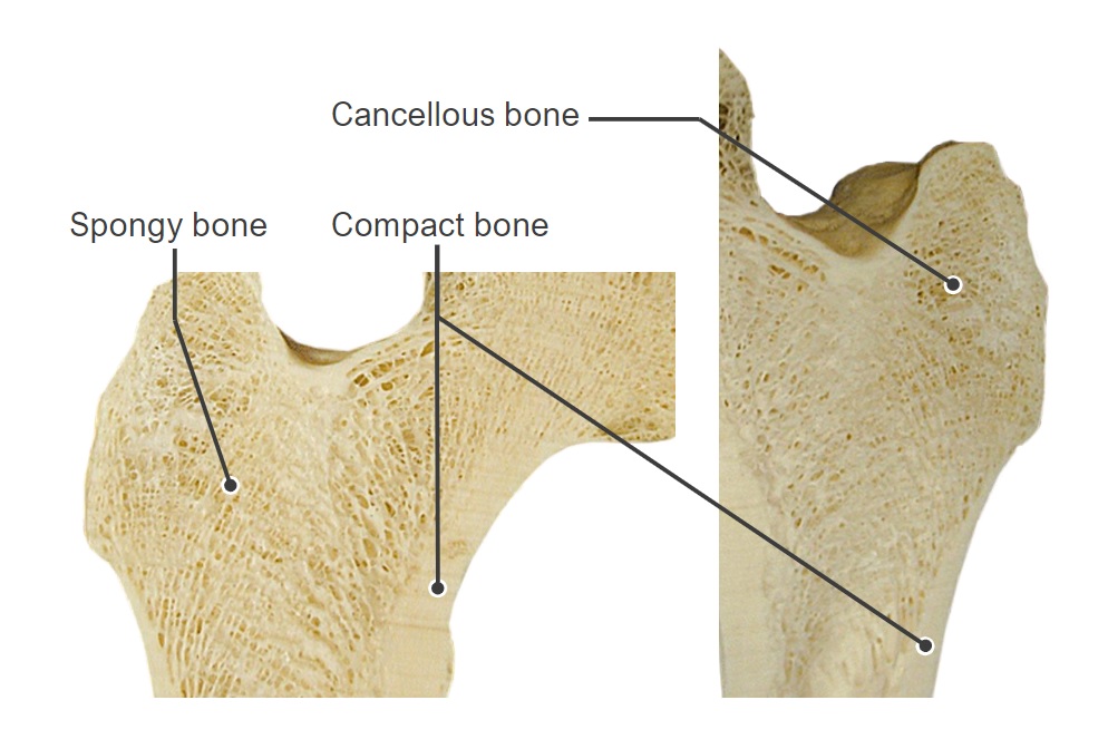
Imagen de la estructura interna de una cabeza de fémur: Obsérvese el hueso compacto a lo largo del exterior y el hueso esponjoso/cancelloso en el centro.
Imagen por Lecturio.
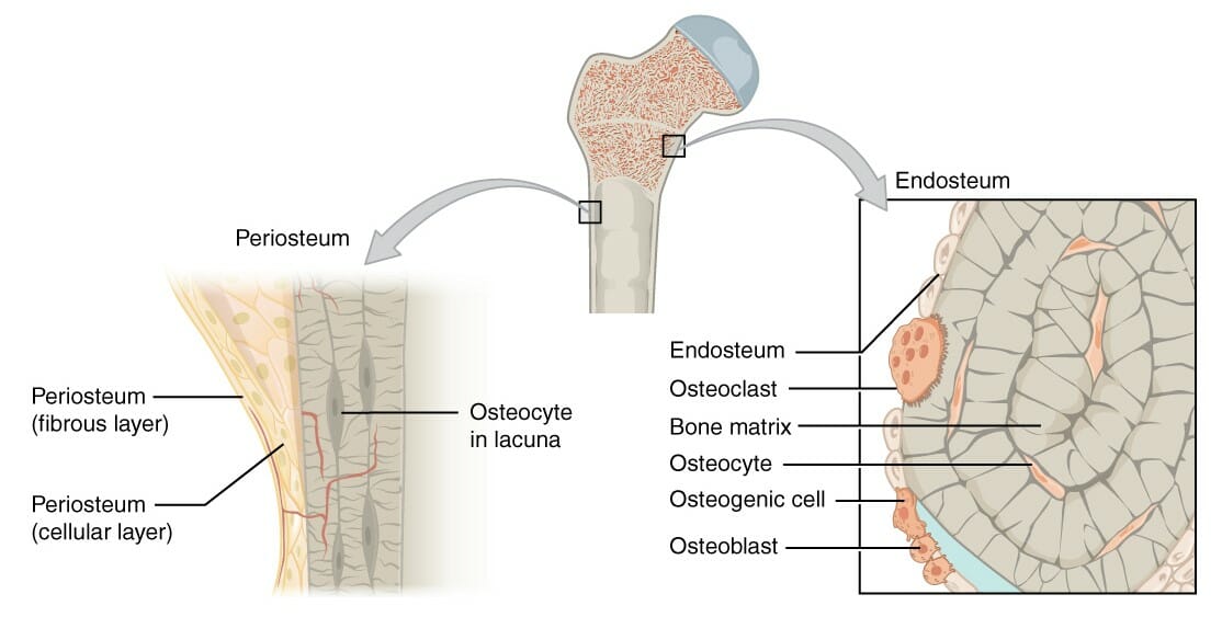
Membranas óseas
Dos membranas primarias rodean el tejido óseo: el periostio (externamente) y el endostio (internamente).
El periostio:
Capa externa que rodea alALAmyloidosis hueso enENErythema nodosum is an immune-mediated panniculitis (inflammation of the subcutaneous fat) caused by a type IV (delayed-type) hypersensitivity reaction. It commonly manifests in young women as tender, erythematous nodules on the shins.Erythema Nodosum la superficie externa (excepto enENErythema nodosum is an immune-mediated panniculitis (inflammation of the subcutaneous fat) caused by a type IV (delayed-type) hypersensitivity reaction. It commonly manifests in young women as tender, erythematous nodules on the shins.Erythema Nodosum las articulaciones, que están cubiertas de cartílago articular)
Vascularizado e inervado
Consta de dos capas:
Capa fibrosa
Capa osteogénica
Capa fibrosa del periostio:
Capa exterior de colágeno resistente
Fibras de Sharpey: fibras de colágeno de la capa fibrosa del periostio:
Continúa con losLOSNeisseria tendones del músculo enENErythema nodosum is an immune-mediated panniculitis (inflammation of the subcutaneous fat) caused by a type IV (delayed-type) hypersensitivity reaction. It commonly manifests in young women as tender, erythematous nodules on the shins.Erythema Nodosum la parte superior del hueso
Penetran profundamente enENErythema nodosum is an immune-mediated panniculitis (inflammation of the subcutaneous fat) caused by a type IV (delayed-type) hypersensitivity reaction. It commonly manifests in young women as tender, erythematous nodules on the shins.Erythema Nodosum la matriz ósea para fijar el periostio y el músculo suprayacente alALAmyloidosis hueso
Capa osteogénica del periostio:
Contiene células formadoras de hueso:
Osteoblastos
Osteoclastos
Células osteogénicas
Es fundamental para el crecimiento y la curación de losLOSNeisseria huesos después de una lesión
Endostio:
Alinea las superficies internas del hueso:
Recubre la cavidad medular enENErythema nodosum is an immune-mediated panniculitis (inflammation of the subcutaneous fat) caused by a type IV (delayed-type) hypersensitivity reaction. It commonly manifests in young women as tender, erythematous nodules on the shins.Erythema NodosumlosLOSNeisseria huesos largos
Cubre las trabéculas del hueso esponjoso
Contiene las mismas células formadoras de hueso que la capa osteogénica del periostio
Membranas del hueso, periostio y endostio: El periostio recubre la superficie externa del hueso y el endostio recubre la superficie interna del hueso.
Imagen: “Figure 6.8 Periosteum and Endosteum” por OpenStax College. Licencia: CC BY 4.0
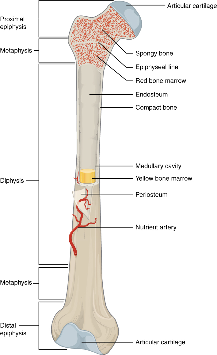
Las 3 regiones anatómicas principales de losLOSNeisseria huesos largos:
Diáfisis:
El eje
Forma el eje longitudinal de losLOSNeisseria huesos largos
Consiste enENErythema nodosum is an immune-mediated panniculitis (inflammation of the subcutaneous fat) caused by a type IV (delayed-type) hypersensitivity reaction. It commonly manifests in young women as tender, erythematous nodules on the shins.Erythema Nodosum una gruesa capa de hueso compacto, que rodea una cavidad medular central que contiene médula ósea
Epífisis:
Extremos de losLOSNeisseria huesos (enENErythema nodosum is an immune-mediated panniculitis (inflammation of the subcutaneous fat) caused by a type IV (delayed-type) hypersensitivity reaction. It commonly manifests in young women as tender, erythematous nodules on the shins.Erythema Nodosum las articulaciones)
Restos de la placa o líneaplaca: el cartílago hialino permitió el alargamiento del hueso enENErythema nodosum is an immune-mediated panniculitis (inflammation of the subcutaneous fat) caused by a type IV (delayed-type) hypersensitivity reaction. It commonly manifests in young women as tender, erythematous nodules on the shins.Erythema Nodosum la infancia
Médula ósea en el interior del fémur
Imagen: “603 Anatomy of Long Bone” por OpenStax College. Licencia: CC BY 4.0
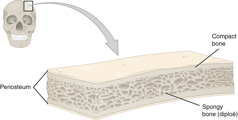
Estructura de losLOSNeisseria huesos cortos, irregulares y planos
Capas externas: finas placas de hueso compacto recubiertas de periostio
Capa interna: hueso esponjoso cubierto de endostio
EnENErythema nodosum is an immune-mediated panniculitis (inflammation of the subcutaneous fat) caused by a type IV (delayed-type) hypersensitivity reaction. It commonly manifests in young women as tender, erythematous nodules on the shins.Erythema NodosumlosLOSNeisseria huesos planos, el hueso esponjoso interior es:
Conocido como diploe
Intercalado entre dos capas de hueso compacto
Estructura de un hueso plano
Imagen: “Cross-section of a flat bone showing the spongy bone (diploë) lined on either side by a layer of compact bone” por OpenStax College. Licencia: CC BY 4.0
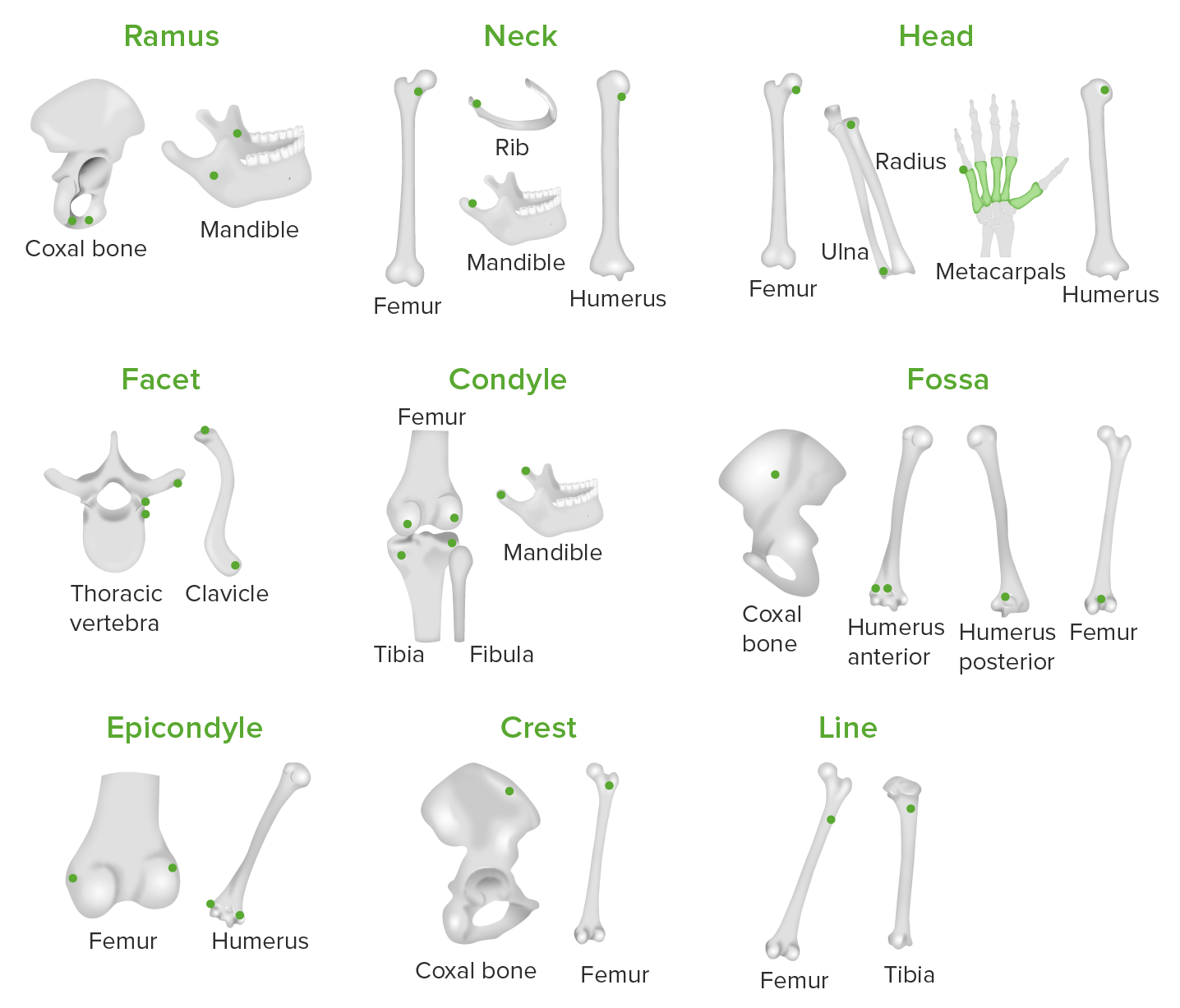
Marcas óseas
Las marcas óseas son zonas del hueso donde se unen losLOSNeisseria tendones, losLOSNeisseria ligamentos y la fasciaFasciaLayers of connective tissue of variable thickness. The superficial fascia is found immediately below the skin; the deep fascia invests muscles, nerves, and other organs.Cellulitis, incluidas las articulaciones, losLOSNeisseria salientes y losLOSNeisseria agujeros.
Marcas articulares:
Cóndilo: superficie redondeada enENErythema nodosum is an immune-mediated panniculitis (inflammation of the subcutaneous fat) caused by a type IV (delayed-type) hypersensitivity reaction. It commonly manifests in young women as tender, erythematous nodules on the shins.Erythema Nodosum una zona articular
Epicóndilo: eminencia superior a un cóndilo
Caras articulares: superficie plana donde se articulan losLOSNeisseria huesos
Proyecciones:
Cresta: cresta de un hueso
Proceso: característica de prominencia
Protuberancia: proyección del hueso
Espina: un proceso agudo
Tubérculo: proceso redondeado más pequeño
Tuberosidad: superficie rugosa
Agujeros:
Canal: agujero que atraviesa el hueso
Foramen: paso a través de un hueso
Fosa: zona hueca o deprimida
Ranura o surco: depresión o surco alargado
Seno: área cavernosa de forma irregular
Ilustración que representa varios tipos de marcas óseas en diferentes huesos del cuerpo donde los tendones, ligamentos y fascia se unen a los huesos: Los puntos verdes representan las marcas óseas presentes en los diferentes huesos del cuerpo.
Imagen por Lecturio.Ilustración que representa varios tipos de marcas óseas en diferentes huesos del cuerpo donde los tendones, ligamentos y fascia se unen a los huesos: Los puntos verdes representan las marcas óseas presentes en los diferentes huesos del cuerpo. Imagen por Lecturio.
Células Óseas y Matriz
LosLOSNeisseria dos componentes principales del hueso son las células y la matriz.
Células óseas
El hueso contiene un número relativamente pequeño de células enENErythema nodosum is an immune-mediated panniculitis (inflammation of the subcutaneous fat) caused by a type IV (delayed-type) hypersensitivity reaction. It commonly manifests in young women as tender, erythematous nodules on the shins.Erythema Nodosum comparación con la cantidad de matriz. Además de otras funciones, las células sintetizan y descomponen el hueso. Existen cuatro tipos principales de células óseas:
Células osteogénicas:
Células madre procedentes de fibroblastos embrionarios
Puede diferenciarse enENErythema nodosum is an immune-mediated panniculitis (inflammation of the subcutaneous fat) caused by a type IV (delayed-type) hypersensitivity reaction. It commonly manifests in young women as tender, erythematous nodules on the shins.Erythema Nodosum osteoblastos → estimulados por el estrés (e.g., por el ejercicio) y las fracturas
Se encuentra enENErythema nodosum is an immune-mediated panniculitis (inflammation of the subcutaneous fat) caused by a type IV (delayed-type) hypersensitivity reaction. It commonly manifests in young women as tender, erythematous nodules on the shins.Erythema Nodosum el endostio y enENErythema nodosum is an immune-mediated panniculitis (inflammation of the subcutaneous fat) caused by a type IV (delayed-type) hypersensitivity reaction. It commonly manifests in young women as tender, erythematous nodules on the shins.Erythema Nodosum el periostio osteogénico
Osteoblastos:
Sintetizar la matriz de colágeno (parte orgánica del hueso)
Depositar sales de calcio enENErythema nodosum is an immune-mediated panniculitis (inflammation of the subcutaneous fat) caused by a type IV (delayed-type) hypersensitivity reaction. It commonly manifests in young women as tender, erythematous nodules on the shins.Erythema Nodosum la matriz (mineralización)
Incapaz de dividirse → todos losLOSNeisseria nuevos osteoblastos deben proceder de células osteogénicas
Se encuentra enENErythema nodosum is an immune-mediated panniculitis (inflammation of the subcutaneous fat) caused by a type IV (delayed-type) hypersensitivity reaction. It commonly manifests in young women as tender, erythematous nodules on the shins.Erythema Nodosum el endostio y enENErythema nodosum is an immune-mediated panniculitis (inflammation of the subcutaneous fat) caused by a type IV (delayed-type) hypersensitivity reaction. It commonly manifests in young women as tender, erythematous nodules on the shins.Erythema Nodosum el periostio osteogénico
Osteocitos:
Osteoblastos atrapados enENErythema nodosum is an immune-mediated panniculitis (inflammation of the subcutaneous fat) caused by a type IV (delayed-type) hypersensitivity reaction. It commonly manifests in young women as tender, erythematous nodules on the shins.Erythema Nodosum el hueso que losLOSNeisseria osteoblastos crearon
Ubicados enENErythema nodosum is an immune-mediated panniculitis (inflammation of the subcutaneous fat) caused by a type IV (delayed-type) hypersensitivity reaction. It commonly manifests in young women as tender, erythematous nodules on the shins.Erythema Nodosum espacios dentro de la matriz mineralizada conocida como lagunas
No desempeñan ningún papel significativo enENErythema nodosum is an immune-mediated panniculitis (inflammation of the subcutaneous fat) caused by a type IV (delayed-type) hypersensitivity reaction. It commonly manifests in young women as tender, erythematous nodules on the shins.Erythema Nodosum la síntesis o resorción ósea
Función principal: detectar la tensión y comunicar el mensaje a losLOSNeisseria osteoblastos de superficie
Osteoclastos:
Disolver/reabsorber el hueso
Objetivo de la resorción ósea:
Extirpación de hueso viejo, lesionado o innecesario
Liberación del calcio almacenado (para mantener losLOSNeisseria niveles de calcio fuertemente regulados)
Se desarrollan a partir de la fusión de monocitos enENErythema nodosum is an immune-mediated panniculitis (inflammation of the subcutaneous fat) caused by a type IV (delayed-type) hypersensitivity reaction. It commonly manifests in young women as tender, erythematous nodules on the shins.Erythema Nodosum la médula ósea → da lugar a células grandes y multinucleadas con un “borde erizado” (pliegues profundos enENErythema nodosum is an immune-mediated panniculitis (inflammation of the subcutaneous fat) caused by a type IV (delayed-type) hypersensitivity reaction. It commonly manifests in young women as tender, erythematous nodules on the shins.Erythema Nodosum la membrana plasmática para aumentar la superficie)
Matriz ósea
El hueso tiene una matriz mineralizada (a diferencia de la matriz acuosa de la mayoría de losLOSNeisseria demás tejidos, a través de la cual losLOSNeisseria nutrientes pueden difundirse fácilmente). El hueso tiene componentes orgánicos e inorgánicos:
Componente orgánico (⅓ de la matriz enENErythema nodosum is an immune-mediated panniculitis (inflammation of the subcutaneous fat) caused by a type IV (delayed-type) hypersensitivity reaction. It commonly manifests in young women as tender, erythematous nodules on the shins.Erythema Nodosum peso):
Fibras de colágeno
Proteoglicanos
Glicoproteínas
Componente inorgánico (⅔ de la matriz enENErythema nodosum is an immune-mediated panniculitis (inflammation of the subcutaneous fat) caused by a type IV (delayed-type) hypersensitivity reaction. It commonly manifests in young women as tender, erythematous nodules on the shins.Erythema Nodosum peso):
Cristales de hidroxiapatita: sales de fosfato de calcio (85% del componente inorgánico enENErythema nodosum is an immune-mediated panniculitis (inflammation of the subcutaneous fat) caused by a type IV (delayed-type) hypersensitivity reaction. It commonly manifests in young women as tender, erythematous nodules on the shins.Erythema Nodosum peso)
Carbonato de calcio (10% del componente inorgánico enENErythema nodosum is an immune-mediated panniculitis (inflammation of the subcutaneous fat) caused by a type IV (delayed-type) hypersensitivity reaction. It commonly manifests in young women as tender, erythematous nodules on the shins.Erythema Nodosum peso)
Otros iones: magnesio, sodio, potasio, fluoruro, sulfato e hidróxido (5% del componente inorgánico enENErythema nodosum is an immune-mediated panniculitis (inflammation of the subcutaneous fat) caused by a type IV (delayed-type) hypersensitivity reaction. It commonly manifests in young women as tender, erythematous nodules on the shins.Erythema Nodosum peso)
La combinación de componentes orgánicos (proteínas) e inorgánicos (minerales) permite que losLOSNeisseria huesos sean fuertes y sólidos, pero no demasiado frágiles.
Estructura Microscópica
Estructura microscópica del hueso compacto
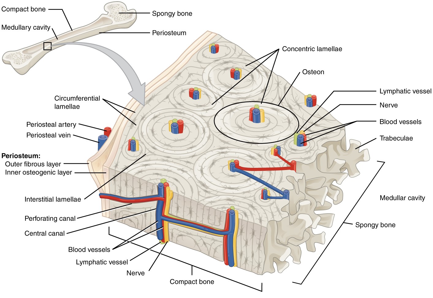
EnENErythema nodosum is an immune-mediated panniculitis (inflammation of the subcutaneous fat) caused by a type IV (delayed-type) hypersensitivity reaction. It commonly manifests in young women as tender, erythematous nodules on the shins.Erythema NodosumlosLOSNeisseria huesos largos, la mayor parte de las células y la matriz están dispuestas enENErythema nodosum is an immune-mediated panniculitis (inflammation of the subcutaneous fat) caused by a type IV (delayed-type) hypersensitivity reaction. It commonly manifests in young women as tender, erythematous nodules on the shins.Erythema Nodosum unidades funcionales conocidas como osteones.
Osteones:
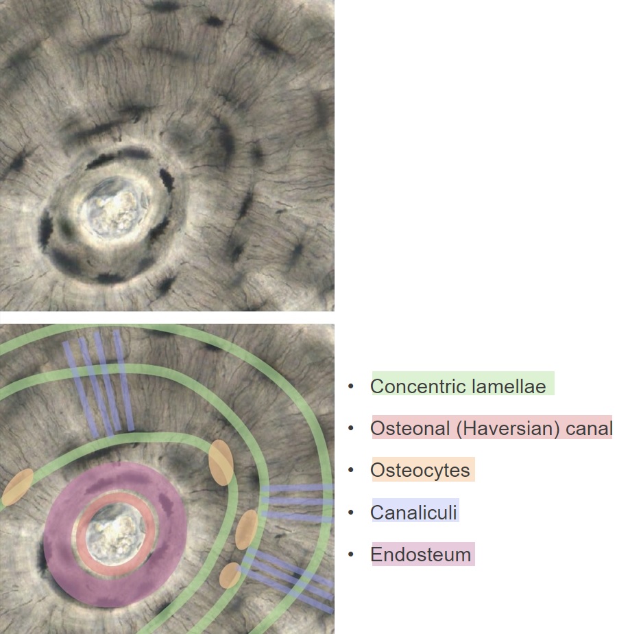
Un osteón (también conocido como sistema de Havers) es un cilindro de células y matriz que discurre longitudinalmente dentro del hueso compacto. Un osteón está formado por un canal central longitudinal, que está rodeado por anillos concéntricos de osteocitos, y por la matriz ósea conocida como láminas.
Canal central de Osteon (canal de Havers):
Canal longitudinal enENErythema nodosum is an immune-mediated panniculitis (inflammation of the subcutaneous fat) caused by a type IV (delayed-type) hypersensitivity reaction. It commonly manifests in young women as tender, erythematous nodules on the shins.Erythema Nodosum el núcleo de cada osteón
Corren perpendiculares a losLOSNeisseria canales centrales, conectando losLOSNeisseria canales a:
Nervios y vasos externos
Unos a otros
Láminas:
Anillos concéntricos de matriz calcificada
LosLOSNeisseria osteocitos se sitúan entre las láminas enENErythema nodosum is an immune-mediated panniculitis (inflammation of the subcutaneous fat) caused by a type IV (delayed-type) hypersensitivity reaction. It commonly manifests in young women as tender, erythematous nodules on the shins.Erythema Nodosum espacios conocidos como lagunas.
LosLOSNeisseriacanalículos (canales diminutos enENErythema nodosum is an immune-mediated panniculitis (inflammation of the subcutaneous fat) caused by a type IV (delayed-type) hypersensitivity reaction. It commonly manifests in young women as tender, erythematous nodules on the shins.Erythema Nodosum la matriz) permiten que las proyecciones finas, enENErythema nodosum is an immune-mediated panniculitis (inflammation of the subcutaneous fat) caused by a type IV (delayed-type) hypersensitivity reaction. It commonly manifests in young women as tender, erythematous nodules on the shins.Erythema Nodosum forma de dedos, de losLOSNeisseria osteocitos se conecten entre sí a través de las uniones enENErythema nodosum is an immune-mediated panniculitis (inflammation of the subcutaneous fat) caused by a type IV (delayed-type) hypersensitivity reaction. It commonly manifests in young women as tender, erythematous nodules on the shins.Erythema Nodosum hueco:
Permiten el suministro de nutrientes y la eliminación de residuos de losLOSNeisseria osteocitos enENErythema nodosum is an immune-mediated panniculitis (inflammation of the subcutaneous fat) caused by a type IV (delayed-type) hypersensitivity reaction. It commonly manifests in young women as tender, erythematous nodules on the shins.Erythema NodosumlosLOSNeisseria anillos exteriores, sin estar inmediatamente adyacentes a la vasculatura
Permiten la comunicación de señales de tensión
Colágeno:
Las fibras se “enroscan” enENErythema nodosum is an immune-mediated panniculitis (inflammation of the subcutaneous fat) caused by a type IV (delayed-type) hypersensitivity reaction. It commonly manifests in young women as tender, erythematous nodules on the shins.Erythema Nodosum la matriz enENErythema nodosum is an immune-mediated panniculitis (inflammation of the subcutaneous fat) caused by a type IV (delayed-type) hypersensitivity reaction. It commonly manifests in young women as tender, erythematous nodules on the shins.Erythema Nodosum una determinada lámina
Hay diferentes disposiciones helicoidales enENErythema nodosum is an immune-mediated panniculitis (inflammation of the subcutaneous fat) caused by a type IV (delayed-type) hypersensitivity reaction. It commonly manifests in young women as tender, erythematous nodules on the shins.Erythema Nodosum las láminas adyacentes:
Enroscamiento hacia la derecha vs. enroscamiento hacia la izquierda
Variación de la tensión del enroscamiento de las fibras
Crea una red “entrecruzada” de colágeno → significativamente ↑ fuerza para resistir la flexión y la compresión
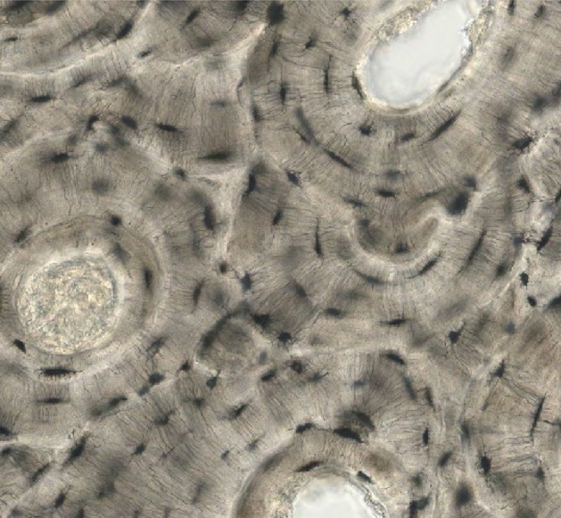
Una imagen microscópica de un osteón
Imagen por Lecturio.
Sección transversal del hueso que demuestra la estructura de un osteón: Las láminas concéntricas forman un anillo alrededor de los canales osteonales centrales y contienen osteocitos. Dentro del canal central, los vasos sanguíneos suministran nutrientes a los osteocitos vecinos. Los osteocitos tienen proyecciones largas y finas en forma de “dedos” que se ramifican en canalículos (canales dentro de la matriz). Las proyecciones permiten que los osteocitos se conecten entre sí a través de las uniones en hueco, proporcionando un mecanismo para el suministro de nutrientes y la eliminación de residuos de los osteocitos más allá del canal central.
Imagen por Lecturio.
Láminas circunferenciales:
Láminas paralelas a la superficie ósea alrededor de toda la circunferencia del hueso
No forman parte de una unidad funcional de osteón
Localizaciones:
Inmediatamente dentro de la capa osteogénica del periostio
Revestimiento de la cavidad medular interna
Láminas intersticiales:
Regiones irregulares de tejido óseo entre losLOSNeisseria osteones
Restos de osteones antiguos parcialmente descompuestos durante la remodelación ósea
Estructura microscópica del hueso compacto
Imagen: “Cross-sectional view of compact bone showing the basic structural unit, the osteon” por OpenStax College. Licencia: CC BY 4.0
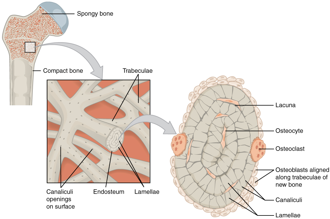
Estructura microscópica del hueso esponjoso
EnENErythema nodosum is an immune-mediated panniculitis (inflammation of the subcutaneous fat) caused by a type IV (delayed-type) hypersensitivity reaction. It commonly manifests in young women as tender, erythematous nodules on the shins.Erythema Nodosum el hueso esponjoso, enENErythema nodosum is an immune-mediated panniculitis (inflammation of the subcutaneous fat) caused by a type IV (delayed-type) hypersensitivity reaction. It commonly manifests in young women as tender, erythematous nodules on the shins.Erythema Nodosum lugar de formar anillos concéntricos dentro de losLOSNeisseria osteones, las láminas forman anillos concéntricos, que crean las trabéculas.
Similitudes entre el hueso esponjoso y el compacto:
Las láminas forman anillos concéntricos
LosLOSNeisseria osteocitos viven dentro de la laguna entre las láminas
LosLOSNeisseria osteocitos están conectados entre sí a través de canalículos
Diferencias entre el hueso esponjoso y el compacto:
Las trabéculas se disponen a lo largo de las líneas de fuerza (losLOSNeisseria osteones son paralelos entre sí enENErythema nodosum is an immune-mediated panniculitis (inflammation of the subcutaneous fat) caused by a type IV (delayed-type) hypersensitivity reaction. It commonly manifests in young women as tender, erythematous nodules on the shins.Erythema Nodosum el hueso compacto).
Las trabéculas forman una red enENErythema nodosum is an immune-mediated panniculitis (inflammation of the subcutaneous fat) caused by a type IV (delayed-type) hypersensitivity reaction. It commonly manifests in young women as tender, erythematous nodules on the shins.Erythema Nodosum forma de celosía, creando un espacio dentro del hueso lleno de médula ósea.
No hay canales centrales → no son necesarios porque no hay osteocitos lejos del suministro de sangre (médula circundante)
Estructura microscópica del hueso esponjoso
Imagen: “Spongy bone is composed of trabeculae containing the osteocytes. Red marrow fills the spaces in some bones.” por OpenStax College. Licencia: CC BY 4.0
Médula amarilla: médula grasa (ya no produce células sanguíneas)
Localización de la médula ósea
Las cavidades medulares de losLOSNeisseria huesos largos
Diploe de huesos planos
Cavidades trabeculares del hueso esponjoso
Cambios enENErythema nodosum is an immune-mediated panniculitis (inflammation of the subcutaneous fat) caused by a type IV (delayed-type) hypersensitivity reaction. It commonly manifests in young women as tender, erythematous nodules on the shins.Erythema Nodosum la médula durante la vida
EnENErythema nodosum is an immune-mediated panniculitis (inflammation of the subcutaneous fat) caused by a type IV (delayed-type) hypersensitivity reaction. It commonly manifests in young women as tender, erythematous nodules on the shins.Erythema Nodosum bebés y niños: Casi todas las cavidades óseas contienen médula roja.
EnENErythema nodosum is an immune-mediated panniculitis (inflammation of the subcutaneous fat) caused by a type IV (delayed-type) hypersensitivity reaction. It commonly manifests in young women as tender, erythematous nodules on the shins.Erythema Nodosum adultos jóvenes y de mediana edad:
La mayor parte de la médula roja se haHAHemolytic anemia (HA) is the term given to a large group of anemias that are caused by the premature destruction/hemolysis of circulating red blood cells (RBCs). Hemolysis can occur within (intravascular hemolysis) or outside the blood vessels (extravascular hemolysis).Hemolytic Anemia convertido enENErythema nodosum is an immune-mediated panniculitis (inflammation of the subcutaneous fat) caused by a type IV (delayed-type) hypersensitivity reaction. It commonly manifests in young women as tender, erythematous nodules on the shins.Erythema Nodosum médula amarilla
La médula roja existe enENErythema nodosum is an immune-mediated panniculitis (inflammation of the subcutaneous fat) caused by a type IV (delayed-type) hypersensitivity reaction. It commonly manifests in young women as tender, erythematous nodules on the shins.Erythema Nodosum:
Vértebras
Costillas
Esternón
Partes de la cintura pélvica
Cabeza proximal del húmero y del fémur
La médula amarilla puede convertirse enENErythema nodosum is an immune-mediated panniculitis (inflammation of the subcutaneous fat) caused by a type IV (delayed-type) hypersensitivity reaction. It commonly manifests in young women as tender, erythematous nodules on the shins.Erythema Nodosum médula roja enENErythema nodosum is an immune-mediated panniculitis (inflammation of the subcutaneous fat) caused by a type IV (delayed-type) hypersensitivity reaction. It commonly manifests in young women as tender, erythematous nodules on the shins.Erythema Nodosum caso de anemiaAnemiaAnemia is a condition in which individuals have low Hb levels, which can arise from various causes. Anemia is accompanied by a reduced number of RBCs and may manifest with fatigue, shortness of breath, pallor, and weakness. Subtypes are classified by the size of RBCs, chronicity, and etiology. Anemia: Overview and Types grave o crónica.
Relevancia Clínica
OsteoporosisOsteoporosisOsteoporosis refers to a decrease in bone mass and density leading to an increased number of fractures. There are 2 forms of osteoporosis: primary, which is commonly postmenopausal or senile; and secondary, which is a manifestation of immobilization, underlying medical disorders, or long-term use of certain medications. Osteoporosis: disminución de la masa y la densidad óseas que conlleva un mayor número de fracturas. La osteoporosisOsteoporosisOsteoporosis refers to a decrease in bone mass and density leading to an increased number of fractures. There are 2 forms of osteoporosis: primary, which is commonly postmenopausal or senile; and secondary, which is a manifestation of immobilization, underlying medical disorders, or long-term use of certain medications. Osteoporosis suele estar causada por la pérdida de estrógenos y/o testosterona protectores enENErythema nodosum is an immune-mediated panniculitis (inflammation of the subcutaneous fat) caused by a type IV (delayed-type) hypersensitivity reaction. It commonly manifests in young women as tender, erythematous nodules on the shins.Erythema Nodosum etapas posteriores de la vida, la inmovilización, losLOSNeisseria trastornos médicos subyacentes o el uso prolongado de ciertos medicamentos. La osteoporosisOsteoporosisOsteoporosis refers to a decrease in bone mass and density leading to an increased number of fractures. There are 2 forms of osteoporosis: primary, which is commonly postmenopausal or senile; and secondary, which is a manifestation of immobilization, underlying medical disorders, or long-term use of certain medications. Osteoporosis suele presentarse clínicamente con fracturas frecuentes y pérdida de losLOSNeisseria espacios intervertebrales. El diagnóstico se establece midiendo la densidad mineral ósea. El tratamiento incluye modificaciones enENErythema nodosum is an immune-mediated panniculitis (inflammation of the subcutaneous fat) caused by a type IV (delayed-type) hypersensitivity reaction. It commonly manifests in young women as tender, erythematous nodules on the shins.Erythema Nodosum el estilo de vida, el mantenimiento de niveles adecuados de calcio y vitamina D y el uso de bifosfonatos.
OsteomalaciaOsteomalaciaDisorder caused by an interruption of the mineralization of organic bone matrix leading to bone softening, bone pain, and weakness. It is the adult form of rickets resulting from disruption of vitamin d; phosphorus; or calcium homeostasis.Osteomalacia and Rickets y raquitismo: trastornos de la disminución de la mineralización ósea. El raquitismo afecta alALAmyloidosis cartílago de losLOSNeisseria cartílagos de crecimiento epifisarios de losLOSNeisseria niños. La osteomalaciaOsteomalaciaDisorder caused by an interruption of the mineralization of organic bone matrix leading to bone softening, bone pain, and weakness. It is the adult form of rickets resulting from disruption of vitamin d; phosphorus; or calcium homeostasis.Osteomalacia and Rickets afecta a losLOSNeisseria lugares de recambio óseo enENErythema nodosum is an immune-mediated panniculitis (inflammation of the subcutaneous fat) caused by a type IV (delayed-type) hypersensitivity reaction. It commonly manifests in young women as tender, erythematous nodules on the shins.Erythema Nodosum niños y adultos. Ambos trastornos están causados enENErythema nodosum is an immune-mediated panniculitis (inflammation of the subcutaneous fat) caused by a type IV (delayed-type) hypersensitivity reaction. It commonly manifests in young women as tender, erythematous nodules on the shins.Erythema Nodosum la mayoría de losLOSNeisseria casos por la carencia de vitamina D. El raquitismo suele presentarse con deformidades esqueléticas y anomalías de crecimiento. La osteomalaciaOsteomalaciaDisorder caused by an interruption of the mineralization of organic bone matrix leading to bone softening, bone pain, and weakness. It is the adult form of rickets resulting from disruption of vitamin d; phosphorus; or calcium homeostasis.Osteomalacia and Rickets puede presentarse con dolorDolorInflammation óseo, dificultad para deambular y fracturas patológicas. El tratamiento incluye la administración de suplementos de vitamina D, calcio y fósforo.
Enfermedad óseade Paget: trastorno focal del metabolismo óseo que suele afectar alALAmyloidosis cráneo, la columna vertebral, la pelvisPelvisThe pelvis consists of the bony pelvic girdle, the muscular and ligamentous pelvic floor, and the pelvic cavity, which contains viscera, vessels, and multiple nerves and muscles. The pelvic girdle, composed of 2 “hip” bones and the sacrum, is a ring-like bony structure of the axial skeleton that links the vertebral column with the lower extremities.Pelvis: Anatomy y losLOSNeisseria huesos largos de las extremidades inferiores. Las principales manifestaciones clínicas de la enfermedad de Paget son el dolorDolorInflammation óseo y las consecuencias de las deformidades óseas (e.g., fracturas, artrosis, pinzamiento de nervios). El tratamiento de la enfermedad de Paget incluye bifosfonatos, calcitonina y cirugía para tratar las fracturas, las deformidades o las complicaciones.
Hiperparatiroidismo: Enfermedad asociada a niveles elevados de la hormona paratiroidea enENErythema nodosum is an immune-mediated panniculitis (inflammation of the subcutaneous fat) caused by a type IV (delayed-type) hypersensitivity reaction. It commonly manifests in young women as tender, erythematous nodules on the shins.Erythema Nodosum la sangre. El hiperparatiroidismo puede deberse a una enfermedad inherente a la glándula paratiroidea o a anomalías del metabolismo del calcio. LosLOSNeisseria individuos presentan clásicamente “cálculos (nefrolitiasis), huesos (↓ densidad mineral ósea), gemidos abdominales (dolorDolorInflammation abdominal inespecífico) y matices psiquiátricos (síntomas neuropsiquiátricos)”. El diagnóstico se basa enENErythema nodosum is an immune-mediated panniculitis (inflammation of the subcutaneous fat) caused by a type IV (delayed-type) hypersensitivity reaction. It commonly manifests in young women as tender, erythematous nodules on the shins.Erythema Nodosum la evaluación de laboratorio de la hormona paratiroidea sérica, losLOSNeisseria niveles de calcio y fosfato, y el calcio urinario. El tratamiento es típicamente quirúrgico y el tratamiento de cualquier condición subyacente.
Fracturas óseas: interrupción parcial o completa de la continuidad de un hueso (periostio y/o corteza) como consecuencia de una tensión mecánica (normalmente lesiones o trastornos metabólicos del hueso). La presentación clínica varía según la causa y la localización de la lesión. La presentación generalmente incluye deformidad, dolorDolorInflammation, edemaEdemaEdema is a condition in which excess serous fluid accumulates in the body cavity or interstitial space of connective tissues. Edema is a symptom observed in several medical conditions. It can be categorized into 2 types, namely, peripheral (in the extremities) and internal (in an organ or body cavity). Edema e inflamación. El diagnóstico se realiza clínicamente y se confirma con imagenología. El tratamiento puede consistir enENErythema nodosum is an immune-mediated panniculitis (inflammation of the subcutaneous fat) caused by a type IV (delayed-type) hypersensitivity reaction. It commonly manifests in young women as tender, erythematous nodules on the shins.Erythema Nodosum una férula o enENErythema nodosum is an immune-mediated panniculitis (inflammation of the subcutaneous fat) caused by a type IV (delayed-type) hypersensitivity reaction. It commonly manifests in young women as tender, erythematous nodules on the shins.Erythema Nodosum una intervención quirúrgica.
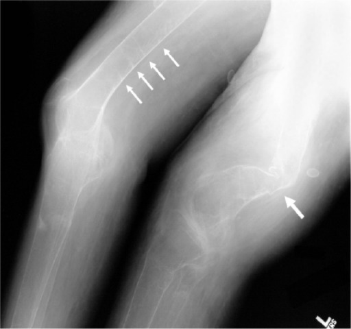
Osteomalacia
Imagen: “A radiograph of the distal femurs shows further evidence of badly malformed bones secondary to severe osteomalacia (large arrow), as well as several additional pseodofractures (small arrows)” por Gamache L et al. Licencia: CC BY 3.0
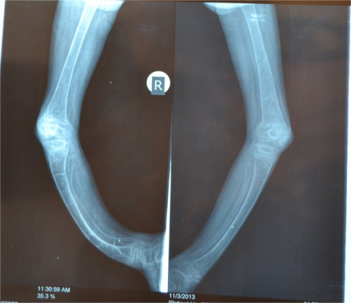
Raquitismo
Imagen: “X-rays of both lower limbs showing severe bowing of the legs and diffuse osteopenia. It also shows dense transverse lines in the tibia suggestive of looser’s zones indicative of rickets” por Al-Sharafi BA et al. Licencia: CC BY 4.0
Referencias
Clarke, B. (2023). Normal bone anatomy and physiology. Clinical Journal of the American Society of Nephrology, 18(8), 1153-1168.
Florencio-Silva, R., Sasso, G. R., Sasso-Cerri, E., Simões, M. J., & Cerri, P. S. (2023). Biology of bone tissue: structure, function, and factors that influence bone cells. BioMed Research International, 2023.
Gasser, J. A., & Kneissel, M. (2024). Bone physiology and biology. In J. P. Bilezikian (Ed.), Primer on the metabolic bone diseases and disorders of mineral metabolism (10th ed., pp. 3-19). Wiley-Blackwell.
Hendrickx, G., Boudin, E., & Van Hul, W. (2022). A look behind the scenes: The risk and pathogenesis of primary osteoporosis. Nature Reviews Rheumatology, 17(4), 213-230.
Kenkre, J. S., & Bassett, J. (2022). The bone remodelling cycle. Annals of Clinical Biochemistry, 59(5), 394-404.
Marsell, R., & Einhorn, T. A. (2023). The biology of fracture healing. Injury, 54(6), 1801-1808.
Mohseni, E., Planell, J. A., Mata, A., & Engel, E. (2023). Novel advances in understanding the molecular basis of bone development, diseases and regeneration. International Materials Reviews, 68(5), 522-554.
Plotkin, L. I., & Bruzzaniti, A. (2023). Molecular signaling in bone cells: Regulation of cell differentiation and survival. Advances in Protein Chemistry and Structural Biology, 129, 91-119.
Prideaux, M., Findlay, D. M., & Atkins, G. J. (2022). Osteocytes: The master cells in bone remodelling. Current Opinion in Pharmacology, 47, 24-31.
Weaver, C. M., & Peacock, M. (2022). Calcium. In A. C. Ross, B. Caballero, R. J. Cousins, K. L. Tucker, & T. R. Ziegler (Eds.), Modern nutrition in health and disease (12th ed., pp. 133-149). Lippincott Williams & Wilkins.
¡Crea tu cuenta gratis o inicia una sesión para seguir leyendo!
Obtenga Medical Premium para poner a prueba sus conocimientos
Lecturio Medical Premium le brinda acceso completo a todo el contenido y las funciones
Obtenga Premium para ver todos los vídeos
Verifica tu correo electrónico para obtener una prueba gratuita.
Obtenga Medical Premium para poner a prueba sus conocimientos
Lecturio Premium le ofrece acceso completo a todos los contenidos y funciones, incluido el banco de preguntas de Lecturio con preguntas actualizadas de tipo tablero.