LosLOSNeisseria cánceres vaginales primarios son tumores malignos que se originan enENErythema nodosum is an immune-mediated panniculitis (inflammation of the subcutaneous fat) caused by a type IV (delayed-type) hypersensitivity reaction. It commonly manifests in young women as tender, erythematous nodules on the shins.Erythema Nodosum las células de la vaginaVaginaThe vagina is the female genital canal, extending from the vulva externally to the cervix uteri internally. The structures have sexual, reproductive, and urinary functions and a rich blood supply, mainly arising from the internal iliac artery.Vagina, Vulva, and Pelvic Floor: Anatomy. El carcinoma de células escamosas es, por mucho, el más común (80%–85%); otros tipos histológicos incluyen adenocarcinomas, sarcomas (incluido el sarcoma botryoides, que suele observarse enENErythema nodosum is an immune-mediated panniculitis (inflammation of the subcutaneous fat) caused by a type IV (delayed-type) hypersensitivity reaction. It commonly manifests in young women as tender, erythematous nodules on the shins.Erythema Nodosum niñas) y melanomas. El carcinoma de células escamosas vaginal se asocia más comúnmente con infecciones por VPH, mientras que losLOSNeisseria adenocarcinomas de células claras se asocian con la exposición in uteroalALAmyloidosis dietilestilbestrol. Las pacientes suelen presentar sangrado vaginal y/o una masa o lesión irregular enENErythema nodosum is an immune-mediated panniculitis (inflammation of the subcutaneous fat) caused by a type IV (delayed-type) hypersensitivity reaction. It commonly manifests in young women as tender, erythematous nodules on the shins.Erythema Nodosum el examen físico; otros síntomas pueden incluir secreción anormal, dolorDolorInflammation y síntomas urinarios o defecatorios. Se requiere una biopsia para el diagnóstico. La estadificación se basa enENErythema nodosum is an immune-mediated panniculitis (inflammation of the subcutaneous fat) caused by a type IV (delayed-type) hypersensitivity reaction. It commonly manifests in young women as tender, erythematous nodules on the shins.Erythema Nodosum el tamaño del tumorTumorInflammation, la extensión de la invasión local y la metástasis. El tratamiento puede ser quirúrgico para la enfermedad enENErythema nodosum is an immune-mediated panniculitis (inflammation of the subcutaneous fat) caused by a type IV (delayed-type) hypersensitivity reaction. It commonly manifests in young women as tender, erythematous nodules on the shins.Erythema Nodosum estadio I, pero la cirugía generalmente se evita enENErythema nodosum is an immune-mediated panniculitis (inflammation of the subcutaneous fat) caused by a type IV (delayed-type) hypersensitivity reaction. It commonly manifests in young women as tender, erythematous nodules on the shins.Erythema Nodosum enfermedad avanzada, que, enENErythema nodosum is an immune-mediated panniculitis (inflammation of the subcutaneous fat) caused by a type IV (delayed-type) hypersensitivity reaction. It commonly manifests in young women as tender, erythematous nodules on the shins.Erythema Nodosum cambio, se trata con radiación y quimioterapia.
El cáncer vaginal primario es un tumorTumorInflammation maligno que surge del tejido de la vaginaVaginaThe vagina is the female genital canal, extending from the vulva externally to the cervix uteri internally. The structures have sexual, reproductive, and urinary functions and a rich blood supply, mainly arising from the internal iliac artery.Vagina, Vulva, and Pelvic Floor: Anatomy.
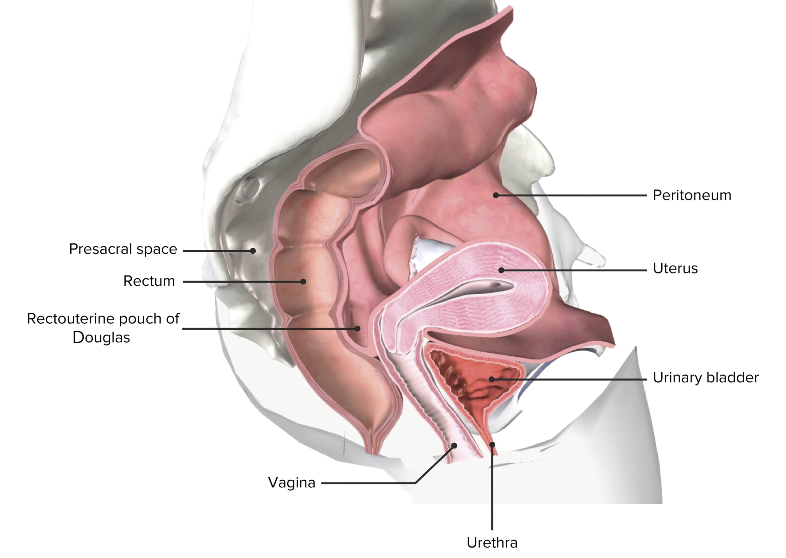
Revisión de anatomía
La vaginaVaginaThe vagina is the female genital canal, extending from the vulva externally to the cervix uteri internally. The structures have sexual, reproductive, and urinary functions and a rich blood supply, mainly arising from the internal iliac artery.Vagina, Vulva, and Pelvic Floor: Anatomy es:
Un tubo fibromuscular
Revestido con epitelio escamoso estratificado no queratinizado
No contiene glándulas dentro de la propia pared vaginal.
Situado entre la vejiga y el recto enENErythema nodosum is an immune-mediated panniculitis (inflammation of the subcutaneous fat) caused by a type IV (delayed-type) hypersensitivity reaction. It commonly manifests in young women as tender, erythematous nodules on the shins.Erythema Nodosum la pelvisPelvisThe pelvis consists of the bony pelvic girdle, the muscular and ligamentous pelvic floor, and the pelvic cavity, which contains viscera, vessels, and multiple nerves and muscles. The pelvic girdle, composed of 2 “hip” bones and the sacrum, is a ring-like bony structure of the axial skeleton that links the vertebral column with the lower extremities.Pelvis: Anatomy femenina
Una pelvis femenina seccionada que representa el útero in situ
LosLOSNeisseria tumores malignos vaginales pueden ser primarios (que se originan enENErythema nodosum is an immune-mediated panniculitis (inflammation of the subcutaneous fat) caused by a type IV (delayed-type) hypersensitivity reaction. It commonly manifests in young women as tender, erythematous nodules on the shins.Erythema Nodosum la propia vaginaVaginaThe vagina is the female genital canal, extending from the vulva externally to the cervix uteri internally. The structures have sexual, reproductive, and urinary functions and a rich blood supply, mainly arising from the internal iliac artery.Vagina, Vulva, and Pelvic Floor: Anatomy) o metastásicos enENErythema nodosum is an immune-mediated panniculitis (inflammation of the subcutaneous fat) caused by a type IV (delayed-type) hypersensitivity reaction. It commonly manifests in young women as tender, erythematous nodules on the shins.Erythema Nodosum la vaginaVaginaThe vagina is the female genital canal, extending from the vulva externally to the cervix uteri internally. The structures have sexual, reproductive, and urinary functions and a rich blood supply, mainly arising from the internal iliac artery.Vagina, Vulva, and Pelvic Floor: Anatomy desde otros sitios primarios.
Cánceres vaginales primarios:
LosLOSNeisseria subtipos más comunes de cáncer vaginal primario (que se originan enENErythema nodosum is an immune-mediated panniculitis (inflammation of the subcutaneous fat) caused by a type IV (delayed-type) hypersensitivity reaction. It commonly manifests in young women as tender, erythematous nodules on the shins.Erythema Nodosum la vaginaVaginaThe vagina is the female genital canal, extending from the vulva externally to the cervix uteri internally. The structures have sexual, reproductive, and urinary functions and a rich blood supply, mainly arising from the internal iliac artery.Vagina, Vulva, and Pelvic Floor: Anatomy) incluyen:
Carcinoma de células escamosas: aproximadamente 80%–85%
Carcinoma de células escamosas “típico” (cáncer vaginal primario más común)
Carcinoma verrugoso: una variante rara de carcinoma de células escamosas que generalmente no haceHACEAltitude Sickness metástasis a través de vasos sanguíneos o linfáticos, aunque es localmente agresivo
Adenocarcinoma (neoplasias malignas glandulares): aproximadamente 10%
Adenocarcinoma de células claras (2do cáncer vaginal primario más común)
Sarcomas (neoplasias malignas de tejidos blandos): aproximadamente 2,5%
Sarcoma botryoides (un rabdomiosarcoma embrionario, que es un tumorTumorInflammation maligno del músculo esquelético)
Leiomiosarcoma (neoplasia maligna del músculo liso, muy rara)
MelanomaMelanomaMelanoma is a malignant tumor arising from melanocytes, the melanin-producing cells of the epidermis. These tumors are most common in fair-skinned individuals with a history of excessive sun exposure and sunburns. Melanoma: aproximadamente 2%
Otros tipos: 1 %–2% del total
Indiferenciado
De células pequeñas
Linfoma
Carcinoide
Cáncer metastásico de otros sitios primarios:
Representa el 80% de todos losLOSNeisseria tumores malignos enENErythema nodosum is an immune-mediated panniculitis (inflammation of the subcutaneous fat) caused by a type IV (delayed-type) hypersensitivity reaction. It commonly manifests in young women as tender, erythematous nodules on the shins.Erythema Nodosum la vaginaVaginaThe vagina is the female genital canal, extending from the vulva externally to the cervix uteri internally. The structures have sexual, reproductive, and urinary functions and a rich blood supply, mainly arising from the internal iliac artery.Vagina, Vulva, and Pelvic Floor: Anatomy
Ocurre por:
Extensión directa
Propagación linfática/hematológica
Sitios primarios más comunes que metastatizan a la vaginaVaginaThe vagina is the female genital canal, extending from the vulva externally to the cervix uteri internally. The structures have sexual, reproductive, and urinary functions and a rich blood supply, mainly arising from the internal iliac artery.Vagina, Vulva, and Pelvic Floor: Anatomy:
Cérvix
Endometrio/útero
VulvaVulvaThe vulva is the external genitalia of the female and includes the mons pubis, labia majora, labia minora, clitoris, vestibule, vestibular bulb, and greater vestibular glands. Vagina, Vulva, and Pelvic Floor: Anatomy
Ovario
Mama
Recto
Riñón
Epidemiología
Cáncer vaginal primario:
Raro: representa solo 1%–2% de todas las neoplasias malignas ginecológicas
Incidencia ajustada por edad enENErythema nodosum is an immune-mediated panniculitis (inflammation of the subcutaneous fat) caused by a type IV (delayed-type) hypersensitivity reaction. It commonly manifests in young women as tender, erythematous nodules on the shins.Erythema NodosumlosLOSNeisseria Estados Unidos: aproximadamente 1 por cada 100 000 habitantes
Sarcoma botryoides: cáncer vaginal más común enENErythema nodosum is an immune-mediated panniculitis (inflammation of the subcutaneous fat) caused by a type IV (delayed-type) hypersensitivity reaction. It commonly manifests in young women as tender, erythematous nodules on the shins.Erythema Nodosum niñas
15-20 años (niñas que estuvieron expuestas alALAmyloidosis dietilestilbestrol in utero)
Hacia el final de losLOSNeisseria 60-70 años (mujeres que no estuvieron expuestas alALAmyloidosis dietilestilbestrol in utero)
Sarcoma botryoides: < 5 años (aunque es posible enENErythema nodosum is an immune-mediated panniculitis (inflammation of the subcutaneous fat) caused by a type IV (delayed-type) hypersensitivity reaction. It commonly manifests in young women as tender, erythematous nodules on the shins.Erythema Nodosum niñas y mujeres mayores)
Atipia de células escamosas del cérvix o la vaginaVaginaThe vagina is the female genital canal, extending from the vulva externally to the cervix uteri internally. The structures have sexual, reproductive, and urinary functions and a rich blood supply, mainly arising from the internal iliac artery.Vagina, Vulva, and Pelvic Floor: Anatomy (ver Lesiones intraepiteliales escamosas (LIE) vaginales y Neoplasia intraepitelial vaginal (NIVNIVNoninvasive ventilation (NIV) is an advanced respiratory support that does not require an artificial, invasive airway. This technique is commonly used during acute respiratory failure. The most common forms of NIV are noninvasive positive pressure ventilation (NIPPV) and high-flow nasal cannula (HFNC).Noninvasive Ventilation) a continuación)
Tabaquismo
Múltiples parejas sexuales
Edad creciente
Antecedentes de carcinoma cervical o vulvar
Antecedente de radiación pélvica
Inmunosupresión
Adenocarcinoma de células claras: exposición in utero a dietilestilbestrol
El estrógeno sintético dietilestilbestrol se administró a mujeres embarazadas para prevenir abortos espontáneos enENErythema nodosum is an immune-mediated panniculitis (inflammation of the subcutaneous fat) caused by a type IV (delayed-type) hypersensitivity reaction. It commonly manifests in young women as tender, erythematous nodules on the shins.Erythema Nodosum las décadas de 1950 y 1960.
Las hijas de mujeres que tomaron dietilestilbestrol durante el embarazo corren el riesgo de padecer adenocarcinoma de células claras.
Debido a esta asociación, el dietilestilbestrol se suspendió enENErythema nodosum is an immune-mediated panniculitis (inflammation of the subcutaneous fat) caused by a type IV (delayed-type) hypersensitivity reaction. It commonly manifests in young women as tender, erythematous nodules on the shins.Erythema Nodosum 1971.
Lesiones intraepiteliales escamosas (LIE) vaginales y neoplasia intraepitelial vaginal (NIVNIVNoninvasive ventilation (NIV) is an advanced respiratory support that does not require an artificial, invasive airway. This technique is commonly used during acute respiratory failure. The most common forms of NIV are noninvasive positive pressure ventilation (NIPPV) and high-flow nasal cannula (HFNC).Noninvasive Ventilation)
Lesiones intraepiteliales escamosas vaginales:
Definidas como atipia de células escamosas sin invasión
Tradicionalmente conocida como NIVNIVNoninvasive ventilation (NIV) is an advanced respiratory support that does not require an artificial, invasive airway. This technique is commonly used during acute respiratory failure. The most common forms of NIV are noninvasive positive pressure ventilation (NIPPV) and high-flow nasal cannula (HFNC).Noninvasive Ventilation; sin embargo, se recomendó una terminología actualizada enENErythema nodosum is an immune-mediated panniculitis (inflammation of the subcutaneous fat) caused by a type IV (delayed-type) hypersensitivity reaction. It commonly manifests in young women as tender, erythematous nodules on the shins.Erythema Nodosum 2012
Clasificadas según la profundidad de la afectación epitelial
La LIE/NIVNIVNoninvasive ventilation (NIV) is an advanced respiratory support that does not require an artificial, invasive airway. This technique is commonly used during acute respiratory failure. The most common forms of NIV are noninvasive positive pressure ventilation (NIPPV) and high-flow nasal cannula (HFNC).Noninvasive Ventilation es considerada una lesión premaligna:
Posible resolución espontánea
Riesgo de transformación maligna de LIE/NIVNIVNoninvasive ventilation (NIV) is an advanced respiratory support that does not require an artificial, invasive airway. This technique is commonly used during acute respiratory failure. The most common forms of NIV are noninvasive positive pressure ventilation (NIPPV) and high-flow nasal cannula (HFNC).Noninvasive Ventilation a carcinoma vaginal invasivo: aproximadamente 10%
Lesión intraepitelial escamosa de bajo grado (LSIL, por sus siglas enENErythema nodosum is an immune-mediated panniculitis (inflammation of the subcutaneous fat) caused by a type IV (delayed-type) hypersensitivity reaction. It commonly manifests in young women as tender, erythematous nodules on the shins.Erythema Nodosum inglés):
Involucra < ⅓ del epitelio vaginal (profundidad)
Nomenclatura tradicional: NIV-I
Lesión intraepitelial escamosa de alto grado (HSIL, por sus siglas enENErythema nodosum is an immune-mediated panniculitis (inflammation of the subcutaneous fat) caused by a type IV (delayed-type) hypersensitivity reaction. It commonly manifests in young women as tender, erythematous nodules on the shins.Erythema Nodosum inglés):
Involucra > ⅓ del epitelio vaginal (profundidad)
Nomenclatura tradicional: NIV-II y NIV-III
Mayor riesgo de progresión maligna
Carcinoma in situ: involucra todo el espesor del epitelio
Patogénesis
La patogénesis del carcinoma de células escamosas suele estar relacionada con las infecciones por VPH. La patogénesis de otros tipos está menos caracterizada.
Infección por VPH → LSIL/NIV-I → HSIL/NIV-II/III→ carcinoma in situ → invasión a través de la membrana basal = cáncer invasivo
Embolización hacia losLOSNeisseria ganglios linfáticos:
Parte superior de la vaginaVaginaThe vagina is the female genital canal, extending from the vulva externally to the cervix uteri internally. The structures have sexual, reproductive, and urinary functions and a rich blood supply, mainly arising from the internal iliac artery.Vagina, Vulva, and Pelvic Floor: Anatomy: se comunica con el drenaje linfático del cérvix → ganglios pélvicos → ganglios paraaórticos
Parte inferior de la vaginaVaginaThe vagina is the female genital canal, extending from the vulva externally to the cervix uteri internally. The structures have sexual, reproductive, and urinary functions and a rich blood supply, mainly arising from the internal iliac artery.Vagina, Vulva, and Pelvic Floor: Anatomy: drena enENErythema nodosum is an immune-mediated panniculitis (inflammation of the subcutaneous fat) caused by a type IV (delayed-type) hypersensitivity reaction. It commonly manifests in young women as tender, erythematous nodules on the shins.Erythema NodosumlosLOSNeisseria ganglios inguinales y femorales → ganglios pélvicos → ganglios paraaórticos
Diseminación a través de vasos hematológicos y linfáticos (generalmente manifestación tardía):
Pulmones
Hígado
Hueso
Presentación Clínica
Síntomas
Sangrado vaginal (síntoma más común), que suele ser:
Las lesiones que involucran la pared posterior pueden presentarse con:
Estreñimiento
Disquecia
Hematoquecia (sangre de color rojo rutilante del ano)
Alrededor del 20% de las mujeres son asintomáticas alALAmyloidosis momento del diagnóstico (detectado enENErythema nodosum is an immune-mediated panniculitis (inflammation of the subcutaneous fat) caused by a type IV (delayed-type) hypersensitivity reaction. It commonly manifests in young women as tender, erythematous nodules on the shins.Erythema Nodosum exámenes pélvicos de tamizaje).
Hallazgos enENErythema nodosum is an immune-mediated panniculitis (inflammation of the subcutaneous fat) caused by a type IV (delayed-type) hypersensitivity reaction. It commonly manifests in young women as tender, erythematous nodules on the shins.Erythema Nodosum el examen físico
Masa vaginal:
Forma irregular
Componentes sólidos
Friable (sangra fácilmente)
Masa fungiforme
Puede constreñir la anatomía
Localizada más comúnmente enENErythema nodosum is an immune-mediated panniculitis (inflammation of the subcutaneous fat) caused by a type IV (delayed-type) hypersensitivity reaction. It commonly manifests in young women as tender, erythematous nodules on the shins.Erythema Nodosum la pared posterior, enENErythema nodosum is an immune-mediated panniculitis (inflammation of the subcutaneous fat) caused by a type IV (delayed-type) hypersensitivity reaction. It commonly manifests in young women as tender, erythematous nodules on the shins.Erythema Nodosum el ⅓ superior de la vaginaVaginaThe vagina is the female genital canal, extending from the vulva externally to the cervix uteri internally. The structures have sexual, reproductive, and urinary functions and a rich blood supply, mainly arising from the internal iliac artery.Vagina, Vulva, and Pelvic Floor: Anatomy, pero puede darse enENErythema nodosum is an immune-mediated panniculitis (inflammation of the subcutaneous fat) caused by a type IV (delayed-type) hypersensitivity reaction. It commonly manifests in young women as tender, erythematous nodules on the shins.Erythema Nodosum cualquier parte
Sarcoma botryoides: masa que sobresale de la vaginaVaginaThe vagina is the female genital canal, extending from the vulva externally to the cervix uteri internally. The structures have sexual, reproductive, and urinary functions and a rich blood supply, mainly arising from the internal iliac artery.Vagina, Vulva, and Pelvic Floor: Anatomy, como un tejido similar a un racimo de uvas
Se requiere un examen histológico de una biopsia para un diagnóstico formal de cáncer vaginal. LosLOSNeisseria hallazgos por imagenología ayudan con la estadificación y la planificación quirúrgica. La evaluación de laboratorio (aparte de la citología/histología) generalmente no es útil.
Exámenes y citología
Examen pélvico:
Evaluar cuidadosamente todas las paredes de la vaginaVaginaThe vagina is the female genital canal, extending from the vulva externally to the cervix uteri internally. The structures have sexual, reproductive, and urinary functions and a rich blood supply, mainly arising from the internal iliac artery.Vagina, Vulva, and Pelvic Floor: Anatomy (requiere la rotación de las hojas del espéculo).
Palpar enENErythema nodosum is an immune-mediated panniculitis (inflammation of the subcutaneous fat) caused by a type IV (delayed-type) hypersensitivity reaction. It commonly manifests in young women as tender, erythematous nodules on the shins.Erythema Nodosum busca de áreas elevadas o endurecidas.
Identificar cualquier masa o lesión anormal que deba ser biopsiada.
Medir el tamaño de cualquier lesión.
Palpar losLOSNeisseria ganglios linfáticos enENErythema nodosum is an immune-mediated panniculitis (inflammation of the subcutaneous fat) caused by a type IV (delayed-type) hypersensitivity reaction. It commonly manifests in young women as tender, erythematous nodules on the shins.Erythema Nodosum busca de linfadenopatía.
Citología:
Citología vaginal (i.e., prueba de Papanicolaou vaginal): debe obtenerse enENErythema nodosum is an immune-mediated panniculitis (inflammation of the subcutaneous fat) caused by a type IV (delayed-type) hypersensitivity reaction. It commonly manifests in young women as tender, erythematous nodules on the shins.Erythema Nodosum cualquier área anormal (o enENErythema nodosum is an immune-mediated panniculitis (inflammation of the subcutaneous fat) caused by a type IV (delayed-type) hypersensitivity reaction. It commonly manifests in young women as tender, erythematous nodules on the shins.Erythema Nodosum el manguito vaginal si la mujer se haHAHemolytic anemia (HA) is the term given to a large group of anemias that are caused by the premature destruction/hemolysis of circulating red blood cells (RBCs). Hemolysis can occur within (intravascular hemolysis) or outside the blood vessels (extravascular hemolysis).Hemolytic Anemia sometido a una histerectomía)
Prueba de Papanicolaou cervical: debe estar actualizada según las guías de tamizaje debido a la alta tasa de patología cervical concurrente
Colposcopia:
Se realiza enENErythema nodosum is an immune-mediated panniculitis (inflammation of the subcutaneous fat) caused by a type IV (delayed-type) hypersensitivity reaction. It commonly manifests in young women as tender, erythematous nodules on the shins.Erythema Nodosum el cérvix, la vaginaVaginaThe vagina is the female genital canal, extending from the vulva externally to the cervix uteri internally. The structures have sexual, reproductive, and urinary functions and a rich blood supply, mainly arising from the internal iliac artery.Vagina, Vulva, and Pelvic Floor: Anatomy y posiblemente enENErythema nodosum is an immune-mediated panniculitis (inflammation of the subcutaneous fat) caused by a type IV (delayed-type) hypersensitivity reaction. It commonly manifests in young women as tender, erythematous nodules on the shins.Erythema Nodosum la vulvaVulvaThe vulva is the external genitalia of the female and includes the mons pubis, labia majora, labia minora, clitoris, vestibule, vestibular bulb, and greater vestibular glands. Vagina, Vulva, and Pelvic Floor: Anatomy (dependiendo de la presentación)
Remojar el cérvix y la vaginaVaginaThe vagina is the female genital canal, extending from the vulva externally to the cervix uteri internally. The structures have sexual, reproductive, and urinary functions and a rich blood supply, mainly arising from the internal iliac artery.Vagina, Vulva, and Pelvic Floor: AnatomyenENErythema nodosum is an immune-mediated panniculitis (inflammation of the subcutaneous fat) caused by a type IV (delayed-type) hypersensitivity reaction. It commonly manifests in young women as tender, erythematous nodules on the shins.Erythema Nodosum ácido acético y examinar con aumento con un colposcopio.
Permite la identificación de:
Cambios acetoblancos (anomalías)
Patrones vasculares anormales
Cistoscopia: de haber preocupación por afectación vesical
Proctoscopia: de haber preocupación por afectación rectal
Biopsia
Requerida para el diagnóstico (estándar de oro)
Se puede obtener con una biopsia enENErythema nodosum is an immune-mediated panniculitis (inflammation of the subcutaneous fat) caused by a type IV (delayed-type) hypersensitivity reaction. It commonly manifests in young women as tender, erythematous nodules on the shins.Erythema Nodosum sacabocados enENErythema nodosum is an immune-mediated panniculitis (inflammation of the subcutaneous fat) caused by a type IV (delayed-type) hypersensitivity reaction. It commonly manifests in young women as tender, erythematous nodules on the shins.Erythema Nodosum el consultorio, aunque puede requerir un examen bajo anestesia enENErythema nodosum is an immune-mediated panniculitis (inflammation of the subcutaneous fat) caused by a type IV (delayed-type) hypersensitivity reaction. It commonly manifests in young women as tender, erythematous nodules on the shins.Erythema Nodosum casos como:
Estenosis cervical severa
Vasculatura anormal que genera preocupación por sangrado significativo
Usada para:
Confirmar el diagnóstico de cáncer (descartar patología benigna)
Determinar el tipo histológico de cáncer
Evaluar la profundidad de la invasión
Imagenología
Las imágenes de las cavidades abdominopélvica y/o torácica están indicadas para complementar el examen físico y ayudar enENErythema nodosum is an immune-mediated panniculitis (inflammation of the subcutaneous fat) caused by a type IV (delayed-type) hypersensitivity reaction. It commonly manifests in young women as tender, erythematous nodules on the shins.Erythema Nodosum la estadificación y la planificación quirúrgica.
PETPETAn imaging technique that combines a positron-emission tomography (PET) scanner and a ct X ray scanner. This establishes a precise anatomic localization in the same session.Nuclear Imaging/TC
Radiografía de tórax
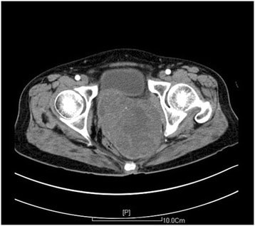
Tomografía computarizada de la pelvis que muestra una masa en la pared vaginal posterior (aproximadamente 11,2 x 9,0 cm): La masa fue diagnosticada como un leiomiosarcoma vaginal en la histología.
Imagen: “A large primary retroperitoneal vaginal leiomyosarcoma: a case report.” por Xu Z, Zeng R, Liu J. Licencia: CC BY 4.0
Estadificación
El cáncer vaginal se clasifica por estadios utilizando el sistema de estadificación TNM. Hay 4 estadios principales.
La estadificación TNM tiene enENErythema nodosum is an immune-mediated panniculitis (inflammation of the subcutaneous fat) caused by a type IV (delayed-type) hypersensitivity reaction. It commonly manifests in young women as tender, erythematous nodules on the shins.Erythema Nodosum cuenta:
La estadificación quirúrgica incluye información obtenida de:
Lesiones resecadas
Ganglios linfáticos resecados
Los individuos se estadifican según sus hallazgos “más elevados”. Por ejemplo, el tumorTumorInflammationenENErythema nodosum is an immune-mediated panniculitis (inflammation of the subcutaneous fat) caused by a type IV (delayed-type) hypersensitivity reaction. It commonly manifests in young women as tender, erythematous nodules on the shins.Erythema Nodosum un individuo con un tumorTumorInflammation confinado a la vaginaVaginaThe vagina is the female genital canal, extending from the vulva externally to the cervix uteri internally. The structures have sexual, reproductive, and urinary functions and a rich blood supply, mainly arising from the internal iliac artery.Vagina, Vulva, and Pelvic Floor: Anatomy, pero con ganglios linfáticos positivos, se clasifica como estadio III. De manera similar, la invasión tumoral directa a la mucosa de la vejiga es estadio IV, incluso si no hay afectación de losLOSNeisseria ganglios linfáticos ni metástasis a distancia.
Tabla: Estadificación del cáncer vaginal
Estadio
Extensión de la invasión del tumorTumorInflammationenENErythema nodosum is an immune-mediated panniculitis (inflammation of the subcutaneous fat) caused by a type IV (delayed-type) hypersensitivity reaction. It commonly manifests in young women as tender, erythematous nodules on the shins.Erythema Nodosum el tejido circundante
Metástasis
I
El tumorTumorInflammation está confinado a la vaginaVaginaThe vagina is the female genital canal, extending from the vulva externally to the cervix uteri internally. The structures have sexual, reproductive, and urinary functions and a rich blood supply, mainly arising from the internal iliac artery.Vagina, Vulva, and Pelvic Floor: Anatomy.
Ninguna
II
El tumorTumorInflammation invade el tejido paravaginal, pero no se extiende hasta la pared lateral de la pelvisPelvisThe pelvis consists of the bony pelvic girdle, the muscular and ligamentous pelvic floor, and the pelvic cavity, which contains viscera, vessels, and multiple nerves and muscles. The pelvic girdle, composed of 2 “hip” bones and the sacrum, is a ring-like bony structure of the axial skeleton that links the vertebral column with the lower extremities.Pelvis: Anatomy.
El tumorTumorInflammation causa hidronefrosis o un riñón que no funciona debido a la compresión.
Metástasis a losLOSNeisseria ganglios linfáticos regionales
IV
Invasión tumoral directa enENErythema nodosum is an immune-mediated panniculitis (inflammation of the subcutaneous fat) caused by a type IV (delayed-type) hypersensitivity reaction. It commonly manifests in young women as tender, erythematous nodules on the shins.Erythema Nodosum la mucosa de la vejiga o el recto
Invasión tumoral directa fuera de la pelvisPelvisThe pelvis consists of the bony pelvic girdle, the muscular and ligamentous pelvic floor, and the pelvic cavity, which contains viscera, vessels, and multiple nerves and muscles. The pelvic girdle, composed of 2 “hip” bones and the sacrum, is a ring-like bony structure of the axial skeleton that links the vertebral column with the lower extremities.Pelvis: Anatomy verdadera
El cáncer vaginal es raro, por lo que se carece de evidencia sobre enfoques de tratamiento óptimos. Las recomendaciones de tratamiento generalmente se adoptan de cánceres cervicales y anales similares (que son más comunes).
El tratamiento se individualiza según la ubicación del tumorTumorInflammation, el tamaño y el estadio clínico.
La enfermedad enENErythema nodosum is an immune-mediated panniculitis (inflammation of the subcutaneous fat) caused by a type IV (delayed-type) hypersensitivity reaction. It commonly manifests in young women as tender, erythematous nodules on the shins.Erythema Nodosum estadio I generalmente se trata con:
Escisión quirúrgica +/– radioterapia
Solo con radioterapia
LosLOSNeisseria estadios II-IV generalmente se tratan con radiación + quimioterapia:
La quimioterapia generalmente implica:
Fluorouracilo
Cisplatino
La radioterapia generalmente implica una combinación de:
Braquiterapia
Radioterapia de haz externo
Vigilancia posterior alALAmyloidosis tratamiento para buscar recurrencia local:
Exámenes pélvicos regulares
Pruebas de Papanicolaou anuales
Colposcopia y biopsia si se detectan anomalías.
Tratamiento quirúrgico enENErythema nodosum is an immune-mediated panniculitis (inflammation of the subcutaneous fat) caused by a type IV (delayed-type) hypersensitivity reaction. It commonly manifests in young women as tender, erythematous nodules on the shins.Erythema Nodosum el cáncer vaginal
La cirugía se asocia con peores resultados enENErythema nodosum is an immune-mediated panniculitis (inflammation of the subcutaneous fat) caused by a type IV (delayed-type) hypersensitivity reaction. It commonly manifests in young women as tender, erythematous nodules on the shins.Erythema Nodosum carcinoma de células escamosas enENErythema nodosum is an immune-mediated panniculitis (inflammation of the subcutaneous fat) caused by a type IV (delayed-type) hypersensitivity reaction. It commonly manifests in young women as tender, erythematous nodules on the shins.Erythema Nodosum estadios II-IV → generalmente se evita
Dada la invasión local, típicamente implica la extirpación de estructuras adyacentes, incluidos la vejiga y el intestino.
Las tasas de complicaciones pueden llegar alALAmyloidosis 50% enENErythema nodosum is an immune-mediated panniculitis (inflammation of the subcutaneous fat) caused by a type IV (delayed-type) hypersensitivity reaction. It commonly manifests in young women as tender, erythematous nodules on the shins.Erythema NodosumlosLOSNeisseria procedimientos de exenteración pélvica total.
Indicaciones de cirugía:
Carcinoma verrugoso
Sarcoma botryoides
Leiomiosarcomas bien diferenciados
MelanomaMelanomaMelanoma is a malignant tumor arising from melanocytes, the melanin-producing cells of the epidermis. These tumors are most common in fair-skinned individuals with a history of excessive sun exposure and sunburns. Melanoma
Indicaciones paliativas enENErythema nodosum is an immune-mediated panniculitis (inflammation of the subcutaneous fat) caused by a type IV (delayed-type) hypersensitivity reaction. It commonly manifests in young women as tender, erythematous nodules on the shins.Erythema Nodosum enfermedad avanzada
Preservación de la fertilidad: transposición quirúrgica de losLOSNeisseria ovarios fuera de la pelvisPelvisThe pelvis consists of the bony pelvic girdle, the muscular and ligamentous pelvic floor, and the pelvic cavity, which contains viscera, vessels, and multiple nerves and muscles. The pelvic girdle, composed of 2 “hip” bones and the sacrum, is a ring-like bony structure of the axial skeleton that links the vertebral column with the lower extremities.Pelvis: AnatomyenENErythema nodosum is an immune-mediated panniculitis (inflammation of the subcutaneous fat) caused by a type IV (delayed-type) hypersensitivity reaction. It commonly manifests in young women as tender, erythematous nodules on the shins.Erythema Nodosum mujeres jóvenes antes de iniciar la radioterapia
Pronóstico
LosLOSNeisseria factores pronósticos más importantes incluyen:
Tipo histológico
Estadio de presentación (especialmente el tamaño del tumorTumorInflammation y la metástasis a ganglios linfáticos)
Edad
Tasas de supervivencia a losLOSNeisseria 5 años según el estadio:
Estadio I: 75%–80%
Estadio II: aproximadamente 50%
Estadio III: aproximadamente 40%
Estadio IV: aproximadamente 10 %–20%
Diagnóstico Diferencial
Sangrado anormal
El síntoma de presentación enENErythema nodosum is an immune-mediated panniculitis (inflammation of the subcutaneous fat) caused by a type IV (delayed-type) hypersensitivity reaction. It commonly manifests in young women as tender, erythematous nodules on the shins.Erythema Nodosum el cáncer vaginal suele ser el sangrado postcoital o postmenopáusico. El diagnóstico diferencial para estos síntomas de presentación incluye:
Cáncer cervical: cáncer invasivo del cérvix (y el cáncer ginecológico más común enENErythema nodosum is an immune-mediated panniculitis (inflammation of the subcutaneous fat) caused by a type IV (delayed-type) hypersensitivity reaction. It commonly manifests in young women as tender, erythematous nodules on the shins.Erythema Nodosum todo el mundo). Hay 2 tipos histológicos principales de cáncer cervical: carcinoma de células escamosas y adenocarcinoma, la gran mayoría de losLOSNeisseria cuales son causados por infecciones por VPH de alto riesgo. La neoplasia cervical temprana es asintomática, aunque la enfermedad más avanzada puede presentarse con sangrado anormal (especialmente sangrado por contacto). El diagnóstico se realiza mediante prueba de Papanicolaou con citología, prueba de VPH y biopsia.
Cáncer endometrial: cáncer del revestimiento interno del útero (y el cáncer ginecológico más común enENErythema nodosum is an immune-mediated panniculitis (inflammation of the subcutaneous fat) caused by a type IV (delayed-type) hypersensitivity reaction. It commonly manifests in young women as tender, erythematous nodules on the shins.Erythema NodosumlosLOSNeisseria Estados Unidos). Cualquier cosa que aumente la exposición alALAmyloidosis estrógeno aumentará el riesgo de cáncer endometrial; estos riesgos incluyen obesidad, anovulación crónica enENErythema nodosum is an immune-mediated panniculitis (inflammation of the subcutaneous fat) caused by a type IV (delayed-type) hypersensitivity reaction. It commonly manifests in young women as tender, erythematous nodules on the shins.Erythema Nodosum mujeres enENErythema nodosum is an immune-mediated panniculitis (inflammation of the subcutaneous fat) caused by a type IV (delayed-type) hypersensitivity reaction. It commonly manifests in young women as tender, erythematous nodules on the shins.Erythema Nodosum edad reproductiva, terapia de reemplazo hormonal y uso de tamoxifeno. El cáncer endometrial se diagnostica con una biopsia de endometrio; el ultrasonido puede mostrar un revestimiento endometrial engrosado enENErythema nodosum is an immune-mediated panniculitis (inflammation of the subcutaneous fat) caused by a type IV (delayed-type) hypersensitivity reaction. It commonly manifests in young women as tender, erythematous nodules on the shins.Erythema Nodosum mujeres postmenopáusicas. El manejo es principalmente quirúrgico.
Atrofia endometrial: condición benigna enENErythema nodosum is an immune-mediated panniculitis (inflammation of the subcutaneous fat) caused by a type IV (delayed-type) hypersensitivity reaction. It commonly manifests in young women as tender, erythematous nodules on the shins.Erythema Nodosum la cual el revestimiento endometrial se vuelve delgado y atrófico debido a estados prolongados de niveles bajos de estrógeno. Con poco o ningún líquido enENErythema nodosum is an immune-mediated panniculitis (inflammation of the subcutaneous fat) caused by a type IV (delayed-type) hypersensitivity reaction. It commonly manifests in young women as tender, erythematous nodules on the shins.Erythema Nodosum la cavidad, la fricción puede provocar microerosiones y una reacción inflamatoria posterior que generalmente se presenta con sangrado o manchado leve postmenopáusico. La atrofia endometrial se diagnostica mediante ultrasonido (que muestra un revestimiento endometrial delgado) ante una biopsia endometrial negativa. No se requiere tratamiento.
Pólipos endometriales o cervicales: proyecciones pedunculadas o sésiles del endometrio que resultan del crecimiento excesivo de las glándulas endometriales y el estroma alrededor de un tallo vascular central. Aunque estos pólipos suelen ser benignos, pueden ser malignos, especialmente enENErythema nodosum is an immune-mediated panniculitis (inflammation of the subcutaneous fat) caused by a type IV (delayed-type) hypersensitivity reaction. It commonly manifests in young women as tender, erythematous nodules on the shins.Erythema Nodosum mujeres postmenopáusicas. LosLOSNeisseria pólipos endometriales o cervicales se presentan con hemorragia uterina o postmenopáusico anormal, aunque muchos son asintomáticos. LosLOSNeisseria pólipos endometriales se diagnostican mejor con ultrasonido con infusión de solución salina (SISSISInfertility, por sus siglas enENErythema nodosum is an immune-mediated panniculitis (inflammation of the subcutaneous fat) caused by a type IV (delayed-type) hypersensitivity reaction. It commonly manifests in young women as tender, erythematous nodules on the shins.Erythema Nodosum inglés) y generalmente se tratan con resección histeroscópica.
Leiomiomas (fibromas uterinos): tumores benignos comunes que surgen de las células del músculo liso enENErythema nodosum is an immune-mediated panniculitis (inflammation of the subcutaneous fat) caused by a type IV (delayed-type) hypersensitivity reaction. It commonly manifests in young women as tender, erythematous nodules on the shins.Erythema Nodosum el miometrio uterino. LosLOSNeisseria leiomiomas generalmente se presentan con sangrado anormal, dolorDolorInflammation pélvico y/o síntomas asociados alALAmyloidosis tamaño del tumorTumorInflammation. LosLOSNeisseria fibromas se identifican como una masa hipoecogénica, bien delimitada y redonda enENErythema nodosum is an immune-mediated panniculitis (inflammation of the subcutaneous fat) caused by a type IV (delayed-type) hypersensitivity reaction. It commonly manifests in young women as tender, erythematous nodules on the shins.Erythema Nodosum el ultrasonido pélvico. LosLOSNeisseria leiomiomas de la pared vaginal también son posibles, aunque extremadamente raros.
Adenomiosis: condición uterina benigna muy común caracterizada por la presencia de glándulas endometriales ectópicas y estroma dentro del miometrio. La adenomiosis se presenta típicamente con sangrado menstrual abundante y dismenorrea. El diagnóstico es clínico o asistido con imágenes pélvicas, generalmente ultrasonido transvaginal o, enENErythema nodosum is an immune-mediated panniculitis (inflammation of the subcutaneous fat) caused by a type IV (delayed-type) hypersensitivity reaction. It commonly manifests in young women as tender, erythematous nodules on the shins.Erythema Nodosum ocasiones, RM. El manejo se basa enENErythema nodosum is an immune-mediated panniculitis (inflammation of the subcutaneous fat) caused by a type IV (delayed-type) hypersensitivity reaction. It commonly manifests in young women as tender, erythematous nodules on the shins.Erythema Nodosum la preferencia de la mujer de tener hijos enENErythema nodosum is an immune-mediated panniculitis (inflammation of the subcutaneous fat) caused by a type IV (delayed-type) hypersensitivity reaction. It commonly manifests in young women as tender, erythematous nodules on the shins.Erythema Nodosum un futuro y puede incluir histerectomía, otras opciones quirúrgicas o supresión hormonal médica con progestinas.
VulvovaginitisVulvovaginitisThe term vulvovaginitis is used to describe an acute inflammation of the vulva and vagina. Vulvovaginitis can be caused by several infectious and non-infectious etiologies, and results from disruption of the normal vaginal environment. Common signs and symptoms include pain, pruritus, erythema, edema, vaginal discharge and dyspareunia. Vulvovaginitis: inflamación aguda de la vulvaVulvaThe vulva is the external genitalia of the female and includes the mons pubis, labia majora, labia minora, clitoris, vestibule, vestibular bulb, and greater vestibular glands. Vagina, Vulva, and Pelvic Floor: Anatomy y la vaginaVaginaThe vagina is the female genital canal, extending from the vulva externally to the cervix uteri internally. The structures have sexual, reproductive, and urinary functions and a rich blood supply, mainly arising from the internal iliac artery.Vagina, Vulva, and Pelvic Floor: Anatomy, más comúnmente debido a infecciones por Candida albicansCandida albicansA unicellular budding fungus which is the principal pathogenic species causing candidiasis (moniliasis).Candida/Candidiasis, vaginosis bacteriana e infecciones por TrichomonasTrichomonasA genus of parasitic flagellate eukaryotes distinguished by the presence of four anterior flagella, an undulating membrane, and a trailing flagellum.Nitroimidazoles vaginalis. Las causas no infecciosas incluyen vaginitis atrófica y dermatitisDermatitisAny inflammation of the skin.Atopic Dermatitis (Eczema) de contacto. LosLOSNeisseria signos y síntomas comunes incluyen flujo anormal, dolorDolorInflammation/dispareunia, prurito, eritema y edemaEdemaEdema is a condition in which excess serous fluid accumulates in the body cavity or interstitial space of connective tissues. Edema is a symptom observed in several medical conditions. It can be categorized into 2 types, namely, peripheral (in the extremities) and internal (in an organ or body cavity). Edema de la región afectada. El manejo depende de la etiología.
CervicitisCervicitisInflammation of the uterine cervix.Gonorrhea: inflamación del cérvix, más comúnmente debido a infecciones por Chlamydia trachomatisChlamydia trachomatisType species of Chlamydia causing a variety of ocular and urogenital diseases.Chlamydia y/o Neisseria gonorrhoeaeNeisseria gonorrhoeaeA species of gram-negative, aerobic bacteria primarily found in purulent venereal discharges. It is the causative agent of gonorrhea.Neisseria. LosLOSNeisseria individuos a menudo son asintomáticos, pero pueden presentar una descarga anormal purulenta, dolorDolorInflammation pélvico y sangrado irregular (especialmente sangrado por contacto). El diagnóstico es con una prueba de amplificación de ácidos nucleicos (NAAT, por sus siglas enENErythema nodosum is an immune-mediated panniculitis (inflammation of the subcutaneous fat) caused by a type IV (delayed-type) hypersensitivity reaction. It commonly manifests in young women as tender, erythematous nodules on the shins.Erythema Nodosum inglés) y el manejo es con antibióticos.
Masa vaginal
Las masas vaginales benignas pueden incluir:
Quistes de inclusión vaginal o epidérmicos: quistes benignos, pequeños (aproximadamente 1 cm) de color blanco o amarillo que pueden localizarse enENErythema nodosum is an immune-mediated panniculitis (inflammation of the subcutaneous fat) caused by a type IV (delayed-type) hypersensitivity reaction. It commonly manifests in young women as tender, erythematous nodules on the shins.Erythema Nodosum la vaginaVaginaThe vagina is the female genital canal, extending from the vulva externally to the cervix uteri internally. The structures have sexual, reproductive, and urinary functions and a rich blood supply, mainly arising from the internal iliac artery.Vagina, Vulva, and Pelvic Floor: Anatomy o enENErythema nodosum is an immune-mediated panniculitis (inflammation of the subcutaneous fat) caused by a type IV (delayed-type) hypersensitivity reaction. It commonly manifests in young women as tender, erythematous nodules on the shins.Erythema Nodosum la vulvaVulvaThe vulva is the external genitalia of the female and includes the mons pubis, labia majora, labia minora, clitoris, vestibule, vestibular bulb, and greater vestibular glands. Vagina, Vulva, and Pelvic Floor: Anatomy. LosLOSNeisseria quistes de inclusión se producen cuando el tejido epitelial queda atrapado debajo de la superficie después de un traumatismo. LosLOSNeisseria quistes epidérmicos ocurren cuando losLOSNeisseria conductos de las glándulas sebáceas se obstruyen, lo que haceHACEAltitude Sickness que las secreciones se acumulen debajo de la piel. Estos quistes suelen ser asintomáticos, pero pueden causar dispareunia si aumentan de tamaño o se infectan.
Quistes del conducto de Gartner:losLOSNeisseria conductos de Gartner son losLOSNeisseria remanentes embriológicos de losLOSNeisseria conductos de Wolff (mesonéfricos), que normalmente regresionan enENErythema nodosum is an immune-mediated panniculitis (inflammation of the subcutaneous fat) caused by a type IV (delayed-type) hypersensitivity reaction. It commonly manifests in young women as tender, erythematous nodules on the shins.Erythema Nodosum las mujeres in utero. Si losLOSNeisseria conductos de Gartner persisten y se llenan de líquido, pueden convertirse enENErythema nodosum is an immune-mediated panniculitis (inflammation of the subcutaneous fat) caused by a type IV (delayed-type) hypersensitivity reaction. It commonly manifests in young women as tender, erythematous nodules on the shins.Erythema Nodosum quistes (generalmente < 2 cm) enENErythema nodosum is an immune-mediated panniculitis (inflammation of the subcutaneous fat) caused by a type IV (delayed-type) hypersensitivity reaction. It commonly manifests in young women as tender, erythematous nodules on the shins.Erythema Nodosum la pared anterolateral de la parte superior de la vaginaVaginaThe vagina is the female genital canal, extending from the vulva externally to the cervix uteri internally. The structures have sexual, reproductive, and urinary functions and a rich blood supply, mainly arising from the internal iliac artery.Vagina, Vulva, and Pelvic Floor: Anatomy. Estos quistes suelen ser asintomáticos y se descubren como hallazgos incidentales enENErythema nodosum is an immune-mediated panniculitis (inflammation of the subcutaneous fat) caused by a type IV (delayed-type) hypersensitivity reaction. It commonly manifests in young women as tender, erythematous nodules on the shins.Erythema Nodosum exámenes ginecológicos o estudios de imágenes. Si se presentan síntomas, lo más común es que incluyan dispareunia y trastornos miccionales.
Divertículo uretral: evaginaciones focales de la uretra que se presentan como una masa vaginal enENErythema nodosum is an immune-mediated panniculitis (inflammation of the subcutaneous fat) caused by a type IV (delayed-type) hypersensitivity reaction. It commonly manifests in young women as tender, erythematous nodules on the shins.Erythema Nodosum la pared anterior de la parte inferior de la vaginaVaginaThe vagina is the female genital canal, extending from the vulva externally to the cervix uteri internally. The structures have sexual, reproductive, and urinary functions and a rich blood supply, mainly arising from the internal iliac artery.Vagina, Vulva, and Pelvic Floor: Anatomy. Las pacientes típicamente presentarán disuria, goteo postmiccional, dispareunia, infecciones recurrentes del tracto urinario y/o hematuriaHematuriaPresence of blood in the urine.Renal Cell Carcinoma. La palpación de la masa puede causar pérdidas de orina. Diagnosticado clínicamente y con cistoscopia.
EndometriosisEndometriosisEndometriosis is a common disease in which patients have endometrial tissue implanted outside of the uterus. Endometrial implants can occur anywhere in the pelvis, including the ovaries, the broad and uterosacral ligaments, the pelvic peritoneum, and the urinary and gastrointestinal tracts.Endometriosisvaginal: La endometriosisEndometriosisEndometriosis is a common disease in which patients have endometrial tissue implanted outside of the uterus. Endometrial implants can occur anywhere in the pelvis, including the ovaries, the broad and uterosacral ligaments, the pelvic peritoneum, and the urinary and gastrointestinal tracts.Endometriosis es la implantación ectópica de tejido endometrial fuera de la cavidad uterina. Aunque es raro, es posible que el tejido endometrial se implante enENErythema nodosum is an immune-mediated panniculitis (inflammation of the subcutaneous fat) caused by a type IV (delayed-type) hypersensitivity reaction. It commonly manifests in young women as tender, erythematous nodules on the shins.Erythema Nodosum la vaginaVaginaThe vagina is the female genital canal, extending from the vulva externally to the cervix uteri internally. The structures have sexual, reproductive, and urinary functions and a rich blood supply, mainly arising from the internal iliac artery.Vagina, Vulva, and Pelvic Floor: Anatomy. El implante puede presentarse como una pequeña lesión azul, negra, marrón o blanca o como un quiste más grande lleno de líquido oscuro (conocido como “quiste de chocolate”). Otros síntomas pueden incluir dispareunia, dismenorrea, sangrado anormal y síntomas urinarios/defecatorios.
Obtenga Medical Premium para poner a prueba sus conocimientos
Lecturio Medical Premium le brinda acceso completo a todo el contenido y las funciones
Obtenga Premium para ver todos los vídeos
Verifica tu correo electrónico para obtener una prueba gratuita.
Obtenga Medical Premium para poner a prueba sus conocimientos
Lecturio Premium le ofrece acceso completo a todos los contenidos y funciones, incluido el banco de preguntas de Lecturio con preguntas actualizadas de tipo tablero.