Los LOS Neisseria bloqueos de rama y fasciculares se producen cuando se interrumpe la actividad eléctrica normal del sistema de His-Purkinje. Estos bloqueos pueden deberse a muchas etiologías que pueden afectar a la estructura del corazón o al AL Amyloidosis sistema de conducción directamente. Los LOS Neisseria bloqueos se clasifican en EN Erythema nodosum is an immune-mediated panniculitis (inflammation of the subcutaneous fat) caused by a type IV (delayed-type) hypersensitivity reaction. It commonly manifests in young women as tender, erythematous nodules on the shins. Erythema Nodosum bloqueo de rama derecha, bloqueo de rama izquierda, bloqueo del fascículo anterior izquierdo y bloqueo del fascículo posterior izquierdo en EN Erythema nodosum is an immune-mediated panniculitis (inflammation of the subcutaneous fat) caused by a type IV (delayed-type) hypersensitivity reaction. It commonly manifests in young women as tender, erythematous nodules on the shins. Erythema Nodosum función de la localización de la alteración. La mayoría de los LOS Neisseria individuos son asintomáticos. El ECG ECG An electrocardiogram (ECG) is a graphic representation of the electrical activity of the heart plotted against time. Adhesive electrodes are affixed to the skin surface allowing measurement of cardiac impulses from many angles. The ECG provides 3-dimensional information about the conduction system of the heart, the myocardium, and other cardiac structures. Electrocardiogram (ECG) proporcionará el diagnóstico. Algunos hallazgos comunes del ECG ECG An electrocardiogram (ECG) is a graphic representation of the electrical activity of the heart plotted against time. Adhesive electrodes are affixed to the skin surface allowing measurement of cardiac impulses from many angles. The ECG provides 3-dimensional information about the conduction system of the heart, the myocardium, and other cardiac structures. Electrocardiogram (ECG) incluyen un intervalo QRS prolongado, cambios en EN Erythema nodosum is an immune-mediated panniculitis (inflammation of the subcutaneous fat) caused by a type IV (delayed-type) hypersensitivity reaction. It commonly manifests in young women as tender, erythematous nodules on the shins. Erythema Nodosum la onda R, desviación del eje y ( en EN Erythema nodosum is an immune-mediated panniculitis (inflammation of the subcutaneous fat) caused by a type IV (delayed-type) hypersensitivity reaction. It commonly manifests in young women as tender, erythematous nodules on the shins. Erythema Nodosum algunos casos) cambios en EN Erythema nodosum is an immune-mediated panniculitis (inflammation of the subcutaneous fat) caused by a type IV (delayed-type) hypersensitivity reaction. It commonly manifests in young women as tender, erythematous nodules on the shins. Erythema Nodosum la onda S. No está indicado ningún tratamiento específico.
Last updated: Jul 14, 2022
Los LOS Neisseria bloqueos de rama y fasciculares se clasifican en EN Erythema nodosum is an immune-mediated panniculitis (inflammation of the subcutaneous fat) caused by a type IV (delayed-type) hypersensitivity reaction. It commonly manifests in young women as tender, erythematous nodules on the shins. Erythema Nodosum función del lugar donde se produce la alteración dentro del sistema de His-Purkinje.

Los bloqueos de rama y fasciculares surgen debido a la obstrucción de la corriente eléctrica a través del sistema de His-Purkinje y se denominan en función de la localización de dicha interrupción.
Imagen por Lecturio.Estos bloqueos fasciculares pueden producirse por muchas de las mismas causas del bloqueo de rama izquierda o derecha, sobre todo:
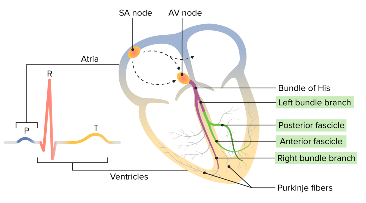
Electrofisiología normal del corazón:
El impulso de conducción comienza en el nodo sinoauricular y viaja a través de la aurícula hasta el nodo auriculoventricular. Desde ahí, se desplaza por el haz de His hacia abajo a través de las dos ramas del haz (y los fascículos) hasta las fibras de Purkinje. El movimiento de este impulso eléctrico puede registrarse en un ECG.
Onda P (azul): despolarización del miocardio auricular
Complejo QRS (naranja): despolarización del miocardio ventricular
Onda T (amarilla): repolarización del miocardio ventricular
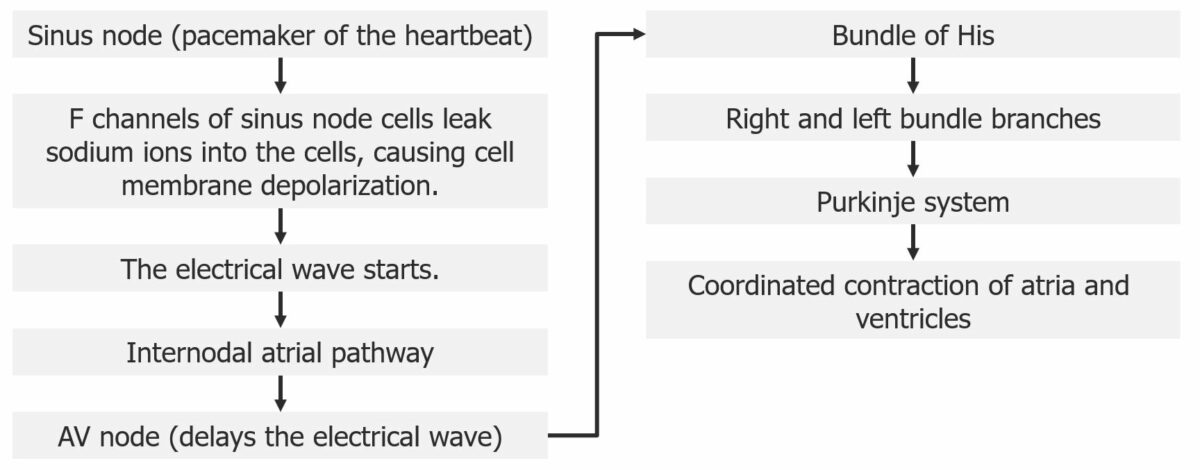
Diagrama del recorrido eléctrico a través del corazón
Imagen por Lecturio.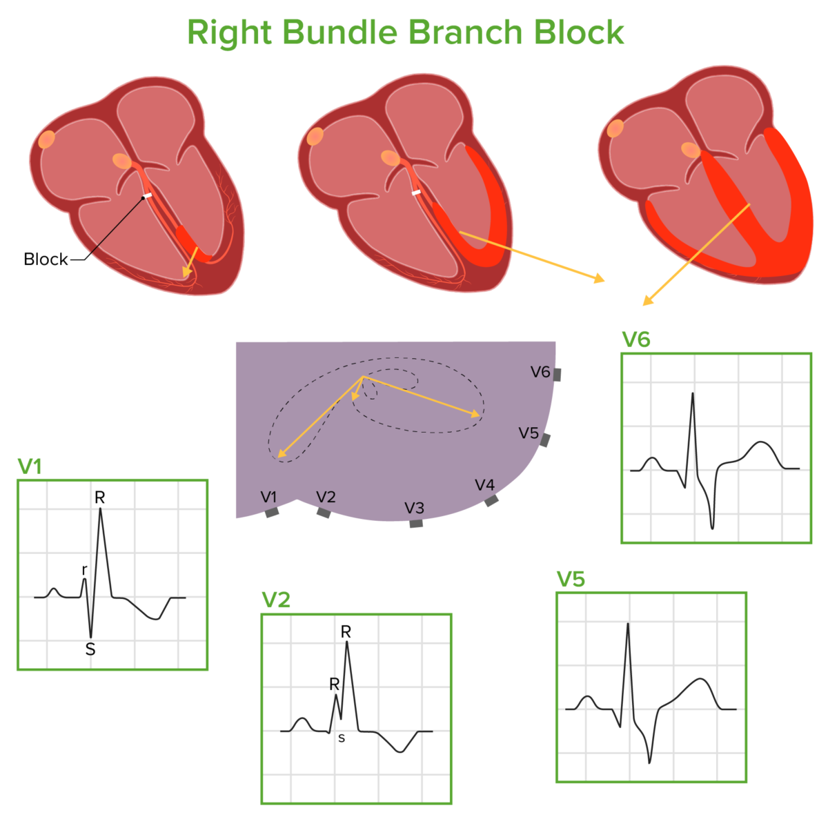
Diagrama que demuestra cómo se mueve el impulso eléctrico a través de los ventrículos en un bloqueo de rama derecha:
El impulso eléctrico pasa por la rama izquierda del haz, por el tabique y el ventrículo izquierdo, y luego por el ventrículo derecho. Estas fases dan lugar a los vectores de conducción eléctrica que se muestran arriba (dibujados en relación con una sección transversal del tórax con las derivaciones precordiales adheridas), que se correlacionan con las correspondientes formas de onda del ECG.
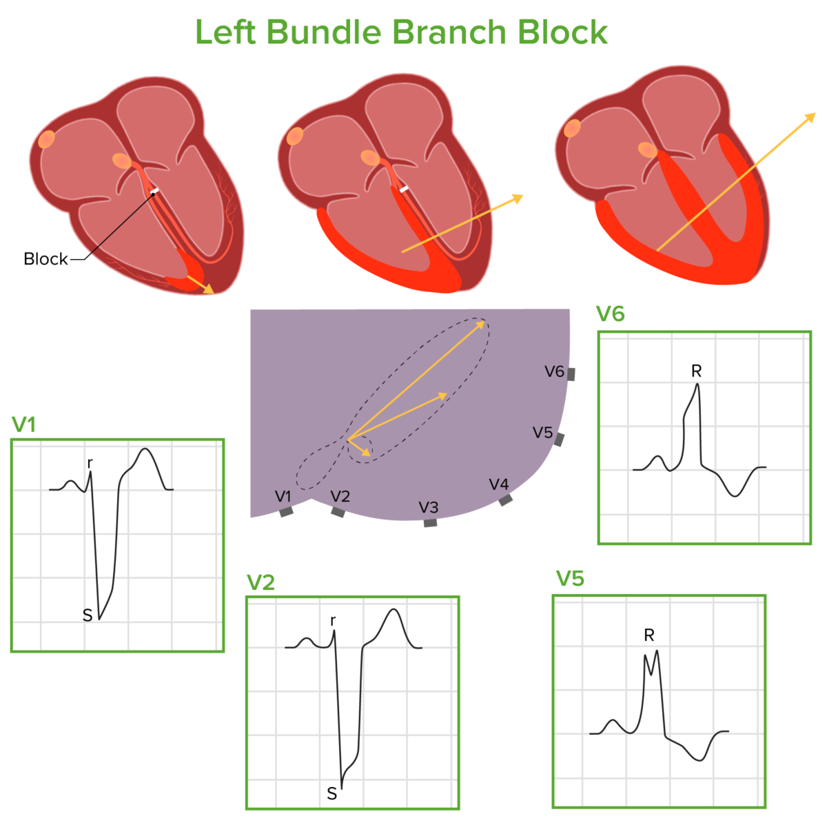
Diagrama que demuestra cómo se mueve el impulso eléctrico a través de los ventrículos en un bloqueo de rama izquierda:
El impulso eléctrico recorre la rama derecha del haz, atraviesa el tabique y el ventrículo derecho, y luego el ventrículo izquierdo. Estas fases dan lugar a los vectores de conducción eléctrica que se muestran arriba (dibujados en relación con una sección transversal del tórax con las derivaciones precordiales adheridas), que se correlacionan con las correspondientes formas de onda del ECG.
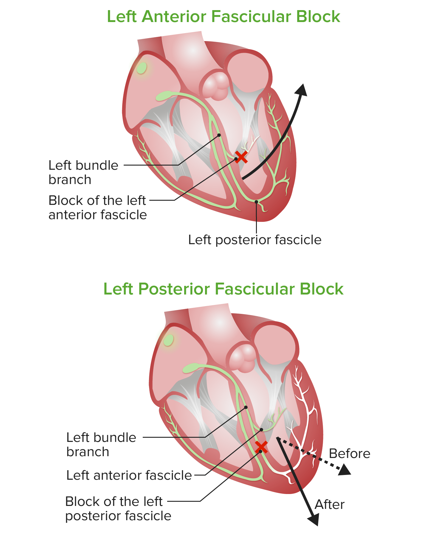
Esquema de los bloqueos fasciculares anterior y posterior izquierdos:
En el bloqueo fascicular anterior izquierdo o hemibloqueo, el vector eléctrico resultante provoca una desviación significativa del eje izquierdo. En el bloqueo fascicular posterior izquierdo o hemibloqueo, el vector eléctrico se desvía un poco hacia la derecha pero no se desplaza significativamente del rango normal del eje QRS.
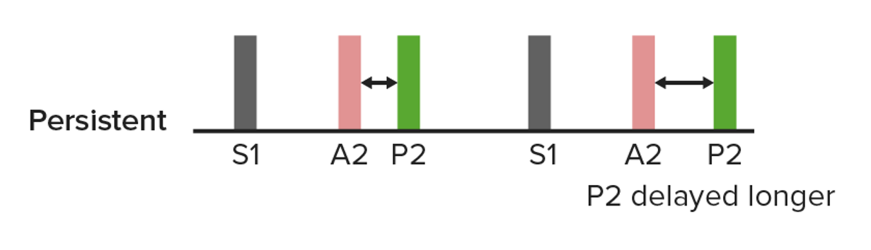
Un diagrama de un S2 dividido persistente, en el que el cierre de la válvula pulmonar se retrasa aún más por la inspiración (derecha). Esto puede ocurrir en un bloqueo de rama derecha.
Imagen por Lecturio.Audio:
Este clip de audio es un ejemplo de un S2 S2 Heart Sounds dividido en EN Erythema nodosum is an immune-mediated panniculitis (inflammation of the subcutaneous fat) caused by a type IV (delayed-type) hypersensitivity reaction. It commonly manifests in young women as tender, erythematous nodules on the shins. Erythema Nodosum el contexto de un bloqueo de rama derecha. Los LOS Neisseria dos ruidos que se producen durante la S2 S2 Heart Sounds son el resultado de un retraso en EN Erythema nodosum is an immune-mediated panniculitis (inflammation of the subcutaneous fat) caused by a type IV (delayed-type) hypersensitivity reaction. It commonly manifests in young women as tender, erythematous nodules on the shins. Erythema Nodosum el cierre de la válvula pulmonar con respecto a la válvula aórtica.
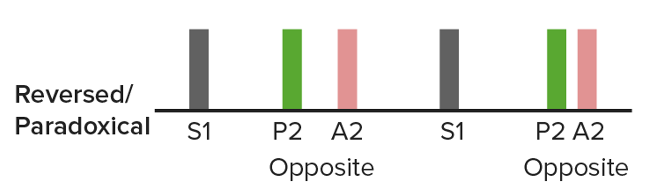
Un diagrama de una división paradójica en el cierre de la válvula aórtica que se retrasa:
El nombre “paradójico” se debe a que la división se estrecha con la inspiración (derecha). Esto puede oírse en algunos individuos con un bloqueo de rama izquierda.
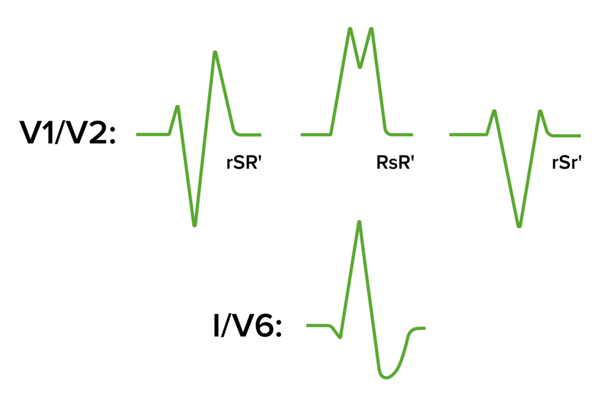
Morfología del QRS observada en el bloqueo de rama derecha:
Los RsR’ (y sus variaciones) dan la apariencia de “orejas de conejo”. La onda S en las derivaciones I y V6 aparecerá amplia, profunda y desplazada.
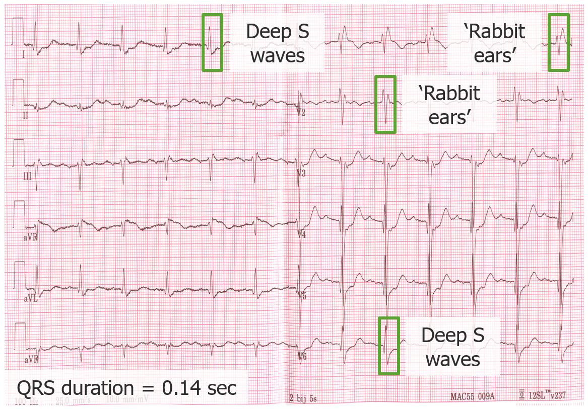
ECG que demuestra un bloqueo de rama derecha:
La duración del QRS es prolongada, a 140 mseg. Obsérvese la rSR’ y la RSr’ en las derivaciones V1 y 2, junto con las ondas S profundas y amplias en I y V6.
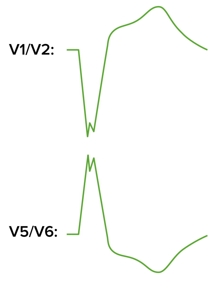
Patrones en el ECG en el bloqueo de rama izquierda:
Se observará una gran onda S en V1, mientras que en V5 y V6 se produce una gran onda R con muescas. Obsérvese que las direcciones del segmento ST y de la onda T son discordantes con el QRS.
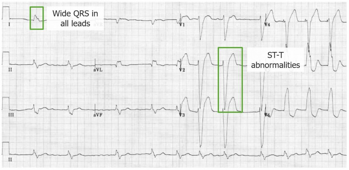
ECG que demuestra un bloqueo de rama izquierda:
Obsérvese el QRS ensanchado; las ondas R grandes y dentadas en V5 y V6; y las ondas S grandes y anchas en V1 y 2. Los segmentos ST y las ondas T también suelen ser discordantes con el complejo QRS.
Un bloqueo de rama puede considerarse incompleto si se observa el patrón habitual de bloqueo de rama izquierda o derecha, pero la duración del QRS es de 110–119 mseg.

ECG que demuestra el bloqueo fascicular anterior izquierdo:
Aquí, el eje se desvía a –60 grados y se observa una pequeña onda Q en aVL. El QRS está ligeramente prolongado, pero todavía < 120 mseg.
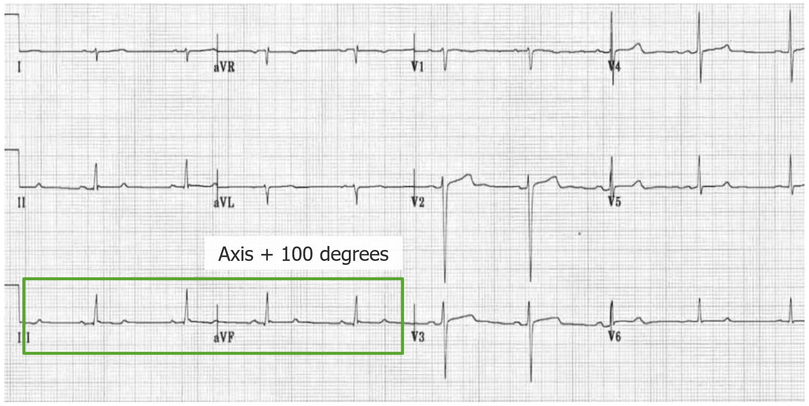
ECG que demuestra un bloqueo fascicular posterior izquierdo:
Hay desviación del eje derecho (+ 100 grados), pequeñas ondas Q en II, III y aVF, complejos rS en I y aVL. La duración del complejo QRS también es < 120 mseg.