La articulación de la rodilla está formada por las articulaciones entre los LOS Neisseria huesos del fémur, la tibia Tibia The second longest bone of the skeleton. It is located on the medial side of the lower leg, articulating with the fibula laterally, the talus distally, and the femur proximally. Knee Joint: Anatomy y la rótula, y es una de las articulaciones más grandes y complejas del cuerpo humano. La rodilla se clasifica como una articulación de bisagra sinovial, que principalmente permite la flexión y extensión con un grado más limitado de traslación y rotación. Las estructuras de soporte de la articulación de la rodilla incluyen una cápsula articular, los LOS Neisseria meniscos lateral y medial y múltiples ligamentos que ayudan a garantizar la movilidad y la estabilidad de la rodilla.
Last updated: Dec 15, 2025
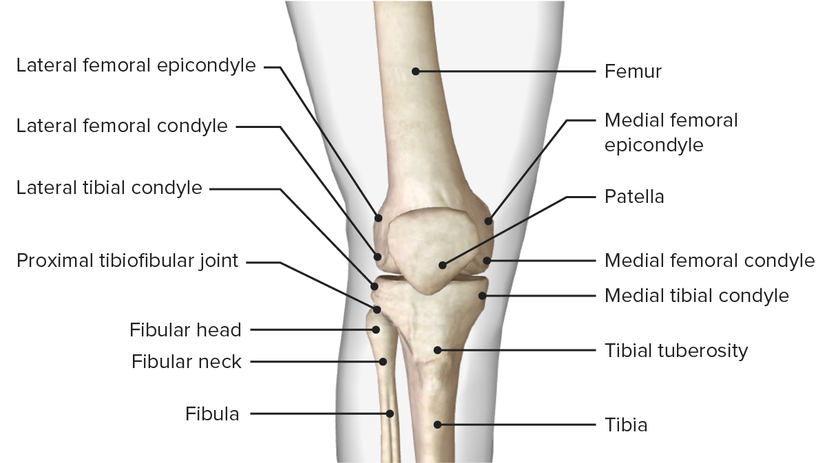
Vista anterior de la estructura ósea de la rodilla, mostrando los puntos de referencia óseos de los huesos del fémur, la tibia y la rótula.
Imagen por BioDigital, editado por Lecturio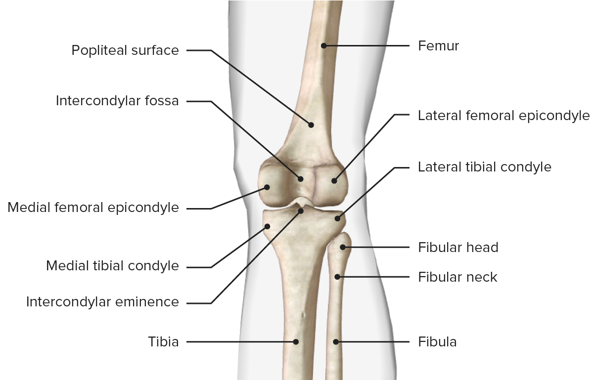
Vista posterior de la estructura ósea de la rodilla, mostrando los puntos de referencia óseos de los huesos del fémur, la tibia y la rótula.
Imagen por BioDigital, editado por LecturioLa rodilla es una articulación de bisagra modificada; una doble articulación condiloidea. Aunque los LOS Neisseria movimientos de la rodilla son principalmente de flexión y extensión, tiene un patrón de movimiento complejo que consta de 6 grados de movimiento durante las actividades dinámicas:
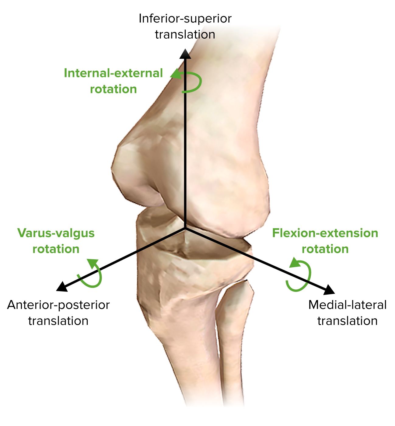
Movimientos de la rodilla
Imagen por BioDigital, editado por Lecturio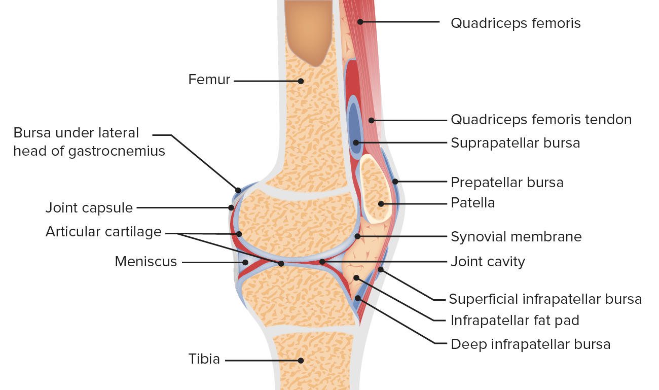
Anatomía de la articulación rotulofemoral
Imagen por Lecturio. Licencia: CC BY-NC-SA 4.0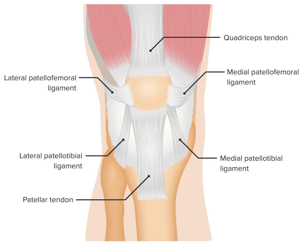
Ligamentos de sostén de la articulación rotulofemoral
Imagen por Lecturio. Licencia: CC BY-NC-SA 4.0
Vistas anterior y posterior de la tibia, el peroné y las articulaciones tibioperoneas
Imagen por BioDigital, editado por LecturioLos LOS Neisseria meniscos son cuñas de fibrocartílago de forma semilunar entre el fémur y la tibia Tibia The second longest bone of the skeleton. It is located on the medial side of the lower leg, articulating with the fibula laterally, the talus distally, and the femur proximally. Knee Joint: Anatomy y están hechos de fibras de colágeno tipo I. Los LOS Neisseria meniscos son amortiguadores, mejoran la congruencia de la articulación tibiofemoral y son vitales para el normal funcionamiento y salud de la rodilla.
Meniscos de la rodilla
Imagen por Lecturio. Licencia: CC BY-NC-SA 4.0
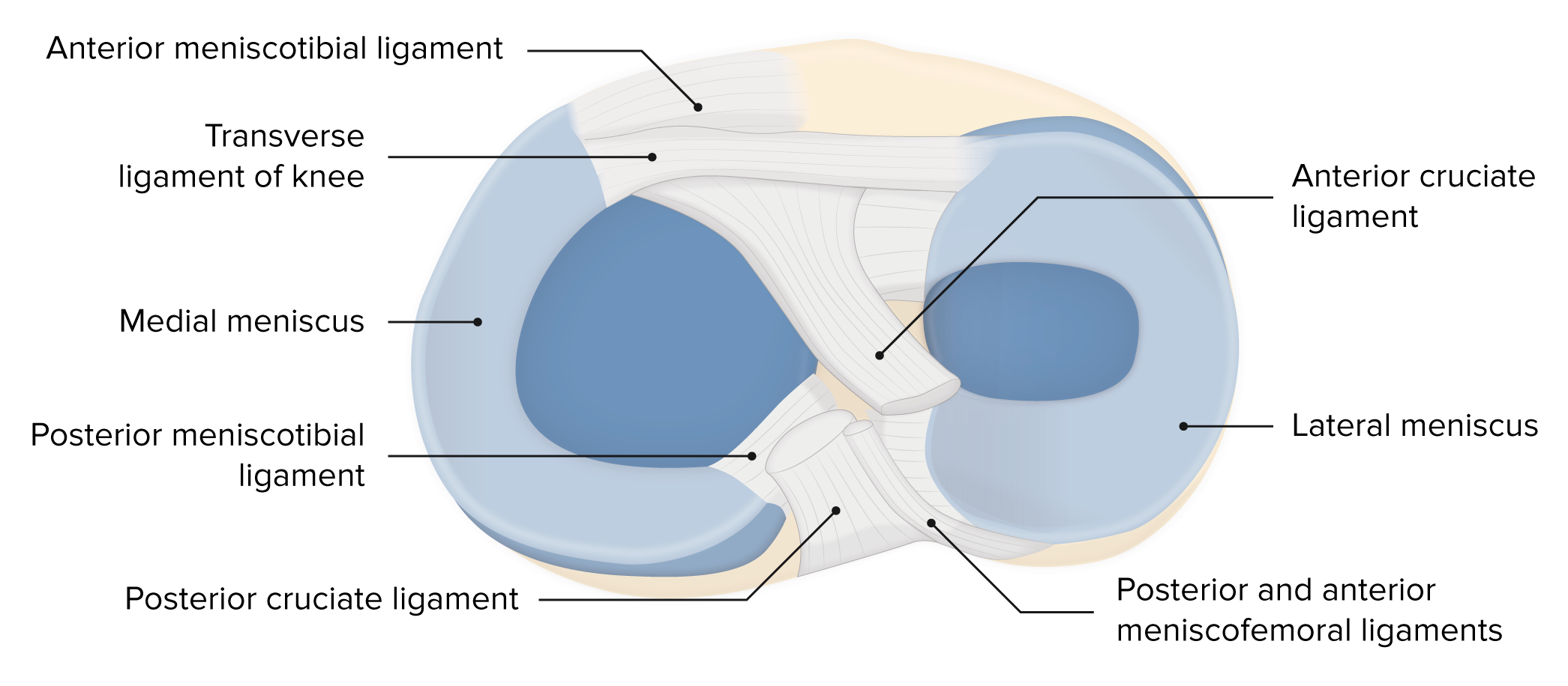
Imagen que muestra los meniscos, los ligamentos y las superficies óseas, y su relación entre sí
Imagen por Lecturio. Licencia: CC BY-NC-SA 4.0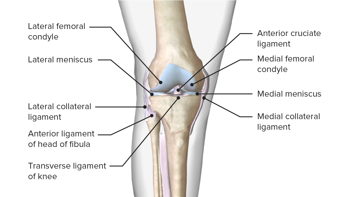
Imagen que muestra los meniscos y su relación con otras superficies articulares que componen la articulación de la rodilla
Imagen por BioDigital, editado por LecturioUna cápsula articular de 2 capas proporciona soporte a la rodilla. Estas capas son la membrana fibrosa externa y la membrana sinovial interna.
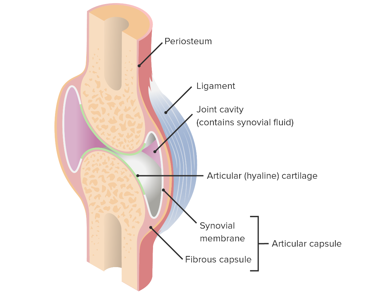
Cápsula articular y sus componentes
Imagen por Lecturio. Licencia: CC BY-NC-SA 4.0Los LOS Neisseria ligamentos y tendones de la rodilla brindan estabilidad a la rodilla durante el movimiento y aumentan la eficiencia de la rodilla.
| Ligamento/tendón | Origen | Inserción | Función |
|---|---|---|---|
| Ligamento cruzado anterior (LCA) se compone de 2 haces:
|
Cóndilo femoral lateral interno | Techo de la fosa intercondílea |
|
| Ligamento cruzado posterior (LCP) se compone de 2 haces:
|
Superficie interna del cóndilo femoral medial | Área intercondílea posterior de la tibia Tibia The second longest bone of the skeleton. It is located on the medial side of the lower leg, articulating with the fibula laterally, the talus distally, and the femur proximally. Knee Joint: Anatomy |
|
| Ligamento colateral medial (LCM) | Epicóndilo femoral medial | Cóndilo medial de la tibia Tibia The second longest bone of the skeleton. It is located on the medial side of the lower leg, articulating with the fibula laterally, the talus distally, and the femur proximally. Knee Joint: Anatomy | Estabiliza la articulación de la rodilla frente al AL Amyloidosis estrés en EN Erythema nodosum is an immune-mediated panniculitis (inflammation of the subcutaneous fat) caused by a type IV (delayed-type) hypersensitivity reaction. It commonly manifests in young women as tender, erythematous nodules on the shins. Erythema Nodosum valgo |
| Ligamento colateral lateral/peroneo | Epicóndilo femoral lateral | Cabeza del peroné | Estabiliza la articulación de la rodilla frente al AL Amyloidosis estrés en EN Erythema nodosum is an immune-mediated panniculitis (inflammation of the subcutaneous fat) caused by a type IV (delayed-type) hypersensitivity reaction. It commonly manifests in young women as tender, erythematous nodules on the shins. Erythema Nodosum varo. |
| Ligamento rotuliano | Rótula distal | Tuberosidad de la tibia Tibia The second longest bone of the skeleton. It is located on the medial side of the lower leg, articulating with the fibula laterally, the talus distally, and the femur proximally. Knee Joint: Anatomy | Componente del mecanismo extensor de la rodilla |
| Tendón del cuádriceps | Músculos cuádriceps | Rótula proximal | Componente del mecanismo extensor de la rodilla |
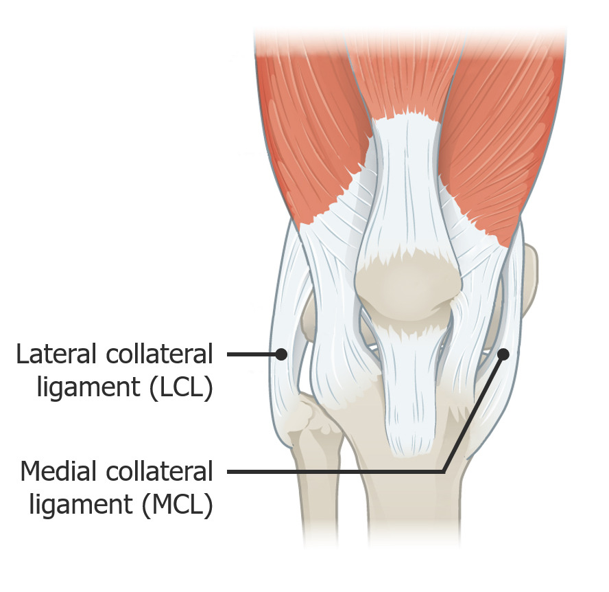
Imagen que muestra los ligamentos colaterales lateral y medial
Imagen: “917 Knee Joint” por OpenStax College. Licencia: CC BY 3.0, editado por Lecturio.Imagen que muestra los meniscos, los ligamentos y las superficies óseas, y su relación entre sí
Imagen por Lecturio. Licencia: CC BY-NC-SA 4.0Las bursas son membranas serosas/sacos llenos de líquido sinovial con una pequeña cantidad de líquido que facilita el movimiento alrededor de una articulación. Hay múltiples bursas descritas alrededor de la articulación de la rodilla.
| Bursas | Localización |
|---|---|
| Suprapatelar | Entre el fémur y el tendón del músculo cuádriceps |
| Prerrotuliana | Entre la rótula y la piel |
| Infrapatelar (superficial y profunda) |
|
| Pata de ganso | Rodilla medial/ tibia Tibia The second longest bone of the skeleton. It is located on the medial side of the lower leg, articulating with the fibula laterally, the talus distally, and the femur proximally. Knee Joint: Anatomy medial proximal |
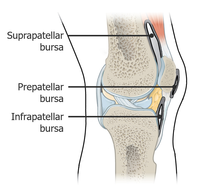
Imagen que muestra las bursas
Imagen: “917 Knee Joint” por OpenStax College. Licencia: CC BY 3.0, editado por Lecturio.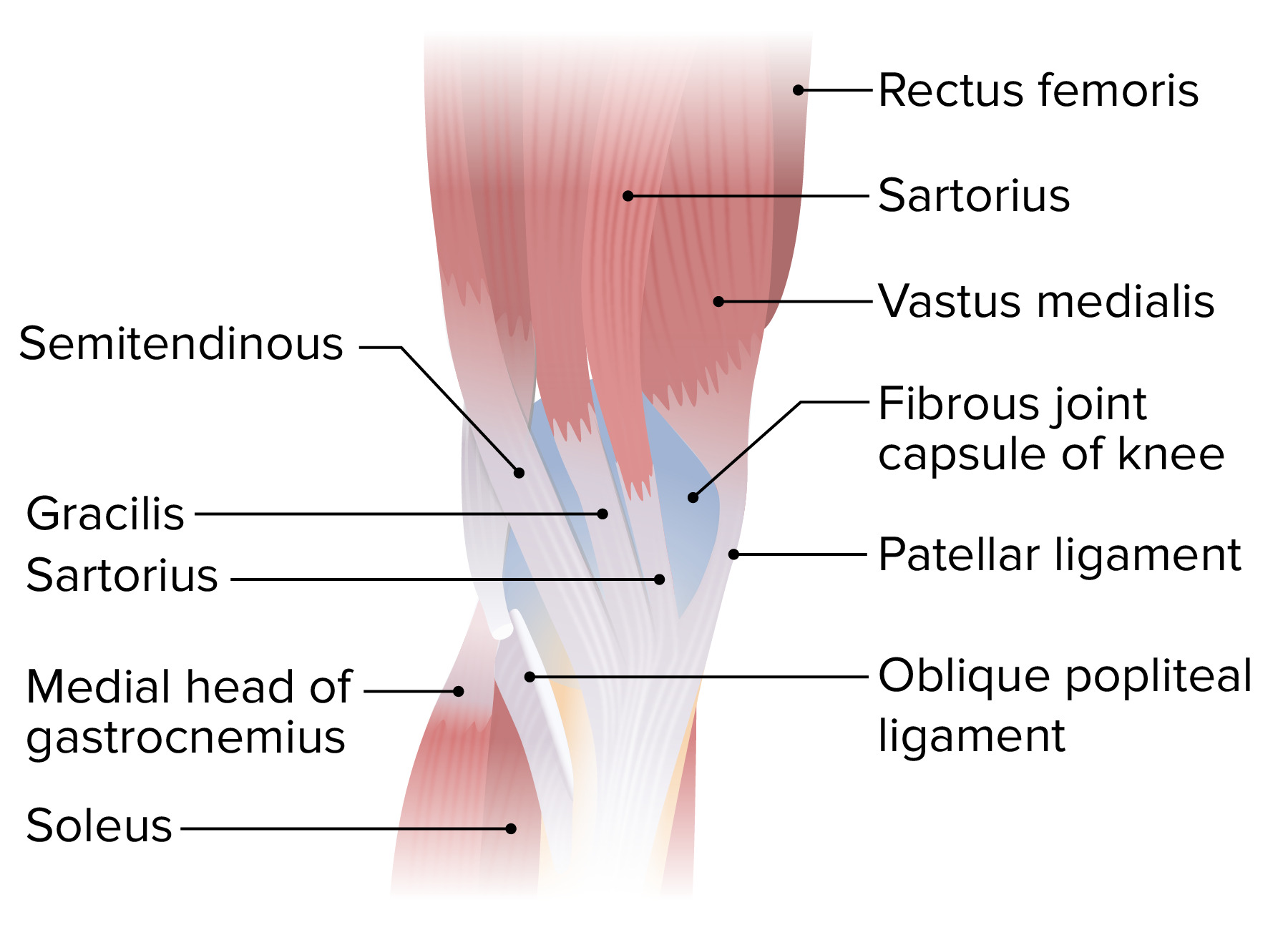
Vista medial de la parte inferior del muslo y la articulación de la rodilla que muestra la inserción de los músculos sartorio, grácil y semitendinoso, formando la pata de ganso (pes anserinus)
Imagen por Lecturio. Licencia: CC BY-NC-SA 4.0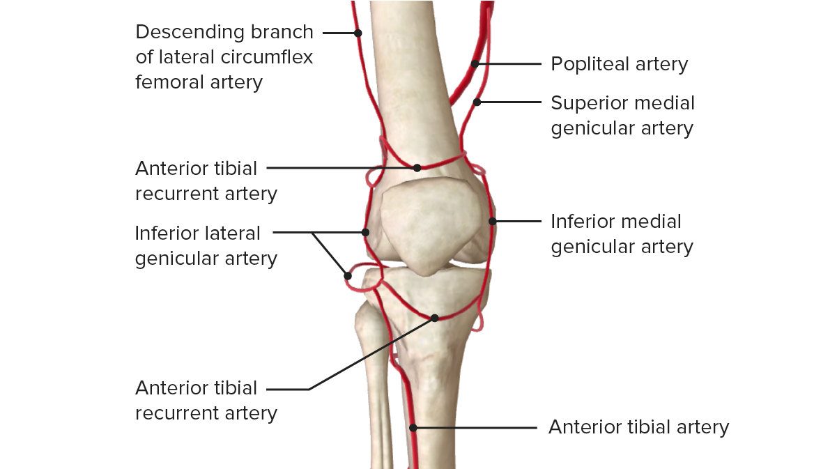
Vista anterior de la irrigación de la articulación de la rodilla.
Imagen por BioDigital, editado por Lecturio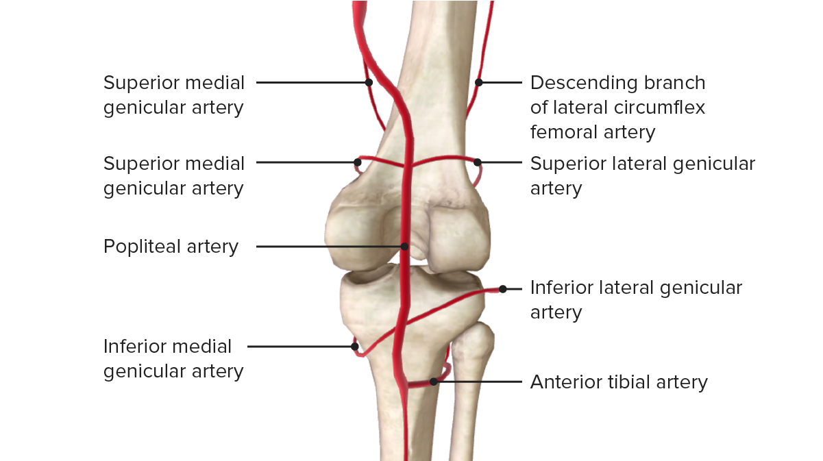
Vista posterior de la irrigación de la articulación de la rodilla.
Imagen por BioDigital, editado por LecturioLas siguientes afecciones comunes están asociadas con la rodilla: