Wiskott-Aldrich Syndrome

Wiskott-Aldrich syndrome (WAS), also known as eczema-thrombocytopenia-immunodeficiency syndrome, IMD2, or immunodeficiency Immunodeficiency Chédiak-Higashi Syndrome 2, is a rare genetic mixed disorder of B- and T-cell deficiency that follows an X-linked recessive X-Linked Recessive Duchenne Muscular Dystrophy inheritance pattern. It is caused by a WAS gene mutation Gene Mutation Myotonic Dystrophies that leads to impaired actin Actin Filamentous proteins that are the main constituent of the thin filaments of muscle fibers. The filaments (known also as filamentous or f-actin) can be dissociated into their globular subunits; each subunit is composed of a single polypeptide 375 amino acids long. This is known as globular or g-actin. In conjunction with myosins, actin is responsible for the contraction and relaxation of muscle. Skeletal Muscle Contraction cytoskeleton Cytoskeleton The network of filaments, tubules, and interconnecting filamentous bridges which give shape, structure, and organization to the cytoplasm. The Cell: Cytosol and Cytoskeleton, phagocytosis Phagocytosis The engulfing and degradation of microorganisms; other cells that are dead, dying, or pathogenic; and foreign particles by phagocytic cells (phagocytes). Innate Immunity: Phagocytes and Antigen Presentation and chemotaxis Chemotaxis The movement of leukocytes in response to a chemical concentration gradient or to products formed in an immunologic reaction. Leukocyte Adhesion Deficiency Type 1, impaired platelet development, and, in general, a loss of humoral and cellular responses. Wiskott-Aldrich syndrome presents as a classic triad of eczema Eczema Atopic dermatitis, also known as eczema, is a chronic, relapsing, pruritic, inflammatory skin disease that occurs more frequently in children, although adults can also be affected. The condition is often associated with elevated serum levels of IgE and a personal or family history of atopy. Skin dryness, erythema, oozing, crusting, and lichenification are present. Atopic Dermatitis (Eczema), bleeding diathesis, and recurrent opportunistic infections Infections Invasion of the host organism by microorganisms or their toxins or by parasites that can cause pathological conditions or diseases. Chronic Granulomatous Disease, but also carries a high risk of development of autoimmunity Autoimmunity Autoimmunity is a pathologic immune response toward self-antigens, resulting from a combination of factors: immunologic, genetic, and environmental. The immune system is equipped with self-tolerance, allowing immune cells such as T cells and B cells to recognize self-antigens and to not mount a reaction against them. Defects in this mechanism, along with environmental triggers (such as infections) and genetic susceptibility factors (most notable of which are the HLA genes) can lead to autoimmune diseases. Autoimmunity and malignancy Malignancy Hemothorax. Management includes hematopoietic stem cell transplantation Hematopoietic stem cell transplantation Transfer of hematopoietic stem cells from bone marrow or blood between individuals within the same species (homologous transplantation) or transfer within the same individual (autologous transplantation). Hematopoietic stem cell transplantation has been used as an alternative to bone marrow transplantation in the treatment of a variety of neoplasms. Organ Transplantation (HSCT), immunoglobulin replacement therapy, prophylactic and therapeutic antibiotics, and systemic steroids Steroids A group of polycyclic compounds closely related biochemically to terpenes. They include cholesterol, numerous hormones, precursors of certain vitamins, bile acids, alcohols (sterols), and certain natural drugs and poisons. Steroids have a common nucleus, a fused, reduced 17-carbon atom ring system, cyclopentanoperhydrophenanthrene. Most steroids also have two methyl groups and an aliphatic side-chain attached to the nucleus. Benign Liver Tumors. Without HSCT, prognosis Prognosis A prediction of the probable outcome of a disease based on a individual's condition and the usual course of the disease as seen in similar situations. Non-Hodgkin Lymphomas is poor and life expectancy Life expectancy Based on known statistical data, the number of years which any person of a given age may reasonably expected to live. Population Pyramids is shortened.

Last updated: Dec 20, 2024

Editorial responsibility: Stanley Oiseth, Lindsay Jones, Evelin Maza

Etiology and Pathophysiology

Epidemiology

Etiology

  • Mutations in the WAS gene Gene A category of nucleic acid sequences that function as units of heredity and which code for the basic instructions for the development, reproduction, and maintenance of organisms. Basic Terms of Genetics on the short arm Arm The arm, or “upper arm” in common usage, is the region of the upper limb that extends from the shoulder to the elbow joint and connects inferiorly to the forearm through the cubital fossa. It is divided into 2 fascial compartments (anterior and posterior). Arm: Anatomy of the X-chromosome (Xp11.22-23) 
  • The WAS gene Gene A category of nucleic acid sequences that function as units of heredity and which code for the basic instructions for the development, reproduction, and maintenance of organisms. Basic Terms of Genetics encodes the WASp (Wiskott-Aldrich syndrome protein) that is mainly expressed in hematopoietic cells and leads to defective cytoskeleton-dependent activation signals in lymphocytes Lymphocytes Lymphocytes are heterogeneous WBCs involved in immune response. Lymphocytes develop from the bone marrow, starting from hematopoietic stem cells (HSCs) and progressing to common lymphoid progenitors (CLPs). B and T lymphocytes and natural killer (NK) cells arise from the lineage. Lymphocytes: Histology, defective antigen Antigen Substances that are recognized by the immune system and induce an immune reaction. Vaccination presentation, and leukocyte migration.
  • Over 300 mutations in the WAS gene Gene A category of nucleic acid sequences that function as units of heredity and which code for the basic instructions for the development, reproduction, and maintenance of organisms. Basic Terms of Genetics have been identified, including missense mutations, nonsense mutations, splice-site mutations, and small deletions.
    • Classic WAS: caused by mutations leading to absent or nonfunctional WASp expression
    • X-linked X-linked Genetic diseases that are linked to gene mutations on the X chromosome in humans or the X chromosome in other species. Included here are animal models of human X-linked diseases. Common Variable Immunodeficiency (CVID) thrombocytopenia Thrombocytopenia Thrombocytopenia occurs when the platelet count is < 150,000 per microliter. The normal range for platelets is usually 150,000-450,000/µL of whole blood. Thrombocytopenia can be a result of decreased production, increased destruction, or splenic sequestration of platelets. Patients are often asymptomatic until platelet counts are < 50,000/µL. Thrombocytopenia (XLT): caused by mutations leading to reduced, partially functional WASp expression
    • X-linked X-linked Genetic diseases that are linked to gene mutations on the X chromosome in humans or the X chromosome in other species. Included here are animal models of human X-linked diseases. Common Variable Immunodeficiency (CVID) neutropenia Neutropenia Neutrophils are an important component of the immune system and play a significant role in the eradication of infections. Low numbers of circulating neutrophils, referred to as neutropenia, predispose the body to recurrent infections or sepsis, though patients can also be asymptomatic. Neutropenia (XLN): caused by gain-of-function mutations in the guanosine triphosphatase (GTPase) binding domain
    • Hotspot mutations (T45M, R86C, and R211X) are associated with distinct phenotypes.
    • Defects in the CD43 molecules are also associated with WAS.

Pathophysiology

  • The WAS gene mutation Gene Mutation Myotonic Dystrophies leads to microthrombocytes (small, defective platelets Platelets Platelets are small cell fragments involved in hemostasis. Thrombopoiesis takes place primarily in the bone marrow through a series of cell differentiation and is influenced by several cytokines. Platelets are formed after fragmentation of the megakaryocyte cytoplasm. Platelets: Histology) in which defective platelets Platelets Platelets are small cell fragments involved in hemostasis. Thrombopoiesis takes place primarily in the bone marrow through a series of cell differentiation and is influenced by several cytokines. Platelets are formed after fragmentation of the megakaryocyte cytoplasm. Platelets: Histology are removed by the spleen Spleen The spleen is the largest lymphoid organ in the body, located in the LUQ of the abdomen, superior to the left kidney and posterior to the stomach at the level of the 9th-11th ribs just below the diaphragm. The spleen is highly vascular and acts as an important blood filter, cleansing the blood of pathogens and damaged erythrocytes. Spleen: Anatomy and/or liver Liver The liver is the largest gland in the human body. The liver is found in the superior right quadrant of the abdomen and weighs approximately 1.5 kilograms. Its main functions are detoxification, metabolism, nutrient storage (e.g., iron and vitamins), synthesis of coagulation factors, formation of bile, filtration, and storage of blood. Liver: Anatomy, resulting in lower platelet counts Platelet counts The number of platelets per unit volume in a sample of venous blood. Coagulation Studies → bleeding diathesis 
  • Defective antigen Antigen Substances that are recognized by the immune system and induce an immune reaction. Vaccination presentation and leukocyte signaling → higher susceptibility to infections Infections Invasion of the host organism by microorganisms or their toxins or by parasites that can cause pathological conditions or diseases. Chronic Granulomatous Disease by opportunistic pathogens, most commonly in the respiratory tract, gastrointestinal (GI) tract, and skin Skin The skin, also referred to as the integumentary system, is the largest organ of the body. The skin is primarily composed of the epidermis (outer layer) and dermis (deep layer). The epidermis is primarily composed of keratinocytes that undergo rapid turnover, while the dermis contains dense layers of connective tissue. Skin: Structure and Functions

Clinical Presentation

  • Classic triad (present from birth in 30% of cases):
    1. Eczema Eczema Atopic dermatitis, also known as eczema, is a chronic, relapsing, pruritic, inflammatory skin disease that occurs more frequently in children, although adults can also be affected. The condition is often associated with elevated serum levels of IgE and a personal or family history of atopy. Skin dryness, erythema, oozing, crusting, and lichenification are present. Atopic Dermatitis (Eczema) (80% of cases)
    2. Bleeding diathesis (resulting from low platelet count):
      • Petechiae Petechiae Primary Skin Lesions
      • Bruising
      • Purpura
      • Spontaneous nose Nose The nose is the human body’s primary organ of smell and functions as part of the upper respiratory system. The nose may be best known for inhaling oxygen and exhaling carbon dioxide, but it also contributes to other important functions, such as tasting. The anatomy of the nose can be divided into the external nose and the nasal cavity. Nose Anatomy (External & Internal) bleeds
      • Bloody diarrhea Bloody diarrhea Diarrhea
    3. Recurrent opportunistic infections Infections Invasion of the host organism by microorganisms or their toxins or by parasites that can cause pathological conditions or diseases. Chronic Granulomatous Disease (usually by encapsulated Encapsulated Klebsiella organisms):
      • Streptococcus Streptococcus Streptococcus is one of the two medically important genera of gram-positive cocci, the other being Staphylococcus. Streptococci are identified as different species on blood agar on the basis of their hemolytic pattern and sensitivity to optochin and bacitracin. There are many pathogenic species of streptococci, including S. pyogenes, S. agalactiae, S. pneumoniae, and the viridans streptococci. Streptococcus pneumoniae (most commonly, otitis media)
      • Neisseria meningitidis Neisseria meningitidis A species of gram-negative, aerobic bacteria. It is a commensal and pathogen only of humans, and can be carried asymptomatically in the nasopharynx. When found in cerebrospinal fluid it is the causative agent of cerebrospinal meningitis. It is also found in venereal discharges and blood. There are at least 13 serogroups based on antigenic differences in the capsular polysaccharides; the ones causing most meningitis infections being a, b, c, y, and w-135. Each serogroup can be further classified by serotype, serosubtype, and immunotype. Neisseria
      • Haemophilus influenzae Haemophilus Influenzae A species of Haemophilus found on the mucous membranes of humans and a variety of animals. The species is further divided into biotypes I through viii. Haemophilus
      • Also, candidal infections Infections Invasion of the host organism by microorganisms or their toxins or by parasites that can cause pathological conditions or diseases. Chronic Granulomatous Disease of the respiratory tract, GI tract, or skin Skin The skin, also referred to as the integumentary system, is the largest organ of the body. The skin is primarily composed of the epidermis (outer layer) and dermis (deep layer). The epidermis is primarily composed of keratinocytes that undergo rapid turnover, while the dermis contains dense layers of connective tissue. Skin: Structure and Functions
  • Increased risk of autoimmune manifestations (4070% of cases, due to an elevated eosinophil count), including:
    • Autoimmune hemolytic anemia Autoimmune Hemolytic Anemia Autoimmune hemolytic anemia (AIHA) is a rare type of hemolytic anemia characterized by antibody production against self RBCs, leading to destruction of these cells in the spleen and other reticuloendothelial tissues. The disease is generally categorized as warm or cold, depending on the thermal reactivity of the autoantibodies. Autoimmune Hemolytic Anemia
    • Neutropenia Neutropenia Neutrophils are an important component of the immune system and play a significant role in the eradication of infections. Low numbers of circulating neutrophils, referred to as neutropenia, predispose the body to recurrent infections or sepsis, though patients can also be asymptomatic. Neutropenia
    • Vasculitis Vasculitis Inflammation of any one of the blood vessels, including the arteries; veins; and rest of the vasculature system in the body. Systemic Lupus Erythematosus
    • Inflammatory bowel disease
    • Renal disease
    • Rheumatoid arthritis Arthritis Acute or chronic inflammation of joints. Osteoarthritis
  • Higher risk of hematological malignancies (1020% of cases) such as B-cell lymphoma Lymphoma A general term for various neoplastic diseases of the lymphoid tissue. Imaging of the Mediastinum
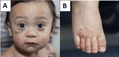
Wiskott-aldrich syndrome: petechiae, hematoma and eczema

Young male patient with Wiskott–Aldrich syndrome presenting with multiple face petechiae and a hematoma under the right eye (A) and eczema of the foot (B)

Image: “Wiskott–Aldrich syndrome” by Michael H. Albert and Alexandra F. Freeman, on behalf of the Inborn Errors Working Party (IEWP) of the European Society for Blood and Marrow Transplantation (EBMT). License: CC BY 4.0

Diagnosis

  • Check immunoglobulin levels:
    • Normal or low levels of IgG IgG The major immunoglobulin isotype class in normal human serum. There are several isotype subclasses of igg, for example, igg1, igg2a, and igg2b. Hypersensitivity Pneumonitis and IgM IgM A class of immunoglobulin bearing mu chains (immunoglobulin mu-chains). Igm can fix complement. The name comes from its high molecular weight and originally being called a macroglobulin. Immunoglobulins: Types and Functions (low specificity since IgG IgG The major immunoglobulin isotype class in normal human serum. There are several isotype subclasses of igg, for example, igg1, igg2a, and igg2b. Hypersensitivity Pneumonitis may also be elevated)
    • Increased levels of IgE IgE An immunoglobulin associated with mast cells. Overexpression has been associated with allergic hypersensitivity. Immunoglobulins: Types and Functions and IgA IgA Represents 15-20% of the human serum immunoglobulins, mostly as the 4-chain polymer in humans or dimer in other mammals. Secretory iga is the main immunoglobulin in secretions. Immunoglobulins: Types and Functions
  • Check CBC:
    • Low platelet count of 20,000/mm3–50,000/mm3
    • Small platelet size (microthrombocytopenia)
  • Genetic testing Genetic Testing Detection of a mutation; genotype; karyotype; or specific alleles associated with genetic traits, heritable diseases, or predisposition to a disease, or that may lead to the disease in descendants. It includes prenatal genetic testing. Myotonic Dystrophies for mutated WAS gene Gene A category of nucleic acid sequences that function as units of heredity and which code for the basic instructions for the development, reproduction, and maintenance of organisms. Basic Terms of Genetics confirms the diagnosis
  • Additional optional tests:
    • Serology Serology The study of serum, especially of antigen-antibody reactions in vitro. Yellow Fever Virus: decreased T- and B-cell count
    • Skin Skin The skin, also referred to as the integumentary system, is the largest organ of the body. The skin is primarily composed of the epidermis (outer layer) and dermis (deep layer). The epidermis is primarily composed of keratinocytes that undergo rapid turnover, while the dermis contains dense layers of connective tissue. Skin: Structure and Functions immunologic testing may reveal hyposensitivity or delayed sensitivity. 
    • Decreased levels of WASp are observed

Management

  • Hematopoietic stem cell transplantation Hematopoietic stem cell transplantation Transfer of hematopoietic stem cells from bone marrow or blood between individuals within the same species (homologous transplantation) or transfer within the same individual (autologous transplantation). Hematopoietic stem cell transplantation has been used as an alternative to bone marrow transplantation in the treatment of a variety of neoplasms. Organ Transplantation (HSCT) is the only possible cure. 
  • General measures:
    • Protective helmets should be worn to prevent cerebral bleeding in children. 
    • Patients Patients Individuals participating in the health care system for the purpose of receiving therapeutic, diagnostic, or preventive procedures. Clinician–Patient Relationship may benefit from transfusions for severe thrombocytopenia Severe Thrombocytopenia Thrombocytopenia
    • Iron Iron A metallic element with atomic symbol fe, atomic number 26, and atomic weight 55. 85. It is an essential constituent of hemoglobins; cytochromes; and iron-binding proteins. It plays a role in cellular redox reactions and in the transport of oxygen. Trace Elements supplements are recommended for anemia Anemia Anemia is a condition in which individuals have low Hb levels, which can arise from various causes. Anemia is accompanied by a reduced number of RBCs and may manifest with fatigue, shortness of breath, pallor, and weakness. Subtypes are classified by the size of RBCs, chronicity, and etiology. Anemia: Overview and Types due to chronic blood loss.
    • Topical or systemic steroids Steroids A group of polycyclic compounds closely related biochemically to terpenes. They include cholesterol, numerous hormones, precursors of certain vitamins, bile acids, alcohols (sterols), and certain natural drugs and poisons. Steroids have a common nucleus, a fused, reduced 17-carbon atom ring system, cyclopentanoperhydrophenanthrene. Most steroids also have two methyl groups and an aliphatic side-chain attached to the nucleus. Benign Liver Tumors are recommended for severe eczema Eczema Atopic dermatitis, also known as eczema, is a chronic, relapsing, pruritic, inflammatory skin disease that occurs more frequently in children, although adults can also be affected. The condition is often associated with elevated serum levels of IgE and a personal or family history of atopy. Skin dryness, erythema, oozing, crusting, and lichenification are present. Atopic Dermatitis (Eczema) but should be used with caution due to immunosuppressive effects.
    • Prophylactic antibiotics to prevent infection
    • Immunoglobulin replacement therapy, with IV preferred over subcutaneous injection due to higher risk of bleeding
    • Acetylsalicylic acid ( ASA ASA Anterior Cord Syndrome) and nonsteroidal anti-inflammatory drugs ( NSAIDs NSAIDS Primary vs Secondary Headaches) are contraindicated due to their effects on platelet function.
  • Prognosis Prognosis A prediction of the probable outcome of a disease based on a individual’s condition and the usual course of the disease as seen in similar situations. Non-Hodgkin Lymphomas:
    • HSCT leads to a 90% survival rate.
    • Without HSCT, the average life expectancy Life expectancy Based on known statistical data, the number of years which any person of a given age may reasonably expected to live. Population Pyramids is 1520 years.

Differential Diagnosis

The following conditions are differential diagnoses for Wiskott-Aldrich syndrome: 

  • Idiopathic Idiopathic Dermatomyositis thrombocytopenic purpura ( ITP ITP Immune thrombocytopenic purpura (ITP), formerly known as idiopathic thrombocytopenic purpura, is a condition that develops secondary to immune-mediated destruction of platelets, resulting in thrombocytopenia (platelet count < 100,000/mm³). Immune thrombocytopenic purpura can be either primary or secondary due to drugs or underlying disease. Immune Thrombocytopenic Purpura): a condition that develops secondary to immune-mediated destruction of platelets Platelets Platelets are small cell fragments involved in hemostasis. Thrombopoiesis takes place primarily in the bone marrow through a series of cell differentiation and is influenced by several cytokines. Platelets are formed after fragmentation of the megakaryocyte cytoplasm. Platelets: Histology, resulting in thrombocytopenia Thrombocytopenia Thrombocytopenia occurs when the platelet count is < 150,000 per microliter. The normal range for platelets is usually 150,000-450,000/µL of whole blood. Thrombocytopenia can be a result of decreased production, increased destruction, or splenic sequestration of platelets. Patients are often asymptomatic until platelet counts are < 50,000/µL. Thrombocytopenia (platelet count < 150 X 10⁹/L). The condition can be primary with an isolated decreased platelet count. Secondary causes are drug-induced or due to infections Infections Invasion of the host organism by microorganisms or their toxins or by parasites that can cause pathological conditions or diseases. Chronic Granulomatous Disease such as HIV HIV Anti-HIV Drugs or hepatitis C Hepatitis C Hepatitis C is an infection of the liver caused by the hepatitis C virus (HCV). The infection can be transmitted through infectious blood or body fluids and may be transmitted during childbirth or through IV drug use or sexual intercourse. Hepatitis C virus can cause both acute and chronic hepatitis, ranging from a mild to a serious, lifelong illness including liver cirrhosis and hepatocellular carcinoma (HCC). Hepatitis C Virus. Idiopathic Idiopathic Dermatomyositis thrombocytopenic purpura is usually a diagnosis of exclusion.
  • Platelet alloimmunization in neonates: a condition characterized by a low platelet count occurring in infants due to maternal antibodies Antibodies Immunoglobulins (Igs), also known as antibodies, are glycoprotein molecules produced by plasma cells that act in immune responses by recognizing and binding particular antigens. The various Ig classes are IgG (the most abundant), IgM, IgE, IgD, and IgA, which differ in their biologic features, structure, target specificity, and distribution. Immunoglobulins: Types and Functions raised against alloantigens of the fetal platelets Platelets Platelets are small cell fragments involved in hemostasis. Thrombopoiesis takes place primarily in the bone marrow through a series of cell differentiation and is influenced by several cytokines. Platelets are formed after fragmentation of the megakaryocyte cytoplasm. Platelets: Histology
  • Severe combined immunodeficiency Immunodeficiency Chédiak-Higashi Syndrome ( SCID SCID Severe combined immunodeficiency (SCID), also called “bubble boy disease,” is a rare genetic disorder in which the development of functional B and T cells is disturbed due to several genetic mutations that result in reduced or absent immune function. Severe Combined Immunodeficiency (SCID)): the most severe form of primary immunodeficiency Immunodeficiency Chédiak-Higashi Syndrome, SCID SCID Severe combined immunodeficiency (SCID), also called “bubble boy disease,” is a rare genetic disorder in which the development of functional B and T cells is disturbed due to several genetic mutations that result in reduced or absent immune function. Severe Combined Immunodeficiency (SCID) is a genetic disorder that involves defective antibody response due to either direct involvement with B lymphocytes B lymphocytes Lymphoid cells concerned with humoral immunity. They are short-lived cells resembling bursa-derived lymphocytes of birds in their production of immunoglobulin upon appropriate stimulation. B cells: Types and Functions or through improper B-lymphocyte activation due to non-functional T-helper cells. Presents as severe and recurrent opportunistic infections Infections Invasion of the host organism by microorganisms or their toxins or by parasites that can cause pathological conditions or diseases. Chronic Granulomatous Disease and is diagnosed through quantitative polymerase chain reaction Polymerase chain reaction Polymerase chain reaction (PCR) is a technique that amplifies DNA fragments exponentially for analysis. The process is highly specific, allowing for the targeting of specific genomic sequences, even with minuscule sample amounts. The PCR cycles multiple times through 3 phases: denaturation of the template DNA, annealing of a specific primer to the individual DNA strands, and synthesis/elongation of new DNA molecules. Polymerase Chain Reaction (PCR) ( PCR PCR Polymerase chain reaction (PCR) is a technique that amplifies DNA fragments exponentially for analysis. The process is highly specific, allowing for the targeting of specific genomic sequences, even with minuscule sample amounts. The PCR cycles multiple times through 3 phases: denaturation of the template DNA, annealing of a specific primer to the individual DNA strands, and synthesis/elongation of new DNA molecules. Polymerase Chain Reaction (PCR)) and flow cytometry Flow cytometry Technique using an instrument system for making, processing, and displaying one or more measurements on individual cells obtained from a cell suspension. Cells are usually stained with one or more fluorescent dyes specific to cell components of interest, e.g., DNA, and fluorescence of each cell is measured as it rapidly transverses the excitation beam (laser or mercury arc lamp). Fluorescence provides a quantitative measure of various biochemical and biophysical properties of the cell, as well as a basis for cell sorting. Other measurable optical parameters include light absorption and light scattering, the latter being applicable to the measurement of cell size, shape, density, granularity, and stain uptake. X-linked Agammaglobulinemia.
  • Thrombotic thrombocytopenic purpura Thrombotic thrombocytopenic purpura Thrombotic thrombocytopenic purpura (TTP) is a life-threatening condition due to either a congenital or an acquired deficiency of ADAMTS-13, a metalloproteinase that cleaves multimers of von Willebrand factor (VWF). The large multimers then aggregate excessive platelets resulting in microvascular thrombosis and an increase in consumption of platelets. Thrombotic Thrombocytopenic Purpura ( TTP TTP Thrombotic thrombocytopenic purpura (TTP) is a life-threatening condition due to either a congenital or an acquired deficiency of adamts-13, a metalloproteinase that cleaves multimers of von Willebrand factor (vWF). The large multimers then aggregate excessive platelets resulting in microvascular thrombosis and an increase in consumption of platelets. Thrombotic Thrombocytopenic Purpura): a life-threatening condition due to either a congenital deficiency or an acquired deficiency of ADAMTS-13, a metalloproteinase that cleaves multimers of von Willebrand Factor von Willebrand factor A high-molecular-weight plasma protein, produced by endothelial cells and megakaryocytes, that is part of the factor VIII/von Willebrand factor complex. The von Willebrand factor has receptors for collagen, platelets, and ristocetin activity as well as the immunologically distinct antigenic determinants. It functions in adhesion of platelets to collagen and hemostatic plug formation. The prolonged bleeding time in von Willebrand diseases is due to the deficiency of this factor. Hemostasis (vWF). These large multimers then aggregate excessive platelets Platelets Platelets are small cell fragments involved in hemostasis. Thrombopoiesis takes place primarily in the bone marrow through a series of cell differentiation and is influenced by several cytokines. Platelets are formed after fragmentation of the megakaryocyte cytoplasm. Platelets: Histology, resulting in thrombosis Thrombosis Formation and development of a thrombus or blood clot in the blood vessel. Epidemic Typhus plus increased usage of platelets Platelets Platelets are small cell fragments involved in hemostasis. Thrombopoiesis takes place primarily in the bone marrow through a series of cell differentiation and is influenced by several cytokines. Platelets are formed after fragmentation of the megakaryocyte cytoplasm. Platelets: Histology that leads to thrombocytopenia Thrombocytopenia Thrombocytopenia occurs when the platelet count is < 150,000 per microliter. The normal range for platelets is usually 150,000-450,000/µL of whole blood. Thrombocytopenia can be a result of decreased production, increased destruction, or splenic sequestration of platelets. Patients are often asymptomatic until platelet counts are < 50,000/µL. Thrombocytopenia.

References

  1. Ochs, H. (2024). Wiskott-Aldrich syndrome. UpToDate. Retrieved November 26, 2024, from: https://www.uptodate.com/contents/wiskott-aldrich-syndrome
  2. Huynh, P. (2024). Wiskott-Aldrich Syndrome. Medscape. Retrieved November 26, 2024, from: https://emedicine.medscape.com/article/137015-overview
  3. Wiskott-Aldrich Syndrome: Medlineplus genetics. (n.d.-b). Retrieved November 26, 2024, from https://medlineplus.gov/genetics/condition/wiskott-aldrich-syndrome/#frequency
  4. Bohm, S., et al. (2023). Outcomes of hematopoietic stem cell gene therapy for Wiskott-Aldrich syndrome. Blood, 142(15), 1281-1292. https://doi.org/10.1182/blood.2022019117
  5. Jiang, Y., et al. (2023). Immunological features and treatment of Wiskott-Aldrich syndrome. Frontiers in Immunology, 14, 123456.
  6. Michels, A., et al. (2023). Excellent outcomes following hematopoietic cell transplantation for Wiskott-Aldrich syndrome. Blood, 135(23), 2094-2104. https://doi.org/10.1182/blood.2019002939
  7. Porteus, M., et al. (2023). Gene editing shows promise for Wiskott-Aldrich syndrome. Immune Deficiency Foundation.
  8. Vallée, T., et al. (2024). Wiskott-Aldrich syndrome: A study of 577 patients defines the genotype as a predictive biomarker for disease severity. Blood, 143(24), 2504-2514. https://doi.org/10.1182/blood.2023021411
  9. Vallée, T., et al. (2024). Recent advances in understanding the pathophysiology of Wiskott-Aldrich syndrome. Blood, American Society of Hematology. https://doi.org/10.1182/blood-2008-12-115253

Create your free account or log in to continue reading!

Sign up now and get free access to Lecturio with concept pages, medical videos, and questions for your medical education.

User Reviews

Details