Vesicoureteral Reflux

Vesicoureteral reflux (VUR) is the retrograde flow Retrograde flow Veins: Histology of urine from the bladder Bladder A musculomembranous sac along the urinary tract. Urine flows from the kidneys into the bladder via the ureters, and is held there until urination. Pyelonephritis and Perinephric Abscess into the upper urinary tract Urinary tract The urinary tract is located in the abdomen and pelvis and consists of the kidneys, ureters, urinary bladder, and urethra. The structures permit the excretion of urine from the body. Urine flows from the kidneys through the ureters to the urinary bladder and out through the urethra. Urinary Tract: Anatomy. Primary VUR often results from the incomplete closure of the ureterovesical junction, whereas secondary VUR is due to an anatomic or physiologic obstruction. Vesicoureteral reflux does not cause specific symptoms, but it is suspected after detecting hydronephrosis Hydronephrosis Hydronephrosis is dilation of the renal collecting system as a result of the obstruction of urine outflow. Hydronephrosis can be unilateral or bilateral. Nephrolithiasis is the most common cause of hydronephrosis in young adults, while prostatic hyperplasia and neoplasm are seen in older patients. Hydronephrosis on prenatal ultrasonography or in a young child presenting with a urinary tract Urinary tract The urinary tract is located in the abdomen and pelvis and consists of the kidneys, ureters, urinary bladder, and urethra. The structures permit the excretion of urine from the body. Urine flows from the kidneys through the ureters to the urinary bladder and out through the urethra. Urinary Tract: Anatomy infection. A voiding cystourethrogram Voiding Cystourethrogram Urinary Tract Infections (UTIs) in Children should be performed to diagnose the condition and assess its severity. The majority of patients Patients Individuals participating in the health care system for the purpose of receiving therapeutic, diagnostic, or preventive procedures. Clinician–Patient Relationship will have spontaneous resolution of VUR. Some patients Patients Individuals participating in the health care system for the purpose of receiving therapeutic, diagnostic, or preventive procedures. Clinician–Patient Relationship may require surgical management, particularly individuals with high-grade reflux.

Last updated: Aug 29, 2022

Editorial responsibility: Stanley Oiseth, Lindsay Jones, Evelin Maza

Overview

Definition

Vesicoureteral reflux (VUR) is the retrograde flow Retrograde flow Veins: Histology of urine from the bladder Bladder A musculomembranous sac along the urinary tract. Urine flows from the kidneys into the bladder via the ureters, and is held there until urination. Pyelonephritis and Perinephric Abscess into the upper urinary tract Urinary tract The urinary tract is located in the abdomen and pelvis and consists of the kidneys, ureters, urinary bladder, and urethra. The structures permit the excretion of urine from the body. Urine flows from the kidneys through the ureters to the urinary bladder and out through the urethra. Urinary Tract: Anatomy.

Epidemiology

  • The most common urinary finding in children:
    • Occurs in 1%–2% of children < 2 years of age
    • Prevalence Prevalence The total number of cases of a given disease in a specified population at a designated time. It is differentiated from incidence, which refers to the number of new cases in the population at a given time. Measures of Disease Frequency is 15% in children with prenatal hydronephrosis Hydronephrosis Hydronephrosis is dilation of the renal collecting system as a result of the obstruction of urine outflow. Hydronephrosis can be unilateral or bilateral. Nephrolithiasis is the most common cause of hydronephrosis in young adults, while prostatic hyperplasia and neoplasm are seen in older patients. Hydronephrosis
  • 3 times more common in white children than Black children
  • 2 times more common in girls than boys

Etiology

Primary VUR:

  • Genetic predisposition
  • Congenital Congenital Chorioretinitis defect of the terminal portion of the ureter (e.g., short intravesical ureter)

Secondary VUR:

Pathophysiology

Normal physiology

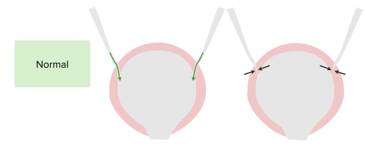
  • The distal ureters Ureters One of a pair of thick-walled tubes that transports urine from the kidney pelvis to the urinary bladder. Urinary Tract: Anatomy course through the bladder Bladder A musculomembranous sac along the urinary tract. Urine flows from the kidneys into the bladder via the ureters, and is held there until urination. Pyelonephritis and Perinephric Abscess wall at an oblique angle.
  • During micturition, the bladder Bladder A musculomembranous sac along the urinary tract. Urine flows from the kidneys into the bladder via the ureters, and is held there until urination. Pyelonephritis and Perinephric Abscess and the surrounding muscles contract and compress the intravesical ureter → prevention of urine reflux into the ureters Ureters One of a pair of thick-walled tubes that transports urine from the kidney pelvis to the urinary bladder. Urinary Tract: Anatomy
The normal function of the bladder and ureteres

Normal function of the bladder and ureters:
The ureters enter at an angle, allowing the bladder to squeeze the ureteral opening, which is closed during micturition to prevent reflux.
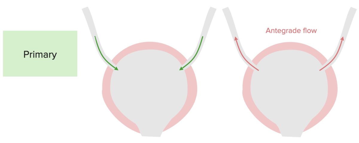
Image by Lecturio.

Primary VUR

  • A short intravesical ureter → ureter enters at a different angle
  • Malfunction of the valve-like mechanism during micturition → reflux of urine into the ureter and collecting system
The pathophysiology of primary vesicoureteral reflux

Pathophysiology of primary vesicoureteral reflux:
A defect in the terminal ureter and in the ability to close the ureterovesicular junction results in the antegrade flow up the ureters during micturition.

Image by Lecturio.

Potential consequences

  • Persistent intrarenal reflux → renal scarring Scarring Inflammation → reflux nephropathy
  • VUR → ↑ risk of urinary tract Urinary tract The urinary tract is located in the abdomen and pelvis and consists of the kidneys, ureters, urinary bladder, and urethra. The structures permit the excretion of urine from the body. Urine flows from the kidneys through the ureters to the urinary bladder and out through the urethra. Urinary Tract: Anatomy infections Infections Invasion of the host organism by microorganisms or their toxins or by parasites that can cause pathological conditions or diseases. Chronic Granulomatous Disease (UTIs), including pyelonephritis Pyelonephritis Pyelonephritis is infection affecting the renal pelvis and the renal parenchyma. This condition arises mostly as a complication of bladder infection that ascends to the upper urinary tract. Pyelonephritis can be acute or chronic (which results from persistent or chronic infections). Typical acute symptoms are flank pain, fever, and nausea with vomiting. T Pyelonephritis and Perinephric Abscess

Clinical Presentation and Diagnosis

Clinical presentation

There are no specific signs or symptoms for VUR, and the condition may be suspected in the following circumstances:

Supporting laboratory evaluation

There are no laboratory tests that can diagnose VUR. The following assessments may be performed to evaluate for complications:

  • BUN and creatinine → renal dysfunction
  • Urinalysis Urinalysis Examination of urine by chemical, physical, or microscopic means. Routine urinalysis usually includes performing chemical screening tests, determining specific gravity, observing any unusual color or odor, screening for bacteriuria, and examining the sediment microscopically. Urinary Tract Infections (UTIs) in Children with culture → UTI UTI Urinary tract infections (UTIs) represent a wide spectrum of diseases, from self-limiting simple cystitis to severe pyelonephritis that can result in sepsis and death. Urinary tract infections are most commonly caused by Escherichia coli, but may also be caused by other bacteria and fungi. Urinary Tract Infections (UTIs)

Imaging

Renal and bladder Bladder A musculomembranous sac along the urinary tract. Urine flows from the kidneys into the bladder via the ureters, and is held there until urination. Pyelonephritis and Perinephric Abscess US:

  • Indications:
    • After 1st febrile UTI UTI Urinary tract infections (UTIs) represent a wide spectrum of diseases, from self-limiting simple cystitis to severe pyelonephritis that can result in sepsis and death. Urinary tract infections are most commonly caused by Escherichia coli, but may also be caused by other bacteria and fungi. Urinary Tract Infections (UTIs) in all children aged 2–24 months
    • In all children with recurrent cases of UTIs
  • Potential findings:
    • Dilated ureters Ureters One of a pair of thick-walled tubes that transports urine from the kidney pelvis to the urinary bladder. Urinary Tract: Anatomy
    • Hydronephrosis Hydronephrosis Hydronephrosis is dilation of the renal collecting system as a result of the obstruction of urine outflow. Hydronephrosis can be unilateral or bilateral. Nephrolithiasis is the most common cause of hydronephrosis in young adults, while prostatic hyperplasia and neoplasm are seen in older patients. Hydronephrosis

Voiding cystourethrogram Voiding Cystourethrogram Urinary Tract Infections (UTIs) in Children:
  • Test of choice for the diagnosis and degree of VUR
  • Indications:
    • Bilateral hydronephrosis Hydronephrosis Hydronephrosis is dilation of the renal collecting system as a result of the obstruction of urine outflow. Hydronephrosis can be unilateral or bilateral. Nephrolithiasis is the most common cause of hydronephrosis in young adults, while prostatic hyperplasia and neoplasm are seen in older patients. Hydronephrosis or renal scarring Scarring Inflammation found on US 
    • 1st UTI UTI Urinary tract infections (UTIs) represent a wide spectrum of diseases, from self-limiting simple cystitis to severe pyelonephritis that can result in sepsis and death. Urinary tract infections are most commonly caused by Escherichia coli, but may also be caused by other bacteria and fungi. Urinary Tract Infections (UTIs) with abnormal US
    • Recurrent UTIs
  • Technique:
    • Contrast is placed in the bladder Bladder A musculomembranous sac along the urinary tract. Urine flows from the kidneys into the bladder via the ureters, and is held there until urination. Pyelonephritis and Perinephric Abscess through a catheter. 
    • Fluoroscopy Fluoroscopy Production of an image when x-rays strike a fluorescent screen. X-rays images are taken during voiding.
    • Reflux of contrast into the ureter during voiding → VUR
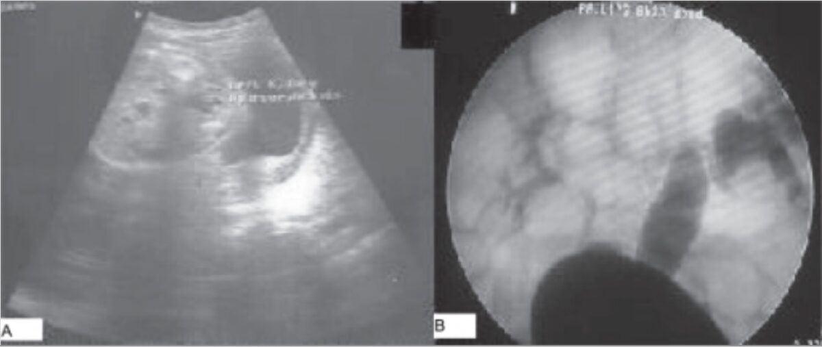
Imaging findings in vesicoureteral reflux

Imaging findings in vesicoureteral reflux:
A: Postnatal ultrasound showing unilateral hydronephrosis
B: Voiding cystourethrogram showing contrast reflux in the right and left ureters and collecting systems (right: grade III, left: grade V)

Image: “Imaging findings in vesicoureteral reflux” by Department of Urology, Kles Kidney Foundation, Nehru Nagar, Belgaum, India. License: CC BY 2.0

Severity classification

Severity classification helps grade the severity of VUR based on imaging findings.
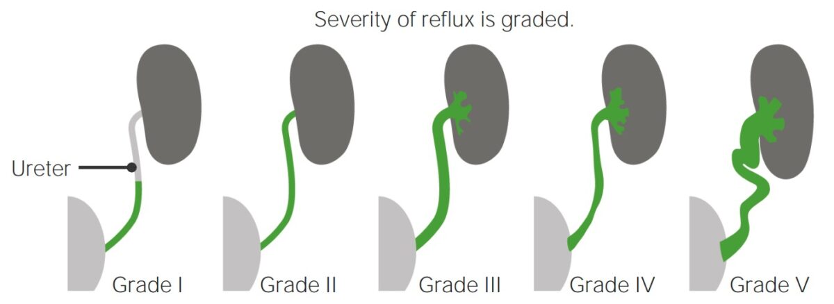
  • Grade I: reflux into the ureter
  • Grade II: reflux into the entirety of the ureter
  • Grade III: Reflux fills and mildly dilates the ureter with mild blunting of the calyces.
  • Grade IV: Reflux results in grossly dilated ureters Ureters One of a pair of thick-walled tubes that transports urine from the kidney pelvis to the urinary bladder. Urinary Tract: Anatomy and calyces. Some tortuosity of the ureter is also seen.
  • Grade V: massive reflux causing significant dilation of the urinary collecting system and intrarenal reflux (“ megaureter Megaureter Hydronephrosis”)
Vesicoureteral reflux classification

Severity classification of vesicoureteral reflux

Image by Lecturio.

Management and Prognosis

Management

Conservative management:

Invasive management:

  • Goal is to improve the angle of the ureter → ↓ reflux
  • Indications:
  • Procedures:
    • Subureteric transurethral injection
    • Open surgical reimplantation of ureters Ureters One of a pair of thick-walled tubes that transports urine from the kidney pelvis to the urinary bladder. Urinary Tract: Anatomy

Prognosis Prognosis A prediction of the probable outcome of a disease based on a individual’s condition and the usual course of the disease as seen in similar situations. Non-Hodgkin Lymphomas

  • Most patients Patients Individuals participating in the health care system for the purpose of receiving therapeutic, diagnostic, or preventive procedures. Clinician–Patient Relationship show spontaneous resolution of VUR.
  • Factors that increase the likelihood of resolution:
    • Lower severity of reflux
    • Age of diagnosis < 2 years
    • Unilateral disease

Differential Diagnosis

  • Ureteropelvic junction Ureteropelvic junction Urinary Tract: Anatomy obstruction: the most common cause of pediatric hydronephrosis Hydronephrosis Hydronephrosis is dilation of the renal collecting system as a result of the obstruction of urine outflow. Hydronephrosis can be unilateral or bilateral. Nephrolithiasis is the most common cause of hydronephrosis in young adults, while prostatic hyperplasia and neoplasm are seen in older patients. Hydronephrosis. An anatomic lesion or functional disturbance causes an obstruction where the ureter enters the kidney.  Ureteropelvic junction Ureteropelvic junction Urinary Tract: Anatomy obstruction usually presents as a palpable abdominal mass Mass Three-dimensional lesion that occupies a space within the breast Imaging of the Breast, flank pain Flank pain Pain emanating from below the ribs and above the ilium. Renal Cell Carcinoma, hematuria Hematuria Presence of blood in the urine. Renal Cell Carcinoma UTI UTI Urinary tract infections (UTIs) represent a wide spectrum of diseases, from self-limiting simple cystitis to severe pyelonephritis that can result in sepsis and death. Urinary tract infections are most commonly caused by Escherichia coli, but may also be caused by other bacteria and fungi. Urinary Tract Infections (UTIs), or failure to thrive Failure to Thrive Failure to thrive (FTT), or faltering growth, describes suboptimal weight gain and growth in children. The majority of cases are due to inadequate caloric intake; however, genetic, infectious, and oncological etiologies are also common. Failure to Thrive. The diagnosis is made with imaging. A voiding cystourethrogram Voiding Cystourethrogram Urinary Tract Infections (UTIs) in Children should be performed to rule out VUR. Management may include surveillance Surveillance Developmental Milestones and Normal Growth or surgery.
  • UTI UTI Urinary tract infections (UTIs) represent a wide spectrum of diseases, from self-limiting simple cystitis to severe pyelonephritis that can result in sepsis and death. Urinary tract infections are most commonly caused by Escherichia coli, but may also be caused by other bacteria and fungi. Urinary Tract Infections (UTIs): a wide spectrum of diseases, ranging from self-limiting Self-Limiting Meningitis in Children simple cystitis Cystitis Inflammation of the urinary bladder, either from bacterial or non-bacterial causes. Cystitis is usually associated with painful urination (dysuria), increased frequency, urgency, and suprapubic pain. Urinary Tract Infections (UTIs) to severe pyelonephritis Pyelonephritis Pyelonephritis is infection affecting the renal pelvis and the renal parenchyma. This condition arises mostly as a complication of bladder infection that ascends to the upper urinary tract. Pyelonephritis can be acute or chronic (which results from persistent or chronic infections). Typical acute symptoms are flank pain, fever, and nausea with vomiting. T Pyelonephritis and Perinephric Abscess that can result in sepsis Sepsis Systemic inflammatory response syndrome with a proven or suspected infectious etiology. When sepsis is associated with organ dysfunction distant from the site of infection, it is called severe sepsis. When sepsis is accompanied by hypotension despite adequate fluid infusion, it is called septic shock. Sepsis and Septic Shock and death. Depending on the location of the infection, patients Patients Individuals participating in the health care system for the purpose of receiving therapeutic, diagnostic, or preventive procedures. Clinician–Patient Relationship can present with dysuria Dysuria Painful urination. It is often associated with infections of the lower urinary tract. Urinary Tract Infections (UTIs), urinary urgency Urinary Urgency Normal Pressure Hydrocephalus, increased urinary frequency, suprapubic pain Pain An unpleasant sensation induced by noxious stimuli which are detected by nerve endings of nociceptive neurons. Pain: Types and Pathways, and fever Fever Fever is defined as a measured body temperature of at least 38°C (100.4°F). Fever is caused by circulating endogenous and/or exogenous pyrogens that increase levels of prostaglandin E2 in the hypothalamus. Fever is commonly associated with chills, rigors, sweating, and flushing of the skin. Fever. Urinalysis Urinalysis Examination of urine by chemical, physical, or microscopic means. Routine urinalysis usually includes performing chemical screening tests, determining specific gravity, observing any unusual color or odor, screening for bacteriuria, and examining the sediment microscopically. Urinary Tract Infections (UTIs) in Children and urine culture Urine culture Urinary Tract Infections (UTIs) along with the clinical presentation help in the diagnosis of UTIs. Imaging should be ordered to rule out VUR in children. Management options include oral or IV antibiotics.
  • Posterior urethral valves: obstructing membranous folds within the lumen of the posterior urethra Urethra A tube that transports urine from the urinary bladder to the outside of the body in both the sexes. It also has a reproductive function in the male by providing a passage for sperm. Urinary Tract: Anatomy. Infants usually present with UTI UTI Urinary tract infections (UTIs) represent a wide spectrum of diseases, from self-limiting simple cystitis to severe pyelonephritis that can result in sepsis and death. Urinary tract infections are most commonly caused by Escherichia coli, but may also be caused by other bacteria and fungi. Urinary Tract Infections (UTIs), diminished urinary stream, urinary incontinence Urinary incontinence Urinary incontinence (UI) is involuntary loss of bladder control or unintentional voiding, which represents a hygienic or social problem to the patient. Urinary incontinence is a symptom, a sign, and a disorder. The 5 types of UI include stress, urge, mixed, overflow, and functional. Urinary Incontinence, and VUR. The diagnosis of posterior urethral valves can be made using US or a voiding cystourethrogram Voiding Cystourethrogram Urinary Tract Infections (UTIs) in Children. Management is with surgery.
  • Congenital Congenital Chorioretinitis megacalyces: an incidental finding resulting from the underdevelopment of the renal medullary pyramids. The resulting enlargement of the calyces can mimic hydronephrosis Hydronephrosis Hydronephrosis is dilation of the renal collecting system as a result of the obstruction of urine outflow. Hydronephrosis can be unilateral or bilateral. Nephrolithiasis is the most common cause of hydronephrosis in young adults, while prostatic hyperplasia and neoplasm are seen in older patients. Hydronephrosis. The radiographic appearance of the calyces along with the normal caliber of the renal pelvis Renal pelvis Kidneys: Anatomy and ureter aid in the diagnosis. No management is required.

References

  1. Baskin, L.S. (2020). Congenital ureteropelvic junction obstruction. UpToDate. Retrieved March 12, 2021, from https://www.uptodate.com/contents/congenital-ureteropelvic-junction-obstruction
  2. Holmes, N. (2020). Clinical presentation and diagnosis of posterior urethral valves. UpToDate. Retrieved March 12, 2021, from https://www.uptodate.com/contents/clinical-presentation-and-diagnosis-of-posterior-urethral-valves
  3. Mattoo, T.K., Greenfield, S.P. (2021). Clinical presentation, diagnosis, and course of primary vesicoureteral reflux. UpToDate. Retrieved April 19, 2021, from https://www.uptodate.com/contents/clinical-presentation-diagnosis-and-course-of-primary-vesicoureteral-reflux
  4. Mattoo, T.K., Greenfield, S.P. (2021). Management of vesicoureteral reflux. In Kim, M.S. (Ed.), UpToDate. Retrieved April 25, 2021, from https://www.uptodate.com/contents/management-of-vesicoureteral-reflux
  5. Van Why, S.K., Avner, E.D. (2016). Vesicoureteral Reflux. Nelson Textbook of Pediatrics, 2562–67.
  6. Estrada, Jr., C.R., Cendron, M. (2018). Vesicoureteral reflux. In Kim, E.D. (Ed.), Medscape. Retrieved April 25, 2021, from https://emedicine.medscape.com/article/439403-overview
  7. Banker, H., Aeddula, N.R. (2020). Vesicoureteral reflux. [online] StatPearls. Retrieved April 25, 2021, from https://www.ncbi.nlm.nih.gov/books/NBK563262/

Create your free account or log in to continue reading!

Sign up now and get free access to Lecturio with concept pages, medical videos, and questions for your medical education.

User Reviews

Details