Vertigo

Vertigo is defined as the perceived sensation of rotational motion while remaining still. A very common complaint in primary care and the ER, vertigo is more frequently experienced by women and its prevalence Prevalence The total number of cases of a given disease in a specified population at a designated time. It is differentiated from incidence, which refers to the number of new cases in the population at a given time. Measures of Disease Frequency increases with age. Vertigo is classified into peripheral or central based on its etiology. Vertigo is a clinical diagnosis, differentiated through history and physical examination findings, most notably nystagmus Nystagmus Involuntary movements of the eye that are divided into two types, jerk and pendular. Jerk nystagmus has a slow phase in one direction followed by a corrective fast phase in the opposite direction, and is usually caused by central or peripheral vestibular dysfunction. Pendular nystagmus features oscillations that are of equal velocity in both directions and this condition is often associated with visual loss early in life. Albinism. Further testing may be required in malignant cases. Management depends on the etiology but certain maneuvers such as the Epley maneuver can be diagnostic and therapeutic.

Last updated: Jan 13, 2023

Editorial responsibility: Stanley Oiseth, Lindsay Jones, Evelin Maza

Overview

Definition

Vertigo is a clinical symptom described as the perceived sensation of rotational motion while remaining still.

Epidemiology

  • The prevalence Prevalence The total number of cases of a given disease in a specified population at a designated time. It is differentiated from incidence, which refers to the number of new cases in the population at a given time. Measures of Disease Frequency of vertigo increases with age.
  • Accounts for > 50% of the presenting complaints of “ dizziness Dizziness An imprecise term which may refer to a sense of spatial disorientation, motion of the environment, or lightheadedness. Lateral Medullary Syndrome (Wallenberg Syndrome)” in the ED
  • Women are 2–3 times more likely to report vertigo than men.
  • Associated with depression and cardiovascular disease

Classification

  • Peripheral: due to pathology of the vestibular labyrinth or vestibular nerve
  • Central: due to pathology of the brainstem or cerebellum Cerebellum The cerebellum, Latin for “little brain,” is located in the posterior cranial fossa, dorsal to the pons and midbrain, and its principal role is in the coordination of movements. The cerebellum consists of 3 lobes on either side of its 2 hemispheres and is connected in the middle by the vermis. Cerebellum: Anatomy

Etiology

Peripheral:

  • Benign Benign Fibroadenoma paroxysmal positional vertigo (BPPV) (93% of cases)
  • Acute labyrinthitis (due to viral infection)
  • Ménière‌ ‌disease
  • Vestibular neuritis
  • Ramsay Hunt syndrome Ramsay Hunt syndrome A syndrome characterized by facial palsy in association with a herpetic eruption of the external auditory meatus. This may occasionally be associated with tinnitus, vertigo, deafness, severe otalgia, and inflammation of the pinna. The condition is caused by reactivation of a latent herpesvirus 3, human infection which causes inflammation of the facial and vestibular nerves, and may occasionally involve additional cranial nerves. Herpes Zoster (Shingles) (also known as herpes zoster oticus Herpes zoster oticus A syndrome characterized by facial palsy in association with a herpetic eruption of the external auditory meatus. This may occasionally be associated with tinnitus, vertigo, deafness, severe otalgia, and inflammation of the pinna. The condition is caused by reactivation of a latent herpesvirus 3, human infection which causes inflammation of the facial and vestibular nerves, and may occasionally involve additional cranial nerves. Herpes Zoster (Shingles))
  • Cholesteatomas
  • Otosclerosis
  • Otomastoiditis
  • Perilymphatic fistula Fistula Abnormal communication most commonly seen between two internal organs, or between an internal organ and the surface of the body. Anal Fistula
  • Vestibular schwannoma Vestibular schwannoma Acoustic neuroma, also referred to as vestibular schwannoma, is a benign tumor arising from Schwann cells of the vestibular component of the cranial nerve VIII. Acoustic neuroma forms within the internal auditory meatus and extends into the cerebellopontine angle. Acoustic Neuroma

Central:

  • Ischemic or hemorrhagic strokes
  • CNS tumors ( cerebellopontine angle Cerebellopontine angle Junction between the cerebellum and the pons. Acoustic Neuroma):
    • Meningioma Meningioma Meningiomas are slow-growing tumors that arise from the meninges of the brain and spinal cord. The vast majority are benign. These tumors commonly occur in individuals with a history of high doses of skull radiation, head trauma, and neurofibromatosis 2. Meningioma
    • Brainstem glioma
    • Medulloblastoma Medulloblastoma Medulloblastomas are malignant primitive neuroectodermal tumors that arise in the posterior fossa in children. Medulloblastomas are the most common malignant brain tumors in children. Patients with medulloblastoma present with symptoms of increased intracranial pressure and cerebellar signs, which both evolve and worsen over weeks to a few months. Medulloblastoma
    • Metastases
  • Vestibular migraines
  • Medication-induced vertigo (e.g., phenytoin Phenytoin An anticonvulsant that is used to treat a wide variety of seizures. The mechanism of therapeutic action is not clear, although several cellular actions have been described including effects on ion channels, active transport, and general membrane stabilization. Phenytoin has been proposed for several other therapeutic uses, but its use has been limited by its many adverse effects and interactions with other drugs. First-Generation Anticonvulsant Drugs and salicylates)

Both: multiple sclerosis Sclerosis A pathological process consisting of hardening or fibrosis of an anatomical structure, often a vessel or a nerve. Wilms Tumor

Pathophysiology

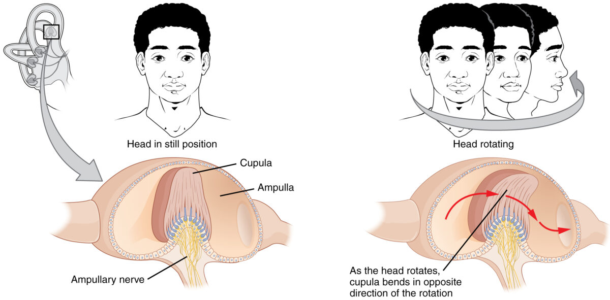
To understand the causes of vertigo, it is important to understand how the human body perceives and maintains balance.
  • Vestibular nuclei receive a signal corresponding to the acceleration of the head.
    • The signal is carried to cranial nerves Cranial nerves There are 12 pairs of cranial nerves (CNs), which run from the brain to various parts of the head, neck, and trunk. The CNs can be sensory or motor or both. The CNs are named and numbered in Roman numerals according to their location, from the front to the back of the brain. The 12 Cranial Nerves: Overview and Functions (CNs) III, IV, and VI → coordination Coordination Cerebellar Disorders of the movements of the eyes and head
    • Eyes move in a direction opposite to that of head rotation Rotation Motion of an object in which either one or more points on a line are fixed. It is also the motion of a particle about a fixed point. X-rays to maintain fixation.
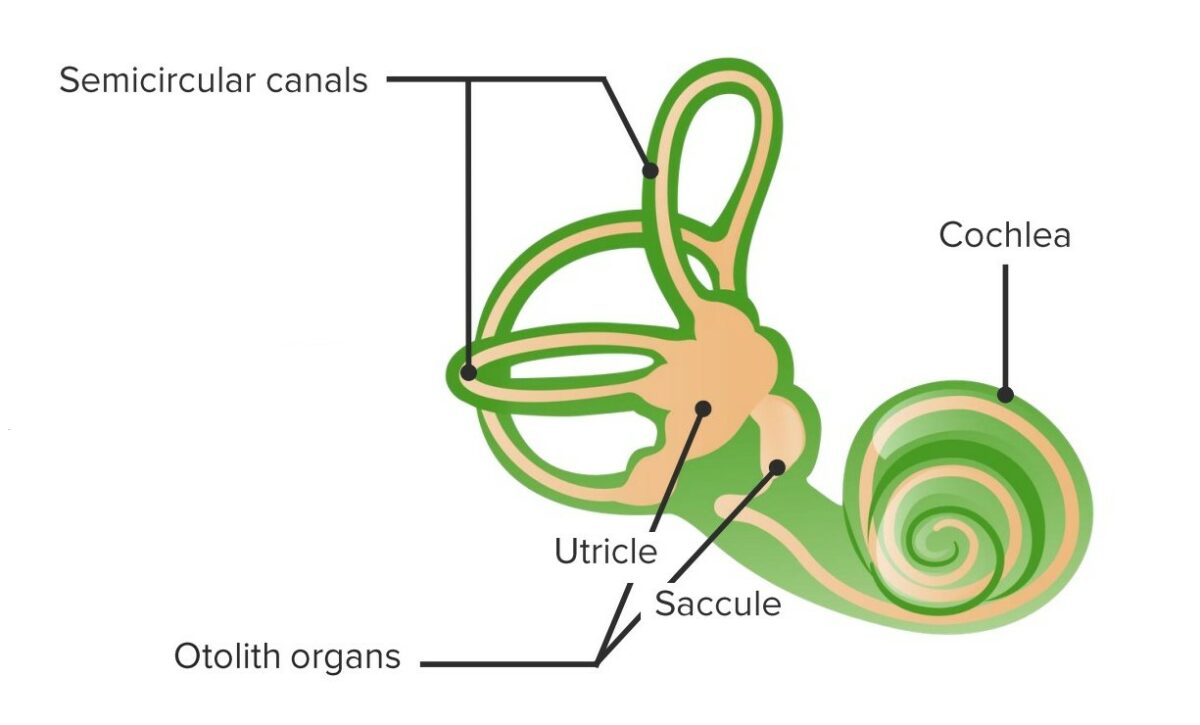
  • Semicircular canals Semicircular canals Three long canals (anterior, posterior, and lateral) of the bony labyrinth. They are set at right angles to each other and are situated posterosuperior to the vestibule of the bony labyrinth (vestibular labyrinth). The semicircular canals have five openings into the vestibule with one shared by the anterior and the posterior canals. Within the canals are the semicircular ducts. Auditory and Vestibular Pathways: Anatomy are involved with rotational acceleration.
    • When the head is rotated, endolymph courses through the semicircular canals Semicircular canals Three long canals (anterior, posterior, and lateral) of the bony labyrinth. They are set at right angles to each other and are situated posterosuperior to the vestibule of the bony labyrinth (vestibular labyrinth). The semicircular canals have five openings into the vestibule with one shared by the anterior and the posterior canals. Within the canals are the semicircular ducts. Auditory and Vestibular Pathways: Anatomy.
    • Endolymph causes the cupula, which encases the hair cells Hair cells Auditory sensory cells of organ of corti, usually placed in one row medially to the core of spongy bone (the modiolus). Inner hair cells are in fewer numbers than the outer auditory hair cells, and their stereocilia are approximately twice as thick as those of the outer hair cells. Auditory and Vestibular Pathways: Anatomy, to bend in a direction opposite to that of the rotation Rotation Motion of an object in which either one or more points on a line are fixed. It is also the motion of a particle about a fixed point. X-rays.
    • While bending, hair cells Hair cells Auditory sensory cells of organ of corti, usually placed in one row medially to the core of spongy bone (the modiolus). Inner hair cells are in fewer numbers than the outer auditory hair cells, and their stereocilia are approximately twice as thick as those of the outer hair cells. Auditory and Vestibular Pathways: Anatomy depolarize or hyperpolarize → transmission of the signal corresponding to the rotational movement via the vestibular pathway of CN VIII to the vestibular nuclei
    • Once the endolymph reaches the same rate of acceleration as that of the rotation Rotation Motion of an object in which either one or more points on a line are fixed. It is also the motion of a particle about a fixed point. X-rays of the head, the cupula returns to the upright position and signal transmission stops.
  • Otolith organs are involved with linear and horizontal acceleration.
    • Head begins to accelerate in a linear direction → otoliths embedded within the endolymph are displaced in the opposite direction
    • Displacement Displacement The process by which an emotional or behavioral response that is appropriate for one situation appears in another situation for which it is inappropriate. Defense Mechanisms of the otoliths makes the endolymph accelerate with them → hair cells Hair cells Auditory sensory cells of organ of corti, usually placed in one row medially to the core of spongy bone (the modiolus). Inner hair cells are in fewer numbers than the outer auditory hair cells, and their stereocilia are approximately twice as thick as those of the outer hair cells. Auditory and Vestibular Pathways: Anatomy underneath bend → hair cells Hair cells Auditory sensory cells of organ of corti, usually placed in one row medially to the core of spongy bone (the modiolus). Inner hair cells are in fewer numbers than the outer auditory hair cells, and their stereocilia are approximately twice as thick as those of the outer hair cells. Auditory and Vestibular Pathways: Anatomy depolarize or hyperpolarize
    • Results in the generation of a signal carried by the vestibular pathway of CN VIII to the vestibular nuclei
    • Once the endolymph reaches the same rate of acceleration as the rest of the head, the stereocilia of hair cells Hair cells Auditory sensory cells of organ of corti, usually placed in one row medially to the core of spongy bone (the modiolus). Inner hair cells are in fewer numbers than the outer auditory hair cells, and their stereocilia are approximately twice as thick as those of the outer hair cells. Auditory and Vestibular Pathways: Anatomy return to the upright position and signal transmission stops.

History of Present Illnesses

Chief complaint

The chief complaint is dizziness Dizziness An imprecise term which may refer to a sense of spatial disorientation, motion of the environment, or lightheadedness. Lateral Medullary Syndrome (Wallenberg Syndrome).

  • Affected individuals may misidentify a feeling of lightheadedness Lightheadedness Hypotension, near-fainting, or a lack of balance as “vertigo.”
  • Ask: “Does it feel like the room is spinning around you?”

Duration and frequency

  • A few minutes or less: BPPV
  • Minutes to hours: vestibular migraine Migraine Migraine headache is a primary headache disorder and is among the most prevalent disorders in the world. Migraine is characterized by episodic, moderate to severe headaches that may be associated with increased sensitivity to light and sound, as well as nausea and/or vomiting. Migraine Headache, transient ischemic attack Transient ischemic attack Transient ischemic attack (TIA) is a temporary episode of neurologic dysfunction caused by ischemia without infarction that resolves completely when blood supply is restored. Transient ischemic attack is a neurologic emergency that warrants urgent medical attention. Transient Ischemic Attack (TIA) ( TIA TIA Transient ischemic attack (TIA) is a temporary episode of neurologic dysfunction caused by ischemia without infarction that resolves completely when blood supply is restored. Transient ischemic attack is a neurologic emergency that warrants urgent medical attention. Transient Ischemic Attack (TIA))
  • Hours or longer: vestibular neuritis, stroke

Associated symptoms

  • Nausea Nausea An unpleasant sensation in the stomach usually accompanied by the urge to vomit. Common causes are early pregnancy, sea and motion sickness, emotional stress, intense pain, food poisoning, and various enteroviruses. Antiemetics and vomiting Vomiting The forcible expulsion of the contents of the stomach through the mouth. Hypokalemia: common and nonspecific
  • Focal neurologic deficits Neurologic Deficits High-Risk Headaches: indication of vertebrobasilar stroke or multiple sclerosis Sclerosis A pathological process consisting of hardening or fibrosis of an anatomical structure, often a vessel or a nerve. Wilms Tumor
  • Cardiovascular risk factors (stroke): diabetes Diabetes Diabetes mellitus (DM) is a metabolic disease characterized by hyperglycemia and dysfunction of the regulation of glucose metabolism by insulin. Type 1 DM is diagnosed mostly in children and young adults as the result of autoimmune destruction of β cells in the pancreas and the resulting lack of insulin. Type 2 DM has a significant association with obesity and is characterized by insulin resistance. Diabetes Mellitus, hypertension Hypertension Hypertension, or high blood pressure, is a common disease that manifests as elevated systemic arterial pressures. Hypertension is most often asymptomatic and is found incidentally as part of a routine physical examination or during triage for an unrelated medical encounter. Hypertension, and hyperlipidemia
  • Headache Headache The symptom of pain in the cranial region. It may be an isolated benign occurrence or manifestation of a wide variety of headache disorders. Brain Abscess with aura Aura Reversible neurological phenomena that often precede or coincide with headache onset. Migraine Headache (e.g., photophobia Photophobia Abnormal sensitivity to light. This may occur as a manifestation of eye diseases; migraine; subarachnoid hemorrhage; meningitis; and other disorders. Photophobia may also occur in association with depression and other mental disorders. Migraine Headache): indicative of vestibular migraine Migraine Migraine headache is a primary headache disorder and is among the most prevalent disorders in the world. Migraine is characterized by episodic, moderate to severe headaches that may be associated with increased sensitivity to light and sound, as well as nausea and/or vomiting. Migraine Headache
  • Hearing loss Hearing loss Hearing loss, also known as hearing impairment, is any degree of impairment in the ability to apprehend sound as determined by audiometry to be below normal hearing thresholds. Clinical presentation may occur at birth or as a gradual loss of hearing with age, including a short-term or sudden loss at any point. Hearing Loss: may indicate Ménière disease Ménière disease Ménière disease is a condition characterized by episodes of vertigo, tinnitus, and hearing loss, likely caused by endolymphatic hydrops of the labyrinthine system in the inner ear. The risk factors include a family history of Ménière disease, preexisting autoimmune disorders, allergies, and trauma to the head or ear. Ménière Disease

Triggers

  • Positional changes
  • Pressure changes

Recent history

  • Recent use of medications:
    • Administration of aminoglycosides Aminoglycosides Aminoglycosides are a class of antibiotics including gentamicin, tobramycin, amikacin, neomycin, plazomicin, and streptomycin. The class binds the 30S ribosomal subunit to inhibit bacterial protein synthesis. Unlike other medications with a similar mechanism of action, aminoglycosides are bactericidal. Aminoglycosides:
      • Preference for the cochlea Cochlea The part of the inner ear (labyrinth) that is concerned with hearing. It forms the anterior part of the labyrinth, as a snail-like structure that is situated almost horizontally anterior to the vestibular labyrinth. Ear: Anatomy: neomycin, kanamycin, dihydrostreptomycin, and amikacin
      • Preference for the vestibular system: tobramycin, gentamicin Gentamicin Aminoglycosides, and streptomycin
    • Anticonvulsants: e.g., phenytoin Phenytoin An anticonvulsant that is used to treat a wide variety of seizures. The mechanism of therapeutic action is not clear, although several cellular actions have been described including effects on ion channels, active transport, and general membrane stabilization. Phenytoin has been proposed for several other therapeutic uses, but its use has been limited by its many adverse effects and interactions with other drugs. First-Generation Anticonvulsant Drugs
    • Salicylates: e.g., aspirin Aspirin The prototypical analgesic used in the treatment of mild to moderate pain. It has anti-inflammatory and antipyretic properties and acts as an inhibitor of cyclooxygenase which results in the inhibition of the biosynthesis of prostaglandins. Aspirin also inhibits platelet aggregation and is used in the prevention of arterial and venous thrombosis. Nonsteroidal Antiinflammatory Drugs (NSAIDs)
  • Recent intoxication: alcohol
  • Trauma: recent trauma to the head

Clinical pearl

Vertigo may apparently “decrease” in severity over time, as the affected individual adapts over days to weeks following the onset.

Related videos

Physical Examination

The main goal of physical examination is to differentiate central from peripheral vertigo. If the affected individual additionally complains of hearing loss Hearing loss Hearing loss, also known as hearing impairment, is any degree of impairment in the ability to apprehend sound as determined by audiometry to be below normal hearing thresholds. Clinical presentation may occur at birth or as a gradual loss of hearing with age, including a short-term or sudden loss at any point. Hearing Loss, “Weber” and “Rinne” tests should be performed.
General exam

Nystagmus Nystagmus Involuntary movements of the eye that are divided into two types, jerk and pendular. Jerk nystagmus has a slow phase in one direction followed by a corrective fast phase in the opposite direction, and is usually caused by central or peripheral vestibular dysfunction. Pendular nystagmus features oscillations that are of equal velocity in both directions and this condition is often associated with visual loss early in life. Albinism

  • Fast and rhythmic “beating” of the eyes returning to the point of fixation after a slight drift
  • In peripheral lesions (and causes of vertigo): beating (fast phase) toward the affected site
    • Instructing the individual to look toward the affected side will increase the amplitude and frequency of nystagmus Nystagmus Involuntary movements of the eye that are divided into two types, jerk and pendular. Jerk nystagmus has a slow phase in one direction followed by a corrective fast phase in the opposite direction, and is usually caused by central or peripheral vestibular dysfunction. Pendular nystagmus features oscillations that are of equal velocity in both directions and this condition is often associated with visual loss early in life. Albinism.
    • Can also present with torsion
    • Inhibited by visual fixation
  • In central lesions: beating in any direction
    • Not suppressed by visual fixation

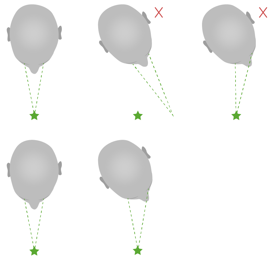
Head impulse test

  • Eyes are fixed on a target and the examiner quickly turns the subject’s head by 15º to the side.
  • Normal: Eyes remain on target.
  • Abnormal: Eyes drift off the target to later return with a saccade.
    • Indicates deficient vestibulo-ocular reflex Vestibulo-Ocular Reflex A reflex wherein impulses are conveyed from the cupulas of the semicircular canals and from the otolithic membrane of the saccule and utricle via the vestibular nuclei of the brain stem and the median longitudinal fasciculus to the oculomotor nerve nuclei. It functions to maintain a stable retinal image during head rotation by generating appropriate compensatory eye movements. Cranial Nerve Palsies → peripheral lesion
Diagram of an abnormal and normal head impulse test

Diagram of abnormal (above) and normal (below) head impulse tests:
Notice that in abnormal situations, the individual’s focal point shifts with the head to later return to the initial point of focus with a saccade.
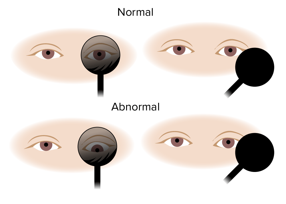
Image by Lecturio.

Skew deviation test

  • The examiner covers 1 eye of the individual for a few seconds.
  • Normal: The covered eye remains fixed when uncovered.
  • Abnormal: The covered eye shifts vertically when uncovered.
    • Indicates central lesion
Vertigo

Skew deviation test:
Normally, the eye remains fixed when uncovered. With central lesions, the eye shifts vertically when uncovered.
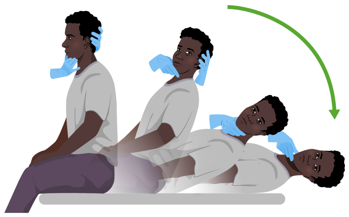
Image by Lecturio.

Dix-Hallpike maneuver

  • The subject sits on an examination table and quickly adopts a supine position while the examiner supports their head (to the right or to the left) at a 20º angle below the edge of the bed.
  • The position is held for 30 seconds.
  • Normal: no symptoms of vertigo or nystagmus Nystagmus Involuntary movements of the eye that are divided into two types, jerk and pendular. Jerk nystagmus has a slow phase in one direction followed by a corrective fast phase in the opposite direction, and is usually caused by central or peripheral vestibular dysfunction. Pendular nystagmus features oscillations that are of equal velocity in both directions and this condition is often associated with visual loss early in life. Albinism
  • Abnormal: Vertigo with/without nystagmus Nystagmus Involuntary movements of the eye that are divided into two types, jerk and pendular. Jerk nystagmus has a slow phase in one direction followed by a corrective fast phase in the opposite direction, and is usually caused by central or peripheral vestibular dysfunction. Pendular nystagmus features oscillations that are of equal velocity in both directions and this condition is often associated with visual loss early in life. Albinism is evoked.
Dix-hallpike maneuver

Dix-Hallpike maneuver:
Both diagnostic and curative in benign paroxysmal positional vertigo (BPPV). The subject sits on an examination table and quickly adopts a supine position while the examiner supports their head (to the right or to the left) at a 20º angle below the edge of the bed. The position is held for 30 seconds. In individuals with BPPV, symptoms of vertigo with or without nystagmus become evident.

Image by Lecturio.

Romberg test Romberg Test Cranial Nerve Palsies

  • The subject stands with their feet together and with arms to the sides or crossed.
  • The subject is instructed to close their eyes for 30 seconds.
  • In peripheral lesions: leaning or falling toward the side of the lesion
  • In central lesions: Direction of the lean LEAN Quality Measurement and Improvement or fall is variable Variable Variables represent information about something that can change. The design of the measurement scales, or of the methods for obtaining information, will determine the data gathered and the characteristics of that data. As a result, a variable can be qualitative or quantitative, and may be further classified into subgroups. Types of Variables, and some affected individuals may not be able to stand without assistance.

Diagnostic Evaluation

Vertigo itself is a clinical diagnosis. Further studies are only carried out if more malignant etiologies are suspected.

Neuroimaging Neuroimaging Non-invasive methods of visualizing the central nervous system, especially the brain, by various imaging modalities. Febrile Infant

  • Indicated in suspicion of a central lesion (e.g., stroke, focal neurologic deficits Neurologic Deficits High-Risk Headaches, headache Headache The symptom of pain in the cranial region. It may be an isolated benign occurrence or manifestation of a wide variety of headache disorders. Brain Abscess)
  • MRI: preferred modality
  • CT:
    • When MRI is not available or contraindicated
    • When thin cuts focusing on the brainstem and cerebellum Cerebellum The cerebellum, Latin for “little brain,” is located in the posterior cranial fossa, dorsal to the pons and midbrain, and its principal role is in the coordination of movements. The cerebellum consists of 3 lobes on either side of its 2 hemispheres and is connected in the middle by the vermis. Cerebellum: Anatomy are needed

Audiogram

  • Performed by an audiologist or an otolaryngologist
  • Indicated in any individual with unilateral or bilateral hearing loss Bilateral hearing loss Partial hearing loss in both ears. Hearing Loss

Vestibular testing

  • Also called videonystagmography
  • Measures nystagmus Nystagmus Involuntary movements of the eye that are divided into two types, jerk and pendular. Jerk nystagmus has a slow phase in one direction followed by a corrective fast phase in the opposite direction, and is usually caused by central or peripheral vestibular dysfunction. Pendular nystagmus features oscillations that are of equal velocity in both directions and this condition is often associated with visual loss early in life. Albinism via ocular, positional, and caloric testing
  • Used to confirm inner ear Inner ear The essential part of the hearing organ consists of two labyrinthine compartments: the bony labyrinthine and the membranous labyrinth. Ear: Anatomy pathology

Management

Definitive management depends on the etiology.
Pharmacological management

  • Focused on symptomatic relief:
    • Antihistamines Antihistamines Antihistamines are drugs that target histamine receptors, particularly H1 and H2 receptors. H1 antagonists are competitive and reversible inhibitors of H1 receptors. First-generation antihistamines cross the blood-brain barrier and can cause sedation. Antihistamines
    • Benzodiazepines Benzodiazepines Benzodiazepines work on the gamma-aminobutyric acid type A (GABAA) receptor to produce inhibitory effects on the CNS. Benzodiazepines do not mimic GABA, the main inhibitory neurotransmitter in humans, but instead potentiate GABA activity. Benzodiazepines
    • Antiemetics Antiemetics Antiemetics are medications used to treat and/or prevent nausea and vomiting. These drugs act on different target receptors. The main classes include benzodiazepines, corticosteroids, atypical antipsychotics, cannabinoids, and antagonists of the following receptors: serotonin, dopamine, and muscarinic and neurokinin receptors. Antiemetics

Nonpharmacological management

  • Vestibular rehabilitation Vestibular Rehabilitation Ménière Disease: training on maintaining balance based on visual and proprioceptive clues
  • Lifestyle recommendations: avoiding caffeine Caffeine A methylxanthine naturally occurring in some beverages and also used as a pharmacological agent. Caffeine’s most notable pharmacological effect is as a central nervous system stimulant, increasing alertness and producing agitation. Several cellular actions of caffeine have been observed, but it is not entirely clear how each contributes to its pharmacological profile. Among the most important are inhibition of cyclic nucleotide phosphodiesterases, antagonism of adenosine receptors, and modulation of intracellular calcium handling. Stimulants and alcohol (in Ménière disease Ménière disease Ménière disease is a condition characterized by episodes of vertigo, tinnitus, and hearing loss, likely caused by endolymphatic hydrops of the labyrinthine system in the inner ear. The risk factors include a family history of Ménière disease, preexisting autoimmune disorders, allergies, and trauma to the head or ear. Ménière Disease), avoiding triggers
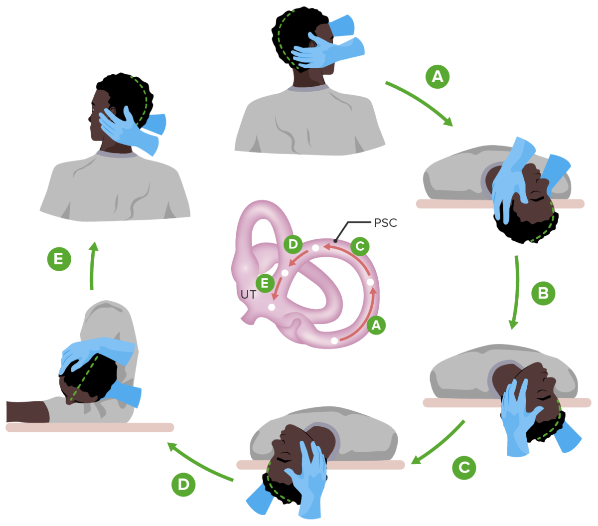
  • Canalith repositioning procedure: Epley maneuver can be curative in individuals with BPPV.
  • For debilitating symptoms, surgical procedures or intratympanic gentamicin Gentamicin Aminoglycosides injections may be used.

Complications

  • High risk for falls!
  • Some affected individuals may require home-safety evaluation and supervision.
A diagram of epley maneuver (bppv)

Diagram showing the Epley maneuver for the management of benign paroxysmal positional vertigo (BPPV):
Each position is carried out as shown while being held for 30 seconds.
PSC: posterior semicircular canal
UT: utricle

Image by Lecturio.

Clinical Relevance

The following conditions can cause dizziness Dizziness An imprecise term which may refer to a sense of spatial disorientation, motion of the environment, or lightheadedness. Lateral Medullary Syndrome (Wallenberg Syndrome) similar to vertigo:

  • Syncope Syncope Syncope is a short-term loss of consciousness and loss of postural stability followed by spontaneous return of consciousness to the previous neurologic baseline without the need for resuscitation. The condition is caused by transient interruption of cerebral blood flow that may be benign or related to a underlying life-threatening condition. Syncope: a short-term loss of consciousness caused by inadequate cerebral blood flow Inadequate Cerebral Blood Flow Syncope. Syncope Syncope Syncope is a short-term loss of consciousness and loss of postural stability followed by spontaneous return of consciousness to the previous neurologic baseline without the need for resuscitation. The condition is caused by transient interruption of cerebral blood flow that may be benign or related to a underlying life-threatening condition. Syncope has a wide range of etiologies. Affected individuals may have prodromal symptoms associated with imminent syncope Syncope Syncope is a short-term loss of consciousness and loss of postural stability followed by spontaneous return of consciousness to the previous neurologic baseline without the need for resuscitation. The condition is caused by transient interruption of cerebral blood flow that may be benign or related to a underlying life-threatening condition. Syncope or presyncope Presyncope Syncope, such as lightheadedness Lightheadedness Hypotension, sweating, palpitations Palpitations Ebstein’s Anomaly, nausea Nausea An unpleasant sensation in the stomach usually accompanied by the urge to vomit. Common causes are early pregnancy, sea and motion sickness, emotional stress, intense pain, food poisoning, and various enteroviruses. Antiemetics, feeling warm or cold, and blurred vision Blurred Vision Retinal Detachment. Diagnosis is clinical, and management involves the identification Identification Defense Mechanisms and treatment of the underlying disorders.
  • Anemia Anemia Anemia is a condition in which individuals have low Hb levels, which can arise from various causes. Anemia is accompanied by a reduced number of RBCs and may manifest with fatigue, shortness of breath, pallor, and weakness. Subtypes are classified by the size of RBCs, chronicity, and etiology. Anemia: Overview and Types: a condition characterized by low hemoglobin levels that can arise due to various causes. Anemia Anemia Anemia is a condition in which individuals have low Hb levels, which can arise from various causes. Anemia is accompanied by a reduced number of RBCs and may manifest with fatigue, shortness of breath, pallor, and weakness. Subtypes are classified by the size of RBCs, chronicity, and etiology. Anemia: Overview and Types is accompanied by a reduced RBC count and may manifest with fatigue Fatigue The state of weariness following a period of exertion, mental or physical, characterized by a decreased capacity for work and reduced efficiency to respond to stimuli. Fibromyalgia, lightheadedness Lightheadedness Hypotension, shortness of breath Shortness of breath Dyspnea is the subjective sensation of breathing discomfort. Dyspnea is a normal manifestation of heavy physical or psychological exertion, but also may be caused by underlying conditions (both pulmonary and extrapulmonary). Dyspnea, pallor, and weakness. Diagnosis is made based on CBC and peripheral blood smear Peripheral Blood Smear Anemia: Overview and Types. Management involves treatment of the underlying disorder and transfusion in severe cases.
  • Ménière disease Ménière disease Ménière disease is a condition characterized by episodes of vertigo, tinnitus, and hearing loss, likely caused by endolymphatic hydrops of the labyrinthine system in the inner ear. The risk factors include a family history of Ménière disease, preexisting autoimmune disorders, allergies, and trauma to the head or ear. Ménière Disease: a disorder of the inner ear Inner ear The essential part of the hearing organ consists of two labyrinthine compartments: the bony labyrinthine and the membranous labyrinth. Ear: Anatomy characterized by hearing loss Hearing loss Hearing loss, also known as hearing impairment, is any degree of impairment in the ability to apprehend sound as determined by audiometry to be below normal hearing thresholds. Clinical presentation may occur at birth or as a gradual loss of hearing with age, including a short-term or sudden loss at any point. Hearing Loss, fluctuating aural symptoms (e.g., tinnitus Tinnitus A nonspecific symptom of hearing disorder characterized by the sensation of buzzing, ringing, clicking, pulsations, and other noises in the ear. Objective tinnitus refers to noises generated from within the ear or adjacent structures that can be heard by other individuals. The term subjective tinnitus is used when the sound is audible only to the affected individual. Tinnitus may occur as a manifestation of cochlear diseases; vestibulocochlear nerve diseases; intracranial hypertension; craniocerebral trauma; and other conditions. Cranial Nerve Palsies), and spontaneous episodes of vertigo. Diagnosis is made based on a thorough history and physical examination, including a full otologic exam. An audiogram is also a key component of the evaluation. Management is centered around noninvasive techniques to maintain function; however, there is no cure.
  • Multiple sclerosis Sclerosis A pathological process consisting of hardening or fibrosis of an anatomical structure, often a vessel or a nerve. Wilms Tumor: a chronic inflammatory autoimmune disease leading to demyelination Demyelination Multiple Sclerosis of the CNS. The clinical presentation varies widely depending on the site of lesions, but typically involves neurological symptoms affecting vision Vision Ophthalmic Exam, motor Motor Neurons which send impulses peripherally to activate muscles or secretory cells. Nervous System: Histology function, sensation, and autonomic function. Management involves corticosteroids Corticosteroids Chorioretinitis for acute exacerbations and disease-modifying agents to reduce exacerbations and slow disease progression.
  • Ischemic stroke Ischemic Stroke An ischemic stroke (also known as cerebrovascular accident) is an acute neurologic injury that occurs as a result of brain ischemia; this condition may be due to cerebral blood vessel occlusion by thrombosis or embolism, or rarely due to systemic hypoperfusion. Ischemic Stroke: also known as a cerebrovascular accident Cerebrovascular accident An ischemic stroke (also known as cerebrovascular accident) is an acute neurologic injury that occurs as a result of brain ischemia; this condition may be due to cerebral blood vessel occlusion by thrombosis or embolism, or rarely due to systemic hypoperfusion. Ischemic Stroke (CVA), ischemic stroke Ischemic Stroke An ischemic stroke (also known as cerebrovascular accident) is an acute neurologic injury that occurs as a result of brain ischemia; this condition may be due to cerebral blood vessel occlusion by thrombosis or embolism, or rarely due to systemic hypoperfusion. Ischemic Stroke is an acute neurologic injury resulting from brain ischemia Brain Ischemia Localized reduction of blood flow to brain tissue due to arterial obstruction or systemic hypoperfusion. This frequently occurs in conjunction with brain hypoxia. Prolonged ischemia is associated with brain infarction. Ischemic Stroke. The clinical presentation includes neurologic symptoms with varying degrees of motor Motor Neurons which send impulses peripherally to activate muscles or secretory cells. Nervous System: Histology and sensory Sensory Neurons which conduct nerve impulses to the central nervous system. Nervous System: Histology loss, which corresponds to the area of the brain Brain The part of central nervous system that is contained within the skull (cranium). Arising from the neural tube, the embryonic brain is comprised of three major parts including prosencephalon (the forebrain); mesencephalon (the midbrain); and rhombencephalon (the hindbrain). The developed brain consists of cerebrum; cerebellum; and other structures in the brain stem. Nervous System: Anatomy, Structure, and Classification affected and the extent of tissue damage. Management is with the timely restoration of blood flow Blood flow Blood flow refers to the movement of a certain volume of blood through the vasculature over a given unit of time (e.g., mL per minute). Vascular Resistance, Flow, and Mean Arterial Pressure and prevention of a 2nd stroke.
  • Wernicke encephalopathy Encephalopathy Hyper-IgM Syndrome: an acute, reversible condition caused by severe thiamine Thiamine Also known as thiamine or thiamin, it is a vitamin C12H17N4OSCl of the vitamin B complex that is essential to normal metabolism and nerve function and is widespread in plants and animals Water-soluble Vitamins and their Deficiencies deficiency. Wernicke encephalopathy Encephalopathy Hyper-IgM Syndrome is most commonly seen in individuals with severe alcohol-use disorder Alcohol-use disorder A primary, chronic disease with genetic, psychosocial, and environmental factors influencing its development and manifestations. The disease is often progressive and fatal. It is characterized by impaired control over drinking, preoccupation with the drug alcohol, use of alcohol despite adverse consequences, and distortions in thinking, most notably denial. Each of these symptoms may be continuous or periodic. Klebsiella and is characterized by the classic triad of encephalopathy Encephalopathy Hyper-IgM Syndrome, oculomotor dysfunction, and gait ataxia Gait ataxia Impairment of the ability to coordinate the movements required for normal ambulation (walking) which may result from impairments of motor function or sensory feedback. This condition may be associated with brain diseases (including cerebellar diseases and basal ganglia diseases); spinal cord diseases; or peripheral nervous system diseases. Friedreich Ataxia, although all 3 features are only present in ⅓ of the affected population. Diagnosis is made clinically. Management includes thiamine Thiamine Also known as thiamine or thiamin, it is a vitamin C12H17N4OSCl of the vitamin B complex that is essential to normal metabolism and nerve function and is widespread in plants and animals Water-soluble Vitamins and their Deficiencies supplementation and recommendations for alcohol cessation.

References

  1. Stanton, M., Freeman, A.M. (2021). Vertigo. StatPearls. Treasure Island (FL): StatPearls Publishing. Retrieved September 15, 2021, from http://www.ncbi.nlm.nih.gov/books/NBK482356/ 
  2. Barrett, K.E., Barman, S.M., Boitano, S., Reckelhoff, J.F. (2017). Hearing & Equilibrium. In Ganong’s Medical Physiology Examination and Board Review. McGraw-Hill Education. Retrieved September 15, 2021, from http://accessmedicine.mhmedical.com/content.aspx?aid=1142554680 
  3. Wipperman, J. (2021). Dizziness and Vertigo. In Kellerman, R.D., Rakel, D.P. (Eds.), Conn’s Current Therapy 2021, pp. 9–14. Elsevier. Retrieved September 15, 2021, from https://www.clinicalkey.es/#!/content/book/3-s2.0-B9780323790062000045 
  4. Walker, M.F., Daroff, R.B. (2018). Dizziness and vertigo. Jameson, J., et al. (Eds.), Harrison’s Principles of Internal Medicine, 20 ed. McGraw Hill. Retrieved September 14, 2021, from https://accessmedicine.mhmedical.com/content.aspx?sectionid=192011330&bookid=2129&Resultclick=2
  5. Kerber, K. (2021). Dizziness. DeckerMed Medicine. Retrieved September 15, 2021, from doi:10.2310/PSYCH.6089
  6. Kroenke, K., Lucas, C.A., Rosenberg, M.L., et al. Causes of persistent dizziness. A prospective study of 100 patients in ambulatory care. Ann Intern Med. 1992, 117: pp. 898–904.

Create your free account or log in to continue reading!

Sign up now and get free access to Lecturio with concept pages, medical videos, and questions for your medical education.

User Reviews

Details