Urologic cancer is a broad term that involves cancer of the male and female urinary tracts and male reproductive organs. Risk factors for urologic cancer are smoking Smoking Willful or deliberate act of inhaling and exhaling smoke from burning substances or agents held by hand. Interstitial Lung Diseases; exposure to chemicals such as benzidine and beta-naphthylamine, and arsenic Arsenic A shiny gray element with atomic symbol as, atomic number 33, and atomic weight 75. It occurs throughout the universe, mostly in the form of metallic arsenides. Most forms are toxic. According to the fourth annual report on carcinogens, arsenic and certain arsenic compounds have been listed as known carcinogens. Metal Poisoning (Lead, Arsenic, Iron); genetic predisposition; and chronic irritation of the urinary system. Clinical presentation includes painless hematuria Hematuria Presence of blood in the urine. Renal Cell Carcinoma, flank and/or suprapubic pain Pain An unpleasant sensation induced by noxious stimuli which are detected by nerve endings of nociceptive neurons. Pain: Types and Pathways, dysuria Dysuria Painful urination. It is often associated with infections of the lower urinary tract. Urinary Tract Infections (UTIs), and unexplained significant weight loss Weight loss Decrease in existing body weight. Bariatric Surgery. The gold standard for diagnosis is endoscopy Endoscopy Procedures of applying endoscopes for disease diagnosis and treatment. Endoscopy involves passing an optical instrument through a small incision in the skin i.e., percutaneous; or through a natural orifice and along natural body pathways such as the digestive tract; and/or through an incision in the wall of a tubular structure or organ, i.e. Transluminal, to examine or perform surgery on the interior parts of the body. Gastroesophageal Reflux Disease (GERD) of the urologic structures (cystoscopy, cystourethroscopy, ureteropyeloscopy) with biopsy Biopsy Removal and pathologic examination of specimens from the living body. Ewing Sarcoma. Additional studies include radiologic imaging, which gives information about the tumor Tumor Inflammation invasion and spread of the disease to other sites or organs. Management includes surgery, chemotherapy Chemotherapy Osteosarcoma, radiotherapy, and supportive treatment, depending on the location, extent, and histology.
Last updated: Dec 15, 2025
Growth of abnormal cells from the lining of organs of the male and female urinary tracts and the male reproductive organs.
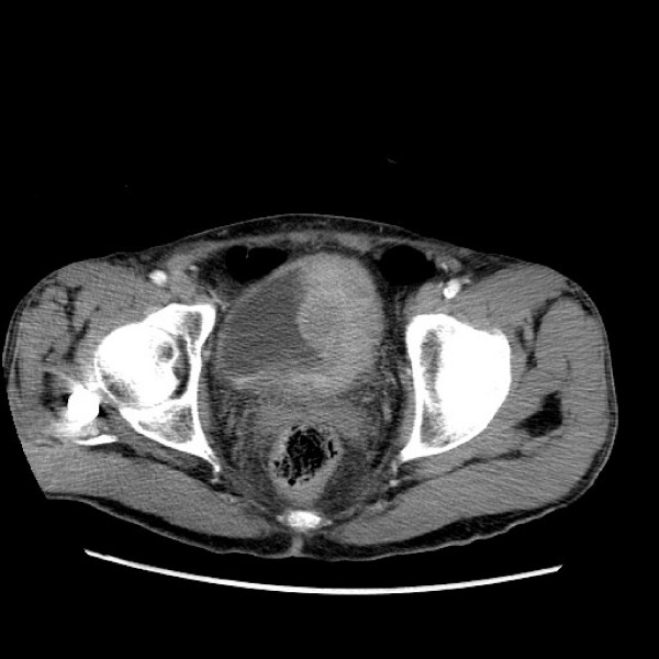
CT scan of bladder cancer:
Left-sided lobulated enhancing soft tissue mass in the bladder.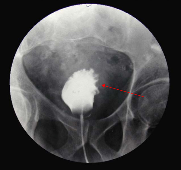
Cystogram (phase in intravenous pyelography), showing marked trabecular thickening in an individual with bladder carcinoma.
Image: “Cystogram showing marked trabecular thickening” by Gaurav K, Fitch J, Panda M. License: CC BY 2.0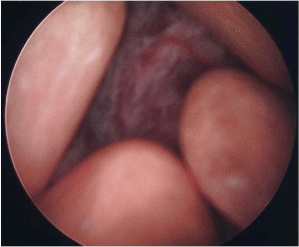
Cystoscopic image showing a bladder mass and multiple calculi
Image: “Cystoscopy showing a bladder mass and multiple calculi.” by Cho JH, Holley JL. License: CC BY 2.0Bladder Bladder A musculomembranous sac along the urinary tract. Urine flows from the kidneys into the bladder via the ureters, and is held there until urination. Pyelonephritis and Perinephric Abscess cancer pathologic stages are based on the TNM staging system TNM staging system Grading, Staging, and Metastasis.
|
Tumor
Tumor
Inflammation (T) category |
Description |
|---|---|
| Tx | Primary tumor Tumor Inflammation cannot be assessed. |
| T0 | No evidence of primary tumor Tumor Inflammation |
| Ta TA Thyrotoxicosis and Hyperthyroidism | Noninvasive papillary or exophytic Exophytic Retinoblastoma lesions |
| Tis | Carcinoma in situ (“flat tumor Tumor Inflammation”) |
| T1 | Tumor Tumor Inflammation invades lamina propria Lamina propria Whipple’s Disease (or submucosa). |
| T2 | Tumor Tumor Inflammation invades muscularis propria. |
| T2a: Tumor Tumor Inflammation invades superficial muscularis propria (inner half). | |
| T2b: Tumor Tumor Inflammation invades deep muscularis propria (outer half). | |
| T3 T3 A T3 thyroid hormone normally synthesized and secreted by the thyroid gland in much smaller quantities than thyroxine (T4). Most T3 is derived from peripheral monodeiodination of T4 at the 5′ position of the outer ring of the iodothyronine nucleus. The hormone finally delivered and used by the tissues is mainly t3. Thyroid Hormones | Tumor Tumor Inflammation invades perivesical fat. |
| T3a: Microscopic invasion | |
| T3b: Macroscopic (extravesical mass Mass Three-dimensional lesion that occupies a space within the breast Imaging of the Breast) invasion | |
| T4 T4 The major hormone derived from the thyroid gland. Thyroxine is synthesized via the iodination of tyrosines (monoiodotyrosine) and the coupling of iodotyrosines (diiodotyrosine) in the thyroglobulin. Thyroxine is released from thyroglobulin by proteolysis and secreted into the blood. Thyroxine is peripherally deiodinated to form triiodothyronine which exerts a broad spectrum of stimulatory effects on cell metabolism. Thyroid Hormones | Tumor Tumor Inflammation invades 1 of the following: prostatic stroma, seminal vesicles Vesicles Female Genitourinary Examination, uterus Uterus The uterus, cervix, and fallopian tubes are part of the internal female reproductive system. The uterus has a thick wall made of smooth muscle (the myometrium) and an inner mucosal layer (the endometrium). The most inferior portion of the uterus is the cervix, which connects the uterine cavity to the vagina. Uterus, Cervix, and Fallopian Tubes: Anatomy, vagina Vagina The vagina is the female genital canal, extending from the vulva externally to the cervix uteri internally. The structures have sexual, reproductive, and urinary functions and a rich blood supply, mainly arising from the internal iliac artery. Vagina, Vulva, and Pelvic Floor: Anatomy, pelvic wall, abdominal wall Abdominal wall The outer margins of the abdomen, extending from the osteocartilaginous thoracic cage to the pelvis. Though its major part is muscular, the abdominal wall consists of at least seven layers: the skin, subcutaneous fat, deep fascia; abdominal muscles, transversalis fascia, extraperitoneal fat, and the parietal peritoneum. Surgical Anatomy of the Abdomen. |
| T4a: Tumor Tumor Inflammation invades prostatic stroma, uterus Uterus The uterus, cervix, and fallopian tubes are part of the internal female reproductive system. The uterus has a thick wall made of smooth muscle (the myometrium) and an inner mucosal layer (the endometrium). The most inferior portion of the uterus is the cervix, which connects the uterine cavity to the vagina. Uterus, Cervix, and Fallopian Tubes: Anatomy, vagina Vagina The vagina is the female genital canal, extending from the vulva externally to the cervix uteri internally. The structures have sexual, reproductive, and urinary functions and a rich blood supply, mainly arising from the internal iliac artery. Vagina, Vulva, and Pelvic Floor: Anatomy (adjacent organs). | |
| T4b: Tumor Tumor Inflammation invades pelvic wall, abdominal wall Abdominal wall The outer margins of the abdomen, extending from the osteocartilaginous thoracic cage to the pelvis. Though its major part is muscular, the abdominal wall consists of at least seven layers: the skin, subcutaneous fat, deep fascia; abdominal muscles, transversalis fascia, extraperitoneal fat, and the parietal peritoneum. Surgical Anatomy of the Abdomen, or other organs. |
| Node (N) category | Description |
|---|---|
| Nx | Lymph nodes Lymph Nodes They are oval or bean shaped bodies (1 – 30 mm in diameter) located along the lymphatic system. Lymphatic Drainage System: Anatomy cannot be assessed. |
| N0 | No lymph Lymph The interstitial fluid that is in the lymphatic system. Secondary Lymphatic Organs node metastasis Metastasis The transfer of a neoplasm from one organ or part of the body to another remote from the primary site. Grading, Staging, and Metastasis |
| N1 | Single regional lymph Lymph The interstitial fluid that is in the lymphatic system. Secondary Lymphatic Organs node metastasis Metastasis The transfer of a neoplasm from one organ or part of the body to another remote from the primary site. Grading, Staging, and Metastasis in the true pelvis Pelvis The pelvis consists of the bony pelvic girdle, the muscular and ligamentous pelvic floor, and the pelvic cavity, which contains viscera, vessels, and multiple nerves and muscles. The pelvic girdle, composed of 2 “hip” bones and the sacrum, is a ring-like bony structure of the axial skeleton that links the vertebral column with the lower extremities. Pelvis: Anatomy (perivesical, obturator, internal and external iliac, or sacral lymph nodes Lymph Nodes They are oval or bean shaped bodies (1 – 30 mm in diameter) located along the lymphatic system. Lymphatic Drainage System: Anatomy) |
| N2 | Multiple regional lymph Lymph The interstitial fluid that is in the lymphatic system. Secondary Lymphatic Organs node metastases in the true pelvis Pelvis The pelvis consists of the bony pelvic girdle, the muscular and ligamentous pelvic floor, and the pelvic cavity, which contains viscera, vessels, and multiple nerves and muscles. The pelvic girdle, composed of 2 “hip” bones and the sacrum, is a ring-like bony structure of the axial skeleton that links the vertebral column with the lower extremities. Pelvis: Anatomy (perivesical, obturator, internal and external iliac, or sacral lymph Lymph The interstitial fluid that is in the lymphatic system. Secondary Lymphatic Organs node metastasis Metastasis The transfer of a neoplasm from one organ or part of the body to another remote from the primary site. Grading, Staging, and Metastasis) |
| N3 | Metastasis Metastasis The transfer of a neoplasm from one organ or part of the body to another remote from the primary site. Grading, Staging, and Metastasis to the common iliac lymph nodes Lymph Nodes They are oval or bean shaped bodies (1 – 30 mm in diameter) located along the lymphatic system. Lymphatic Drainage System: Anatomy |
| Metastasis Metastasis The transfer of a neoplasm from one organ or part of the body to another remote from the primary site. Grading, Staging, and Metastasis (M) category | Description |
|---|---|
| M0 | No distant metastasis Metastasis The transfer of a neoplasm from one organ or part of the body to another remote from the primary site. Grading, Staging, and Metastasis |
| M1 | Distant metastasis Metastasis The transfer of a neoplasm from one organ or part of the body to another remote from the primary site. Grading, Staging, and Metastasis |
| M1a: distant metastasis Metastasis The transfer of a neoplasm from one organ or part of the body to another remote from the primary site. Grading, Staging, and Metastasis limited to lymph nodes Lymph Nodes They are oval or bean shaped bodies (1 – 30 mm in diameter) located along the lymphatic system. Lymphatic Drainage System: Anatomy beyond the common iliac nodes | |
| M1b: non– lymph Lymph The interstitial fluid that is in the lymphatic system. Secondary Lymphatic Organs node distant metastasis Metastasis The transfer of a neoplasm from one organ or part of the body to another remote from the primary site. Grading, Staging, and Metastasis |
| Stage | T | N | M |
|---|---|---|---|
| Stage 0a | Ta TA Thyrotoxicosis and Hyperthyroidism | N0 | M0 |
| Stage 0is | Tis | N0 | M0 |
| Stage I | T1 | N0 | M0 |
| Stage II | T2a | N0 | M0 |
| T2b | N0 | M0 | |
| Stage IIIA | T3a, T3b, T4a | N0 | M0 |
| Stage IIIA | T1–T4a | N1 | M0 |
| Stage IIIB | T1–T4a | N2, N3 | M0 |
| Stage IVA | T4b | Any N | M0 |
| Stage IVA | Any T | Any N | M1a |
| Stage IVB | Any T | Any N | M1b |
Abnormal growth of cells in the inner lining of the ureter/ ureters Ureters One of a pair of thick-walled tubes that transports urine from the kidney pelvis to the urinary bladder. Urinary Tract: Anatomy, of which > 90% are urothelial carcinomas.
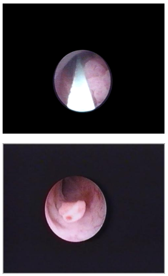
Ureteral tumor noted on endoscopic examination of the ureter.
Image: “Ureteral tumour with elective indication for endoscopic treatment” by Niţă G, Georgescu D, Mulţescu R, Draguţescu M, Mihai B, Geavlete B, Persu C, Geavlete P. License: CC BY 2.0Staging Staging Methods which attempt to express in replicable terms the extent of the neoplasm in the patient. Grading, Staging, and Metastasis applies to cancer involving the renal pelvis Renal pelvis Kidneys: Anatomy and ureter.
| T category | Description |
|---|---|
| TX | Primary tumor Tumor Inflammation cannot be assessed. |
| T0 | No evidence of primary tumor Tumor Inflammation |
| Ta TA Thyrotoxicosis and Hyperthyroidism | Noninvasive papillary or exophytic Exophytic Retinoblastoma lesions |
| Tis | CIS CIS Multiple Sclerosis |
| T1 | Tumor Tumor Inflammation invasion of subepithelial Subepithelial Membranoproliferative Glomerulonephritis connective tissue Connective tissue Connective tissues originate from embryonic mesenchyme and are present throughout the body except inside the brain and spinal cord. The main function of connective tissues is to provide structural support to organs. Connective tissues consist of cells and an extracellular matrix. Connective Tissue: Histology |
| T2 | Tumor Tumor Inflammation invasion of muscularis |
| T3 T3 A T3 thyroid hormone normally synthesized and secreted by the thyroid gland in much smaller quantities than thyroxine (T4). Most T3 is derived from peripheral monodeiodination of T4 at the 5′ position of the outer ring of the iodothyronine nucleus. The hormone finally delivered and used by the tissues is mainly t3. Thyroid Hormones | For ureter only: invasion beyond muscularis into periureteric fat. For renal pelvis Renal pelvis Kidneys: Anatomy only: invasion beyond muscularis into peripelvic fat or into the renal parenchyma |
| T4 T4 The major hormone derived from the thyroid gland. Thyroxine is synthesized via the iodination of tyrosines (monoiodotyrosine) and the coupling of iodotyrosines (diiodotyrosine) in the thyroglobulin. Thyroxine is released from thyroglobulin by proteolysis and secreted into the blood. Thyroxine is peripherally deiodinated to form triiodothyronine which exerts a broad spectrum of stimulatory effects on cell metabolism. Thyroid Hormones | Tumor Tumor Inflammation invasion of adjacent organs or the kidney into the perinephric fat |
| Node category | Description |
|---|---|
| NX | Regional lymph nodes Lymph Nodes They are oval or bean shaped bodies (1 – 30 mm in diameter) located along the lymphatic system. Lymphatic Drainage System: Anatomy cannot be assessed. |
| N0 | No lymph Lymph The interstitial fluid that is in the lymphatic system. Secondary Lymphatic Organs node metastasis Metastasis The transfer of a neoplasm from one organ or part of the body to another remote from the primary site. Grading, Staging, and Metastasis |
| N1 | Metastasis Metastasis The transfer of a neoplasm from one organ or part of the body to another remote from the primary site. Grading, Staging, and Metastasis ≤ 2 cm (single lymph Lymph The interstitial fluid that is in the lymphatic system. Secondary Lymphatic Organs node) |
| N2 | Metastasis Metastasis The transfer of a neoplasm from one organ or part of the body to another remote from the primary site. Grading, Staging, and Metastasis > 2 cm (single lymph Lymph The interstitial fluid that is in the lymphatic system. Secondary Lymphatic Organs node or multiple lymph nodes Lymph Nodes They are oval or bean shaped bodies (1 – 30 mm in diameter) located along the lymphatic system. Lymphatic Drainage System: Anatomy) |
| M category | Description |
|---|---|
| M0 | No distant metastasis Metastasis The transfer of a neoplasm from one organ or part of the body to another remote from the primary site. Grading, Staging, and Metastasis |
| M1 | Distant metastasis Metastasis The transfer of a neoplasm from one organ or part of the body to another remote from the primary site. Grading, Staging, and Metastasis |
| Stage | T | N | M |
|---|---|---|---|
| Stage 0a | Ta TA Thyrotoxicosis and Hyperthyroidism | N0 | M0 |
| Stage 0is | Tis | N0 | M0 |
| Stage I | T1 | N0 | M0 |
| Stage II | T2 | N0 | M0 |
| Stage III | T3 T3 A T3 thyroid hormone normally synthesized and secreted by the thyroid gland in much smaller quantities than thyroxine (T4). Most T3 is derived from peripheral monodeiodination of T4 at the 5′ position of the outer ring of the iodothyronine nucleus. The hormone finally delivered and used by the tissues is mainly t3. Thyroid Hormones | N0 | M0 |
| Stage IV | T4 T4 The major hormone derived from the thyroid gland. Thyroxine is synthesized via the iodination of tyrosines (monoiodotyrosine) and the coupling of iodotyrosines (diiodotyrosine) in the thyroglobulin. Thyroxine is released from thyroglobulin by proteolysis and secreted into the blood. Thyroxine is peripherally deiodinated to form triiodothyronine which exerts a broad spectrum of stimulatory effects on cell metabolism. Thyroid Hormones | NX, N0 | M0 |
| Any T | N1, N2 | M0 | |
| Any T | Any N | M1 |
Treatment of ureteral cancer depends on the site, size, and extent of cancer, with surgery providing curative treatment.
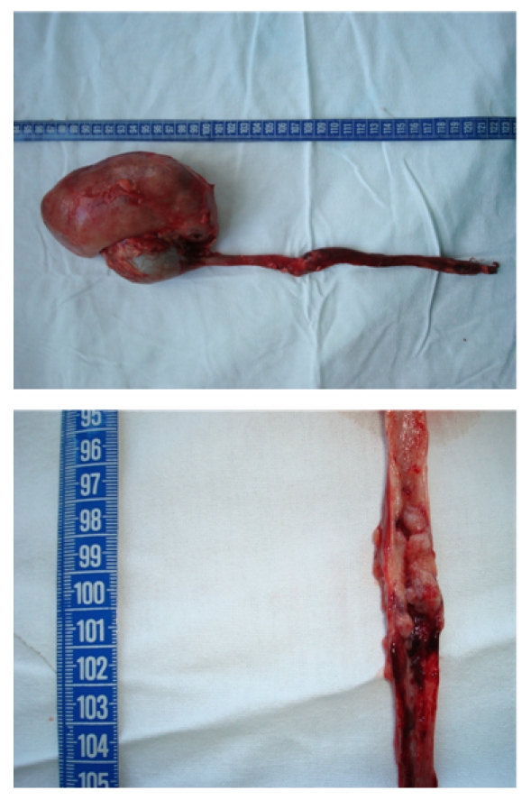
Nephroureterectomy in ureteral cancer
Image: “Nephroureterectomy (with endoscopic desinsertion) for ureteral tumours” by Niţă G, Georgescu D, Mulţescu R, Draguţescu M, Mihai B, Geavlete B, Persu C, Geavlete P. License: CC BY 2.0Urethral cancer is an extremely rare malignancy Malignancy Hemothorax (< 1% of all genitourinary malignancies) that involves the abnormal growth of cells in the lining of the urethra Urethra A tube that transports urine from the urinary bladder to the outside of the body in both the sexes. It also has a reproductive function in the male by providing a passage for sperm. Urinary Tract: Anatomy.
Urethral cancer has an insidious onset and can remain asymptomatic for a long time. Symptoms vary between men and women:
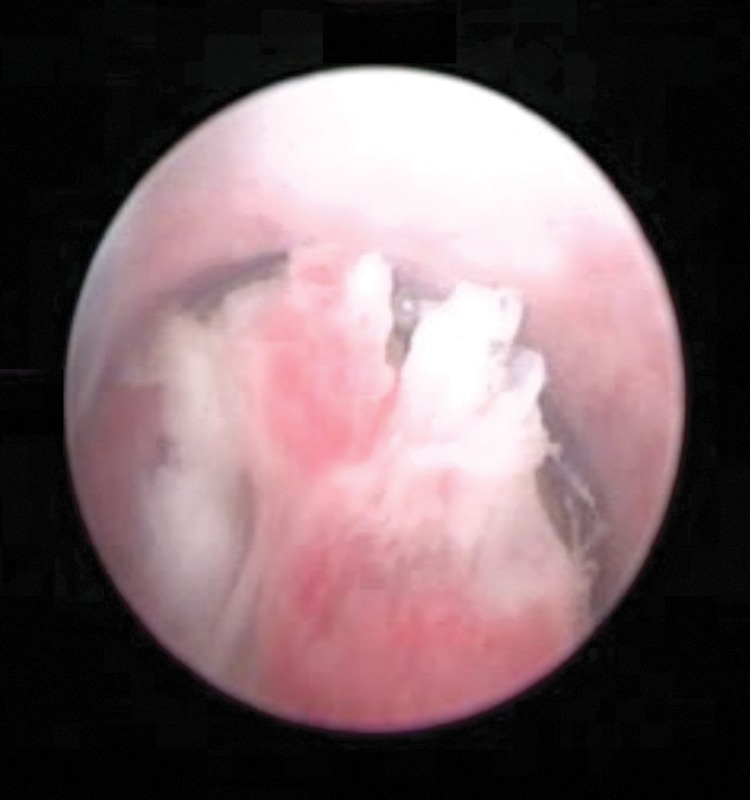
Video urethrocystoscopy showing a papillary tumor in the bulbar urethra
Image: “Video urethrocystoscopy showing papillary tumor at the level of bulbar urethra.” by Journal of Endourology Case Reports. License: CC BY 4.0Urethral cancer is staged according to the TNM criteria.
| T category | Description |
|---|---|
| Tx | Primary tumor Tumor Inflammation cannot be assessed. |
| T0 | No evidence of primary tumor Tumor Inflammation |
| Ta TA Thyrotoxicosis and Hyperthyroidism | Noninvasive papillary carcinoma |
| Tis | Carcinoma in situ Carcinoma in situ A lesion with cytological characteristics associated with invasive carcinoma but the tumor cells are confined to the epithelium of origin, without invasion of the basement membrane. Leukoplakia |
| T1 | Tumor Tumor Inflammation invasion of subepithelial Subepithelial Membranoproliferative Glomerulonephritis connective tissue Connective tissue Connective tissues originate from embryonic mesenchyme and are present throughout the body except inside the brain and spinal cord. The main function of connective tissues is to provide structural support to organs. Connective tissues consist of cells and an extracellular matrix. Connective Tissue: Histology |
| T2 | Tumor Tumor Inflammation invasion of the corpus spongiosum Corpus spongiosum Penis: Anatomy or periurethral muscle |
| T3 T3 A T3 thyroid hormone normally synthesized and secreted by the thyroid gland in much smaller quantities than thyroxine (T4). Most T3 is derived from peripheral monodeiodination of T4 at the 5′ position of the outer ring of the iodothyronine nucleus. The hormone finally delivered and used by the tissues is mainly t3. Thyroid Hormones | Tumor Tumor Inflammation invasion of the corpus cavernosum or anterior vagina Vagina The vagina is the female genital canal, extending from the vulva externally to the cervix uteri internally. The structures have sexual, reproductive, and urinary functions and a rich blood supply, mainly arising from the internal iliac artery. Vagina, Vulva, and Pelvic Floor: Anatomy |
| T4 T4 The major hormone derived from the thyroid gland. Thyroxine is synthesized via the iodination of tyrosines (monoiodotyrosine) and the coupling of iodotyrosines (diiodotyrosine) in the thyroglobulin. Thyroxine is released from thyroglobulin by proteolysis and secreted into the blood. Thyroxine is peripherally deiodinated to form triiodothyronine which exerts a broad spectrum of stimulatory effects on cell metabolism. Thyroid Hormones | Tumor Tumor Inflammation invasion of other adjacent organs |
| T category | Description |
|---|---|
| Tx | Primary tumor Tumor Inflammation cannot be assessed. |
| T0 | No evidence of primary tumor Tumor Inflammation |
| Ta TA Thyrotoxicosis and Hyperthyroidism | Noninvasive papillary carcinoma |
| Tis | Carcinoma in situ (involves the prostatic urethra Prostatic urethra or periurethral or prostatic ducts Prostatic ducts without stromal invasion) |
| T1 | Tumor Tumor Inflammation invasion of the subepithelial Subepithelial Membranoproliferative Glomerulonephritis connective tissue Connective tissue Connective tissues originate from embryonic mesenchyme and are present throughout the body except inside the brain and spinal cord. The main function of connective tissues is to provide structural support to organs. Connective tissues consist of cells and an extracellular matrix. Connective Tissue: Histology immediately underlying the urothelium Urothelium The epithelial lining of the urinary tract. Urinary Tract: Anatomy |
| T2 | Tumor Tumor Inflammation invasion of the prostatic stroma surrounding ducts (by directly extending from the urothelial surface or by invading from prostatic ducts Prostatic ducts ) |
| T3 T3 A T3 thyroid hormone normally synthesized and secreted by the thyroid gland in much smaller quantities than thyroxine (T4). Most T3 is derived from peripheral monodeiodination of T4 at the 5′ position of the outer ring of the iodothyronine nucleus. The hormone finally delivered and used by the tissues is mainly t3. Thyroid Hormones | Tumor Tumor Inflammation invasion of the periprostatic fat |
| T4 T4 The major hormone derived from the thyroid gland. Thyroxine is synthesized via the iodination of tyrosines (monoiodotyrosine) and the coupling of iodotyrosines (diiodotyrosine) in the thyroglobulin. Thyroxine is released from thyroglobulin by proteolysis and secreted into the blood. Thyroxine is peripherally deiodinated to form triiodothyronine which exerts a broad spectrum of stimulatory effects on cell metabolism. Thyroid Hormones | Tumor Tumor Inflammation invasion of the adjacent organs |
| N category | Description |
|---|---|
| Nx | Regional lymph nodes Lymph Nodes They are oval or bean shaped bodies (1 – 30 mm in diameter) located along the lymphatic system. Lymphatic Drainage System: Anatomy cannot be assessed. |
| N0 | Regional lymph Lymph The interstitial fluid that is in the lymphatic system. Secondary Lymphatic Organs node metastasis Metastasis The transfer of a neoplasm from one organ or part of the body to another remote from the primary site. Grading, Staging, and Metastasis is not present. |
| N1 | Single regional lymph Lymph The interstitial fluid that is in the lymphatic system. Secondary Lymphatic Organs node metastasis Metastasis The transfer of a neoplasm from one organ or part of the body to another remote from the primary site. Grading, Staging, and Metastasis in the inguinal region Inguinal region Anterior Abdominal Wall: Anatomy or true pelvis Pelvis The pelvis consists of the bony pelvic girdle, the muscular and ligamentous pelvic floor, and the pelvic cavity, which contains viscera, vessels, and multiple nerves and muscles. The pelvic girdle, composed of 2 “hip” bones and the sacrum, is a ring-like bony structure of the axial skeleton that links the vertebral column with the lower extremities. Pelvis: Anatomy or presacral lymph Lymph The interstitial fluid that is in the lymphatic system. Secondary Lymphatic Organs node |
| N2 | Multiple lymph Lymph The interstitial fluid that is in the lymphatic system. Secondary Lymphatic Organs node metastases in the inguinal region Inguinal region Anterior Abdominal Wall: Anatomy or true pelvis Pelvis The pelvis consists of the bony pelvic girdle, the muscular and ligamentous pelvic floor, and the pelvic cavity, which contains viscera, vessels, and multiple nerves and muscles. The pelvic girdle, composed of 2 “hip” bones and the sacrum, is a ring-like bony structure of the axial skeleton that links the vertebral column with the lower extremities. Pelvis: Anatomy or presacral lymph Lymph The interstitial fluid that is in the lymphatic system. Secondary Lymphatic Organs node |
| M category | Description |
|---|---|
| M0 | No distant metastasis Metastasis The transfer of a neoplasm from one organ or part of the body to another remote from the primary site. Grading, Staging, and Metastasis |
| M1 | Distant metastasis Metastasis The transfer of a neoplasm from one organ or part of the body to another remote from the primary site. Grading, Staging, and Metastasis |
| Stage | T | N | M |
|---|---|---|---|
| Stage 0a | Ta TA Thyrotoxicosis and Hyperthyroidism | N0 | M0 |
| Stage 0is | Tis | N0 | M0 |
| Stage I | T1 | N0 | M0 |
| Stage II | T2 | M0 | |
| Stage III | T1, T2 | N1 | M0 |
| T3 T3 A T3 thyroid hormone normally synthesized and secreted by the thyroid gland in much smaller quantities than thyroxine (T4). Most T3 is derived from peripheral monodeiodination of T4 at the 5′ position of the outer ring of the iodothyronine nucleus. The hormone finally delivered and used by the tissues is mainly t3. Thyroid Hormones | N0, N1 | M0 | |
| Stage IV | T4 T4 The major hormone derived from the thyroid gland. Thyroxine is synthesized via the iodination of tyrosines (monoiodotyrosine) and the coupling of iodotyrosines (diiodotyrosine) in the thyroglobulin. Thyroxine is released from thyroglobulin by proteolysis and secreted into the blood. Thyroxine is peripherally deiodinated to form triiodothyronine which exerts a broad spectrum of stimulatory effects on cell metabolism. Thyroid Hormones | N0, N1 | M0 |
| Any T | N2 | M0 | |
| Any T | Any N | M1 |
Localized disease (up to T2) is generally treated with surgery, while locally advanced conditions are treated with multimodal therapy.
The following differential diagnoses are for an individual presenting with hematuria Hematuria Presence of blood in the urine. Renal Cell Carcinoma: