Upper motor Motor Neurons which send impulses peripherally to activate muscles or secretory cells. Nervous System: Histology neurons Neurons The basic cellular units of nervous tissue. Each neuron consists of a body, an axon, and dendrites. Their purpose is to receive, conduct, and transmit impulses in the nervous system. Nervous System: Histology (UMNs) and lower motor Motor Neurons which send impulses peripherally to activate muscles or secretory cells. Nervous System: Histology neurons Neurons The basic cellular units of nervous tissue. Each neuron consists of a body, an axon, and dendrites. Their purpose is to receive, conduct, and transmit impulses in the nervous system. Nervous System: Histology (LMNs) combine to form a neuronal circuit for movement. UMNs are responsible for carrying motor Motor Neurons which send impulses peripherally to activate muscles or secretory cells. Nervous System: Histology information from the cerebral cortex Cerebral cortex The cerebral cortex is the largest and most developed part of the human brain and CNS. Occupying the upper part of the cranial cavity, the cerebral cortex has 4 lobes and is divided into 2 hemispheres that are joined centrally by the corpus callosum. Cerebral Cortex: Anatomy and brain Brain The part of central nervous system that is contained within the skull (cranium). Arising from the neural tube, the embryonic brain is comprised of three major parts including prosencephalon (the forebrain); mesencephalon (the midbrain); and rhombencephalon (the hindbrain). The developed brain consists of cerebrum; cerebellum; and other structures in the brain stem. Nervous System: Anatomy, Structure, and Classification stem to the spinal cord Spinal cord The spinal cord is the major conduction pathway connecting the brain to the body; it is part of the CNS. In cross section, the spinal cord is divided into an H-shaped area of gray matter (consisting of synapsing neuronal cell bodies) and a surrounding area of white matter (consisting of ascending and descending tracts of myelinated axons). Spinal Cord: Anatomy, and LMNs carry motor Motor Neurons which send impulses peripherally to activate muscles or secretory cells. Nervous System: Histology information from UMNs in the spinal cord Spinal cord The spinal cord is the major conduction pathway connecting the brain to the body; it is part of the CNS. In cross section, the spinal cord is divided into an H-shaped area of gray matter (consisting of synapsing neuronal cell bodies) and a surrounding area of white matter (consisting of ascending and descending tracts of myelinated axons). Spinal Cord: Anatomy to the skeletal muscles Skeletal muscles A subtype of striated muscle, attached by tendons to the skeleton. Skeletal muscles are innervated and their movement can be consciously controlled. They are also called voluntary muscles. Muscle Tissue: Histology for movement. Upper motor Motor Neurons which send impulses peripherally to activate muscles or secretory cells. Nervous System: Histology neuron lesions cause damage to neurons Neurons The basic cellular units of nervous tissue. Each neuron consists of a body, an axon, and dendrites. Their purpose is to receive, conduct, and transmit impulses in the nervous system. Nervous System: Histology above the motor Motor Neurons which send impulses peripherally to activate muscles or secretory cells. Nervous System: Histology nuclei of the cranial nerves Cranial nerves There are 12 pairs of cranial nerves (CNs), which run from the brain to various parts of the head, neck, and trunk. The CNs can be sensory or motor or both. The CNs are named and numbered in Roman numerals according to their location, from the front to the back of the brain. The 12 Cranial Nerves: Overview and Functions in the brain Brain The part of central nervous system that is contained within the skull (cranium). Arising from the neural tube, the embryonic brain is comprised of three major parts including prosencephalon (the forebrain); mesencephalon (the midbrain); and rhombencephalon (the hindbrain). The developed brain consists of cerebrum; cerebellum; and other structures in the brain stem. Nervous System: Anatomy, Structure, and Classification stem or the anterior horn Anterior horn One of three central columns of the spinal cord. It is composed of gray matter spinal laminae VIII and ix. Brown-Séquard Syndrome cells in the spinal cord Spinal cord The spinal cord is the major conduction pathway connecting the brain to the body; it is part of the CNS. In cross section, the spinal cord is divided into an H-shaped area of gray matter (consisting of synapsing neuronal cell bodies) and a surrounding area of white matter (consisting of ascending and descending tracts of myelinated axons). Spinal Cord: Anatomy. These lesions present clinically with symptoms of weakness, spasticity Spasticity Spinal Disk Herniation, clonus Clonus Ehrlichiosis and Anaplasmosis, and hyperreflexia. Causes include stroke, traumatic brain injury Traumatic brain injury A form of acquired brain injury which occurs when a sudden trauma causes damage to the brain. Le Fort Fractures, brain Brain The part of central nervous system that is contained within the skull (cranium). Arising from the neural tube, the embryonic brain is comprised of three major parts including prosencephalon (the forebrain); mesencephalon (the midbrain); and rhombencephalon (the hindbrain). The developed brain consists of cerebrum; cerebellum; and other structures in the brain stem. Nervous System: Anatomy, Structure, and Classification cancer, infectious and inflammatory disorders, and metabolic and neurodegenerative disorders. Lower motor Motor Neurons which send impulses peripherally to activate muscles or secretory cells. Nervous System: Histology neuron lesions affect the nerve fibers Nerve Fibers Slender processes of neurons, including the axons and their glial envelopes (myelin sheath). Nerve fibers conduct nerve impulses to and from the central nervous system. Nervous System: Histology traveling from the anterior horn Anterior horn One of three central columns of the spinal cord. It is composed of gray matter spinal laminae VIII and ix. Brown-Séquard Syndrome of the spinal cord Spinal cord The spinal cord is the major conduction pathway connecting the brain to the body; it is part of the CNS. In cross section, the spinal cord is divided into an H-shaped area of gray matter (consisting of synapsing neuronal cell bodies) and a surrounding area of white matter (consisting of ascending and descending tracts of myelinated axons). Spinal Cord: Anatomy to the peripheral muscle. These lesions present clinically with muscle weakness, atrophy Atrophy Decrease in the size of a cell, tissue, organ, or multiple organs, associated with a variety of pathological conditions such as abnormal cellular changes, ischemia, malnutrition, or hormonal changes. Cellular Adaptation, and hyporeflexia Hyporeflexia Duchenne Muscular Dystrophy, with sensation remaining intact. Diagnosis is made by clinical neurologic exam, and management is mostly supportive, but some newer therapies have recently become available.
Last updated: Aug 8, 2025
Upper
motor
Motor
Neurons which send impulses peripherally to activate muscles or secretory cells.
Nervous System: Histology neuron (UMN) lesions originate in the
cerebral cortex
Cerebral cortex
The cerebral cortex is the largest and most developed part of the human brain and CNS. Occupying the upper part of the cranial cavity, the cerebral cortex has 4 lobes and is divided into 2 hemispheres that are joined centrally by the corpus callosum.
Cerebral Cortex: Anatomy or
brain
Brain
The part of central nervous system that is contained within the skull (cranium). Arising from the neural tube, the embryonic brain is comprised of three major parts including prosencephalon (the forebrain); mesencephalon (the midbrain); and rhombencephalon (the hindbrain). The developed brain consists of cerebrum; cerebellum; and other structures in the brain stem.
Nervous System: Anatomy, Structure, and Classification stem and cause damage to
neurons
Neurons
The basic cellular units of nervous tissue. Each neuron consists of a body, an axon, and dendrites. Their purpose is to receive, conduct, and transmit impulses in the nervous system.
Nervous System: Histology above the
motor
Motor
Neurons which send impulses peripherally to activate muscles or secretory cells.
Nervous System: Histology nuclei of the
cranial nerves
Cranial nerves
There are 12 pairs of cranial nerves (CNs), which run from the brain to various parts of the head, neck, and trunk. The CNs can be sensory or motor or both. The CNs are named and numbered in Roman numerals according to their location, from the front to the back of the brain.
The 12 Cranial Nerves: Overview and Functions (CNs) in the
brain
Brain
The part of central nervous system that is contained within the skull (cranium). Arising from the neural tube, the embryonic brain is comprised of three major parts including prosencephalon (the forebrain); mesencephalon (the midbrain); and rhombencephalon (the hindbrain). The developed brain consists of cerebrum; cerebellum; and other structures in the brain stem.
Nervous System: Anatomy, Structure, and Classification stem or the
anterior horn
Anterior horn
One of three central columns of the spinal cord. It is composed of gray matter spinal laminae VIII and ix.
Brown-Séquard Syndrome cells in the
spinal cord
Spinal cord
The spinal cord is the major conduction pathway connecting the brain to the body; it is part of the CNS. In cross section, the spinal cord is divided into an H-shaped area of gray matter (consisting of synapsing neuronal cell bodies) and a surrounding area of white matter (consisting of ascending and descending tracts of myelinated axons).
Spinal Cord: Anatomy (SC).
Lower
motor
Motor
Neurons which send impulses peripherally to activate muscles or secretory cells.
Nervous System: Histology neuron (LMN) lesions affect the
nerve fibers
Nerve Fibers
Slender processes of neurons, including the axons and their glial envelopes (myelin sheath). Nerve fibers conduct nerve impulses to and from the central nervous system.
Nervous System: Histology traveling from the
anterior horn
Anterior horn
One of three central columns of the spinal cord. It is composed of gray matter spinal laminae VIII and ix.
Brown-Séquard Syndrome of the SC to the peripheral muscle.
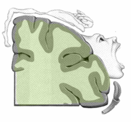
Depiction of the motor homunculus
Image: “Motor homunculus” by Albert Kok. License: Public Domain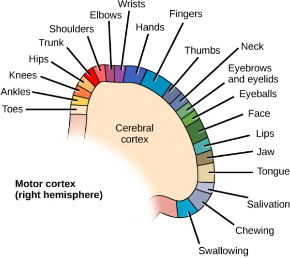
Cortical homunculus showing different parts of the motor cortex that control different muscle groups
Image: “Different parts of the motor cortex control different muscle groups” by CNX OpenStax. License: CC BY 4.0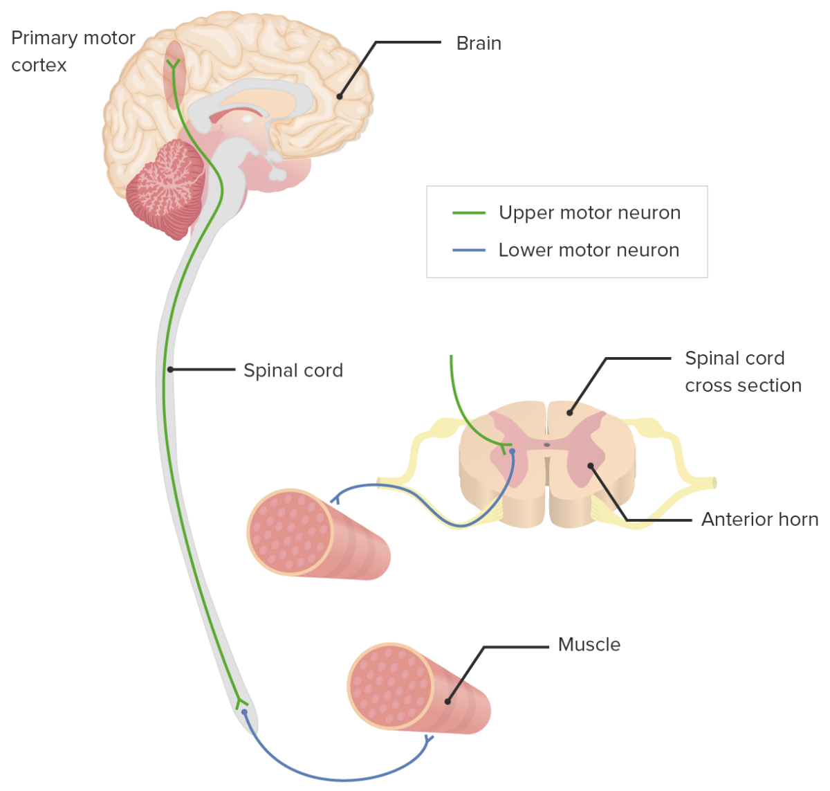
Upper motor neuron (UMN) and lower motor neuron (LMN) pathways
Image by Lecturio.UMN lesions above the decussation manifest as motor Motor Neurons which send impulses peripherally to activate muscles or secretory cells. Nervous System: Histology symptoms on the contralateral side of the body, whereas lesions below the pyramidal decussation present on the ipsilateral side of the body. Pathologic mechanisms of LMN lesions involve axonal degeneration and cell death Cell death Injurious stimuli trigger the process of cellular adaptation, whereby cells respond to withstand the harmful changes in their environment. Overwhelmed adaptive mechanisms lead to cell injury. Mild stimuli produce reversible injury. If the stimulus is severe or persistent, injury becomes irreversible. Apoptosis is programmed cell death, a mechanism with both physiologic and pathologic effects. Cell Injury and Death of the LMN.
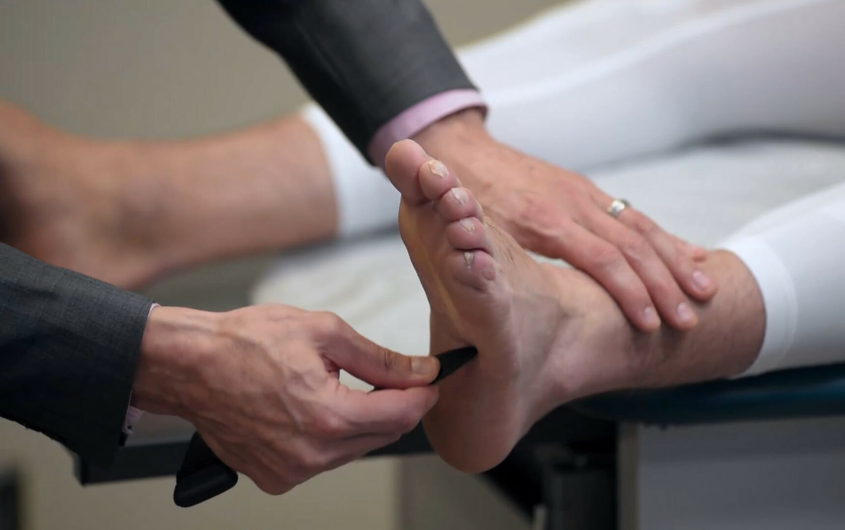
Normal (negative) Babinski reflex:
Toes and feet flare down and inward.
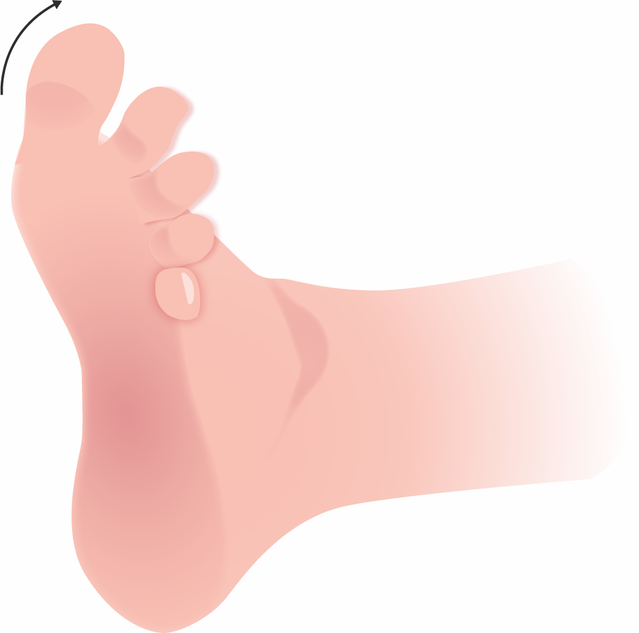
An abnormal (positive) Babinski reflex is a sign of an upper motor neuron lesion. In an abnormal (positive) response, the great toe will dorsiflex as the other toes fan out.
Image by Lecturio.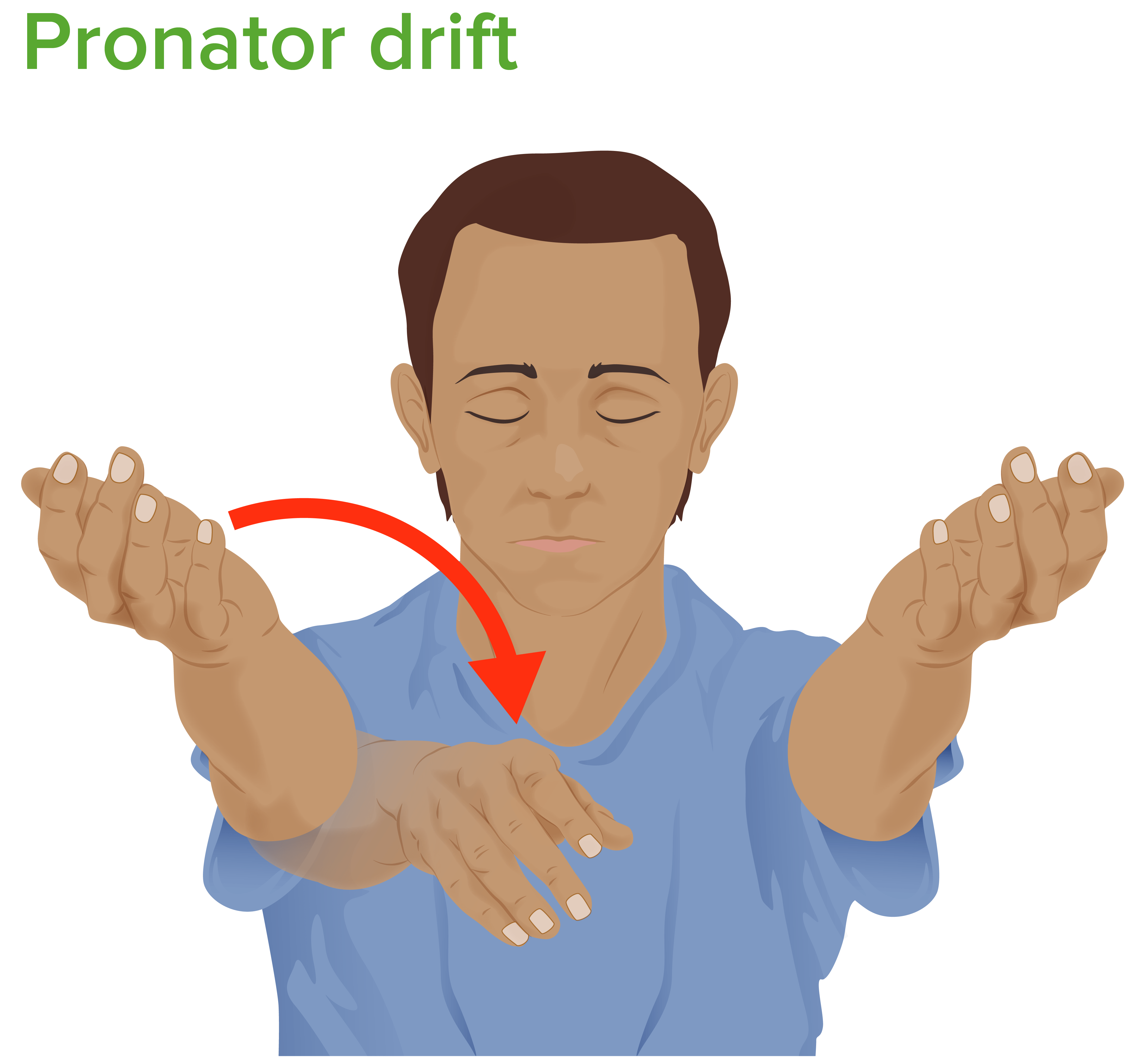
Pronator drift:
The individual is asked to hold both arms horizontally and straight with the palms facing up and the eyes closed. Lowering or pronation of 1 arm is abnormal and indicative of contralateral pyramidal/corticospinal tract injury.
| Clinical finding | UMN lesion | LMN lesion |
|---|---|---|
| Muscle tone Muscle tone The state of activity or tension of a muscle beyond that related to its physical properties, that is, its active resistance to stretch. In skeletal muscle, tonus is dependent upon efferent innervation. Skeletal Muscle Contraction |
|
Hypotonic Hypotonic Solutions that have a lesser osmotic pressure than a reference solution such as blood, plasma, or interstitial fluid. Renal Sodium and Water Regulation |
| Muscle mass Mass Three-dimensional lesion that occupies a space within the breast Imaging of the Breast | No change | Muscle wasting Muscle Wasting Duchenne Muscular Dystrophy present |
| Fasciculation | None | Present |
| Tendon reflexes | Hyperreflexia | Hyporeflexia Hyporeflexia Duchenne Muscular Dystrophy/ areflexia Areflexia Duchenne Muscular Dystrophy |
| Electromyographic findings | Normal nerve conduction | Abnormal nerve conduction |
It is important to differentiate central from peripheral nervous system Peripheral nervous system The nervous system outside of the brain and spinal cord. The peripheral nervous system has autonomic and somatic divisions. The autonomic nervous system includes the enteric, parasympathetic, and sympathetic subdivisions. The somatic nervous system includes the cranial and spinal nerves and their ganglia and the peripheral sensory receptors. Nervous System: Anatomy, Structure, and Classification lesions on exam.
There is no standard treatment for motor Motor Neurons which send impulses peripherally to activate muscles or secretory cells. Nervous System: Histology neuron diseases, but there are some medical and surgical methods available to treat the sequelae of UMN and LMN lesions and help individuals maintain their quality Quality Activities and programs intended to assure or improve the quality of care in either a defined medical setting or a program. The concept includes the assessment or evaluation of the quality of care; identification of problems or shortcomings in the delivery of care; designing activities to overcome these deficiencies; and follow-up monitoring to ensure effectiveness of corrective steps. Quality Measurement and Improvement of life.