Advertisement

Advertisement

Advertisement

Advertisement

Tumor Necrosis Factor (TNF)

Tumor Tumor Inflammation necrosis Necrosis The death of cells in an organ or tissue due to disease, injury or failure of the blood supply. Ischemic Cell Damage factor (TNF) is a major cytokine, released primarily by macrophages Macrophages The relatively long-lived phagocytic cell of mammalian tissues that are derived from blood monocytes. Main types are peritoneal macrophages; alveolar macrophages; histiocytes; kupffer cells of the liver; and osteoclasts. They may further differentiate within chronic inflammatory lesions to epithelioid cells or may fuse to form foreign body giant cells or langhans giant cells. Innate Immunity: Phagocytes and Antigen Presentation in response to stimuli. The presence of microbial products and dead cells and injury are among the stimulating factors. This protein belongs to the TNF superfamily, a group of ligands and receptors Receptors Receptors are proteins located either on the surface of or within a cell that can bind to signaling molecules known as ligands (e.g., hormones) and cause some type of response within the cell. Receptors performing functions in inflammatory response, morphogenesis, and cell proliferation. Tumor Tumor Inflammation necrosis Necrosis The death of cells in an organ or tissue due to disease, injury or failure of the blood supply. Ischemic Cell Damage factor interacts with 2 receptors Receptors Receptors are proteins located either on the surface of or within a cell that can bind to signaling molecules known as ligands (e.g., hormones) and cause some type of response within the cell. Receptors, which initiate signal transduction Transduction The transfer of bacterial DNA by phages from an infected bacterium to another bacterium. This also refers to the transfer of genes into eukaryotic cells by viruses. This naturally occurring process is routinely employed as a gene transfer technique. Bacteriology pathways leading to different cellular responses ( inflammation Inflammation Inflammation is a complex set of responses to infection and injury involving leukocytes as the principal cellular mediators in the body's defense against pathogenic organisms. Inflammation is also seen as a response to tissue injury in the process of wound healing. The 5 cardinal signs of inflammation are pain, heat, redness, swelling, and loss of function. Inflammation, cell survival, or apoptosis Apoptosis A regulated cell death mechanism characterized by distinctive morphologic changes in the nucleus and cytoplasm, including the endonucleolytic cleavage of genomic DNA, at regularly spaced, internucleosomal sites, I.e., DNA fragmentation. It is genetically-programmed and serves as a balance to mitosis in regulating the size of animal tissues and in mediating pathologic processes associated with tumor growth. Ischemic Cell Damage). Inappropriate or unrestrained activation of TNF signaling produces chronic inflammation Chronic Inflammation Inflammation, as seen in autoimmune conditions (e.g., rheumatoid arthritis Arthritis Acute or chronic inflammation of joints. Osteoarthritis, psoriasis Psoriasis Psoriasis is a common T-cell-mediated inflammatory skin condition. The etiology is unknown, but is thought to be due to genetic inheritance and environmental triggers. There are 4 major subtypes, with the most common form being chronic plaque psoriasis. Psoriasis). The mechanism of TNF inhibition has been used in treating these inflammatory diseases.

Last updated: May 17, 2024
Editorial responsibility: Stanley Oiseth, Lindsay Jones, Evelin Maza

Advertisement

Advertisement

Advertisement

Advertisement

Advertisement

Advertisement

Overview

Tumor Tumor Inflammation necrosis Necrosis The death of cells in an organ or tissue due to disease, injury or failure of the blood supply. Ischemic Cell Damage factor (TNF)

  • Other names: TNF-ɑ, cachectin
    • Originally, the protein derived from macrophages Macrophages The relatively long-lived phagocytic cell of mammalian tissues that are derived from blood monocytes. Main types are peritoneal macrophages; alveolar macrophages; histiocytes; kupffer cells of the liver; and osteoclasts. They may further differentiate within chronic inflammatory lesions to epithelioid cells or may fuse to form foreign body giant cells or langhans giant cells. Innate Immunity: Phagocytes and Antigen Presentation was named tumor Tumor Inflammation necrosis Necrosis The death of cells in an organ or tissue due to disease, injury or failure of the blood supply. Ischemic Cell Damage factor and a second, related, one derived from lymphocytes Lymphocytes Lymphocytes are heterogeneous WBCs involved in immune response. Lymphocytes develop from the bone marrow, starting from hematopoietic stem cells (HSCs) and progressing to common lymphoid progenitors (CLPs). B and T lymphocytes and natural killer (NK) cells arise from the lineage. Lymphocytes: Histology was named lymphotoxin. 
    • Being bound to the same receptor Receptor Receptors are proteins located either on the surface of or within a cell that can bind to signaling molecules known as ligands (e.g., hormones) and cause some type of response within the cell. Receptors and sharing similarities in amino acid Amino acid Amino acids (AAs) are composed of a central carbon atom attached to a carboxyl group, an amino group, a hydrogen atom, and a side chain (R group). Basics of Amino Acids sequences, these proteins Proteins Linear polypeptides that are synthesized on ribosomes and may be further modified, crosslinked, cleaved, or assembled into complex proteins with several subunits. The specific sequence of amino acids determines the shape the polypeptide will take, during protein folding, and the function of the protein. Energy Homeostasis were previously called:
      • TNF-α (for TNF)
      • TNF-β (for lymphotoxin)
    • Further studies on the protein sequences now identify multiple members (ligands and receptors Receptors Receptors are proteins located either on the surface of or within a cell that can bind to signaling molecules known as ligands (e.g., hormones) and cause some type of response within the cell. Receptors) of the TNF superfamily.
  • Major cytokine, released primarily by macrophages Macrophages The relatively long-lived phagocytic cell of mammalian tissues that are derived from blood monocytes. Main types are peritoneal macrophages; alveolar macrophages; histiocytes; kupffer cells of the liver; and osteoclasts. They may further differentiate within chronic inflammatory lesions to epithelioid cells or may fuse to form foreign body giant cells or langhans giant cells. Innate Immunity: Phagocytes and Antigen Presentation in response to stimuli (such as lipopolysaccharides Lipopolysaccharides Lipid-containing polysaccharides which are endotoxins and important group-specific antigens. They are often derived from the cell wall of gram-negative bacteria and induce immunoglobulin secretion. The lipopolysaccharide molecule consists of three parts: lipid a, core polysaccharide, and o-specific chains (o antigens). When derived from Escherichia coli, lipopolysaccharides serve as polyclonal b-cell mitogens commonly used in laboratory immunology. Diarrheagenic E. coli or other toll-like receptor-activating stimuli)
  • Other cells (e.g., T cells T cells Lymphocytes responsible for cell-mediated immunity. Two types have been identified – cytotoxic (t-lymphocytes, cytotoxic) and helper T-lymphocytes (t-lymphocytes, helper-inducer). They are formed when lymphocytes circulate through the thymus gland and differentiate to thymocytes. When exposed to an antigen, they divide rapidly and produce large numbers of new T cells sensitized to that antigen. T cells: Types and Functions, mast cells Mast cells Granulated cells that are found in almost all tissues, most abundantly in the skin and the gastrointestinal tract. Like the basophils, mast cells contain large amounts of histamine and heparin. Unlike basophils, mast cells normally remain in the tissues and do not circulate in the blood. Mast cells, derived from the bone marrow stem cells, are regulated by the stem cell factor. Innate Immunity: Phagocytes and Antigen Presentation, fibroblasts Fibroblasts Connective tissue cells which secrete an extracellular matrix rich in collagen and other macromolecules. Sarcoidosis) also produce TNF.

General functions of tumor Tumor Inflammation necrosis Necrosis The death of cells in an organ or tissue due to disease, injury or failure of the blood supply. Ischemic Cell Damage factor

  • Proinflammatory:
    • Endothelial activation:
      • ↑ Endothelial adhesion Adhesion The process whereby platelets adhere to something other than platelets, e.g., collagen; basement membrane; microfibrils; or other ‘foreign’ surfaces. Coagulation Studies molecules (e.g., P-selectin, E-selectin)
      • ↑ Mediators (other cytokines Cytokines Non-antibody proteins secreted by inflammatory leukocytes and some non-leukocytic cells, that act as intercellular mediators. They differ from classical hormones in that they are produced by a number of tissue or cell types rather than by specialized glands. They generally act locally in a paracrine or autocrine rather than endocrine manner. Adaptive Immune Response, chemokines Chemokines Class of pro-inflammatory cytokines that have the ability to attract and activate leukocytes. They can be divided into at least three structural branches: c; cc; and cxc; according to variations in a shared cysteine motif. Adaptive Cell-mediated Immunity, eicosanoids Eicosanoids Eicosanoids are cell-signaling molecules produced from arachidonic acid. With the action of phospholipase A2, arachidonic acid is released from the plasma membrane. The different families of eicosanoids, which are prostaglandins (PGs), thromboxanes (TXA2s), prostacyclin (PGI2), lipoxins (LXs), and leukotrienes (LTs), emerge from a series of reactions catalyzed by different enzymes. Eicosanoids)
      • ↑ Procoagulant activity
    • Activation of immune cells (such as leukocytes Leukocytes White blood cells. These include granular leukocytes (basophils; eosinophils; and neutrophils) as well as non-granular leukocytes (lymphocytes and monocytes). White Myeloid Cells: Histology)
    • Acute-phase response: lipid and protein mobilization and ↓ appetite (leading to weight loss Weight loss Decrease in existing body weight. Bariatric Surgery and cachexia)
  • Cell proliferation and differentiation
  • Apoptosis Apoptosis A regulated cell death mechanism characterized by distinctive morphologic changes in the nucleus and cytoplasm, including the endonucleolytic cleavage of genomic DNA, at regularly spaced, internucleosomal sites, I.e., DNA fragmentation. It is genetically-programmed and serves as a balance to mitosis in regulating the size of animal tissues and in mediating pathologic processes associated with tumor growth. Ischemic Cell Damage 
  • Antitumor activity

Tumor Necrosis Factor Superfamily

Members of the superfamily

  • 19 ligands interacting with 29 receptors Receptors Receptors are proteins located either on the surface of or within a cell that can bind to signaling molecules known as ligands (e.g., hormones) and cause some type of response within the cell. Receptors; each ligand able to interact with > 1 receptor Receptor Receptors are proteins located either on the surface of or within a cell that can bind to signaling molecules known as ligands (e.g., hormones) and cause some type of response within the cell. Receptors 
  • Proinflammatory properties are predominantly present in the ligands.
  • Most members have protective roles but also have harmful effects (with links to a variety of diseases).

Notable members

  • TNF: 
    • Fever Fever Fever is defined as a measured body temperature of at least 38°C (100.4°F). Fever is caused by circulating endogenous and/or exogenous pyrogens that increase levels of prostaglandin E2 in the hypothalamus. Fever is commonly associated with chills, rigors, sweating, and flushing of the skin. Fever (pyrogenic)
    • Antitumor
    • Mediator of inflammation Inflammation Inflammation is a complex set of responses to infection and injury involving leukocytes as the principal cellular mediators in the body’s defense against pathogenic organisms. Inflammation is also seen as a response to tissue injury in the process of wound healing. The 5 cardinal signs of inflammation are pain, heat, redness, swelling, and loss of function. Inflammation and sepsis Sepsis Systemic inflammatory response syndrome with a proven or suspected infectious etiology. When sepsis is associated with organ dysfunction distant from the site of infection, it is called severe sepsis. When sepsis is accompanied by hypotension despite adequate fluid infusion, it is called septic shock. Sepsis and Septic Shock
  • Lymphotoxin:
    • Mediator of inflammation Inflammation Inflammation is a complex set of responses to infection and injury involving leukocytes as the principal cellular mediators in the body’s defense against pathogenic organisms. Inflammation is also seen as a response to tissue injury in the process of wound healing. The 5 cardinal signs of inflammation are pain, heat, redness, swelling, and loss of function. Inflammation
    • Antiviral Antiviral Antivirals for Hepatitis B response
    • Antitumor
    • Control of secondary lymphoid organ development
  • CD40 CD40 Members of the tumor necrosis factor receptor superfamily with specificity for CD40 ligand. They are found on mature B-lymphocytes, some epithelial cells; and lymphoid dendritic cells. Evidence suggests that CD40-dependent activation of B-cells is important for generation of memory B-cells within the germinal centers. Mutations in the CD40 antigen gene result in hyper-igm immunodeficiency syndrome, type 3. Signaling of the receptor occurs through its association with tnf receptor-associated factors. Hyper-IgM Syndrome ligand (CD40L):
    • Expressed in T cells T cells Lymphocytes responsible for cell-mediated immunity. Two types have been identified – cytotoxic (t-lymphocytes, cytotoxic) and helper T-lymphocytes (t-lymphocytes, helper-inducer). They are formed when lymphocytes circulate through the thymus gland and differentiate to thymocytes. When exposed to an antigen, they divide rapidly and produce large numbers of new T cells sensitized to that antigen. T cells: Types and Functions and interacts with the CD40 CD40 Members of the tumor necrosis factor receptor superfamily with specificity for CD40 ligand. They are found on mature B-lymphocytes, some epithelial cells; and lymphoid dendritic cells. Evidence suggests that CD40-dependent activation of B-cells is important for generation of memory B-cells within the germinal centers. Mutations in the CD40 antigen gene result in hyper-igm immunodeficiency syndrome, type 3. Signaling of the receptor occurs through its association with tnf receptor-associated factors. Hyper-IgM Syndrome on B cells B cells Lymphoid cells concerned with humoral immunity. They are short-lived cells resembling bursa-derived lymphocytes of birds in their production of immunoglobulin upon appropriate stimulation. B cells: Types and Functions
    • ​​CD40L– CD40 CD40 Members of the tumor necrosis factor receptor superfamily with specificity for CD40 ligand. They are found on mature B-lymphocytes, some epithelial cells; and lymphoid dendritic cells. Evidence suggests that CD40-dependent activation of B-cells is important for generation of memory B-cells within the germinal centers. Mutations in the CD40 antigen gene result in hyper-igm immunodeficiency syndrome, type 3. Signaling of the receptor occurs through its association with tnf receptor-associated factors. Hyper-IgM Syndrome induces B-cell proliferation, class-switch recombination Recombination Production of new arrangements of DNA by various mechanisms such as assortment and segregation, crossing over; gene conversion; genetic transformation; genetic conjugation; genetic transduction; or mixed infection of viruses. Virology, and somatic hypermutation Somatic hypermutation A programmed mutation process whereby changes are introduced to the nucleotide sequence of immunoglobulin gene DNA during development. B cells: Types and Functions.
  • Fas ligand (FasL):
    • Expressed in cytotoxic Cytotoxic Parvovirus B19 T cells T cells Lymphocytes responsible for cell-mediated immunity. Two types have been identified – cytotoxic (t-lymphocytes, cytotoxic) and helper T-lymphocytes (t-lymphocytes, helper-inducer). They are formed when lymphocytes circulate through the thymus gland and differentiate to thymocytes. When exposed to an antigen, they divide rapidly and produce large numbers of new T cells sensitized to that antigen. T cells: Types and Functions
    • Interaction of FasL and Fas (a death receptor Receptor Receptors are proteins located either on the surface of or within a cell that can bind to signaling molecules known as ligands (e.g., hormones) and cause some type of response within the cell. Receptors) → Fas-associated protein with death domain (FADD) → caspase 8 activation → apoptosis Apoptosis A regulated cell death mechanism characterized by distinctive morphologic changes in the nucleus and cytoplasm, including the endonucleolytic cleavage of genomic DNA, at regularly spaced, internucleosomal sites, I.e., DNA fragmentation. It is genetically-programmed and serves as a balance to mitosis in regulating the size of animal tissues and in mediating pathologic processes associated with tumor growth. Ischemic Cell Damage

Effects of Tumor Necrosis Factor

Induction

  • TNF produced by various cells (predominantly macrophages Macrophages The relatively long-lived phagocytic cell of mammalian tissues that are derived from blood monocytes. Main types are peritoneal macrophages; alveolar macrophages; histiocytes; kupffer cells of the liver; and osteoclasts. They may further differentiate within chronic inflammatory lesions to epithelioid cells or may fuse to form foreign body giant cells or langhans giant cells. Innate Immunity: Phagocytes and Antigen Presentation); stimuli include:
    • Microbial products (e.g., lipopolysaccharide Lipopolysaccharide Lipid-containing polysaccharides which are endotoxins and important group-specific antigens. They are often derived from the cell wall of gram-negative bacteria and induce immunoglobulin secretion. The lipopolysaccharide molecule consists of three parts: lipid a, core polysaccharide, and o-specific chains (o antigens). When derived from Escherichia coli, lipopolysaccharides serve as polyclonal b-cell mitogens commonly used in laboratory immunology. Klebsiella)
    • Dead cells
    • Immune complexes Immune complexes The complex formed by the binding of antigen and antibody molecules. The deposition of large antigen-antibody complexes leading to tissue damage causes immune complex diseases. C3 Deficiency
    • Foreign antigens/bodies
    • Physical injury
    • IL-1
  • Starts initially as membrane-bound (pro-TNF):
    • Then expressed as a transmembrane protein
    • Requires proteolytic cleavage by TNF-α–converting enzyme (TACE) → releasing the soluble form of TNF

Tumor Tumor Inflammation necrosis Necrosis The death of cells in an organ or tissue due to disease, injury or failure of the blood supply. Ischemic Cell Damage factor receptors Receptors Receptors are proteins located either on the surface of or within a cell that can bind to signaling molecules known as ligands (e.g., hormones) and cause some type of response within the cell. Receptors

  • The functions of TNF are mediated by 2 receptor Receptor Receptors are proteins located either on the surface of or within a cell that can bind to signaling molecules known as ligands (e.g., hormones) and cause some type of response within the cell. Receptors types:
    • TNFR1:
      • Extensively studied
      • More prevalent
      • Expressed universally on almost all cell types
      • Contains death domain (DD) and is a member of the death receptor Receptor Receptors are proteins located either on the surface of or within a cell that can bind to signaling molecules known as ligands (e.g., hormones) and cause some type of response within the cell. Receptors family, with capacity to induce apoptotic death
    • TNFR2:
      • Mainly restricted to immune cells and some tumor Tumor Inflammation cells 
      • Does not have DD, but has TNF-receptor–associated factor (TRAF) binding site
      • Believed to have a role in cell survival and regeneration Regeneration The physiological renewal, repair, or replacement of tissue. Wound Healing
  • Receptors Receptors Receptors are proteins located either on the surface of or within a cell that can bind to signaling molecules known as ligands (e.g., hormones) and cause some type of response within the cell. Receptors trigger Trigger The type of signal that initiates the inspiratory phase by the ventilator Invasive Mechanical Ventilation distinct, but also interconnected, signaling pathways.

Effects

  • TNF, via TNFR1:
    • Inflammatory effect:
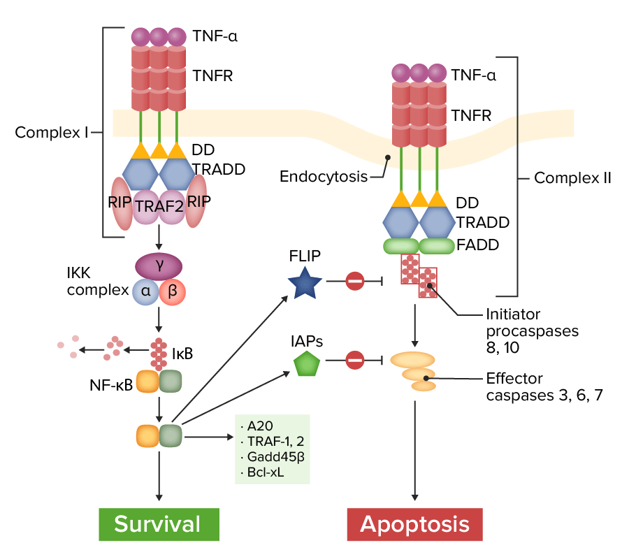
      • Certain proteins Proteins Linear polypeptides that are synthesized on ribosomes and may be further modified, crosslinked, cleaved, or assembled into complex proteins with several subunits. The specific sequence of amino acids determines the shape the polypeptide will take, during protein folding, and the function of the protein. Energy Homeostasis (TNF-receptor type 1 Type 1 Spinal Muscular Atrophy–associated death domain (TRADD), TNF-receptor–associated factor 2 (TRAF2), and receptor Receptor Receptors are proteins located either on the surface of or within a cell that can bind to signaling molecules known as ligands (e.g., hormones) and cause some type of response within the cell. Receptors interacting protein (RIP)) are sequentially recruited.
      • Proinflammatory pathways such as nuclear factor kappa-B Nuclear factor kappa-B Ubiquitous, inducible, nuclear transcriptional activator that binds to enhancer elements in many different cell types and is activated by pathogenic stimuli. The NF-kappa B complex is a heterodimer composed of two DNA-binding subunits: NF-kappa B1 and relA. MALT Lymphoma (NF-κB) and mitogen-activated protein kinase Protein kinase A family of enzymes that catalyze the conversion of ATP and a protein to adp and a phosphoprotein. Interferons (MAPK) pathways are activated.
      • NF-κB activation → transcription Transcription Transcription of genetic information is the first step in gene expression. Transcription is the process by which DNA is used as a template to make mRNA. This process is divided into 3 stages: initiation, elongation, and termination. Stages of Transcription of inflammatory proteins Proteins Linear polypeptides that are synthesized on ribosomes and may be further modified, crosslinked, cleaved, or assembled into complex proteins with several subunits. The specific sequence of amino acids determines the shape the polypeptide will take, during protein folding, and the function of the protein. Energy Homeostasis, cell survival and proliferation, induction of antiapoptotic genes Genes A category of nucleic acid sequences that function as units of heredity and which code for the basic instructions for the development, reproduction, and maintenance of organisms. DNA Types and Structure
    • Apoptotic effect:
      • TRADD and FADD are recruited.
      • Activation of caspase 8 is triggered → proteolytic cascade → apoptosis Apoptosis A regulated cell death mechanism characterized by distinctive morphologic changes in the nucleus and cytoplasm, including the endonucleolytic cleavage of genomic DNA, at regularly spaced, internucleosomal sites, I.e., DNA fragmentation. It is genetically-programmed and serves as a balance to mitosis in regulating the size of animal tissues and in mediating pathologic processes associated with tumor growth. Ischemic Cell Damage
  • TNF, via TNFR2:
    • TRAF2 is recruited → activation of different pathways, such as NF-κB and MAPK
    • Effects:
      • Tissue/cell regeneration Regeneration The physiological renewal, repair, or replacement of tissue. Wound Healing, proliferation, and survival
      • Host defense against pathogens ( inflammation Inflammation Inflammation is a complex set of responses to infection and injury involving leukocytes as the principal cellular mediators in the body’s defense against pathogenic organisms. Inflammation is also seen as a response to tissue injury in the process of wound healing. The 5 cardinal signs of inflammation are pain, heat, redness, swelling, and loss of function. Inflammation)
Tnf receptor (tnfr) pathway of signaling

Tumor necrosis factor receptor 1 (TNFR1) pathway of signaling:
Complex I (on the left): With ligation of TNF-α, TNFR recruits various adapter molecules, resulting in the activation of nuclear factor kappa-B (NF-κB), which induces several anti-apoptotic genes and survival signals. FLICE inhibitory protein (FLIP) and inhibitor of apoptosis proteins (IAPs) modulate and inhibit the apoptosis pathway. Complex II (on the right): Without certain adapter proteins (TRAF-2, RIP), TNFR leads to recruitment of Fas-associated protein with death domain (FADD). Caspase-8 is activated and released into the cytoplasm, activating effector caspases to induce apoptosis.
IκB: inhibitor of NF-κB
TRAF-2: TNF-receptor–associated factor.
RIP: receptor interacting protein
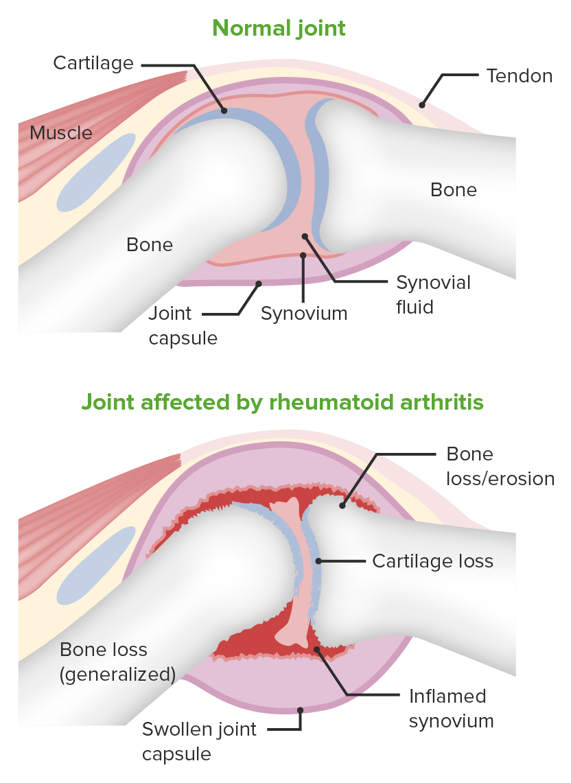
Image by Lecturio.

Tumor Necrosis Factor and Diseases

Diseases

  • Binding to 2 different receptors Receptors Receptors are proteins located either on the surface of or within a cell that can bind to signaling molecules known as ligands (e.g., hormones) and cause some type of response within the cell. Receptors, TNF initiates signal transduction Transduction The transfer of bacterial DNA by phages from an infected bacterium to another bacterium. This also refers to the transfer of genes into eukaryotic cells by viruses. This naturally occurring process is routinely employed as a gene transfer technique. Bacteriology pathways that produce varying cellular responses, including cell survival, differentiation, proliferation, and cell death Cell death Injurious stimuli trigger the process of cellular adaptation, whereby cells respond to withstand the harmful changes in their environment. Overwhelmed adaptive mechanisms lead to cell injury. Mild stimuli produce reversible injury. If the stimulus is severe or persistent, injury becomes irreversible. Apoptosis is programmed cell death, a mechanism with both physiologic and pathologic effects. Cell Injury and Death.
  • Inappropriate or unrestrained activation of TNF signaling produces chronic inflammation Chronic Inflammation Inflammation → pathologic complications such as autoimmune diseases Autoimmune diseases Disorders that are characterized by the production of antibodies that react with host tissues or immune effector cells that are autoreactive to endogenous peptides. Selective IgA Deficiency 
  • Abnormal secretion Secretion Coagulation Studies of TNF has been associated with:
    • Rheumatoid arthritis Arthritis Acute or chronic inflammation of joints. Osteoarthritis
    • Inflammatory bowel disease (Crohn’s disease and ulcerative colitis Colitis Inflammation of the colon section of the large intestine, usually with symptoms such as diarrhea (often with blood and mucus), abdominal pain, and fever. Pseudomembranous Colitis)
    • Psoriatic arthritis Psoriatic Arthritis A type of inflammatory arthritis associated with psoriasis, often involving the axial joints and the peripheral terminal interphalangeal joints. It is characterized by the presence of hla-b27-associated spondyloarthropathy, and the absence of rheumatoid factor. Psoriasis
    • Psoriasis Psoriasis Psoriasis is a common T-cell-mediated inflammatory skin condition. The etiology is unknown, but is thought to be due to genetic inheritance and environmental triggers. There are 4 major subtypes, with the most common form being chronic plaque psoriasis. Psoriasis
    • Noninfectious uveitis Uveitis Uveitis is the inflammation of the uvea, the pigmented middle layer of the eye, which comprises the iris, ciliary body, and choroid. The condition is categorized based on the site of disease; anterior uveitis is the most common. Diseases of the Uvea
Pathology of rheumatoid arthritis

Image demonstrating the pathology of rheumatoid arthritis:
Changes in the joint space are shown.

Image by Lecturio.

Anti– tumor Tumor Inflammation necrosis Necrosis The death of cells in an organ or tissue due to disease, injury or failure of the blood supply. Ischemic Cell Damage factor therapy

Tumor Tumor Inflammation necrosis Necrosis The death of cells in an organ or tissue due to disease, injury or failure of the blood supply. Ischemic Cell Damage factor has multiple biologic effects, and in certain conditions (e.g., rheumatoid arthritis Arthritis Acute or chronic inflammation of joints. Osteoarthritis), elevated levels of TNF have been noted. The mechanism of TNF inhibition has been used in treating inflammatory diseases.

Table: Indications for anti– tumor Tumor Inflammation necrosis Necrosis The death of cells in an organ or tissue due to disease, injury or failure of the blood supply. Ischemic Cell Damage factor therapy
Anti-TNF therapy Mechanism of action Indications
Infliximab Infliximab A chimeric monoclonal antibody to tnf-alpha that is used in the treatment of rheumatoid arthritis; ankylosing spondylitis; psoriatic arthritis and Crohn’s disease. Disease-Modifying Antirheumatic Drugs (DMARDs) Recombinant chimeric antibody (having a murine variable region Variable region That region of the immunoglobulin molecule that varies in its amino acid sequence and composition, and comprises the binding site for a specific antigen. It is located at the n-terminus of the fab fragment of the immunoglobulin. It includes hypervariable regions (complementarity determining regions) and framework regions. Immunoglobulins: Types and Functions and a human IgG1 constant region Constant region The domains of the immunoglobulin molecules that are invariable in their amino acid sequence within any class or subclass of immunoglobulin. They confer biological as well as structural functions to immunoglobulins. One each on both the light chains and the heavy chains comprises the c-terminus half of the immunoglobulin fab fragment and two or three of them make up the rest of the heavy chains (all of the immunoglobulin Fc fragment). Immunoglobulins: Types and Functions) that binds to TNF, preventing receptor Receptor Receptors are proteins located either on the surface of or within a cell that can bind to signaling molecules known as ligands (e.g., hormones) and cause some type of response within the cell. Receptors interaction
  • Crohn’s disease
  • Rheumatoid arthritis Arthritis Acute or chronic inflammation of joints. Osteoarthritis
  • Ankylosing spondylitis Ankylosing spondylitis Ankylosing spondylitis (also known as Bechterew’s disease or Marie-Strümpell disease) is a seronegative spondyloarthropathy characterized by chronic and indolent inflammation of the axial skeleton. Severe disease can lead to fusion and rigidity of the spine. Ankylosing Spondylitis
  • Plaque Plaque Primary Skin Lesions psoriasis Psoriasis Psoriasis is a common T-cell-mediated inflammatory skin condition. The etiology is unknown, but is thought to be due to genetic inheritance and environmental triggers. There are 4 major subtypes, with the most common form being chronic plaque psoriasis. Psoriasis
  • Psoriatic arthritis Psoriatic Arthritis A type of inflammatory arthritis associated with psoriasis, often involving the axial joints and the peripheral terminal interphalangeal joints. It is characterized by the presence of hla-b27-associated spondyloarthropathy, and the absence of rheumatoid factor. Psoriasis
  • Ulcerative colitis Colitis Inflammation of the colon section of the large intestine, usually with symptoms such as diarrhea (often with blood and mucus), abdominal pain, and fever. Pseudomembranous Colitis
Etanercept Etanercept A recombinant version of soluble human tnf receptor fused to an IgG Fc fragment that binds specifically to tumor necrosis factor and inhibits its binding with endogenous tnf receptors. It prevents the inflammatory effect of tnf and is used to treat rheumatoid arthritis; psoriatic arthritis and ankylosing spondylitis. Immunosuppressants Fusion protein Fusion protein Proteins that catalyze membrane fusion. Measles Virus that binds and neutralizes TNF and lymphotoxin
  • Plaque Plaque Primary Skin Lesions psoriasis Psoriasis Psoriasis is a common T-cell-mediated inflammatory skin condition. The etiology is unknown, but is thought to be due to genetic inheritance and environmental triggers. There are 4 major subtypes, with the most common form being chronic plaque psoriasis. Psoriasis
  • Polyarticular juvenile idiopathic Idiopathic Dermatomyositis arthritis Arthritis Acute or chronic inflammation of joints. Osteoarthritis
  • Psoriatic arthritis Psoriatic Arthritis A type of inflammatory arthritis associated with psoriasis, often involving the axial joints and the peripheral terminal interphalangeal joints. It is characterized by the presence of hla-b27-associated spondyloarthropathy, and the absence of rheumatoid factor. Psoriasis
  • Rheumatoid arthritis Arthritis Acute or chronic inflammation of joints. Osteoarthritis
Adalimumab Adalimumab A humanized monoclonal antibody that binds specifically to tnf-alpha and blocks its interaction with endogenous tnf receptors to modulate inflammation. It is used in the treatment of rheumatoid arthritis; psoriatic arthritis; Crohn’s disease and ulcerative colitis. Disease-Modifying Antirheumatic Drugs (DMARDs) Fully humanized IgG1 monoclonal antibody that blocks TNF binding to receptors Receptors Receptors are proteins located either on the surface of or within a cell that can bind to signaling molecules known as ligands (e.g., hormones) and cause some type of response within the cell. Receptors
  • Ankylosing spondylitis Ankylosing spondylitis Ankylosing spondylitis (also known as Bechterew’s disease or Marie-Strümpell disease) is a seronegative spondyloarthropathy characterized by chronic and indolent inflammation of the axial skeleton. Severe disease can lead to fusion and rigidity of the spine. Ankylosing Spondylitis
  • Crohn’s disease
  • Hidradenitis suppurativa Hidradenitis suppurativa Hidradenitis suppurativa (HS) is a chronic skin condition due to the inflammation of apocrine sweat glands and hair follicles. Most commonly, it occurs due to occlusion of the follicular component of pilosebaceous units (PSUs). Hidradenitis Suppurativa
  • Juvenile idiopathic Idiopathic Dermatomyositis arthritis Arthritis Acute or chronic inflammation of joints. Osteoarthritis
  • Psoriatic arthritis Psoriatic Arthritis A type of inflammatory arthritis associated with psoriasis, often involving the axial joints and the peripheral terminal interphalangeal joints. It is characterized by the presence of hla-b27-associated spondyloarthropathy, and the absence of rheumatoid factor. Psoriasis
  • Rheumatoid arthritis Arthritis Acute or chronic inflammation of joints. Osteoarthritis
  • Ulcerative colitis Colitis Inflammation of the colon section of the large intestine, usually with symptoms such as diarrhea (often with blood and mucus), abdominal pain, and fever. Pseudomembranous Colitis
  • Uveitis Uveitis Uveitis is the inflammation of the uvea, the pigmented middle layer of the eye, which comprises the iris, ciliary body, and choroid. The condition is categorized based on the site of disease; anterior uveitis is the most common. Diseases of the Uvea
Golimumab Golimumab Disease-Modifying Antirheumatic Drugs (DMARDs) Fully humanized IgG1 monoclonal antibody with high affinity and specificity for TNF
  • Ankylosing spondylitis Ankylosing spondylitis Ankylosing spondylitis (also known as Bechterew’s disease or Marie-Strümpell disease) is a seronegative spondyloarthropathy characterized by chronic and indolent inflammation of the axial skeleton. Severe disease can lead to fusion and rigidity of the spine. Ankylosing Spondylitis
  • Psoriatic arthritis Psoriatic Arthritis A type of inflammatory arthritis associated with psoriasis, often involving the axial joints and the peripheral terminal interphalangeal joints. It is characterized by the presence of hla-b27-associated spondyloarthropathy, and the absence of rheumatoid factor. Psoriasis
  • Rheumatoid arthritis Arthritis Acute or chronic inflammation of joints. Osteoarthritis
  • Ulcerative colitis Colitis Inflammation of the colon section of the large intestine, usually with symptoms such as diarrhea (often with blood and mucus), abdominal pain, and fever. Pseudomembranous Colitis
Certolizumab Certolizumab Disease-Modifying Antirheumatic Drugs (DMARDs) Pegylated humanized monoclonal antibody specific to TNF
  • Ankylosing spondylitis Ankylosing spondylitis Ankylosing spondylitis (also known as Bechterew’s disease or Marie-Strümpell disease) is a seronegative spondyloarthropathy characterized by chronic and indolent inflammation of the axial skeleton. Severe disease can lead to fusion and rigidity of the spine. Ankylosing Spondylitis
  • Crohn’s disease
  • Psoriatic arthritis Psoriatic Arthritis A type of inflammatory arthritis associated with psoriasis, often involving the axial joints and the peripheral terminal interphalangeal joints. It is characterized by the presence of hla-b27-associated spondyloarthropathy, and the absence of rheumatoid factor. Psoriasis
  • Plaque Plaque Primary Skin Lesions psoriasis Psoriasis Psoriasis is a common T-cell-mediated inflammatory skin condition. The etiology is unknown, but is thought to be due to genetic inheritance and environmental triggers. There are 4 major subtypes, with the most common form being chronic plaque psoriasis. Psoriasis
  • Rheumatoid arthritis Arthritis Acute or chronic inflammation of joints. Osteoarthritis

Clinical Relevance

  • Crohn’s disease (CD): chronic, recurrent condition that causes patchy transmural inflammation Transmural inflammation Crohn’s Disease that can involve any part of the GI tract. The terminal ileum Ileum The distal and narrowest portion of the small intestine, between the jejunum and the ileocecal valve of the large intestine. Small Intestine: Anatomy and proximal colon Colon The large intestines constitute the last portion of the digestive system. The large intestine consists of the cecum, appendix, colon (with ascending, transverse, descending, and sigmoid segments), rectum, and anal canal. The primary function of the colon is to remove water and compact the stool prior to expulsion from the body via the rectum and anal canal. Colon, Cecum, and Appendix: Anatomy are usually affected. Crohn’s disease presents with intermittent, nonbloody diarrhea Diarrhea Diarrhea is defined as ≥ 3 watery or loose stools in a 24-hour period. There are a multitude of etiologies, which can be classified based on the underlying mechanism of disease. The duration of symptoms (acute or chronic) and characteristics of the stools (e.g., watery, bloody, steatorrheic, mucoid) can help guide further diagnostic evaluation. Diarrhea and crampy abdominal pain Abdominal Pain Acute Abdomen. Extraintestinal manifestations may include calcium Calcium A basic element found in nearly all tissues. It is a member of the alkaline earth family of metals with the atomic symbol ca, atomic number 20, and atomic weight 40. Calcium is the most abundant mineral in the body and combines with phosphorus to form calcium phosphate in the bones and teeth. It is essential for the normal functioning of nerves and muscles and plays a role in blood coagulation (as factor IV) and in many enzymatic processes. Electrolytes oxalate renal stones, gallstones Gallstones Cholelithiasis (gallstones) is the presence of stones in the gallbladder. Most gallstones are cholesterol stones, while the rest are composed of bilirubin (pigment stones) and other mixed components. Patients are commonly asymptomatic but may present with biliary colic (intermittent pain in the right upper quadrant). Cholelithiasis, erythema Erythema Redness of the skin produced by congestion of the capillaries. This condition may result from a variety of disease processes. Chalazion nodosum, and arthritis Arthritis Acute or chronic inflammation of joints. Osteoarthritis. Diagnosis is established via endoscopy Endoscopy Procedures of applying endoscopes for disease diagnosis and treatment. Endoscopy involves passing an optical instrument through a small incision in the skin i.e., percutaneous; or through a natural orifice and along natural body pathways such as the digestive tract; and/or through an incision in the wall of a tubular structure or organ, i.e. Transluminal, to examine or perform surgery on the interior parts of the body. Gastroesophageal Reflux Disease (GERD) with biopsy Biopsy Removal and pathologic examination of specimens from the living body. Ewing Sarcoma. Management is with corticosteroids Corticosteroids Chorioretinitis, azathioprine Azathioprine An immunosuppressive agent used in combination with cyclophosphamide and hydroxychloroquine in the treatment of rheumatoid arthritis. According to the fourth annual report on carcinogens, this substance has been listed as a known carcinogen. Immunosuppressants, antibiotics, and anti-TNF agents ( infliximab Infliximab A chimeric monoclonal antibody to tnf-alpha that is used in the treatment of rheumatoid arthritis; ankylosing spondylitis; psoriatic arthritis and Crohn’s disease. Disease-Modifying Antirheumatic Drugs (DMARDs) and adalimumab Adalimumab A humanized monoclonal antibody that binds specifically to tnf-alpha and blocks its interaction with endogenous tnf receptors to modulate inflammation. It is used in the treatment of rheumatoid arthritis; psoriatic arthritis; Crohn’s disease and ulcerative colitis. Disease-Modifying Antirheumatic Drugs (DMARDs)). 
  • Ulcerative colitis Colitis Inflammation of the colon section of the large intestine, usually with symptoms such as diarrhea (often with blood and mucus), abdominal pain, and fever. Pseudomembranous Colitis ( UC UC Ulcerative colitis (UC) is an idiopathic inflammatory condition that involves the mucosal surface of the colon. It is a type of inflammatory bowel disease (IBD), along with Crohn’s disease (CD). The rectum is always involved, and inflammation may extend proximally through the colon. Ulcerative Colitis): idiopathic Idiopathic Dermatomyositis inflammatory condition that involves the mucosal surface of the colon Colon The large intestines constitute the last portion of the digestive system. The large intestine consists of the cecum, appendix, colon (with ascending, transverse, descending, and sigmoid segments), rectum, and anal canal. The primary function of the colon is to remove water and compact the stool prior to expulsion from the body via the rectum and anal canal. Colon, Cecum, and Appendix: Anatomy. The rectum Rectum The rectum and anal canal are the most terminal parts of the lower GI tract/large intestine that form a functional unit and control defecation. Fecal continence is maintained by several important anatomic structures including rectal folds, anal valves, the sling-like puborectalis muscle, and internal and external anal sphincters. Rectum and Anal Canal: Anatomy is always involved in UC UC Ulcerative colitis (UC) is an idiopathic inflammatory condition that involves the mucosal surface of the colon. It is a type of inflammatory bowel disease (IBD), along with Crohn’s disease (CD). The rectum is always involved, and inflammation may extend proximally through the colon. Ulcerative Colitis, and inflammation Inflammation Inflammation is a complex set of responses to infection and injury involving leukocytes as the principal cellular mediators in the body’s defense against pathogenic organisms. Inflammation is also seen as a response to tissue injury in the process of wound healing. The 5 cardinal signs of inflammation are pain, heat, redness, swelling, and loss of function. Inflammation may extend proximally through the colon Colon The large intestines constitute the last portion of the digestive system. The large intestine consists of the cecum, appendix, colon (with ascending, transverse, descending, and sigmoid segments), rectum, and anal canal. The primary function of the colon is to remove water and compact the stool prior to expulsion from the body via the rectum and anal canal. Colon, Cecum, and Appendix: Anatomy. Ulcerative colitis Colitis Inflammation of the colon section of the large intestine, usually with symptoms such as diarrhea (often with blood and mucus), abdominal pain, and fever. Pseudomembranous Colitis causes diffuse friability, erosions Erosions Corneal Abrasions, Erosion, and Ulcers with bleeding, and loss of haustra Haustra Colon, Cecum, and Appendix: Anatomy, which are visible on endoscopy Endoscopy Procedures of applying endoscopes for disease diagnosis and treatment. Endoscopy involves passing an optical instrument through a small incision in the skin i.e., percutaneous; or through a natural orifice and along natural body pathways such as the digestive tract; and/or through an incision in the wall of a tubular structure or organ, i.e. Transluminal, to examine or perform surgery on the interior parts of the body. Gastroesophageal Reflux Disease (GERD). Individuals present with bloody diarrhea Bloody diarrhea Diarrhea, colicky abdominal pain Abdominal Pain Acute Abdomen, tenesmus, and fecal urgency. Diagnosis is established via endoscopy Endoscopy Procedures of applying endoscopes for disease diagnosis and treatment. Endoscopy involves passing an optical instrument through a small incision in the skin i.e., percutaneous; or through a natural orifice and along natural body pathways such as the digestive tract; and/or through an incision in the wall of a tubular structure or organ, i.e. Transluminal, to examine or perform surgery on the interior parts of the body. Gastroesophageal Reflux Disease (GERD) with biopsy Biopsy Removal and pathologic examination of specimens from the living body. Ewing Sarcoma. Management includes topical mesalamine, 6-mercaptopurine 6-Mercaptopurine An antimetabolite antineoplastic agent with immunosuppressant properties. It interferes with nucleic acid synthesis by inhibiting purine metabolism and is used, usually in combination with other drugs, in the treatment of or in remission maintenance programs for leukemia. Antimetabolite Chemotherapy, and anti-TNF agents or colectomy for severe cases.
  • Psoriasis Psoriasis Psoriasis is a common T-cell-mediated inflammatory skin condition. The etiology is unknown, but is thought to be due to genetic inheritance and environmental triggers. There are 4 major subtypes, with the most common form being chronic plaque psoriasis. Psoriasis: common T-cell–mediated inflammatory skin Skin The skin, also referred to as the integumentary system, is the largest organ of the body. The skin is primarily composed of the epidermis (outer layer) and dermis (deep layer). The epidermis is primarily composed of keratinocytes that undergo rapid turnover, while the dermis contains dense layers of connective tissue. Skin: Structure and Functions condition. The etiology is unknown, but psoriasis Psoriasis Psoriasis is a common T-cell-mediated inflammatory skin condition. The etiology is unknown, but is thought to be due to genetic inheritance and environmental triggers. There are 4 major subtypes, with the most common form being chronic plaque psoriasis. Psoriasis is thought to be due to genetics Genetics Genetics is the study of genes and their functions and behaviors. Basic Terms of Genetics and environmental triggers. Plaques are well circumscribed, are salmon-colored, and have silvery scales Scales Dry or greasy masses of keratin that represent thickened stratum corneum. Secondary Skin Lesions. The plaques commonly appear on the scalp and extensor surfaces of the extremities. The diagnosis is clinical. Treatment options include topical corticosteroids Corticosteroids Chorioretinitis, retinoids Retinoids Retinol and derivatives of retinol that play an essential role in metabolic functioning of the retina, the growth of and differentiation of epithelial tissue, the growth of bone, reproduction, and the immune response. Dietary vitamin A is derived from a variety of carotenoids found in plants. It is enriched in the liver, egg yolks, and the fat component of dairy products. Fat-soluble Vitamins and their Deficiencies, calcineurin inhibitors Calcineurin Inhibitors Compounds that inhibit or block the phosphatase activity of calcineurin. Immunosuppressants, disease-modifying antirheumatic drugs Disease-modifying antirheumatic drugs Disease-modifying antirheumatic drugs are antiinflammatory medications used to manage rheumatoid arthritis. The medications slow, but do not cure, the progression of the disease. The medications are classified as either synthetic or biologic agents and each has unique mechanisms of action and side effects. Disease-Modifying Antirheumatic Drugs (DMARDs) ( DMARDs DMARDs Disease-modifying antirheumatic drugs are antiinflammatory medications used to manage rheumatoid arthritis. The medications slow, but do not cure, the progression of the disease. The medications are classified as either synthetic or biologic agents and each has unique mechanisms of action and side effects. Disease-Modifying Antirheumatic Drugs (DMARDs)), biologics such as anti-TNF agents, and phototherapy Phototherapy Treatment of disease by exposure to light, especially by variously concentrated light rays or specific wavelengths. Hyperbilirubinemia of the Newborn
  • Ankylosing spondylitis Ankylosing spondylitis Ankylosing spondylitis (also known as Bechterew’s disease or Marie-Strümpell disease) is a seronegative spondyloarthropathy characterized by chronic and indolent inflammation of the axial skeleton. Severe disease can lead to fusion and rigidity of the spine. Ankylosing Spondylitis: also known as Bechterew’s disease Bechterew’s disease Ankylosing spondylitis (also known as bechterew’s disease or marie-strümpell disease) is a seronegative spondyloarthropathy characterized by chronic and indolent inflammation of the axial skeleton. Severe disease can lead to fusion and rigidity of the spine. Ankylosing Spondylitis or Marie-Strümpell disease Marie-Strümpell disease Ankylosing spondylitis (also known as Bechterew’s disease or Marie-Strümpell disease) is a seronegative spondyloarthropathy characterized by chronic and indolent inflammation of the axial skeleton. Severe disease can lead to fusion and rigidity of the spine. Ankylosing Spondylitis. Ankylosing spondylitis Ankylosing spondylitis Ankylosing spondylitis (also known as Bechterew’s disease or Marie-Strümpell disease) is a seronegative spondyloarthropathy characterized by chronic and indolent inflammation of the axial skeleton. Severe disease can lead to fusion and rigidity of the spine. Ankylosing Spondylitis is a seronegative spondyloarthropathy Spondyloarthropathy Ankylosing Spondylitis characterized by chronic and indolent inflammation Inflammation Inflammation is a complex set of responses to infection and injury involving leukocytes as the principal cellular mediators in the body’s defense against pathogenic organisms. Inflammation is also seen as a response to tissue injury in the process of wound healing. The 5 cardinal signs of inflammation are pain, heat, redness, swelling, and loss of function. Inflammation of the axial Axial Computed Tomography (CT) skeleton. Severe disease can lead to fusion and rigidity Rigidity Continuous involuntary sustained muscle contraction which is often a manifestation of basal ganglia diseases. When an affected muscle is passively stretched, the degree of resistance remains constant regardless of the rate at which the muscle is stretched. This feature helps to distinguish rigidity from muscle spasticity. Megacolon of the spine Spine The human spine, or vertebral column, is the most important anatomical and functional axis of the human body. It consists of 7 cervical vertebrae, 12 thoracic vertebrae, and 5 lumbar vertebrae and is limited cranially by the skull and caudally by the sacrum. Vertebral Column: Anatomy. The condition is most often seen in young men and is associated with HLA-B27. Individuals will have progressive back pain Pain An unpleasant sensation induced by noxious stimuli which are detected by nerve endings of nociceptive neurons. Pain: Types and Pathways (which improves with activity), morning stiffness, and decreased range of motion Range of motion The distance and direction to which a bone joint can be extended. Range of motion is a function of the condition of the joints, muscles, and connective tissues involved. Joint flexibility can be improved through appropriate muscle strength exercises. Examination of the Upper Limbs of the spine Spine The human spine, or vertebral column, is the most important anatomical and functional axis of the human body. It consists of 7 cervical vertebrae, 12 thoracic vertebrae, and 5 lumbar vertebrae and is limited cranially by the skull and caudally by the sacrum. Vertebral Column: Anatomy. Extraarticular manifestations are also noted. Diagnosis is based on the clinical history, exam, and imaging ( sacroiliitis Sacroiliitis Inflammation of the sacroiliac joint. It is characterized by lower back pain, especially upon walking, fever, uveitis; psoriasis; and decreased range of motion. Many factors are associated with and cause sacroiliitis including infection; injury to spine, lower back, and pelvis; degenerative arthritis; and pregnancy. Ankylosing Spondylitis and bridging syndesmophytes Syndesmophytes Ankylosing Spondylitis). Management involves physical therapy Physical Therapy Becker Muscular Dystrophy and NSAIDs NSAIDS Primary vs Secondary Headaches. More severe cases may require TNF inhibitors TNF inhibitors Ankylosing Spondylitis or surgery.
  • Rheumatoid arthritis Arthritis Acute or chronic inflammation of joints. Osteoarthritis: symmetric, inflammatory polyarthritis Polyarthritis Rheumatoid Arthritis. Rheumatoid arthritis Arthritis Acute or chronic inflammation of joints. Osteoarthritis is a chronic, progressive autoimmune disorder Autoimmune Disorder Septic Arthritis. Presentation includes joint swelling Swelling Inflammation, pain Pain An unpleasant sensation induced by noxious stimuli which are detected by nerve endings of nociceptive neurons. Pain: Types and Pathways, and morning stiffness (often in the hands). Prolonged and severe disease can lead to irreversible joint deformities. Systemic inflammation Systemic Inflammation Surgical Site Infections can lead to extraarticular manifestations, including rheumatoid nodules Rheumatoid Nodules Rheumatoid Arthritis, interstitial lung disease, Felty syndrome, and pericarditis Pericarditis Pericarditis is an inflammation of the pericardium, often with fluid accumulation. It can be caused by infection (often viral), myocardial infarction, drugs, malignancies, metabolic disorders, autoimmune disorders, or trauma. Acute, subacute, and chronic forms exist. Pericarditis. Diagnosis is clinical and is confirmed by the presence of rheumatoid factor Rheumatoid factor Antibodies found in adult rheumatoid arthritis patients that are directed against gamma-chain immunoglobulins. Autoimmune Hepatitis, anti–cyclic citrullinated peptide antibodies Antibodies Immunoglobulins (Igs), also known as antibodies, are glycoprotein molecules produced by plasma cells that act in immune responses by recognizing and binding particular antigens. The various Ig classes are IgG (the most abundant), IgM, IgE, IgD, and IgA, which differ in their biologic features, structure, target specificity, and distribution. Immunoglobulins: Types and Functions, and characteristic imaging findings. Management involves physical therapy Physical Therapy Becker Muscular Dystrophy, long-term DMARDs DMARDs Disease-modifying antirheumatic drugs are antiinflammatory medications used to manage rheumatoid arthritis. The medications slow, but do not cure, the progression of the disease. The medications are classified as either synthetic or biologic agents and each has unique mechanisms of action and side effects. Disease-Modifying Antirheumatic Drugs (DMARDs), and biologics such as anti-TNF agents. 
  • Hidradenitis suppurativa Hidradenitis suppurativa Hidradenitis suppurativa (HS) is a chronic skin condition due to the inflammation of apocrine sweat glands and hair follicles. Most commonly, it occurs due to occlusion of the follicular component of pilosebaceous units (PSUs). Hidradenitis Suppurativa ( HS HS Hypertrophic scars and keloids are raised, red, and rigid (3 rs) scars that develop during cutaneous wound healing and are characterized by a local abnormal proliferation of fibroblasts with over-production of collagen. Over-expression of growth factors and decreased production of molecules that promote matrix breakdown appear to be involved in the etiology. Hypertrophic and Keloid Scars): chronic skin Skin The skin, also referred to as the integumentary system, is the largest organ of the body. The skin is primarily composed of the epidermis (outer layer) and dermis (deep layer). The epidermis is primarily composed of keratinocytes that undergo rapid turnover, while the dermis contains dense layers of connective tissue. Skin: Structure and Functions condition due to the inflammation Inflammation Inflammation is a complex set of responses to infection and injury involving leukocytes as the principal cellular mediators in the body’s defense against pathogenic organisms. Inflammation is also seen as a response to tissue injury in the process of wound healing. The 5 cardinal signs of inflammation are pain, heat, redness, swelling, and loss of function. Inflammation of apocrine sweat glands Sweat glands Sweat-producing structures that are embedded in the dermis. Each gland consists of a single tube, a coiled body, and a superficial duct. Soft Tissue Abscess and hair follicles. Hidradenitis suppurativa Hidradenitis suppurativa Hidradenitis suppurativa (HS) is a chronic skin condition due to the inflammation of apocrine sweat glands and hair follicles. Most commonly, it occurs due to occlusion of the follicular component of pilosebaceous units (PSUs). Hidradenitis Suppurativa commonly occurs because of occlusion of the follicular component of pilosebaceous units Pilosebaceous Units Hidradenitis Suppurativa (PSUs). Hidradenitis suppurativa Hidradenitis suppurativa Hidradenitis suppurativa (HS) is a chronic skin condition due to the inflammation of apocrine sweat glands and hair follicles. Most commonly, it occurs due to occlusion of the follicular component of pilosebaceous units (PSUs). Hidradenitis Suppurativa is characterized by the formation of abscesses, fistulas, draining skin Skin The skin, also referred to as the integumentary system, is the largest organ of the body. The skin is primarily composed of the epidermis (outer layer) and dermis (deep layer). The epidermis is primarily composed of keratinocytes that undergo rapid turnover, while the dermis contains dense layers of connective tissue. Skin: Structure and Functions lesions, keloids, and pilonidal sinuses. The diagnosis of HS HS Hypertrophic scars and keloids are raised, red, and rigid (3 rs) scars that develop during cutaneous wound healing and are characterized by a local abnormal proliferation of fibroblasts with over-production of collagen. Over-expression of growth factors and decreased production of molecules that promote matrix breakdown appear to be involved in the etiology. Hypertrophic and Keloid Scars is primarily clinical. Management includes lifestyle changes ( weight loss Weight loss Decrease in existing body weight. Bariatric Surgery and smoking Smoking Willful or deliberate act of inhaling and exhaling smoke from burning substances or agents held by hand. Interstitial Lung Diseases cessation) and medical treatment with antibiotics, retinoids Retinoids Retinol and derivatives of retinol that play an essential role in metabolic functioning of the retina, the growth of and differentiation of epithelial tissue, the growth of bone, reproduction, and the immune response. Dietary vitamin A is derived from a variety of carotenoids found in plants. It is enriched in the liver, egg yolks, and the fat component of dairy products. Fat-soluble Vitamins and their Deficiencies, and anti-TNF agents. Resistant disease requires surgery.

References

  1. Aggarwal, B.B. (2003). Signaling pathways of the TNF superfamily: a double-edged sword. Nat Rev Immunol 3:745–756. 
  2. Aggarwal, B.B., Gupta, S.C., Kim, J.H. (2012). Historical perspectives on tumor necrosis factor and its superfamily: 25 years later, a golden journey. Blood 119:651–665. doi:10.1182/blood-2011-04-325225
  3. Beutler, B., Moresco, E.Y. (2021). Innate immunity. Chapter 19 of Kaushansky, K., Prchal, J.T., Burns, L.J., Lichtman, M.A., Levi, M., Linch, D.C. (Eds.),  Williams Hematology, 10th ed. McGraw-Hill. https://accessmedicine.mhmedical.com/content.aspx?bookid=2962&sectionid=252524927
  4. Burmester, G. (2021). Overview of biologic agents and kinase inhibitors in the rheumatic diseases. UpToDate. Retrieved Aug 7, 2021, from https://www.uptodate.com/contents/overview-of-biologic-agents-and-kinase-inhibitors-in-the-rheumatic-diseases
  5. Grimstad, Ø. (2016). Tumor necrosis factor and the tenacious α. JAMA Dermatol 152:557. https://pubmed.ncbi.nlm.nih.gov/27168212/
  6. Kalliolias, G.D., Ivashkiv, L.B. (2016). TNF biology, pathogenic mechanisms and emerging therapeutic strategies. Nature Reviews Rheumatology 12(1):49–62. https://doi.org/10.1038/nrrheum.2015.169
  7. Krensky, A.M., Azzi, J.R., Hafler, D.A. (2017). Immunosuppressants and tolerogens. Chapter 35 of Brunton, L.L., Hilal-Dandan, R., Knollmann, B.C. (Eds.), Goodman & Gilman’s: The Pharmacological Basis of Therapeutics, 13th ed. McGraw-Hill. https://accessmedicine.mhmedical.com/content.aspx?bookid=2189&sectionid=172480027
  8. Kumar, V., Abbas, A. Aster, J. (2021). Inflammation and repair. In: Robbins and Cotran Pathologic Basis of Disease, 10th ed. Elsevier, pp. 88–90).
  9. Jang,D., Lee, A.-H., Shin, H.-Y.,  Song, H.-R., Park, J.-H., Kang, T.-B., Lee, S.-R., Yang, S.-H. (2021). The role of tumor necrosis factor alpha (TNF-α) in autoimmune disease and current TNF-α inhibitors in therapeutics. Int J Mol Sci 22:2719. https://pubmed.ncbi.nlm.nih.gov/33800290/
  10. Varfolomeev, E., Ashkenazi, A. (2004). Tumor necrosis factor. Cell 116:491–497. https://doi.org/10.1016/S0092-8674(04)00166-7
  11. Wang, X., Lin, Y. (2008). Tumor necrosis factor and cancer, buddies or foes? Acta Pharmacologica Sinica 29:1275–1288. https://doi.org/10.1111/j.1745-7254.2008.00889.x

Create your free account or log in to continue reading!

Sign up now and get free access to Lecturio with concept pages, medical videos, and questions for your medical education.

User Reviews

Details