Advertisement

Advertisement

Advertisement

Advertisement

Treacher Collins Syndrome

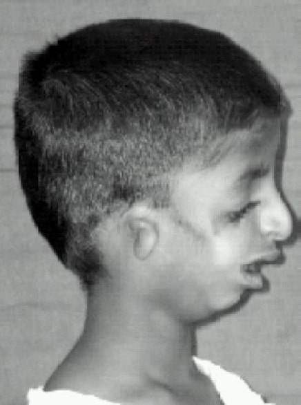
Treacher Collins syndrome is a rare genetic condition with autosomal dominant inheritance Autosomal dominant inheritance Autosomal Recessive and Autosomal Dominant Inheritance. Treacher Collins syndrome is also referred to as mandibulofacial dysostosis or Franceschetti syndrome and is characterized by significant craniofacial deformities and conductive hearing loss Conductive hearing loss Hearing loss due to interference with the mechanical reception or amplification of sound to the cochlea. The interference is in the outer or middle ear involving the ear canal; tympanic membrane; or ear ossicles. Hearing Loss. Treacher Collins syndrome is strictly a physical disease and does not affect cognition or other spheres of development. Diagnosis is confirmed with genetic testing Genetic Testing Detection of a mutation; genotype; karyotype; or specific alleles associated with genetic traits, heritable diseases, or predisposition to a disease, or that may lead to the disease in descendants. It includes prenatal genetic testing. Myotonic Dystrophies. Management of the airway Airway ABCDE Assessment, feeding ability, and hearing loss Hearing loss Hearing loss, also known as hearing impairment, is any degree of impairment in the ability to apprehend sound as determined by audiometry to be below normal hearing thresholds. Clinical presentation may occur at birth or as a gradual loss of hearing with age, including a short-term or sudden loss at any point. Hearing Loss are the primary concerns in the first few years of life. Ultimately, facial reconstructive surgery is needed. Life expectancy Life expectancy Based on known statistical data, the number of years which any person of a given age may reasonably expected to live. Population Pyramids is normal.

Last updated: Dec 13, 2022

Editorial responsibility: Stanley Oiseth, Lindsay Jones, Evelin Maza

Advertisement

Advertisement

Advertisement

Advertisement

Advertisement

Advertisement

Epidemiology and Genetics

Epidemiology

  • Incidence Incidence The number of new cases of a given disease during a given period in a specified population. It also is used for the rate at which new events occur in a defined population. It is differentiated from prevalence, which refers to all cases in the population at a given time. Measures of Disease Frequency: 1 in 25,000–50,000 live births
  • Women and men equally affected

Genetics Genetics Genetics is the study of genes and their functions and behaviors. Basic Terms of Genetics

  • Results from a deletion mutation Mutation Genetic mutations are errors in DNA that can cause protein misfolding and dysfunction. There are various types of mutations, including chromosomal, point, frameshift, and expansion mutations. Types of Mutations or sequence variation impairing proteins Proteins Linear polypeptides that are synthesized on ribosomes and may be further modified, crosslinked, cleaved, or assembled into complex proteins with several subunits. The specific sequence of amino acids determines the shape the polypeptide will take, during protein folding, and the function of the protein. Energy Homeostasis and rRNA rRNA The most abundant form of RNA. Together with proteins, it forms the ribosomes, playing a structural role and also a role in ribosomal binding of mRNA and tRNAs. Individual chains are conventionally designated by their sedimentation coefficients. In eukaryotes, four large chains exist, synthesized in the nucleolus and constituting about 50% of the ribosome. RNA Types and Structure that play a role in early development
  • Most commonly due to loss of function Loss of Function Inflammation mutation Mutation Genetic mutations are errors in DNA that can cause protein misfolding and dysfunction. There are various types of mutations, including chromosomal, point, frameshift, and expansion mutations. Types of Mutations on the TCOF gene Gene A category of nucleic acid sequences that function as units of heredity and which code for the basic instructions for the development, reproduction, and maintenance of organisms. Basic Terms of Genetics on chromosome Chromosome In a prokaryotic cell or in the nucleus of a eukaryotic cell, a structure consisting of or containing DNA which carries the genetic information essential to the cell. Basic Terms of Genetics 5
  • Less commonly due to mutations in the POLR1C gene Gene A category of nucleic acid sequences that function as units of heredity and which code for the basic instructions for the development, reproduction, and maintenance of organisms. Basic Terms of Genetics ( autosomal recessive Autosomal recessive Autosomal inheritance, both dominant and recessive, refers to the transmission of genes from the 22 autosomal chromosomes. Autosomal recessive diseases are only expressed when 2 copies of the recessive allele are inherited. Autosomal Recessive and Autosomal Dominant Inheritance) and POLR1D gene Gene A category of nucleic acid sequences that function as units of heredity and which code for the basic instructions for the development, reproduction, and maintenance of organisms. Basic Terms of Genetics ( autosomal dominant Autosomal dominant Autosomal inheritance, both dominant and recessive, refers to the transmission of genes from the 22 autosomal chromosomes. Autosomal dominant diseases are expressed when only 1 copy of the dominant allele is inherited. Autosomal Recessive and Autosomal Dominant Inheritance)
  • Neural crest Neural crest The two longitudinal ridges along the primitive streak appearing near the end of gastrulation during development of nervous system (neurulation). The ridges are formed by folding of neural plate. Between the ridges is a neural groove which deepens as the fold become elevated. When the folds meet at midline, the groove becomes a closed tube, the neural tube. Hirschsprung Disease cell dysfunction and abnormal 1st and 2nd pharyngeal arch development →  failure of cartilage Cartilage Cartilage is a type of connective tissue derived from embryonic mesenchyme that is responsible for structural support, resilience, and the smoothness of physical actions. Perichondrium (connective tissue membrane surrounding cartilage) compensates for the absence of vasculature in cartilage by providing nutrition and support. Cartilage: Histology, bone Bone Bone is a compact type of hardened connective tissue composed of bone cells, membranes, an extracellular mineralized matrix, and central bone marrow. The 2 primary types of bone are compact and spongy. Bones: Structure and Types, and connective tissues to undergo normal facial development
    • 1st pharyngeal arch (mandibular arch) contributes to mandible Mandible The largest and strongest bone of the face constituting the lower jaw. It supports the lower teeth. Jaw and Temporomandibular Joint: Anatomy, maxilla Maxilla One of a pair of irregularly shaped bones that form the upper jaw. A maxillary bone provides tooth sockets for the superior teeth, forms part of the orbit, and contains the maxillary sinus. Skull: Anatomy, zygomatic bone Zygomatic bone Either of a pair of bones that form the prominent part of the cheek and contribute to the orbit on each side of the skull. Orbit and Extraocular Muscles: Anatomy, and middle ear Middle ear The space and structures directly internal to the tympanic membrane and external to the inner ear (labyrinth). Its major components include the auditory ossicles and the eustachian tube that connects the cavity of middle ear (tympanic cavity) to the upper part of the throat. Acute Otitis Media development.
    • 2nd pharyngeal arch (hyoid arch) also contributes to middle ear Middle ear The space and structures directly internal to the tympanic membrane and external to the inner ear (labyrinth). Its major components include the auditory ossicles and the eustachian tube that connects the cavity of middle ear (tympanic cavity) to the upper part of the throat. Acute Otitis Media development as well as hyoid development.

Clinical Presentation

This syndrome is characterized by a multitude of bilateral and often asymmetric craniofacial structural defects and abnormalities.
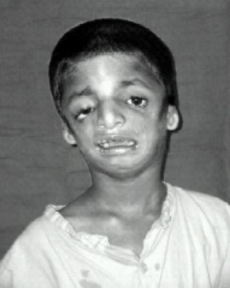
Ocular symptoms

Nasal symptoms

Otologic symptoms

Oropharyngeal symptoms

Cognitive/behavioral symptoms

  • Cognition is typically normal.
  • Failure to thrive Failure to Thrive Failure to thrive (FTT), or faltering growth, describes suboptimal weight gain and growth in children. The majority of cases are due to inadequate caloric intake; however, genetic, infectious, and oncological etiologies are also common. Failure to Thrive may be seen in young children due to feeding difficulties and under-nutrition.
  • Psychological and social development is typically normal, but challenges may arise due to physical appearance.

Diagnosis

  • History and clinical examination
  • Genetic testing Genetic Testing Detection of a mutation; genotype; karyotype; or specific alleles associated with genetic traits, heritable diseases, or predisposition to a disease, or that may lead to the disease in descendants. It includes prenatal genetic testing. Myotonic Dystrophies confirms diagnosis: analysis of TCOF1, POLR1D, and POLR1C genes Genes A category of nucleic acid sequences that function as units of heredity and which code for the basic instructions for the development, reproduction, and maintenance of organisms. DNA Types and Structure
  • Imaging studies to confirm and determine the extent of abnormalities seen:
    • Skull Skull The skull (cranium) is the skeletal structure of the head supporting the face and forming a protective cavity for the brain. The skull consists of 22 bones divided into the viscerocranium (facial skeleton) and the neurocranium. Skull: Anatomy radiographs, particularly with an occipitomental or Waters’ view: aplasia Aplasia Cranial Nerve Palsies or hypoplasia Hypoplasia Hypoplastic Left Heart Syndrome (HLHS) of the zygomatic Zygomatic Either of a pair of bones that form the prominent part of the cheek and contribute to the orbit on each side of the skull. Skull: Anatomy arch and mandibular retrognathia Retrognathia A physical misalignment of the upper (maxilla) and lower (mandibular) jaw bones in which either or both recede relative to the frontal plane of the forehead. Marfan Syndrome
    • Craniofacial computed tomography (CT): malar hypoplasia Malar Hypoplasia Marfan Syndrome
    • Dental radiographs

Management

Craniofacial malformations can result in airway Airway ABCDE Assessment compromise and feeding difficulties, making management of these problems the priority in early years. Once life-threatening issues are resolved, management of hearing loss Hearing loss Hearing loss, also known as hearing impairment, is any degree of impairment in the ability to apprehend sound as determined by audiometry to be below normal hearing thresholds. Clinical presentation may occur at birth or as a gradual loss of hearing with age, including a short-term or sudden loss at any point. Hearing Loss is imperative for speech development. Surgery is needed for correction of facial abnormalities.

Nutritional management

  • Special cleft palate Cleft palate Congenital fissure of the soft and/or hard palate, due to faulty fusion. Cleft Lip and Cleft Palate nursing bottle
  • Manage gastroesophageal reflux 
  • Gastrostomy tube for those incapable of feeding

Surgery

  • Cochlear implants early in life to help facilitate speech development
  • Pediatric plastic surgery is delayed until a significant amount of facial growth has occurred, typically around 7 years of age.
    • Zygomatic Zygomatic Either of a pair of bones that form the prominent part of the cheek and contribute to the orbit on each side of the skull. Skull: Anatomy and orbital reconstruction 
    • External ear reconstruction 
    • Middle ear Middle ear The space and structures directly internal to the tympanic membrane and external to the inner ear (labyrinth). Its major components include the auditory ossicles and the eustachian tube that connects the cavity of middle ear (tympanic cavity) to the upper part of the throat. Acute Otitis Media reconstruction 
    • Maxillomandibular reconstruction (done in adolescent years)
  • Oral surgery: cleft palate Cleft palate Congenital fissure of the soft and/or hard palate, due to faulty fusion. Cleft Lip and Cleft Palate, cleft lip Cleft lip The embryological development of craniofacial structures is an intricate sequential process involving tissue growth and directed cell apoptosis. Disruption of any step in this process may result in the formation of a cleft lip alone or in combination with a cleft palate. As the most common craniofacial malformation of the newborn, the diagnosis of a cleft is clinical and usually apparent at birth. Cleft Lip and Cleft Palate, bifid uvula Uvula A fleshy extension at the back of the soft palate that hangs above the opening of the throat. Peritonsillar Abscess

Other

  • Speech/language therapy
  • Correction of dental abnormalities between 6 and 14 years of age
  • Pediatric ophthalmology evaluation
  • Genetic counseling Genetic Counseling An educational process that provides information and advice to individuals or families about a genetic condition that may affect them. The purpose is to help individuals make informed decisions about marriage, reproduction, and other health management issues based on information about the genetic disease, the available diagnostic tests, and management programs. Psychosocial support is usually offered. Myotonic Dystrophies

Differential Diagnosis

  • Nager syndrome: also called preaxial acrofacial dysostosis, Nager syndrome is a rare genetic condition with mostly autosomal dominant inheritance Autosomal dominant inheritance Autosomal Recessive and Autosomal Dominant Inheritance. Craniofacial malformations are similar to Treacher Collins syndrome. Additionally, patients Patients Individuals participating in the health care system for the purpose of receiving therapeutic, diagnostic, or preventive procedures. Clinician–Patient Relationship have defects of the upper extremities, such as underdeveloped or absent thumbs and shortened forearms Forearms Part of the upper extremity in humans and primates extending from the elbow to the wrist. Bowen Disease and Erythroplasia of Queyrat. Genetic testing Genetic Testing Detection of a mutation; genotype; karyotype; or specific alleles associated with genetic traits, heritable diseases, or predisposition to a disease, or that may lead to the disease in descendants. It includes prenatal genetic testing. Myotonic Dystrophies can help differentiate this condition from Treacher Collins syndrome. Treatment is similar to that for Treacher Collins syndrome.
  • Miller syndrome: also called postaxial acrofacial dysostosis, a rare genetic condition with autosomal recessive inheritance Autosomal recessive inheritance Autosomal Recessive and Autosomal Dominant Inheritance. Craniofacial malformations are seen, similar to Treacher Collins syndrome. Additionally, patients Patients Individuals participating in the health care system for the purpose of receiving therapeutic, diagnostic, or preventive procedures. Clinician–Patient Relationship have supernumerary nipples and upper and lower extremity defects such as missing 5th fingers/toes, shortened forearms Forearms Part of the upper extremity in humans and primates extending from the elbow to the wrist. Bowen Disease and Erythroplasia of Queyrat, and webbed or fused digits. Genetic testing Genetic Testing Detection of a mutation; genotype; karyotype; or specific alleles associated with genetic traits, heritable diseases, or predisposition to a disease, or that may lead to the disease in descendants. It includes prenatal genetic testing. Myotonic Dystrophies helps differentiate this condition from other conditions. Management is similar to that for Treacher Collins syndrome.
  • Pierre Robin sequence Robin sequence Pierre Robin sequence, also known as Pierre Robin syndrome or simply Robin sequence, is a condition in infants that is characterized by a smaller-than-normal mandible, a tongue that retracts back into the throat, and difficulty breathing. Pierre Robin Sequence: refers to a set of maxillofacial malformations that occur due to an abnormality in the embryonic developmental sequence and may or may not be associated with any particular syndrome. Mandibular hypoplasia Hypoplasia Hypoplastic Left Heart Syndrome (HLHS) leads to glossoptosis Glossoptosis Posterior displacement of the tongue toward the pharynx. It is often a feature in syndromes such as in pierre robin syndrome and Down syndrome and associated with airway obstruction during sleep (obstructive sleep apneas). Pierre Robin Sequence (posteriorly displaced tongue Tongue The tongue, on the other hand, is a complex muscular structure that permits tasting and facilitates the process of mastication and communication. The blood supply of the tongue originates from the external carotid artery, and the innervation is through cranial nerves. Lips and Tongue: Anatomy), which results in abnormal palate Palate The palate is the structure that forms the roof of the mouth and floor of the nasal cavity. This structure is divided into soft and hard palates. Palate: Anatomy development and subsequent U-shaped cleft palate Cleft palate Congenital fissure of the soft and/or hard palate, due to faulty fusion. Cleft Lip and Cleft Palate. Diagnosed clinically with possible genetic testing Genetic Testing Detection of a mutation; genotype; karyotype; or specific alleles associated with genetic traits, heritable diseases, or predisposition to a disease, or that may lead to the disease in descendants. It includes prenatal genetic testing. Myotonic Dystrophies to detect any underlying syndrome. Management is similar to that for Treacher Collins syndrome.

References

  1. Chang, Christopher C. & Steinbacher, Derek M. (2012). Treacher Collins Syndrome. Semin Plast Surg. 26(2): 83–90. doi: 10.1055/s-0032-1320066. https://www.ncbi.nlm.nih.gov/pmc/articles/PMC3424693/
  2. Buchanan, Edward P. (2018). Syndromes with craniofacial abnormalities. UpToDate. Retrieved December 1, 2020, from https://www.uptodate.com/contents/syndromes-with-craniofacial-abnormalities?search=mandibulofacial%20dysostosis&source=search_result&selectedTitle=1~23&usage_type=default&display_rank=1
  3. Aljerian, A., & Gilardino, M. S. (2019). Treacher Collins Syndrome. Clinics in plastic surgery, 46(2), 197–205. https://doi.org/10.1016/j.cps.2018.11.005
  4. Plomp, R. G., van Lieshout, M. J., Joosten, K. F., Wolvius, E. B., van der Schroeff, M. P., Versnel, S. L., Poublon, R. M., & Mathijssen, I. M. (2016). Treacher Collins Syndrome: A Systematic Review of Evidence-Based Treatment and Recommendations. Plastic and reconstructive surgery, 137(1), 191–204. https://doi.org/10.1097/PRS.0000000000001896

Create your free account or log in to continue reading!

Sign up now and get free access to Lecturio with concept pages, medical videos, and questions for your medical education.

User Reviews

Details