Advertisement

Advertisement

Advertisement

Advertisement

Toxocariasis

Toxocariasis is caused by the nematodes Toxocara canis and T. cati. These species frequently infect dogs and cats and are most commonly transmitted to humans via accidental ingestion of eggs through the fecal–oral route. Toxocara are not able to complete their life cycle in humans, but they do migrate to organs (including the liver Liver The liver is the largest gland in the human body. The liver is found in the superior right quadrant of the abdomen and weighs approximately 1.5 kilograms. Its main functions are detoxification, metabolism, nutrient storage (e.g., iron and vitamins), synthesis of coagulation factors, formation of bile, filtration, and storage of blood. Liver: Anatomy, lungs Lungs Lungs are the main organs of the respiratory system. Lungs are paired viscera located in the thoracic cavity and are composed of spongy tissue. The primary function of the lungs is to oxygenate blood and eliminate CO2. Lungs: Anatomy, heart, brain Brain The part of central nervous system that is contained within the skull (cranium). Arising from the neural tube, the embryonic brain is comprised of three major parts including prosencephalon (the forebrain); mesencephalon (the midbrain); and rhombencephalon (the hindbrain). The developed brain consists of cerebrum; cerebellum; and other structures in the brain stem. Nervous System: Anatomy, Structure, and Classification, and eyes), where they cause inflammation Inflammation Inflammation is a complex set of responses to infection and injury involving leukocytes as the principal cellular mediators in the body's defense against pathogenic organisms. Inflammation is also seen as a response to tissue injury in the process of wound healing. The 5 cardinal signs of inflammation are pain, heat, redness, swelling, and loss of function. Inflammation and tissue damage. Depending on the affected site, patients Patients Individuals participating in the health care system for the purpose of receiving therapeutic, diagnostic, or preventive procedures. Clinician–Patient Relationship may develop visceral larva migrans or ocular larva migrans. Visceral larva migrans can present with flu-like symptoms Flu-Like Symptoms Babesia/Babesiosis, as well as hepatic, pulmonary, neurologic, and cardiac manifestations. Ocular larva migrans presents with unilateral vision Vision Ophthalmic Exam impairment, and a white, elevated granuloma may be seen on fundus Fundus The superior portion of the body of the stomach above the level of the cardiac notch. Stomach: Anatomy examination. The diagnosis is guided by clinical suspicion and may be supported with serology Serology The study of serum, especially of antigen-antibody reactions in vitro. Yellow Fever Virus. Management includes anthelmintic therapy and steroids Steroids A group of polycyclic compounds closely related biochemically to terpenes. They include cholesterol, numerous hormones, precursors of certain vitamins, bile acids, alcohols (sterols), and certain natural drugs and poisons. Steroids have a common nucleus, a fused, reduced 17-carbon atom ring system, cyclopentanoperhydrophenanthrene. Most steroids also have two methyl groups and an aliphatic side-chain attached to the nucleus. Benign Liver Tumors for severe disease.

Last updated: Oct 31, 2022

Editorial responsibility: Stanley Oiseth, Lindsay Jones, Evelin Maza

Advertisement

Advertisement

Advertisement

Advertisement

Advertisement

Advertisement

General Characteristics and Epidemiology

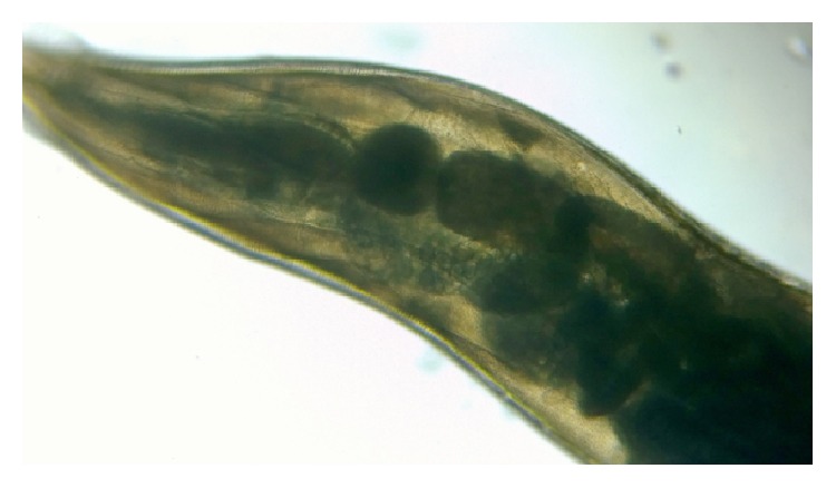
Basic features of Toxocara

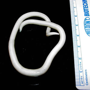
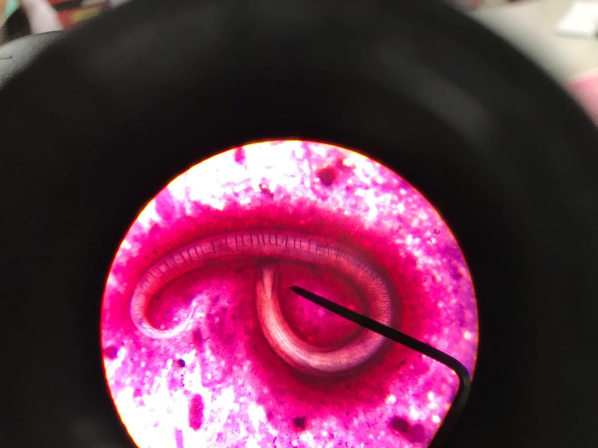
  • Nematode (roundworm)
  • A dioecious helminth (males and females have different morphologies):
    • Females are longer than males.
    • Males have a curved tail with paired spicules.
  • Size: 4–12 cm long
  • Have complete digestive systems
  • Move in contractile waves
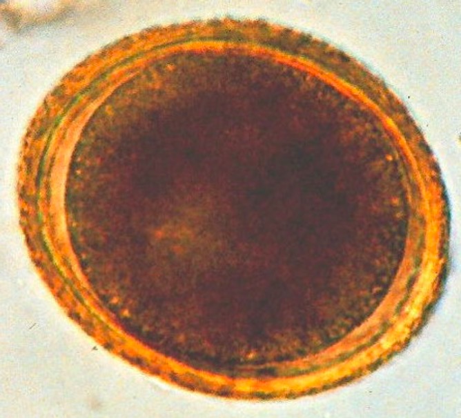
  • Eggs:
    • Brownish
    • Spherical 
    • Granulated or pitted surface
    • Very resistant to adverse environmental conditions
Toxocara (roundworm)

Spherical Toxocara egg with a brown, granulated appearance

Image: “Toxocara (roundworm)” by SuSanA Secretariat. License: CC BY 2.0

Clinically relevant species

Toxocariasis is caused by the following species:

  • Toxocara canis (dog roundworm)
  • Toxocara cati (cat roundworm)

Epidemiology

  • United States:
    • Prevalence Prevalence The total number of cases of a given disease in a specified population at a designated time. It is differentiated from incidence, which refers to the number of new cases in the population at a given time. Measures of Disease Frequency: 5%–15% of the population
    • Approximately 10,000 cases are diagnosed per year.
  • International:
    • 2%–5% positive rate in healthy adults from urban Western countries
    • 14%–37% in rural areas
    • More common in tropical and subtropical regions
  • Race:
    • No ethnic predisposition has been reported
    • More common in non-Hispanic Blacks
  • Age: common in young children and persons < 20 years of age

Pathogenesis

Reservoir Reservoir Animate or inanimate sources which normally harbor disease-causing organisms and thus serve as potential sources of disease outbreaks. Reservoirs are distinguished from vectors (disease vectors) and carriers, which are agents of disease transmission rather than continuing sources of potential disease outbreaks. Humans may serve both as disease reservoirs and carriers. Escherichia coli

  • T. canis:
    • Dogs (most puppies are infected soon after birth)
    • Foxes
  • T. cati: cats

Transmission

Transmission occurs through:

  • Fecal–oral route
  • Ingestion of infected animals Animals Unicellular or multicellular, heterotrophic organisms, that have sensation and the power of voluntary movement. Under the older five kingdom paradigm, animalia was one of the kingdoms. Under the modern three domain model, animalia represents one of the many groups in the domain eukaryota. Cell Types: Eukaryotic versus Prokaryotic

Host risk factors

  • Young age
  • Living with dogs and cats
  • Poor sanitation Sanitation The development and establishment of environmental conditions favorable to the health of the public. Hepatitis E Virus and handwashing
  • Consumption of:
    • Undercooked meat
    • Unwashed fruits and vegetables
  • Contact with infested soil from a yard, sandbox, park, or playground
  • Living in:
    • Rural areas
    • Poverty
    • Hot, humid environment (favors the survival of eggs)

Life cycle and pathophysiology

In animals Animals Unicellular or multicellular, heterotrophic organisms, that have sensation and the power of voluntary movement. Under the older five kingdom paradigm, animalia was one of the kingdoms. Under the modern three domain model, animalia represents one of the many groups in the domain eukaryota. Cell Types: Eukaryotic versus Prokaryotic:

  • Adult Toxocara worms live in the small intestines of dogs and cats.
  • Eggs are excreted in feces into the environment → transmission to a new host
  • Eggs hatch in the intestine and release Release Release of a virus from the host cell following virus assembly and maturation. Egress can occur by host cell lysis, exocytosis, or budding through the plasma membrane. Virology larvae Larvae Wormlike or grublike stage, following the egg in the life cycle of insects, worms, and other metamorphosing animals. Ascaris/Ascariasis → burrow through the bowel wall → enter the bloodstream 
  • Larvae Larvae Wormlike or grublike stage, following the egg in the life cycle of insects, worms, and other metamorphosing animals. Ascaris/Ascariasis migrate through the lungs Lungs Lungs are the main organs of the respiratory system. Lungs are paired viscera located in the thoracic cavity and are composed of spongy tissue. The primary function of the lungs is to oxygenate blood and eliminate CO2. Lungs: Anatomy and trachea Trachea The trachea is a tubular structure that forms part of the lower respiratory tract. The trachea is continuous superiorly with the larynx and inferiorly becomes the bronchial tree within the lungs. The trachea consists of a support frame of semicircular, or C-shaped, rings made out of hyaline cartilage and reinforced by collagenous connective tissue. Trachea: Anatomy → coughed up → swallowed into the GI tract 
  • Adult worms develop in the small intestine Small intestine The small intestine is the longest part of the GI tract, extending from the pyloric orifice of the stomach to the ileocecal junction. The small intestine is the major organ responsible for chemical digestion and absorption of nutrients. It is divided into 3 segments: the duodenum, the jejunum, and the ileum. Small Intestine: Anatomy → lay eggs → excreted in the stool

In humans:

  • Infection occurs through fecal–oral transmission.
  • Eggs hatch in the intestine and release Release Release of a virus from the host cell following virus assembly and maturation. Egress can occur by host cell lysis, exocytosis, or budding through the plasma membrane. Virology larvae Larvae Wormlike or grublike stage, following the egg in the life cycle of insects, worms, and other metamorphosing animals. Ascaris/Ascariasis → burrow through the bowel wall → enter the bloodstream
  • Invade organs throughout the body, most notably:
    • Muscles
    • Liver Liver The liver is the largest gland in the human body. The liver is found in the superior right quadrant of the abdomen and weighs approximately 1.5 kilograms. Its main functions are detoxification, metabolism, nutrient storage (e.g., iron and vitamins), synthesis of coagulation factors, formation of bile, filtration, and storage of blood. Liver: Anatomy
    • Heart
    • Lung
    • Eyes
    • Brain Brain The part of central nervous system that is contained within the skull (cranium). Arising from the neural tube, the embryonic brain is comprised of three major parts including prosencephalon (the forebrain); mesencephalon (the midbrain); and rhombencephalon (the hindbrain). The developed brain consists of cerebrum; cerebellum; and other structures in the brain stem. Nervous System: Anatomy, Structure, and Classification
  • Immune response against larvae Larvae Wormlike or grublike stage, following the egg in the life cycle of insects, worms, and other metamorphosing animals. Ascaris/Ascariasis antigens → inflammation Inflammation Inflammation is a complex set of responses to infection and injury involving leukocytes as the principal cellular mediators in the body’s defense against pathogenic organisms. Inflammation is also seen as a response to tissue injury in the process of wound healing. The 5 cardinal signs of inflammation are pain, heat, redness, swelling, and loss of function. Inflammation → tissue damage → clinical signs and symptoms
  • Toxocara cannot complete the life cycle in humans.

Clinical Presentation

Most Toxocara infections Infections Invasion of the host organism by microorganisms or their toxins or by parasites that can cause pathological conditions or diseases. Chronic Granulomatous Disease are asymptomatic and have a benign Benign Fibroadenoma course. The 2 major forms of toxocariasis are visceral larva migrans and ocular larva migrans.

Visceral larva migrans

General:

  • Fever Fever Fever is defined as a measured body temperature of at least 38°C (100.4°F). Fever is caused by circulating endogenous and/or exogenous pyrogens that increase levels of prostaglandin E2 in the hypothalamus. Fever is commonly associated with chills, rigors, sweating, and flushing of the skin. Fever
  • Anorexia Anorexia The lack or loss of appetite accompanied by an aversion to food and the inability to eat. It is the defining characteristic of the disorder anorexia nervosa. Anorexia Nervosa
  • Malaise Malaise Tick-borne Encephalitis Virus
  • Lymphadenopathy Lymphadenopathy Lymphadenopathy is lymph node enlargement (> 1 cm) and is benign and self-limited in most patients. Etiologies include malignancy, infection, and autoimmune disorders, as well as iatrogenic causes such as the use of certain medications. Generalized lymphadenopathy often indicates underlying systemic disease. Lymphadenopathy
  • Pruritic urticaria-like lesions

Hepatic:

  • Abdominal pain Abdominal Pain Acute Abdomen
  • Hepatomegaly 
  • Liver Liver The liver is the largest gland in the human body. The liver is found in the superior right quadrant of the abdomen and weighs approximately 1.5 kilograms. Its main functions are detoxification, metabolism, nutrient storage (e.g., iron and vitamins), synthesis of coagulation factors, formation of bile, filtration, and storage of blood. Liver: Anatomy nodularity

Pulmonary:

  • Dyspnea Dyspnea Dyspnea is the subjective sensation of breathing discomfort. Dyspnea is a normal manifestation of heavy physical or psychological exertion, but also may be caused by underlying conditions (both pulmonary and extrapulmonary). Dyspnea
  • Nonproductive cough
  • Wheezing Wheezing Wheezing is an abnormal breath sound characterized by a whistling noise that can be relatively high-pitched and shrill (more common) or coarse. Wheezing is produced by the movement of air through narrowed or compressed small (intrathoracic) airways. Wheezing
  • Chest tightness

Less common manifestations:

  • Muscle involvement
  • CNS involvement:
    • Eosinophilic meningitis Meningitis Meningitis is inflammation of the meninges, the protective membranes of the brain, and spinal cord. The causes of meningitis are varied, with the most common being bacterial or viral infection. The classic presentation of meningitis is a triad of fever, altered mental status, and nuchal rigidity. Meningitis
    • Meningoencephalitis Meningoencephalitis Encephalitis
    • Cerebral vasculitis Vasculitis Inflammation of any one of the blood vessels, including the arteries; veins; and rest of the vasculature system in the body. Systemic Lupus Erythematosus
    • Space-occupying lesions
  • Cardiac involvement:
    • Myocarditis Myocarditis Myocarditis is an inflammatory disease of the myocardium, which may occur alone or in association with a systemic process. There are numerous etiologies of myocarditis, but all lead to inflammation and myocyte injury, most often leading to signs and symptoms of heart failure. Myocarditis
    • Pericarditis Pericarditis Pericarditis is an inflammation of the pericardium, often with fluid accumulation. It can be caused by infection (often viral), myocardial infarction, drugs, malignancies, metabolic disorders, autoimmune disorders, or trauma. Acute, subacute, and chronic forms exist. Pericarditis
    • Loeffler (eosinophilic) endocarditis Endocarditis Endocarditis is an inflammatory disease involving the inner lining (endometrium) of the heart, most commonly affecting the cardiac valves. Both infectious and noninfectious etiologies lead to vegetations on the valve leaflets. Patients may present with nonspecific symptoms such as fever and fatigue. Endocarditis
    • Heart failure Heart Failure A heterogeneous condition in which the heart is unable to pump out sufficient blood to meet the metabolic need of the body. Heart failure can be caused by structural defects, functional abnormalities (ventricular dysfunction), or a sudden overload beyond its capacity. Chronic heart failure is more common than acute heart failure which results from sudden insult to cardiac function, such as myocardial infarction. Total Anomalous Pulmonary Venous Return (TAPVR)
    • Cardiac tamponade Tamponade Pericardial effusion, usually of rapid onset, exceeding ventricular filling pressures and causing collapse of the heart with a markedly reduced cardiac output. Pericarditis (from pericardial effusion Pericardial effusion Fluid accumulation within the pericardium. Serous effusions are associated with pericardial diseases. Hemopericardium is associated with trauma. Lipid-containing effusion (chylopericardium) results from leakage of thoracic duct. Severe cases can lead to cardiac tamponade. Pericardial Effusion and Cardiac Tamponade)

Ocular larva migrans

Unilateral symptoms:

  • Vision Vision Ophthalmic Exam impairment
  • Floaters Floaters Chorioretinitis
  • Strabismus Strabismus Strabismus is the misalignment of the eyes while fixating the gaze on an object. Strabismus can be idiopathic, but it may also be caused by cerebral palsy, uncorrected refractive errors, and extraocular muscle or cranial nerve dysfunction. Strabismus

Findings:

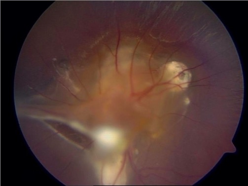
  • White, elevated granuloma (may be confused with retinoblastoma Retinoblastoma Retinoblastoma is a rare tumor but the most common primary intraocular malignancy of childhood. It is believed that the condition arises from a neuronal progenitor cell. Retinoblastoma can be heritable or non-heritable. Retinoblastoma)
  • Uveitis Uveitis Uveitis is the inflammation of the uvea, the pigmented middle layer of the eye, which comprises the iris, ciliary body, and choroid. The condition is categorized based on the site of disease; anterior uveitis is the most common. Diseases of the Uvea
  • Retinitis
  • Endophthalmitis Endophthalmitis Endophthalmitis is an inflammatory process of the inner layers of the eye, which may be either infectious or sterile. Infectious endophthalmitis can lead to irreversible vision loss if not treated quickly. Based on the entry mode of the infectious source, endophthalmitis is divided into endogenous and exogenous types. Endophthalmitis
  • Scleritis Scleritis Refers to any inflammation of the sclera including episcleritis, a benign condition affecting only the episclera, which is generally short-lived and easily treated. Classic scleritis, on the other hand, affects deeper tissue and is characterized by higher rates of visual acuity loss and even mortality, particularly in necrotizing form. Crohn’s Disease

Complications:

  • Retinal detachment Retinal detachment Retinal detachment is the separation of the neurosensory retina from the retinal pigmented epithelium and choroid. Rhegmatogenous retinal detachment, the most common type, stems from a break in the retina, allowing fluid to accumulate in the subretinal space. Retinal Detachment
  • Blindness Blindness The inability to see or the loss or absence of perception of visual stimuli. This condition may be the result of eye diseases; optic nerve diseases; optic chiasm diseases; or brain diseases affecting the visual pathways or occipital lobe. Retinopathy of Prematurity

Diagnosis

The diagnosis of toxocariasis requires a high index of suspicion.

Laboratory evaluation

Serologic testing:

  • ELISA ELISA An immunoassay utilizing an antibody labeled with an enzyme marker such as horseradish peroxidase. While either the enzyme or the antibody is bound to an immunosorbent substrate, they both retain their biologic activity; the change in enzyme activity as a result of the enzyme-antibody-antigen reaction is proportional to the concentration of the antigen and can be measured spectrophotometrically or with the naked eye. Many variations of the method have been developed. St. Louis Encephalitis Virus:
    • Detects antibodies Antibodies Immunoglobulins (Igs), also known as antibodies, are glycoprotein molecules produced by plasma cells that act in immune responses by recognizing and binding particular antigens. The various Ig classes are IgG (the most abundant), IgM, IgE, IgD, and IgA, which differ in their biologic features, structure, target specificity, and distribution. Immunoglobulins: Types and Functions to Toxocara excretory/secretory antigens
    • A positive test does not indicate active infection.
    • May be negative in ocular larva migrans
  • Western blot:
    • More sensitive and specific
    • Expensive
    • Can be used as a confirmatory test

Supporting evaluation:

Biopsy Biopsy Removal and pathologic examination of specimens from the living body. Ewing Sarcoma

  • Provides a definitive diagnosis but is rarely indicated
  • Findings: Toxocara larvae Larvae Wormlike or grublike stage, following the egg in the life cycle of insects, worms, and other metamorphosing animals. Ascaris/Ascariasis within eosinophilic granulomatous lesions

Imaging studies

Imaging is usually guided by the patient’s clinical presentation.
  • Chest X-ray X-ray Penetrating electromagnetic radiation emitted when the inner orbital electrons of an atom are excited and release radiant energy. X-ray wavelengths range from 1 pm to 10 nm. Hard x-rays are the higher energy, shorter wavelength x-rays. Soft x-rays or grenz rays are less energetic and longer in wavelength. The short wavelength end of the x-ray spectrum overlaps the gamma rays wavelength range. The distinction between gamma rays and x-rays is based on their radiation source. Pulmonary Function Tests:
    • Bilateral peribronchial infiltration
    • Parenchymal infiltrates
    • Pleural effusion Pleural Effusion Pleural effusion refers to the accumulation of fluid between the layers of the parietal and visceral pleura. Common causes of this condition include infection, malignancy, autoimmune disorders, or volume overload. Clinical manifestations include chest pain, cough, and dyspnea. Pleural Effusion
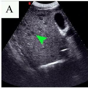
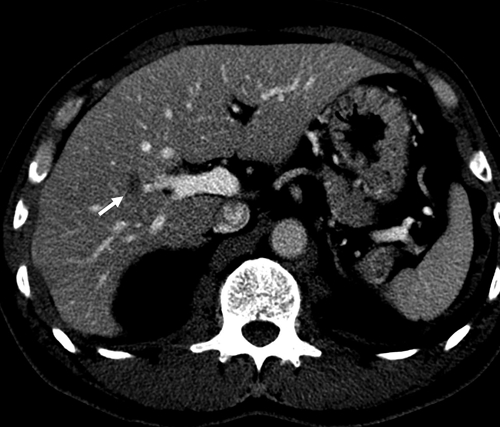
  • Ultrasonography: multiple hypoechoic Hypoechoic A structure that produces a low-amplitude echo (darker grays) Ultrasound (Sonography) areas in the liver Liver The liver is the largest gland in the human body. The liver is found in the superior right quadrant of the abdomen and weighs approximately 1.5 kilograms. Its main functions are detoxification, metabolism, nutrient storage (e.g., iron and vitamins), synthesis of coagulation factors, formation of bile, filtration, and storage of blood. Liver: Anatomy
  • CT scan:
    • Lungs Lungs Lungs are the main organs of the respiratory system. Lungs are paired viscera located in the thoracic cavity and are composed of spongy tissue. The primary function of the lungs is to oxygenate blood and eliminate CO2. Lungs: Anatomy: multiple pulmonary nodules Pulmonary nodules A number of small lung lesions characterized by small round masses of 2- to 3-mm in diameter. They are usually detected by chest ct scans. Such nodules can be associated with metastases of malignancies inside or outside the lung, benign granulomas, or other lesions. Granulomatosis with Polyangiitis with surrounding ground-glass opacities
    • Hepatic: low-density lesions
    • Brain Brain The part of central nervous system that is contained within the skull (cranium). Arising from the neural tube, the embryonic brain is comprised of three major parts including prosencephalon (the forebrain); mesencephalon (the midbrain); and rhombencephalon (the hindbrain). The developed brain consists of cerebrum; cerebellum; and other structures in the brain stem. Nervous System: Anatomy, Structure, and Classification: hyperintense Hyperintense Magnetic Resonance Imaging (MRI) cortical or subcortical granulomas Granulomas A relatively small nodular inflammatory lesion containing grouped mononuclear phagocytes, caused by infectious and noninfectious agents. Sarcoidosis

Management and Prevention

Management of visceral larva migrans

Patients Patients Individuals participating in the health care system for the purpose of receiving therapeutic, diagnostic, or preventive procedures. Clinician–Patient Relationship with mild symptoms do not require therapy, since the disease is self-limited. Patients Patients Individuals participating in the health care system for the purpose of receiving therapeutic, diagnostic, or preventive procedures. Clinician–Patient Relationship with moderate to severe disease may be given:

  • Anthelmintic therapy:
    • Albendazole Albendazole A benzimidazole broad-spectrum anthelmintic structurally related to mebendazole that is effective against many diseases. Anthelmintic Drugs
    • Mebendazole Mebendazole A benzimidazole that acts by interfering with carbohydrate metabolism and inhibiting polymerization of microtubules. Anthelmintic Drugs
  • Prednisone Prednisone A synthetic anti-inflammatory glucocorticoid derived from cortisone. It is biologically inert and converted to prednisolone in the liver. Immunosuppressants for severe respiratory, myocardial, or CNS disease.

Management of ocular larva migrans

  • Antihelmintic therapy
  • Prednisone Prednisone A synthetic anti-inflammatory glucocorticoid derived from cortisone. It is biologically inert and converted to prednisolone in the liver. Immunosuppressants for sight-threatening inflammation Inflammation Inflammation is a complex set of responses to infection and injury involving leukocytes as the principal cellular mediators in the body’s defense against pathogenic organisms. Inflammation is also seen as a response to tissue injury in the process of wound healing. The 5 cardinal signs of inflammation are pain, heat, redness, swelling, and loss of function. Inflammation

Prevention

  • Proper disposal of dog and cat feces
  • Deworming pets
  • Good hygiene practices
  • Cover sandboxes when not in use.

Comparison of Similar Helminths

Table: Comparison of similar helminths Helminths Commonly known as parasitic worms, this group includes the acanthocephala; nematoda; and platyhelminths. Some authors consider certain species of leeches that can become temporarily parasitic as helminths. Anthelmintic Drugs and their associated diseases
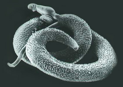
Organism Enterobius vermicularis Enterobius Vermicularis Enterobius/Enterobiasis Toxocara canis Ascaris Ascaris Ascaris is a genus of parasitic nematodes. The infection, ascariasis, is most often caused by A. lumbricoides. Transmission occurs primarily via ingestion of water or food contaminated with Ascaris eggs. Most patients with ascariasis are asymptomatic. Ascaris/Ascariasis lumbricoides Strongyloides stercoralis Strongyloides stercoralis A species of parasitic nematode widely distributed in tropical and subtropical countries. The females and their larvae inhabit the mucosa of the intestinal tract, where they cause ulceration and diarrhea. Strongyloidiasis Schistosoma Schistosoma Schistosomiasis is an infection caused by Schistosoma, a trematode. Schistosomiasis occurs in developing countries with poor sanitation. Freshwater snails are the intermediate host and are transmitted to humans through skin contact with contaminated fresh water. The clinical presentation occurs as a result of the host’s immune response to antigens from the eggs. Schistosoma/Schistosomiasis mansoni
Characteristics Nematode Nematode Nematode Nematode Trematode Trematode Class of parasitic flukes consisting of three subclasses, monogenea, aspidogastrea, and digenea. The digenetic trematodes are the only ones found in man. They are endoparasites and require two hosts to complete their life cycle. Schistosoma/Schistosomiasis
Reservoir Reservoir Animate or inanimate sources which normally harbor disease-causing organisms and thus serve as potential sources of disease outbreaks. Reservoirs are distinguished from vectors (disease vectors) and carriers, which are agents of disease transmission rather than continuing sources of potential disease outbreaks. Humans may serve both as disease reservoirs and carriers. Escherichia coli Humans Dogs Humans
  • Humans
  • Dogs
  • Cats
Humans
Transmission Fecal–oral Fecal–oral Fecal–oral Skin Skin The skin, also referred to as the integumentary system, is the largest organ of the body. The skin is primarily composed of the epidermis (outer layer) and dermis (deep layer). The epidermis is primarily composed of keratinocytes that undergo rapid turnover, while the dermis contains dense layers of connective tissue. Skin: Structure and Functions contact with contaminated soil Skin Skin The skin, also referred to as the integumentary system, is the largest organ of the body. The skin is primarily composed of the epidermis (outer layer) and dermis (deep layer). The epidermis is primarily composed of keratinocytes that undergo rapid turnover, while the dermis contains dense layers of connective tissue. Skin: Structure and Functions contact with contaminated water
Clinical
  • Visceral larva migrans
  • Ocular larva migrans
  • Cough
  • Wheezing Wheezing Wheezing is an abnormal breath sound characterized by a whistling noise that can be relatively high-pitched and shrill (more common) or coarse. Wheezing is produced by the movement of air through narrowed or compressed small (intrathoracic) airways. Wheezing
  • Hemoptysis Hemoptysis Hemoptysis is defined as the expectoration of blood originating in the lower respiratory tract. Hemoptysis is a consequence of another disease process and can be classified as either life threatening or non-life threatening. Hemoptysis can result in significant morbidity and mortality due to both drowning (reduced gas exchange as the lungs fill with blood) and hemorrhagic shock. Hemoptysis
  • Abdominal cramping Abdominal cramping Norovirus
  • Nausea Nausea An unpleasant sensation in the stomach usually accompanied by the urge to vomit. Common causes are early pregnancy, sea and motion sickness, emotional stress, intense pain, food poisoning, and various enteroviruses. Antiemetics
  • Malnutrition Malnutrition Malnutrition is a clinical state caused by an imbalance or deficiency of calories and/or micronutrients and macronutrients. The 2 main manifestations of acute severe malnutrition are marasmus (total caloric insufficiency) and kwashiorkor (protein malnutrition with characteristic edema). Malnutrition in children in resource-limited countries
  • Cough
  • Wheezing Wheezing Wheezing is an abnormal breath sound characterized by a whistling noise that can be relatively high-pitched and shrill (more common) or coarse. Wheezing is produced by the movement of air through narrowed or compressed small (intrathoracic) airways. Wheezing
  • Abdominal pain Abdominal Pain Acute Abdomen
  • Diarrhea Diarrhea Diarrhea is defined as ≥ 3 watery or loose stools in a 24-hour period. There are a multitude of etiologies, which can be classified based on the underlying mechanism of disease. The duration of symptoms (acute or chronic) and characteristics of the stools (e.g., watery, bloody, steatorrheic, mucoid) can help guide further diagnostic evaluation. Diarrhea
  • Rash Rash Rocky Mountain Spotted Fever
  • Swimmer’s itch Swimmer’s itch Schistosoma/Schistosomiasis
  • Katayama fever Katayama Fever Schistosoma/Schistosomiasis
  • Chronic infections Infections Invasion of the host organism by microorganisms or their toxins or by parasites that can cause pathological conditions or diseases. Chronic Granulomatous Disease lead to granuloma formation, which causes brain Brain The part of central nervous system that is contained within the skull (cranium). Arising from the neural tube, the embryonic brain is comprised of three major parts including prosencephalon (the forebrain); mesencephalon (the midbrain); and rhombencephalon (the hindbrain). The developed brain consists of cerebrum; cerebellum; and other structures in the brain stem. Nervous System: Anatomy, Structure, and Classification, lung, intestinal, and liver Liver The liver is the largest gland in the human body. The liver is found in the superior right quadrant of the abdomen and weighs approximately 1.5 kilograms. Its main functions are detoxification, metabolism, nutrient storage (e.g., iron and vitamins), synthesis of coagulation factors, formation of bile, filtration, and storage of blood. Liver: Anatomy disease.
Diagnosis Stool analysis
Management
  • Albendazole Albendazole A benzimidazole broad-spectrum anthelmintic structurally related to mebendazole that is effective against many diseases. Anthelmintic Drugs
  • Mebendazole Mebendazole A benzimidazole that acts by interfering with carbohydrate metabolism and inhibiting polymerization of microtubules. Anthelmintic Drugs
  • Ivermectin Ivermectin A mixture of mostly avermectin h2b1a (rn 71827-03-7) with some avermectin h2b1b (rn 70209-81-3), which are macrolides from streptomyces avermitilis. It binds glutamate-gated chloride channel to cause increased permeability and hyperpolarization of nerve and muscle cells. It also interacts with other chloride channels. It is a broad spectrum antiparasitic that is active against microfilariae of onchocerca volvulus but not the adult form. Anthelmintic Drugs
  • Albendazole Albendazole A benzimidazole broad-spectrum anthelmintic structurally related to mebendazole that is effective against many diseases. Anthelmintic Drugs
Praziquantel Praziquantel An anthelmintic used in most schistosome and many cestode infestations. Anthelmintic Drugs
Prevention Good hygiene
  • Good hygiene
  • Deworm dogs.
  • Proper disposal of dog feces
  • Good hygiene
  • Clean raw fruits and vegetables before consuming.
  • Wear shoes and protective clothing.
  • Improve sanitation Sanitation The development and establishment of environmental conditions favorable to the health of the public. Hepatitis E Virus.
  • Avoid swimming or bathing in contaminated water.
  • Drink boiled or bottled water.
  • Improve sanitation Sanitation The development and establishment of environmental conditions favorable to the health of the public. Hepatitis E Virus.

Differential Diagnosis

  • Allergic bronchopulmonary aspergillosis Allergic bronchopulmonary aspergillosis Hypersensitivity reaction (allergic reaction) to fungus aspergillus in an individual with long-standing bronchial asthma. It is characterized by pulmonary infiltrates, eosinophilia, elevated serum immunoglobulin e, and skin reactivity to aspergillus antigen. Aspergillus/Aspergillosis ( ABPA ABPA Hypersensitivity reaction (allergic reaction) to fungus aspergillus in an individual with long-standing bronchial asthma. It is characterized by pulmonary infiltrates, eosinophilia, elevated serum immunoglobulin e, and skin reactivity to aspergillus antigen. Aspergillus/Aspergillosis): hypersensitivity reaction to Aspergillus Aspergillus A genus of mitosporic fungi containing about 100 species and eleven different teleomorphs in the family trichocomaceae. Echinocandins: Patients Patients Individuals participating in the health care system for the purpose of receiving therapeutic, diagnostic, or preventive procedures. Clinician–Patient Relationship may have symptoms of airway Airway ABCDE Assessment obstruction, such as dyspnea Dyspnea Dyspnea is the subjective sensation of breathing discomfort. Dyspnea is a normal manifestation of heavy physical or psychological exertion, but also may be caused by underlying conditions (both pulmonary and extrapulmonary). Dyspnea, wheezing Wheezing Wheezing is an abnormal breath sound characterized by a whistling noise that can be relatively high-pitched and shrill (more common) or coarse. Wheezing is produced by the movement of air through narrowed or compressed small (intrathoracic) airways. Wheezing, productive cough, and fever Fever Fever is defined as a measured body temperature of at least 38°C (100.4°F). Fever is caused by circulating endogenous and/or exogenous pyrogens that increase levels of prostaglandin E2 in the hypothalamus. Fever is commonly associated with chills, rigors, sweating, and flushing of the skin. Fever. Bronchiectasis Bronchiectasis Bronchiectasis is a chronic disease of the airways that results from permanent bronchial distortion. This results from a continuous cycle of inflammation, bronchial damage and dilation, impaired clearance of secretions, and recurrent infections. Bronchiectasis and pulmonary fibrosis Fibrosis Any pathological condition where fibrous connective tissue invades any organ, usually as a consequence of inflammation or other injury. Bronchiolitis Obliterans may occur if left untreated. Eosinophilia Eosinophilia Abnormal increase of eosinophils in the blood, tissues or organs. Autosomal Dominant Hyperimmunoglobulin E Syndrome may be seen. The diagnosis is made with imaging, IgE IgE An immunoglobulin associated with mast cells. Overexpression has been associated with allergic hypersensitivity. Immunoglobulins: Types and Functions levels, skin Skin The skin, also referred to as the integumentary system, is the largest organ of the body. The skin is primarily composed of the epidermis (outer layer) and dermis (deep layer). The epidermis is primarily composed of keratinocytes that undergo rapid turnover, while the dermis contains dense layers of connective tissue. Skin: Structure and Functions prick testing, and serology Serology The study of serum, especially of antigen-antibody reactions in vitro. Yellow Fever Virus. Management includes steroids Steroids A group of polycyclic compounds closely related biochemically to terpenes. They include cholesterol, numerous hormones, precursors of certain vitamins, bile acids, alcohols (sterols), and certain natural drugs and poisons. Steroids have a common nucleus, a fused, reduced 17-carbon atom ring system, cyclopentanoperhydrophenanthrene. Most steroids also have two methyl groups and an aliphatic side-chain attached to the nucleus. Benign Liver Tumors and antifungal Antifungal Azoles therapy.
  • Retinoblastoma Retinoblastoma Retinoblastoma is a rare tumor but the most common primary intraocular malignancy of childhood. It is believed that the condition arises from a neuronal progenitor cell. Retinoblastoma can be heritable or non-heritable. Retinoblastoma: the most common primary intraocular malignancy Malignancy Hemothorax of childhood: Retinoblastoma Retinoblastoma Retinoblastoma is a rare tumor but the most common primary intraocular malignancy of childhood. It is believed that the condition arises from a neuronal progenitor cell. Retinoblastoma can be heritable or non-heritable. Retinoblastoma typically presents as leukocoria Leukocoria Cataracts in Children (abnormal white reflection in the eye) in a child under the age of 2 years. The disease may affect 1 or both eyes Both Eyes Refractive Errors (unlike toxocariasis). The diagnosis is made with fundus Fundus The superior portion of the body of the stomach above the level of the cardiac notch. Stomach: Anatomy examination and imaging. Management may include enucleation, laser photocoagulation, radiation Radiation Emission or propagation of acoustic waves (sound), electromagnetic energy waves (such as light; radio waves; gamma rays; or x-rays), or a stream of subatomic particles (such as electrons; neutrons; protons; or alpha particles). Osteosarcoma, and chemotherapy Chemotherapy Osteosarcoma.
  • Toxoplasmosis Toxoplasmosis Toxoplasmosis is an infectious disease caused by Toxoplasma gondii, an obligate intracellular protozoan parasite. Felines are the definitive host, but transmission to humans can occur through contact with cat feces or the consumption of contaminated foods. The clinical presentation and complications depend on the host’s immune status. Toxoplasma/Toxoplasmosis: infectious disease caused by Toxoplasma Toxoplasma Toxoplasmosis is an infectious disease caused by Toxoplasma gondii, an obligate intracellular protozoan parasite. Felines are the definitive host, but transmission to humans can occur through contact with cat feces or the consumption of contaminated foods. The clinical presentation and complications depend on the host’s immune status. Toxoplasma/Toxoplasmosis gondii: The clinical presentation and complications of toxoplasmosis Toxoplasmosis Toxoplasmosis is an infectious disease caused by Toxoplasma gondii, an obligate intracellular protozoan parasite. Felines are the definitive host, but transmission to humans can occur through contact with cat feces or the consumption of contaminated foods. The clinical presentation and complications depend on the host’s immune status. Toxoplasma/Toxoplasmosis depend on the host’s immune status and can vary greatly, including an acute, flu-like syndrome, CNS toxoplasmosis CNS toxoplasmosis Infections of the brain caused by the protozoan toxoplasma gondii that primarily arise in individuals with immunologic deficiency syndromes. The infection may involve the brain diffusely or form discrete abscesses. Clinical manifestations include seizures, altered mentation, headache, focal neurologic deficits, and intracranial hypertension. AIDS-defining Conditions, chorioretinitis Chorioretinitis Chorioretinitis is the inflammation of the posterior segment of the eye, including the choroid and the retina. The condition is usually caused by infections, the most common of which is toxoplasmosis. Some of these infections can affect the fetus in utero and present as congenital abnormalities. Chorioretinitis, or pneumonia Pneumonia Pneumonia or pulmonary inflammation is an acute or chronic inflammation of lung tissue. Causes include infection with bacteria, viruses, or fungi. In more rare cases, pneumonia can also be caused through toxic triggers through inhalation of toxic substances, immunological processes, or in the course of radiotherapy. Pneumonia. The diagnostic workup depends on the presentation, but can include lumbar puncture Lumbar Puncture Febrile Infant, imaging, biopsy Biopsy Removal and pathologic examination of specimens from the living body. Ewing Sarcoma, serology Serology The study of serum, especially of antigen-antibody reactions in vitro. Yellow Fever Virus, or PCR PCR Polymerase chain reaction (PCR) is a technique that amplifies DNA fragments exponentially for analysis. The process is highly specific, allowing for the targeting of specific genomic sequences, even with minuscule sample amounts. The PCR cycles multiple times through 3 phases: denaturation of the template DNA, annealing of a specific primer to the individual DNA strands, and synthesis/elongation of new DNA molecules. Polymerase Chain Reaction (PCR).  Management is with antimicrobial therapy.
  • Viral hepatitis: liver Liver The liver is the largest gland in the human body. The liver is found in the superior right quadrant of the abdomen and weighs approximately 1.5 kilograms. Its main functions are detoxification, metabolism, nutrient storage (e.g., iron and vitamins), synthesis of coagulation factors, formation of bile, filtration, and storage of blood. Liver: Anatomy inflammation Inflammation Inflammation is a complex set of responses to infection and injury involving leukocytes as the principal cellular mediators in the body’s defense against pathogenic organisms. Inflammation is also seen as a response to tissue injury in the process of wound healing. The 5 cardinal signs of inflammation are pain, heat, redness, swelling, and loss of function. Inflammation caused by infection with the hepatitis virus Virus Viruses are infectious, obligate intracellular parasites composed of a nucleic acid core surrounded by a protein capsid. Viruses can be either naked (non-enveloped) or enveloped. The classification of viruses is complex and based on many factors, including type and structure of the nucleoid and capsid, the presence of an envelope, the replication cycle, and the host range. Virology: Patients Patients Individuals participating in the health care system for the purpose of receiving therapeutic, diagnostic, or preventive procedures. Clinician–Patient Relationship may present with a viral prodrome Prodrome Symptoms that appear 24–48 hours prior to migraine onset. Migraine Headache of fever Fever Fever is defined as a measured body temperature of at least 38°C (100.4°F). Fever is caused by circulating endogenous and/or exogenous pyrogens that increase levels of prostaglandin E2 in the hypothalamus. Fever is commonly associated with chills, rigors, sweating, and flushing of the skin. Fever, anorexia Anorexia The lack or loss of appetite accompanied by an aversion to food and the inability to eat. It is the defining characteristic of the disorder anorexia nervosa. Anorexia Nervosa, and nausea Nausea An unpleasant sensation in the stomach usually accompanied by the urge to vomit. Common causes are early pregnancy, sea and motion sickness, emotional stress, intense pain, food poisoning, and various enteroviruses. Antiemetics. Right upper quadrant Right upper quadrant Anterior Abdominal Wall: Anatomy abdominal pain Abdominal Pain Acute Abdomen, jaundice Jaundice Jaundice is the abnormal yellowing of the skin and/or sclera caused by the accumulation of bilirubin. Hyperbilirubinemia is caused by either an increase in bilirubin production or a decrease in the hepatic uptake, conjugation, or excretion of bilirubin. Jaundice, and transaminitis Transaminitis Tick-borne Encephalitis Virus may also occur. The diagnosis is made with viral serologic testing and will differentiate viral hepatitis from visceral larva migrans. Management of acute hepatitis Acute Hepatitis Autoimmune Hepatitis is supportive.

References

  1. Woodhall DM, Fiore AE. (2014). Toxocariasis: a review for pediatricians. J Pediatric Infect Dis Soc 3(2):154–159.
  2. Woodhall D, Jones JL, Cantey PT, Wilkins PP, Montgomery SP. (2014). Neglected parasitic infections: what every family physician needs to know. Am Fam Physician 89(10):803–811.
  3. Hotez PJ, Wilkins PP. (2009). Toxocariasis: America’s most common neglected infection of poverty and a helminthiasis of global importance? PLoS Negl Trop Dis 3(3).
  4. Despommier D. (2003). Toxocariasis: clinical aspects, epidemiology, medical ecology, and molecular aspects. Clin Microbiol Rev 16:265–272.
  5. Herrmann N, Glickman LT, Schantz PM, Weston EI, Domanski LMA. (1985). Seroprevalence of zoonotic toxocariasis in the United States: 1971-1973. J Epidemiol 122(5):890–896.
  6. Weller PF, Leder K. (2020). Toxocariasis: visceral and ocular larva migrans. In Baron EL (Ed.), UpToDate. Retrieved April 12, 2021, from https://www.uptodate.com/contents/toxocariasis-visceral-and-ocular-larva-migrans
  7. Huh S, Lee S. (2019). Toxocariasis. In Brusch JL (Ed.), Medscape. Retrieved April 12, 2021, from https://emedicine.medscape.com/article/229855-overview
  8. Pearson RD. (2020). Toxocariasis. MSD Manual Professional Version. Retrieved April 12, 2021, from https://www.msdmanuals.com/professional/infectious-diseases/nematodes-roundworms/toxocariasis
  9. Winders WT, Merkin-Smith L. (2020). Toxocara canis. StatPearls. Retrieved April 12, 2021, from https://www.ncbi.nlm.nih.gov/books/NBK538524/

Create your free account or log in to continue reading!

Sign up now and get free access to Lecturio with concept pages, medical videos, and questions for your medical education.

User Reviews

Details