Advertisement

Advertisement

Advertisement

Advertisement

Thromboangiitis Obliterans (Buerger’s Disease)

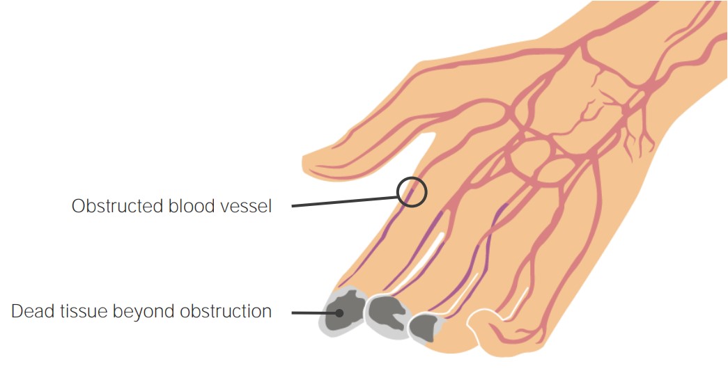
Thromboangiitis obliterans (TAO), also known as Buerger’s disease, is a rare condition causing inflammatory thrombosis Thrombosis Formation and development of a thrombus or blood clot in the blood vessel. Epidemic Typhus of the small- to medium-sized arteries Medium-Sized Arteries Kawasaki Disease and veins Veins Veins are tubular collections of cells, which transport deoxygenated blood and waste from the capillary beds back to the heart. Veins are classified into 3 types: small veins/venules, medium veins, and large veins. Each type contains 3 primary layers: tunica intima, tunica media, and tunica adventitia. Veins: Histology of the upper and lower extremities. Patients Patients Individuals participating in the health care system for the purpose of receiving therapeutic, diagnostic, or preventive procedures. Clinician–Patient Relationship are typically young smokers presenting with distal extremity ischemia Ischemia A hypoperfusion of the blood through an organ or tissue caused by a pathologic constriction or obstruction of its blood vessels, or an absence of blood circulation. Ischemic Cell Damage, ulcers, or gangrene Gangrene Death and putrefaction of tissue usually due to a loss of blood supply. Small Bowel Obstruction. Superficial thrombophlebitis and Raynaud's phenomenon can be early manifestations. Diagnosis is based on clinical findings, vascular testing, and angiography Angiography Radiography of blood vessels after injection of a contrast medium. Cardiac Surgery. Other potential diagnoses must be ruled out. The use of tobacco products is strongly associated with the disease; therefore, smoking Smoking Willful or deliberate act of inhaling and exhaling smoke from burning substances or agents held by hand. Interstitial Lung Diseases cessation is an essential part of management and decreases the risk of amputation Amputation An amputation is the separation of a portion of the limb or the entire limb from the body, along with the bone. Amputations are generally indicated for conditions that compromise the viability of the limb or promote the spread of a local process that could manifest systemically. Amputation.

Last updated: Aug 29, 2022

Editorial responsibility: Stanley Oiseth, Lindsay Jones, Evelin Maza

Advertisement

Advertisement

Advertisement

Advertisement

Advertisement

Advertisement

Epidemiology and Etiology

Epidemiology

  • Thromboangiitis obliterans (TAO) is most prevalent in regions and countries with heavy tobacco use:
  • Decreasing rates in North America and Western Europe are due to the decline in cigarette smoking Smoking Willful or deliberate act of inhaling and exhaling smoke from burning substances or agents held by hand. Interstitial Lung Diseases:
    • Current rate of 12.6‒20 cases per 100,000 
    • Unclear how vaping will affect TAO cases
  • Men > women (3:1)
  • The majority of patients Patients Individuals participating in the health care system for the purpose of receiving therapeutic, diagnostic, or preventive procedures. Clinician–Patient Relationship present before the age of 45.

Etiology

  • Tobacco use is seen in all patients Patients Individuals participating in the health care system for the purpose of receiving therapeutic, diagnostic, or preventive procedures. Clinician–Patient Relationship with TAO: 
    • Heavy cigarette smokers (most common)
    • Cigar smokers
    • Marijuana users (cannabis arteritis) 
    • Smokeless tobacco users (chewing tobacco and snuff)
  • Potential genetic predisposition: 
    • HLA-A9 
    • HLA-B5
    • HLA-A54

Pathophysiology

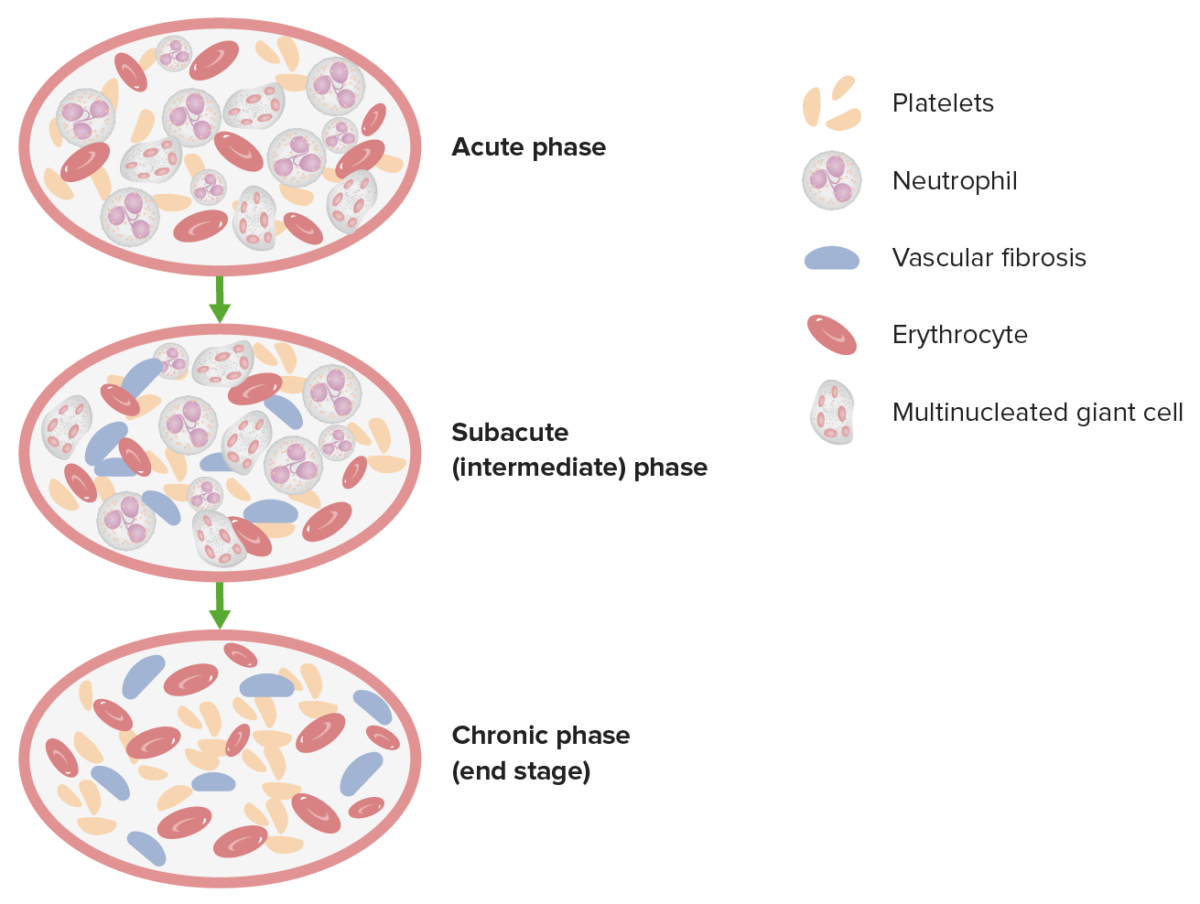
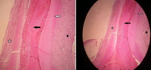
3 pathologic phases in thromboangiitis obliterans

This image demonstrates the 3 pathologic phases in TAO.

Image by Lecturio.
Clinical Presentation

Clinical presentation

  • Young smoker (< 45 years of age)
  • ≥ 2 extremities are usually involved (many times all 4 are involved)
  • Symptoms:
    • Intermittent claudication of the feet, legs, hands, or arms
    • Progresses to pain Pain An unpleasant sensation induced by noxious stimuli which are detected by nerve endings of nociceptive neurons. Pain: Types and Pathways of the digits at rest
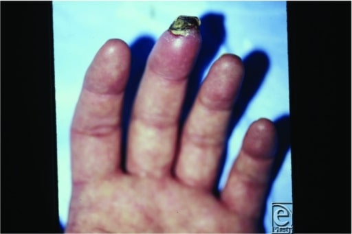
    • Ulcerations on the fingers, toes, or feet
    • Paresthesias Paresthesias Subjective cutaneous sensations (e.g., cold, warmth, tingling, pressure, etc.) that are experienced spontaneously in the absence of stimulation. Posterior Cord Syndrome
    • Color changes of digits
    • Episodic arthralgias or arthritis Arthritis Acute or chronic inflammation of joints. Osteoarthritis (usually wrists and knees)

Physical exam

  • Superficial thrombophlebitis (approximately 50% of cases):
    • An early manifestation
    • Often migratory
    • Tender nodules and cords
    • Follows a venous distribution
  • Raynaud’s phenomenon (vasospasm of the digits in response to cold or emotional stress):
    • Another early manifestation
    • Reversible pallor, cyanosis Cyanosis A bluish or purplish discoloration of the skin and mucous membranes due to an increase in the amount of deoxygenated hemoglobin in the blood or a structural defect in the hemoglobin molecule. Pulmonary Examination, or erythema Erythema Redness of the skin produced by congestion of the capillaries. This condition may result from a variety of disease processes. Chalazion of the digits
    • May be asymmetric
  • Digital ischemia Ischemia A hypoperfusion of the blood through an organ or tissue caused by a pathologic constriction or obstruction of its blood vessels, or an absence of blood circulation. Ischemic Cell Damage (most common presentation):
    • Rubor Rubor Inflammation or cyanosis Cyanosis A bluish or purplish discoloration of the skin and mucous membranes due to an increase in the amount of deoxygenated hemoglobin in the blood or a structural defect in the hemoglobin molecule. Pulmonary Examination of the digit (“Buerger’s color”)
    • Sensory Sensory Neurons which conduct nerve impulses to the central nervous system. Nervous System: Histology abnormalities
    • Cool extremities
    • Diminished distal pulses
    • Ischemic ulcerations
    • Gangrene Gangrene Death and putrefaction of tissue usually due to a loss of blood supply. Small Bowel Obstruction

Diagnosis

Clinical criteria

The diagnosis can be established without biopsy Biopsy Removal and pathologic examination of specimens from the living body. Ewing Sarcoma if the following criteria are met MET Preoperative Care:
  • Age less than 45 years
  • Current or recent tobacco use
  • Distal extremity ischemia Ischemia A hypoperfusion of the blood through an organ or tissue caused by a pathologic constriction or obstruction of its blood vessels, or an absence of blood circulation. Ischemic Cell Damage on objective vascular testing:
    • Allen test: With simultaneous compression Compression Blunt Chest Trauma of the radial and ulnar arteries Arteries Arteries are tubular collections of cells that transport oxygenated blood and nutrients from the heart to the tissues of the body. The blood passes through the arteries in order of decreasing luminal diameter, starting in the largest artery (the aorta) and ending in the small arterioles. Arteries are classified into 3 types: large elastic arteries, medium muscular arteries, and small arteries and arterioles. Arteries: Histology, the hand Hand The hand constitutes the distal part of the upper limb and provides the fine, precise movements needed in activities of daily living. It consists of 5 metacarpal bones and 14 phalanges, as well as numerous muscles innervated by the median and ulnar nerves. Hand: Anatomy remains pale after alternately releasing.
    • Ankle-brachial index Ankle-brachial index Comparison of the blood pressure between the brachial artery and the posterior tibial artery. It is a predictor of peripheral arterial disease. Cardiovascular Examination (ABI) 
    • Wrist-brachial index (WBI)
    • Typical arteriographic findings of TAO 
  • Exclusion of other potential causes

Workup

  • Biopsy Biopsy Removal and pathologic examination of specimens from the living body. Ewing Sarcoma
    • Rarely needed, but is the only modality that will give definitive diagnosis
    • May biopsy Biopsy Removal and pathologic examination of specimens from the living body. Ewing Sarcoma subcutaneous nodules or superficial thrombophlebitis
    • Findings: 
      • Inflammatory intraluminal thrombus, which is highly cellular 
      • Sparing of the vessel wall
    • Biopsy Biopsy Removal and pathologic examination of specimens from the living body. Ewing Sarcoma non-healing ulcers or lesions to rule out cancer.
  • Laboratory tests: 
    • Used to rule out other causes of vascular disease
    • The following should all be normal:
      • Complete blood count (CBC) with differential
      • Complete metabolic panel
      • Urinalysis Urinalysis Examination of urine by chemical, physical, or microscopic means. Routine urinalysis usually includes performing chemical screening tests, determining specific gravity, observing any unusual color or odor, screening for bacteriuria, and examining the sediment microscopically. Urinary Tract Infections (UTIs) in Children
      • Antinuclear antibody, rheumatoid factor Rheumatoid factor Antibodies found in adult rheumatoid arthritis patients that are directed against gamma-chain immunoglobulins. Autoimmune Hepatitis, complement, anti-centromere antibody Anti-Centromere Antibody Scleroderma, anti-scleroderma 70 antibody → rule out vasculitis Vasculitis Inflammation of any one of the blood vessels, including the arteries; veins; and rest of the vasculature system in the body. Systemic Lupus Erythematosus
      • Antiphospholipid antibody, partial thromboplastin time Partial thromboplastin time The time required for the appearance of fibrin strands following the mixing of plasma with phospholipid platelet substitute (e.g., crude cephalins, soybean phosphatides). It is a test of the intrinsic pathway (factors VIII, IX, XI, and XII) and the common pathway (fibrinogen, prothrombin, factors V and X) of blood coagulation. Hemostasis (PTT), prothrombin time Prothrombin time Clotting time of plasma recalcified in the presence of excess tissue thromboplastin. Factors measured are fibrinogen; prothrombin; factor V; factor VII; and factor X. Hemostasis (PT), factor V Leiden Factor V Leiden Hypercoagulable States, antithrombin Antithrombin Endogenous factors and drugs that directly inhibit the action of thrombin, usually by blocking its enzymatic activity. They are distinguished from indirect thrombin inhibitors, such as heparin, which act by enhancing the inhibitory effects of antithrombins. Anticoagulants III, protein C and S, prothrombin Prothrombin A plasma protein that is the inactive precursor of thrombin. It is converted to thrombin by a prothrombin activator complex consisting of factor Xa, factor V, phospholipid, and calcium ions. Hemostasis gene mutation Gene Mutation Myotonic Dystrophies → rule out hypercoagulable Hypercoagulable Hypercoagulable states (also referred to as thrombophilias) are a group of hematologic diseases defined by an increased risk of clot formation (i.e., thrombosis) due to either an increase in procoagulants, a decrease in anticoagulants, or a decrease in fibrinolysis. Hypercoagulable States disorders 
      • Toxicology panel → evaluate for cannabis arteritis
  • Imaging:
    • Angiography Angiography Radiography of blood vessels after injection of a contrast medium. Cardiac Surgery:
      • Evaluates the extent of the disease
      • Non-atherosclerotic, segmental, occlusive lesions in small- to medium-sized vessels
      • Formation of small collateral vessels around the occlusion (“corkscrew collaterals”)
      • Similar findings can be seen in other autoimmune disorders
    • Computed tomography with angiography Angiography Radiography of blood vessels after injection of a contrast medium. Cardiac Surgery ( CTA CTA A non-invasive method that uses a ct scanner for capturing images of blood vessels and tissues. A contrast material is injected, which helps produce detailed images that aid in diagnosing vascular diseases. Pulmonary Function Tests) and magnetic resonance with angiography Angiography Radiography of blood vessels after injection of a contrast medium. Cardiac Surgery ( MRA MRA Imaging of the Heart and Great Vessels): can be used, but may not provide adequate detail
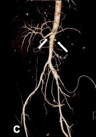
Contrast-enhanced abdominal ct and abdominal aorta ct angiography

Abdominal aorta CT angiography of a patient with TAO showing total occlusion of both renal arteries (white arrows)

Image: “Contrast-enhanced abdominal CT and abdominal aorta CT angiography” by Yun et al. 2015. License: CC BY 4.0, edited by Lecturio.

Management, Complications, and Prognosis

Management

  • Tobacco cessation:
    • Most effective way to “treat” TAO, and decreases the risk of amputation Amputation An amputation is the separation of a portion of the limb or the entire limb from the body, along with the bone. Amputations are generally indicated for conditions that compromise the viability of the limb or promote the spread of a local process that could manifest systemically. Amputation
    • Counseling on complete abstinence of tobacco and cannabis is required.
    • Medications: 
      • Only non-nicotine products are used ( bupropion Bupropion A propiophenone-derived antidepressant and antismoking agent that inhibits the uptake of dopamine. Other Antidepressants, varenicline Varenicline A benzazepine derivative that functions as an alpha4beta2 nicotinic receptor partial agonist. It is used for smoking cessation. Cholinomimetic Drugs).
      • Avoid nicotine Nicotine Nicotine is highly toxic alkaloid. It is the prototypical agonist at nicotinic cholinergic receptors where it dramatically stimulates neurons and ultimately blocks synaptic transmission. Nicotine is also important medically because of its presence in tobacco smoke. Stimulants patches Patches Vitiligo and gum due to a concern that they may continue the disease.
    • Failure of tobacco cessation will lead to disease progression.
  • Intermittent pneumatic compression Compression Blunt Chest Trauma
    • Limited data
    • Useful in small vessel occlusive disease of other etiologies
    • Can be used for painful ulcerations, but improvement is slow
  • Vasodilators: 
    • Alleviate symptoms only
    • Medication options:
      • Prostacyclin Prostacyclin A prostaglandin that is a powerful vasodilator and inhibits platelet aggregation. It is biosynthesized enzymatically from prostaglandin endoperoxides in human vascular tissue. The sodium salt has been also used to treat primary pulmonary hypertension. Eicosanoids analogues ( iloprost Iloprost An eicosanoid, derived from the cyclooxygenase pathway of arachidonic acid metabolism. It is a stable and synthetic analog of epoprostenol, but with a longer half-life than the parent compound. Its actions are similar to prostacyclin. Iloprost produces vasodilation and inhibits platelet aggregation. Pulmonary Hypertension Drugs)
      • Phosphodiesterase (PDE) inhibitors ( cilostazol Cilostazol A quinoline and tetrazole derivative that acts as a phosphodiesterase type 3 inhibitor, with anti-platelet and vasodilating activity. It is used in the treatment of peripheral vascular diseases; ischemic heart disease; and in the prevention of stroke. Phosphodiesterase Inhibitors, pentoxifylline Pentoxifylline A methylxanthine derivative that inhibits phosphodiesterase and affects blood rheology. It improves blood flow by increasing erythrocyte and leukocyte flexibility. It also inhibits platelet aggregation. Pentoxifylline modulates immunologic activity by stimulating cytokine production. Phosphodiesterase Inhibitors)
      • Alpha-adrenergic receptor Receptor Receptors are proteins located either on the surface of or within a cell that can bind to signaling molecules known as ligands (e.g., hormones) and cause some type of response within the cell. Receptors antagonists
      • Nitrates Nitrates Nitrates are a class of medications that cause systemic vasodilation (veins > arteries) by smooth muscle relaxation. Nitrates are primarily indicated for the treatment of angina, where preferential venodilation causes pooling of blood, decreased preload, and ultimately decreased myocardial O2 demand. Nitrates
      • Calcium Calcium A basic element found in nearly all tissues. It is a member of the alkaline earth family of metals with the atomic symbol ca, atomic number 20, and atomic weight 40. Calcium is the most abundant mineral in the body and combines with phosphorus to form calcium phosphate in the bones and teeth. It is essential for the normal functioning of nerves and muscles and plays a role in blood coagulation (as factor IV) and in many enzymatic processes. Electrolytes channel blockers (frequently used for Raynaud’s phenomenon)
  • Revascularization:
    • May not be feasible due to distal disease
    • Arterial bypass surgery:
      • Reserved for severe ischemia Ischemia A hypoperfusion of the blood through an organ or tissue caused by a pathologic constriction or obstruction of its blood vessels, or an absence of blood circulation. Ischemic Cell Damage
      • Requires a suitable target
    • Thrombolytic therapy and angioplasty Angioplasty Reconstruction or repair of a blood vessel, which includes the widening of a pathological narrowing of an artery or vein by the removal of atheromatous plaque material and/or the endothelial lining as well, or by dilatation (balloon angioplasty) to compress an atheroma. Except for endarterectomy, usually these procedures are performed via catheterization as minimally invasive endovascular procedures. Cardiac Surgery are rarely used.

Complications

  • Infection
  • Ulcerations
  • Gangrene Gangrene Death and putrefaction of tissue usually due to a loss of blood supply. Small Bowel Obstruction
  • Amputation Amputation An amputation is the separation of a portion of the limb or the entire limb from the body, along with the bone. Amputations are generally indicated for conditions that compromise the viability of the limb or promote the spread of a local process that could manifest systemically. Amputation
  • Occlusion of coronary, renal, splenic, or mesenteric arteries Arteries Arteries are tubular collections of cells that transport oxygenated blood and nutrients from the heart to the tissues of the body. The blood passes through the arteries in order of decreasing luminal diameter, starting in the largest artery (the aorta) and ending in the small arterioles. Arteries are classified into 3 types: large elastic arteries, medium muscular arteries, and small arteries and arterioles. Arteries: Histology causing organ ischemia Ischemia A hypoperfusion of the blood through an organ or tissue caused by a pathologic constriction or obstruction of its blood vessels, or an absence of blood circulation. Ischemic Cell Damage

Prognosis Prognosis A prediction of the probable outcome of a disease based on a individual’s condition and the usual course of the disease as seen in similar situations. Non-Hodgkin Lymphomas

  • 94% of patients Patients Individuals participating in the health care system for the purpose of receiving therapeutic, diagnostic, or preventive procedures. Clinician–Patient Relationship who stop smoking Smoking Willful or deliberate act of inhaling and exhaling smoke from burning substances or agents held by hand. Interstitial Lung Diseases will avoid amputation Amputation An amputation is the separation of a portion of the limb or the entire limb from the body, along with the bone. Amputations are generally indicated for conditions that compromise the viability of the limb or promote the spread of a local process that could manifest systemically. Amputation.
  • 43% of patients Patients Individuals participating in the health care system for the purpose of receiving therapeutic, diagnostic, or preventive procedures. Clinician–Patient Relationship who continue using tobacco will require amputation Amputation An amputation is the separation of a portion of the limb or the entire limb from the body, along with the bone. Amputations are generally indicated for conditions that compromise the viability of the limb or promote the spread of a local process that could manifest systemically. Amputation within 8 years.

Differential Diagnosis

  • Peripheral artery disease Peripheral artery disease Peripheral artery disease (PAD) is obstruction of the arterial lumen resulting in decreased blood flow to the distal limbs. The disease can be a result of atherosclerosis or thrombosis. Patients may be asymptomatic or have progressive claudication, skin discoloration, ischemic ulcers, or gangrene. Peripheral Artery Disease (PAD): atherosclerotic arterial narrowing resulting in ischemia Ischemia A hypoperfusion of the blood through an organ or tissue caused by a pathologic constriction or obstruction of its blood vessels, or an absence of blood circulation. Ischemic Cell Damage to the distal limbs. Risk factors include hypertension Hypertension Hypertension, or high blood pressure, is a common disease that manifests as elevated systemic arterial pressures. Hypertension is most often asymptomatic and is found incidentally as part of a routine physical examination or during triage for an unrelated medical encounter. Hypertension, hyperlipidemia, diabetes Diabetes Diabetes mellitus (DM) is a metabolic disease characterized by hyperglycemia and dysfunction of the regulation of glucose metabolism by insulin. Type 1 DM is diagnosed mostly in children and young adults as the result of autoimmune destruction of β cells in the pancreas and the resulting lack of insulin. Type 2 DM has a significant association with obesity and is characterized by insulin resistance. Diabetes Mellitus, tobacco use, and increased age. Patients Patients Individuals participating in the health care system for the purpose of receiving therapeutic, diagnostic, or preventive procedures. Clinician–Patient Relationship will have intermittent claudication, cyanosis Cyanosis A bluish or purplish discoloration of the skin and mucous membranes due to an increase in the amount of deoxygenated hemoglobin in the blood or a structural defect in the hemoglobin molecule. Pulmonary Examination, ischemic ulcers, and gangrene Gangrene Death and putrefaction of tissue usually due to a loss of blood supply. Small Bowel Obstruction. The clinical picture, along with abnormal ABIs, will establish the diagnosis and differentiate PAD from TAO. Management includes risk factor modification, antiplatelet agents Antiplatelet agents Antiplatelet agents are medications that inhibit platelet aggregation, a critical step in the formation of the initial platelet plug. Abnormal, or inappropriate, platelet aggregation is a key step in the pathophysiology of arterial ischemic events. The primary categories of antiplatelet agents include aspirin, ADP inhibitors, phosphodiesterase/adenosine uptake inhibitors, and glycoprotein IIb/IIIa inhibitors. Antiplatelet Drugs, PDE inhibitors, and revascularization.
  • Thromboembolic disease: vascular occlusion due to a dislodged thrombus from a more proximal source. The presentation depends on the source, size, and location of the embolism, but includes acute limb ischemia Ischemia A hypoperfusion of the blood through an organ or tissue caused by a pathologic constriction or obstruction of its blood vessels, or an absence of blood circulation. Ischemic Cell Damage and blue toe syndrome Blue toe syndrome A condition that is caused by recurring atheroembolism in the lower extremities. It is characterized by cyanotic discoloration of the toes, usually the first, fourth, and fifth toes. Discoloration may extend to the lateral aspect of the foot. Despite the gangrene-like appearance, blue toes may respond to conservative therapy without amputation. Peripheral Artery Disease. The clinical history, hypercoagulable Hypercoagulable Hypercoagulable states (also referred to as thrombophilias) are a group of hematologic diseases defined by an increased risk of clot formation (i.e., thrombosis) due to either an increase in procoagulants, a decrease in anticoagulants, or a decrease in fibrinolysis. Hypercoagulable States workup, and imaging aid in the diagnosis, and a source will typically be suspected or found on workup, which differentiates thromboembolic disease from TAO. Management includes anticoagulation Anticoagulation Pulmonary Hypertension Drugs and revascularization. 
  • Vasculitis Vasculitis Inflammation of any one of the blood vessels, including the arteries; veins; and rest of the vasculature system in the body. Systemic Lupus Erythematosus: a vascular inflammatory disease, often resulting in ischemia Ischemia A hypoperfusion of the blood through an organ or tissue caused by a pathologic constriction or obstruction of its blood vessels, or an absence of blood circulation. Ischemic Cell Damage, necrosis Necrosis The death of cells in an organ or tissue due to disease, injury or failure of the blood supply. Ischemic Cell Damage, and organ damage. Any vessel can be involved, which differentiates vasculitis Vasculitis Inflammation of any one of the blood vessels, including the arteries; veins; and rest of the vasculature system in the body. Systemic Lupus Erythematosus from TAO. Etiologies include autoimmune disorders, drugs, and infections Infections Invasion of the host organism by microorganisms or their toxins or by parasites that can cause pathological conditions or diseases. Chronic Granulomatous Disease. Patients Patients Individuals participating in the health care system for the purpose of receiving therapeutic, diagnostic, or preventive procedures. Clinician–Patient Relationship present with fever Fever Fever is defined as a measured body temperature of at least 38°C (100.4°F). Fever is caused by circulating endogenous and/or exogenous pyrogens that increase levels of prostaglandin E2 in the hypothalamus. Fever is commonly associated with chills, rigors, sweating, and flushing of the skin. Fever, arthralgias, arthritis Arthritis Acute or chronic inflammation of joints. Osteoarthritis, as well as potential end-organ damage. Diagnosis involves inflammatory markers, autoimmune serology Serology The study of serum, especially of antigen-antibody reactions in vitro. Yellow Fever Virus, infectious workup, and biopsy Biopsy Removal and pathologic examination of specimens from the living body. Ewing Sarcoma. Management depends on the underlying cause.
  • Popliteal artery Popliteal Artery The continuation of the femoral artery coursing through the popliteal fossa; it divides into the anterior and posterior tibial arteries. Popliteal Fossa: Anatomy entrapment syndrome: an uncommon condition in which an abnormally positioned, or enlarged, calf muscle compresses the popliteal artery Popliteal Artery The continuation of the femoral artery coursing through the popliteal fossa; it divides into the anterior and posterior tibial arteries. Popliteal Fossa: Anatomy. The compression Compression Blunt Chest Trauma leads to obstruction of blood flow Blood flow Blood flow refers to the movement of a certain volume of blood through the vasculature over a given unit of time (e.g., mL per minute). Vascular Resistance, Flow, and Mean Arterial Pressure to the lower extremity, and resembles the distal extremity ischemia Ischemia A hypoperfusion of the blood through an organ or tissue caused by a pathologic constriction or obstruction of its blood vessels, or an absence of blood circulation. Ischemic Cell Damage, ulceration Ulceration Corneal Abrasions, Erosion, and Ulcers, or necrosis Necrosis The death of cells in an organ or tissue due to disease, injury or failure of the blood supply. Ischemic Cell Damage of TAO. The diagnosis is made with imaging, which will differentiate popliteal artery Popliteal Artery The continuation of the femoral artery coursing through the popliteal fossa; it divides into the anterior and posterior tibial arteries. Popliteal Fossa: Anatomy entrapment syndrome from TAO. Management includes avoiding any inciting exercise and vascular surgery Vascular surgery Vascular surgery is the specialized field of medicine that focuses on the surgical management of the pathologies of the peripheral circulation. The main goal of most vascular procedures is to restore circulatory function to the affected vessels by relieving occlusions or by redirecting blood flow (e.g., bypass). Vascular Surgery evaluation.
  • Frostbite Frostbite Injuries due to cold weather are common among children and athletes who are involved in sports played in cold conditions. Frostbite is a direct freezing injury to the peripheral tissues and occurs when the skin temperature drops below 0°C (32°F). Common sites of frostbite include the nose, ears, fingers, and toes. Frostbite: injury due to freezing of the tissues that can be associated with vascular thrombosis Thrombosis Formation and development of a thrombus or blood clot in the blood vessel. Epidemic Typhus. Frostbite Frostbite Injuries due to cold weather are common among children and athletes who are involved in sports played in cold conditions. Frostbite is a direct freezing injury to the peripheral tissues and occurs when the skin temperature drops below 0°C (32°F). Common sites of frostbite include the nose, ears, fingers, and toes. Frostbite commonly affects the fingers, toes, nose Nose The nose is the human body’s primary organ of smell and functions as part of the upper respiratory system. The nose may be best known for inhaling oxygen and exhaling carbon dioxide, but it also contributes to other important functions, such as tasting. The anatomy of the nose can be divided into the external nose and the nasal cavity. Nose Anatomy (External & Internal), ears, cheeks Cheeks The part of the face that is below the eye and to the side of the nose and mouth. Melasma, and chin Chin The anatomical frontal portion of the mandible, also known as the mentum, that contains the line of fusion of the two separate halves of the mandible (symphysis menti). This line of fusion divides inferiorly to enclose a triangular area called the mental protuberance. On each side, inferior to the second premolar tooth, is the mental foramen for the passage of blood vessels and a nerve. Melasma. A severe cold exposure history is present, and findings include numbness, white or cyanotic discoloration of the skin Skin The skin, also referred to as the integumentary system, is the largest organ of the body. The skin is primarily composed of the epidermis (outer layer) and dermis (deep layer). The epidermis is primarily composed of keratinocytes that undergo rapid turnover, while the dermis contains dense layers of connective tissue. Skin: Structure and Functions, bullae Bullae Erythema Multiforme, eschar, or tissue necrosis Necrosis The death of cells in an organ or tissue due to disease, injury or failure of the blood supply. Ischemic Cell Damage. The diagnosis is clinical, and management involves rewarming and wound care. Severe injuries receive thrombolysis and prostacyclin Prostacyclin A prostaglandin that is a powerful vasodilator and inhibits platelet aggregation. It is biosynthesized enzymatically from prostaglandin endoperoxides in human vascular tissue. The sodium salt has been also used to treat primary pulmonary hypertension. Eicosanoids therapy.
  • Raynaud’s syndrome: arterial vasospasm that causes episodes of reduced blood flow Blood flow Blood flow refers to the movement of a certain volume of blood through the vasculature over a given unit of time (e.g., mL per minute). Vascular Resistance, Flow, and Mean Arterial Pressure to the fingers and toes, typically due to cold exposure or stress. The affected digits may be transiently pale, cyanotic, or hyperemic, and accompanied by numbness or pain Pain An unpleasant sensation induced by noxious stimuli which are detected by nerve endings of nociceptive neurons. Pain: Types and Pathways. Findings are generally symmetric. Secondary Raynaud’s syndrome can be seen with TAO, which will show asymmetric findings. Diagnosis is clinical, and management includes cold exposure avoidance and vasodilators. 

References

  1. Olin, J.W. (2020). Thromboangiitis obliterans (Beurger’s disease). In Collins, K.A. (Ed.), Uptodate. Retrieved November 13, 2020, from https://www.uptodate.com/contents/thromboangiitis-obliterans-buergers-disease
  2. Nassiri, N. (2020). Thromboangiitis obliterans (Buerger disease). In Rowe, V.L. (Ed.), Medscape. Retrieved November 13, 2020, from https://emedicine.medscape.com/article/460027-overview
  3. Teo, K.K. (2019). Thromboangiitis obliterans. [online] MSD Manual Professional Edition. Retrieved November 13, 2020, from https://www.msdmanuals.com/professional/cardiovascular-disorders/peripheral-arterial-disorders/thromboangiitis-obliterans

Create your free account or log in to continue reading!

Sign up now and get free access to Lecturio with concept pages, medical videos, and questions for your medical education.

User Reviews

Details