Advertisement

Advertisement

Advertisement

Advertisement

Thalamus: Anatomy

The thalamus is a large, ovoid structure in the dorsal part of the diencephalon Diencephalon The paired caudal parts of the prosencephalon from which the thalamus; hypothalamus; epithalamus; and subthalamus are derived. Development of the Nervous System and Face that is located between the cerebral cortex Cerebral cortex The cerebral cortex is the largest and most developed part of the human brain and CNS. Occupying the upper part of the cranial cavity, the cerebral cortex has 4 lobes and is divided into 2 hemispheres that are joined centrally by the corpus callosum. Cerebral Cortex: Anatomy and midbrain Midbrain The middle of the three primitive cerebral vesicles of the embryonic brain. Without further subdivision, midbrain develops into a short, constricted portion connecting the pons and the diencephalon. Midbrain contains two major parts, the dorsal tectum mesencephali and the ventral tegmentum mesencephali, housing components of auditory, visual, and other sensorimotor systems. Brain Stem: Anatomy, consisting of several interconnected nuclei of gray matter Gray matter Region of central nervous system that appears darker in color than the other type, white matter. It is composed of neuronal cell bodies; neuropil; glial cells and capillaries but few myelinated nerve fibers. Cerebral Cortex: Anatomy separated by the laminae of white matter White Matter The region of central nervous system that appears lighter in color than the other type, gray matter. It mainly consists of myelinated nerve fibers and contains few neuronal cell bodies or dendrites. Brown-Séquard Syndrome. The thalamus is the main conductor of information that passes between the cerebral cortex Cerebral cortex The cerebral cortex is the largest and most developed part of the human brain and CNS. Occupying the upper part of the cranial cavity, the cerebral cortex has 4 lobes and is divided into 2 hemispheres that are joined centrally by the corpus callosum. Cerebral Cortex: Anatomy and the periphery, spinal cord Spinal cord The spinal cord is the major conduction pathway connecting the brain to the body; it is part of the CNS. In cross section, the spinal cord is divided into an H-shaped area of gray matter (consisting of synapsing neuronal cell bodies) and a surrounding area of white matter (consisting of ascending and descending tracts of myelinated axons). Spinal Cord: Anatomy, or brain Brain The part of central nervous system that is contained within the skull (cranium). Arising from the neural tube, the embryonic brain is comprised of three major parts including prosencephalon (the forebrain); mesencephalon (the midbrain); and rhombencephalon (the hindbrain). The developed brain consists of cerebrum; cerebellum; and other structures in the brain stem. Nervous System: Anatomy, Structure, and Classification stem and is divided into the anterior, medial, and lateral parts. Each part contains groups of nuclei that function as relay centers for sensory Sensory Neurons which conduct nerve impulses to the central nervous system. Nervous System: Histology impulses and for the modulation of motor Motor Neurons which send impulses peripherally to activate muscles or secretory cells. Nervous System: Histology responses via interconnections with the basal ganglia Basal Ganglia Basal ganglia are a group of subcortical nuclear agglomerations involved in movement, and are located deep to the cerebral hemispheres. Basal ganglia include the striatum (caudate nucleus and putamen), globus pallidus, substantia nigra, and subthalamic nucleus. Basal Ganglia: Anatomy.

Last updated: Dec 8, 2022

Editorial responsibility: Stanley Oiseth, Lindsay Jones, Evelin Maza

Advertisement

Advertisement

Advertisement

Advertisement

Advertisement

Advertisement

Gross Anatomy

Gross features

  • Bilateral structure with identical halves called thalami:
    • The halves are connected by the interthalamic adhesion Adhesion The process whereby platelets adhere to something other than platelets, e.g., collagen; basement membrane; microfibrils; or other ‘foreign’ surfaces. Coagulation Studies.
    • The interthalamic adhesion Adhesion The process whereby platelets adhere to something other than platelets, e.g., collagen; basement membrane; microfibrils; or other ‘foreign’ surfaces. Coagulation Studies is not a commissure and contains no neurons Neurons The basic cellular units of nervous tissue. Each neuron consists of a body, an axon, and dendrites. Their purpose is to receive, conduct, and transmit impulses in the nervous system. Nervous System: Histology.
    • Rather, the interthalamic adhesion Adhesion The process whereby platelets adhere to something other than platelets, e.g., collagen; basement membrane; microfibrils; or other ‘foreign’ surfaces. Coagulation Studies is composed of glial cells Glial Cells The non-neuronal cells of the nervous system. They not only provide physical support, but also respond to injury, regulate the ionic and chemical composition of the extracellular milieu, participate in the blood-brain barrier and blood-retinal barrier, form the myelin insulation of nervous pathways, guide neuronal migration during development, and exchange metabolites with neurons. Neuroglia have high-affinity transmitter uptake systems, voltage-dependent and transmitter-gated ion channels, and can release transmitters, but their role in signaling (as in many other functions) is unclear. Nervous System: Histology that do not participate in interthalamic “crosstalk.”
  • Ovoid in shape
  • Approximately 4 cm in length
  • Location:
    • Superior (cephalad) to the midbrain Midbrain The middle of the three primitive cerebral vesicles of the embryonic brain. Without further subdivision, midbrain develops into a short, constricted portion connecting the pons and the diencephalon. Midbrain contains two major parts, the dorsal tectum mesencephali and the ventral tegmentum mesencephali, housing components of auditory, visual, and other sensorimotor systems. Brain Stem: Anatomy ( mesencephalon Mesencephalon Development of the Nervous System and Face)
    • Inferior (deep) to the cerebral cortex Cerebral cortex The cerebral cortex is the largest and most developed part of the human brain and CNS. Occupying the upper part of the cranial cavity, the cerebral cortex has 4 lobes and is divided into 2 hemispheres that are joined centrally by the corpus callosum. Cerebral Cortex: Anatomy
  • Borders:
    • Anterior: forms the posterior wall of the interventricular foramen Interventricular foramen Ventricular System: Anatomy (of Monro)
    • Posterior: extends beyond the 3rd ventricle
    • Medial: forms the lateral wall of the 3rd ventricle
    • Lateral: contacts the descending posterior limb of the internal capsule Capsule An envelope of loose gel surrounding a bacterial cell which is associated with the virulence of pathogenic bacteria. Some capsules have a well-defined border, whereas others form a slime layer that trails off into the medium. Most capsules consist of relatively simple polysaccharides but there are some bacteria whose capsules are made of polypeptides. Bacteroides
    • Superior: separated from the body of the fornix Fornix Vagina, Vulva, and Pelvic Floor: Anatomy by the tela choroidea
    • Inferior: joins the hypothalamus Hypothalamus The hypothalamus is a collection of various nuclei within the diencephalon in the center of the brain. The hypothalamus plays a vital role in endocrine regulation as the primary regulator of the pituitary gland, and it is the major point of integration between the central nervous and endocrine systems. Hypothalamus and is continuous with the lemnisci of the tegmentum
  • Divided into several gray matter Gray matter Region of central nervous system that appears darker in color than the other type, white matter. It is composed of neuronal cell bodies; neuropil; glial cells and capillaries but few myelinated nerve fibers. Cerebral Cortex: Anatomy (neuronal cell bodies) nuclei by the white matter White Matter The region of central nervous system that appears lighter in color than the other type, gray matter. It mainly consists of myelinated nerve fibers and contains few neuronal cell bodies or dendrites. Brown-Séquard Syndrome ( axons Axons Nerve fibers that are capable of rapidly conducting impulses away from the neuron cell body. Nervous System: Histology) laminae:
    • External medullary laminae cover the lateral surface of the thalamus.
    • Internal laminae divide the thalamic nuclei into separate groups.
  • Thalamic nuclei:
Table: Thalamic nuclei
Nucleus Nucleus Within a eukaryotic cell, a membrane-limited body which contains chromosomes and one or more nucleoli (cell nucleolus). The nuclear membrane consists of a double unit-type membrane which is perforated by a number of pores; the outermost membrane is continuous with the endoplasmic reticulum. A cell may contain more than one nucleus. The Cell: Organelles Major input Major output Function
Anterior Mammillary body and hippocampal formation Hippocampal formation A curved elevation of gray matter extending the entire length of the floor of the temporal horn of the lateral ventricle. The hippocampus proper, subiculum, and dentate gyrus constitute the hippocampal formation. Sometimes authors include the entorhinal cortex in the hippocampal formation. Limbic System: Anatomy Cingulate gyrus Cingulate gyrus One of the convolutions on the medial surface of the cerebral hemispheres. It surrounds the rostral part of the brain and corpus callosum and forms part of the limbic system. Limbic System: Anatomy Limbic pathway
Ventral posteromedial Trigeminothalamic tract and rostral solitary nucleus Nucleus Within a eukaryotic cell, a membrane-limited body which contains chromosomes and one or more nucleoli (cell nucleolus). The nuclear membrane consists of a double unit-type membrane which is perforated by a number of pores; the outermost membrane is continuous with the endoplasmic reticulum. A cell may contain more than one nucleus. The Cell: Organelles Primary somatosensory cortex Somatosensory cortex Area of the parietal lobe concerned with receiving sensations such as movement, pain, pressure, position, temperature, touch, and vibration. It lies posterior to the central sulcus. Cerebral Cortex: Anatomy and gustatory cortex Touch, position, pain Pain An unpleasant sensation induced by noxious stimuli which are detected by nerve endings of nociceptive neurons. Pain: Types and Pathways, and temperature from face; taste
Ventral posterolateral Dorsal column-medial lemniscus and anterolateral tract Primary somatosensory cortex Somatosensory cortex Area of the parietal lobe concerned with receiving sensations such as movement, pain, pressure, position, temperature, touch, and vibration. It lies posterior to the central sulcus. Cerebral Cortex: Anatomy Sense of pain Pain An unpleasant sensation induced by noxious stimuli which are detected by nerve endings of nociceptive neurons. Pain: Types and Pathways and temperature, touch, and position
Ventral anterior Basal ganglia Basal Ganglia Basal ganglia are a group of subcortical nuclear agglomerations involved in movement, and are located deep to the cerebral hemispheres. Basal ganglia include the striatum (caudate nucleus and putamen), globus pallidus, substantia nigra, and subthalamic nucleus. Basal Ganglia: Anatomy Motor Motor Neurons which send impulses peripherally to activate muscles or secretory cells. Nervous System: Histology, premotor, and diffuse Motor Motor Neurons which send impulses peripherally to activate muscles or secretory cells. Nervous System: Histology planning
Ventral lateral Cerebellum Cerebellum The cerebellum, Latin for “little brain,” is located in the posterior cranial fossa, dorsal to the pons and midbrain, and its principal role is in the coordination of movements. The cerebellum consists of 3 lobes on either side of its 2 hemispheres and is connected in the middle by the vermis. Cerebellum: Anatomy Motor Motor Neurons which send impulses peripherally to activate muscles or secretory cells. Nervous System: Histology and premotor cortex Motor Motor Neurons which send impulses peripherally to activate muscles or secretory cells. Nervous System: Histology planning and control
Dorsomedial Amygdala Amygdala Almond-shaped group of basal nuclei anterior to the inferior horn of the lateral ventricle of the temporal lobe. The amygdala is part of the limbic system. Limbic System: Anatomy, olfactory cortex Olfactory cortex Basal forebrain and medial part of temporal lobe areas that receive synaptic inputs from the olfactory bulb. Olfaction: Anatomy, limbic basal ganglia Basal Ganglia Basal ganglia are a group of subcortical nuclear agglomerations involved in movement, and are located deep to the cerebral hemispheres. Basal ganglia include the striatum (caudate nucleus and putamen), globus pallidus, substantia nigra, and subthalamic nucleus. Basal Ganglia: Anatomy Frontal Frontal The bone that forms the frontal aspect of the skull. Its flat part forms the forehead, articulating inferiorly with the nasal bone and the cheek bone on each side of the face. Skull: Anatomy cortex Limbic pathway, major relay to frontal Frontal The bone that forms the frontal aspect of the skull. Its flat part forms the forehead, articulating inferiorly with the nasal bone and the cheek bone on each side of the face. Skull: Anatomy cortex
Pulvinar Visual, auditory, and other sensory pathways Sensory pathways Spinal Cord: Anatomy Parietal Parietal One of a pair of irregularly shaped quadrilateral bones situated between the frontal bone and occipital bone, which together form the sides of the cranium. Skull: Anatomy, occipital Occipital Part of the back and base of the cranium that encloses the foramen magnum. Skull: Anatomy, and areas of temporal association Sensory Sensory Neurons which conduct nerve impulses to the central nervous system. Nervous System: Histology integration and visual attention Attention Focusing on certain aspects of current experience to the exclusion of others. It is the act of heeding or taking notice or concentrating. Psychiatric Assessment
Medial geniculate Inferior colliculus Inferior colliculus The posterior pair of the quadrigeminal bodies which contain centers for auditory function. Brain Stem: Anatomy Primary auditory cortex Auditory cortex The region of the cerebral cortex that receives the auditory radiation from the medial geniculate body. Auditory and Vestibular Pathways: Anatomy Hearing
Lateral geniculate Retina Retina The ten-layered nervous tissue membrane of the eye. It is continuous with the optic nerve and receives images of external objects and transmits visual impulses to the brain. Its outer surface is in contact with the choroid and the inner surface with the vitreous body. The outermost layer is pigmented, whereas the inner nine layers are transparent. Eye: Anatomy ( optic tract Optic Tract Nerve fiber originating from the optic chiasm that connects predominantly to the lateral geniculate bodies. It is the continuation of the visual pathway that conveys the visual information originally from the retina to the optic chiasm via the optic nerves. The Visual Pathway and Related Disorders) Primary visual cortex Primary Visual Cortex The Visual Pathway and Related Disorders Vision Vision Ophthalmic Exam
Intralaminar Reticular formation Reticular Formation A region extending from the pons and medulla oblongata through the mesencephalon, characterized by a diversity of neurons of various sizes and shapes, arranged in different aggregations and enmeshed in a complicated fiber network. Brain Stem: Anatomy, spinal cord Spinal cord The spinal cord is the major conduction pathway connecting the brain to the body; it is part of the CNS. In cross section, the spinal cord is divided into an H-shaped area of gray matter (consisting of synapsing neuronal cell bodies) and a surrounding area of white matter (consisting of ascending and descending tracts of myelinated axons). Spinal Cord: Anatomy, hypothalamus Hypothalamus The hypothalamus is a collection of various nuclei within the diencephalon in the center of the brain. The hypothalamus plays a vital role in endocrine regulation as the primary regulator of the pituitary gland, and it is the major point of integration between the central nervous and endocrine systems. Hypothalamus Limbic cortex and basal ganglia Basal Ganglia Basal ganglia are a group of subcortical nuclear agglomerations involved in movement, and are located deep to the cerebral hemispheres. Basal ganglia include the striatum (caudate nucleus and putamen), globus pallidus, substantia nigra, and subthalamic nucleus. Basal Ganglia: Anatomy Arousal, motivation, affect, pain Pain An unpleasant sensation induced by noxious stimuli which are detected by nerve endings of nociceptive neurons. Pain: Types and Pathways
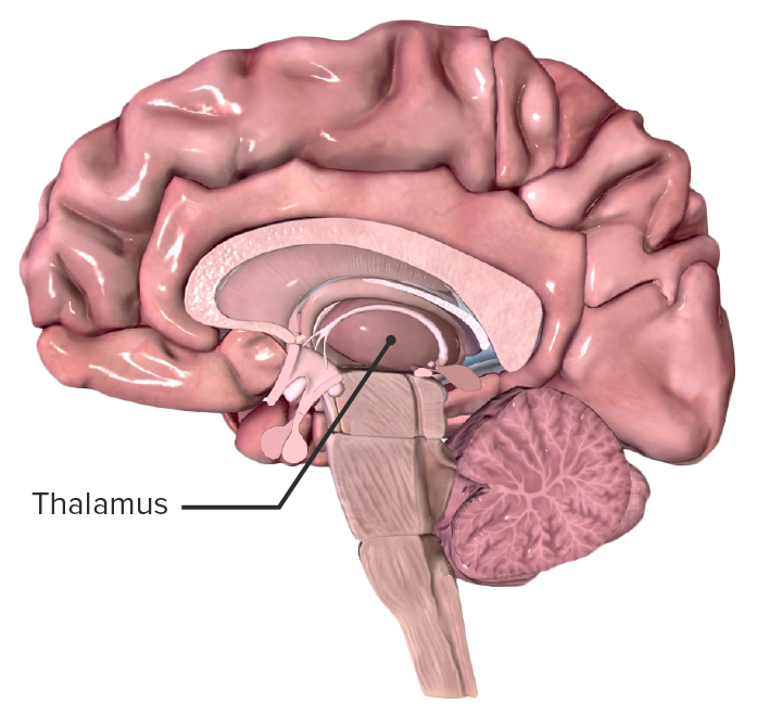
Location of the thalamus in a midsagittal section of the human brain

Location of the thalamus in a midsagittal section of the human brain

Image by BioDigital, edited by Lecturio
Microscopic structure

  • Thalamic nuclei consist of cell bodies of excitatory and inhibitory neurons Neurons The basic cellular units of nervous tissue. Each neuron consists of a body, an axon, and dendrites. Their purpose is to receive, conduct, and transmit impulses in the nervous system. Nervous System: Histology.
  • Dendrites Dendrites Extensions of the nerve cell body. They are short and branched and receive stimuli from other neurons. Nervous System: Histology receive sensory Sensory Neurons which conduct nerve impulses to the central nervous system. Nervous System: Histology information from all over the body.
  • Sensory Sensory Neurons which conduct nerve impulses to the central nervous system. Nervous System: Histology information is “filtered” and presented to the cortex via axons Axons Nerve fibers that are capable of rapidly conducting impulses away from the neuron cell body. Nervous System: Histology known as the thalamocortical radiations.
  • Thalamic neuronal connections also include:
    • Hippocampus
    • Mammillary bodies Mammillary bodies A pair of nuclei and associated gray matter in the interpeduncular space rostral to the posterior perforated substance in the posterior hypothalamus. Limbic System: Anatomy
    • Fornix Fornix Vagina, Vulva, and Pelvic Floor: Anatomy
    • Limbic system Limbic system The limbic system is a neuronal network that mediates emotion and motivation, while also playing a role in learning and memory. The extended neural network is vital to numerous basic psychological functions and plays an invaluable role in processing and responding to environmental stimuli. Limbic System: Anatomy

Functions

  • Regulation and maintenance of consciousness (alertness/wakefulness) and sleep Sleep A readily reversible suspension of sensorimotor interaction with the environment, usually associated with recumbency and immobility. Physiology of Sleep
  • Filtering of sensory Sensory Neurons which conduct nerve impulses to the central nervous system. Nervous System: Histology information for selective presentation to the cerebral cortex Cerebral cortex The cerebral cortex is the largest and most developed part of the human brain and CNS. Occupying the upper part of the cranial cavity, the cerebral cortex has 4 lobes and is divided into 2 hemispheres that are joined centrally by the corpus callosum. Cerebral Cortex: Anatomy:
    • Olfactory inputs exist but are not filtered or modified.
    • All other sensory Sensory Neurons which conduct nerve impulses to the central nervous system. Nervous System: Histology stimuli undergo processing in the thalamus prior to transmission to the cortical centers.
  • Modulation of motor Motor Neurons which send impulses peripherally to activate muscles or secretory cells. Nervous System: Histology responses via interconnections with the basal ganglia Basal Ganglia Basal ganglia are a group of subcortical nuclear agglomerations involved in movement, and are located deep to the cerebral hemispheres. Basal ganglia include the striatum (caudate nucleus and putamen), globus pallidus, substantia nigra, and subthalamic nucleus. Basal Ganglia: Anatomy

Vasculature

  • Supplied by branches of the posterior cerebral artery Posterior cerebral artery Artery formed by the bifurcation of the basilar artery. Branches of the posterior cerebral artery supply portions of the occipital lobe; parietal lobe; inferior temporal gyrus, brainstem, and choroid plexus. Cerebrovascular System: Anatomy
  • Blood supply to the thalamus is divided into 4 regions:
    • Anterior
    • Inferolateral
    • Paramedian
    • Posterior

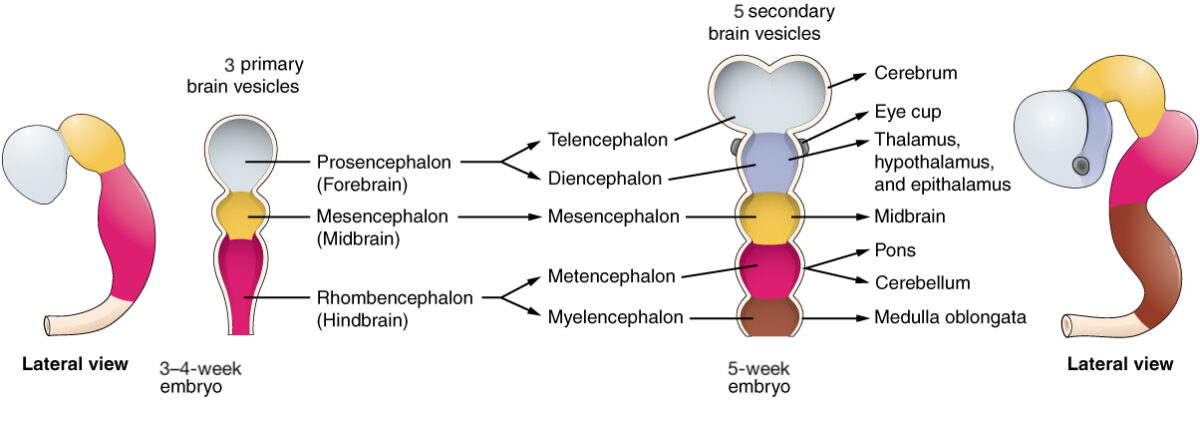
Embryologic development

Development-of-the-thalamus

Development of the thalamus from the prosencephalon, where it forms the diencephalon, comprising the thalamus, hypothalamus, and epithalamus

Image: “Primary and Secondary Vesicle Stages of Development ” by Phil Schatz. License: CC BY 4.0, edited by Lecturio.

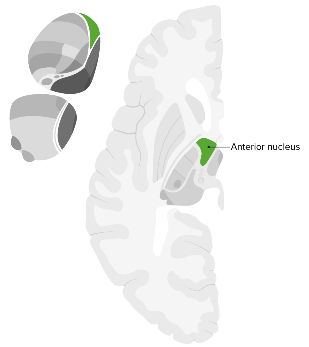
Anterior Nuclei

The anterior nuclei are subdivided into 3 sections with inputs from the mammillary bodies Mammillary bodies A pair of nuclei and associated gray matter in the interpeduncular space rostral to the posterior perforated substance in the posterior hypothalamus. Limbic System: Anatomy and hippocampus and output to the cingulate gyrus Cingulate gyrus One of the convolutions on the medial surface of the cerebral hemispheres. It surrounds the rostral part of the brain and corpus callosum and forms part of the limbic system. Limbic System: Anatomy. The anterior nuclei are associated with learning, memory Memory Complex mental function having four distinct phases: (1) memorizing or learning, (2) retention, (3) recall, and (4) recognition. Clinically, it is usually subdivided into immediate, recent, and remote memory. Psychiatric Assessment, and emotion.
  • Consists of 3 subdivisions:
    • Anteroventral
    • Anteromedial
    • Anterodorsal
  • Inputs:
    • Mammillary body
    • Hippocampal formation Hippocampal formation A curved elevation of gray matter extending the entire length of the floor of the temporal horn of the lateral ventricle. The hippocampus proper, subiculum, and dentate gyrus constitute the hippocampal formation. Sometimes authors include the entorhinal cortex in the hippocampal formation. Limbic System: Anatomy
    • Limbic system Limbic system The limbic system is a neuronal network that mediates emotion and motivation, while also playing a role in learning and memory. The extended neural network is vital to numerous basic psychological functions and plays an invaluable role in processing and responding to environmental stimuli. Limbic System: Anatomy
  • Output: cingulate gyrus Cingulate gyrus One of the convolutions on the medial surface of the cerebral hemispheres. It surrounds the rostral part of the brain and corpus callosum and forms part of the limbic system. Limbic System: Anatomy in the cerebral cortex Cerebral cortex The cerebral cortex is the largest and most developed part of the human brain and CNS. Occupying the upper part of the cranial cavity, the cerebral cortex has 4 lobes and is divided into 2 hemispheres that are joined centrally by the corpus callosum. Cerebral Cortex: Anatomy
  • Functions:
    • Learning
    • Memory Memory Complex mental function having four distinct phases: (1) memorizing or learning, (2) retention, (3) recall, and (4) recognition. Clinically, it is usually subdivided into immediate, recent, and remote memory. Psychiatric Assessment
    • Emotions
Anterior nucleus of thalamus

Location of the anterior nucleus within the thalamus: Note its central and anterior location within this hemisection of the brain.

Image by Lecturio.

Ventral Posteromedial/Posterolateral Nuclei

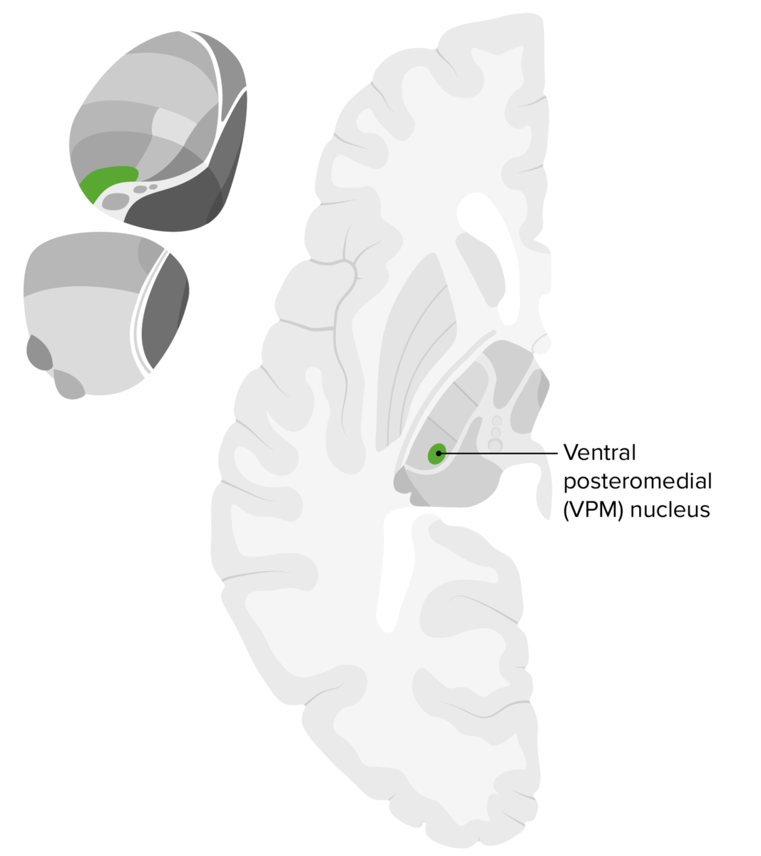
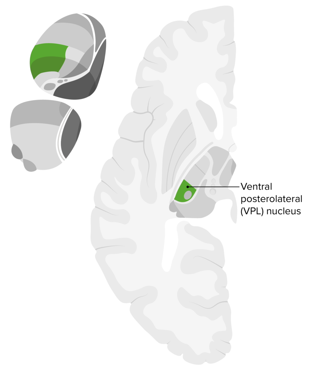
The ventral posteromedial and ventral posterolateral nuclei are associated with inputs from the rostral solitary nucleus Nucleus Within a eukaryotic cell, a membrane-limited body which contains chromosomes and one or more nucleoli (cell nucleolus). The nuclear membrane consists of a double unit-type membrane which is perforated by a number of pores; the outermost membrane is continuous with the endoplasmic reticulum. A cell may contain more than one nucleus. The Cell: Organelles and trigeminothalamic tract and outputs to the primary somatosensory cortex Somatosensory cortex Area of the parietal lobe concerned with receiving sensations such as movement, pain, pressure, position, temperature, touch, and vibration. It lies posterior to the central sulcus. Cerebral Cortex: Anatomy and frontal Frontal The bone that forms the frontal aspect of the skull. Its flat part forms the forehead, articulating inferiorly with the nasal bone and the cheek bone on each side of the face. Skull: Anatomy operculum Operculum Diphyllobothrium/Diphyllobothriasis/insula.

  • Ventral posteromedial nucleus Nucleus Within a eukaryotic cell, a membrane-limited body which contains chromosomes and one or more nucleoli (cell nucleolus). The nuclear membrane consists of a double unit-type membrane which is perforated by a number of pores; the outermost membrane is continuous with the endoplasmic reticulum. A cell may contain more than one nucleus. The Cell: Organelles:
    • Receives and distributes information regarding touch, position, pain Pain An unpleasant sensation induced by noxious stimuli which are detected by nerve endings of nociceptive neurons. Pain: Types and Pathways, and temperature from the face as well as gustatory information
    • Inputs:
      • Trigeminothalamic tract: sensory Sensory Neurons which conduct nerve impulses to the central nervous system. Nervous System: Histology information from the face and oral cavity
      •  Rostral solitary nucleus Nucleus Within a eukaryotic cell, a membrane-limited body which contains chromosomes and one or more nucleoli (cell nucleolus). The nuclear membrane consists of a double unit-type membrane which is perforated by a number of pores; the outermost membrane is continuous with the endoplasmic reticulum. A cell may contain more than one nucleus. The Cell: Organelles: gustatory information
    • Outputs:
      • Primary somatosensory cortex Somatosensory cortex Area of the parietal lobe concerned with receiving sensations such as movement, pain, pressure, position, temperature, touch, and vibration. It lies posterior to the central sulcus. Cerebral Cortex: Anatomy
      • Gustatory cortex ( frontal Frontal The bone that forms the frontal aspect of the skull. Its flat part forms the forehead, articulating inferiorly with the nasal bone and the cheek bone on each side of the face. Skull: Anatomy operculum Operculum Diphyllobothrium/Diphyllobothriasis and insula)
    • Functions:
      • Relays somatosensory cranial nerve inputs (touch, position, pain Pain An unpleasant sensation induced by noxious stimuli which are detected by nerve endings of nociceptive neurons. Pain: Types and Pathways, and temperature from the face)
      • Relays gustation to the cortex
  • Ventral posterolateral nucleus Nucleus Within a eukaryotic cell, a membrane-limited body which contains chromosomes and one or more nucleoli (cell nucleolus). The nuclear membrane consists of a double unit-type membrane which is perforated by a number of pores; the outermost membrane is continuous with the endoplasmic reticulum. A cell may contain more than one nucleus. The Cell: Organelles:
    • Receives and distributes information regarding touch, position, pain Pain An unpleasant sensation induced by noxious stimuli which are detected by nerve endings of nociceptive neurons. Pain: Types and Pathways, and temperature from the body and relays it to the somatosensory cortex Somatosensory cortex Area of the parietal lobe concerned with receiving sensations such as movement, pain, pressure, position, temperature, touch, and vibration. It lies posterior to the central sulcus. Cerebral Cortex: Anatomy
    • Includes sensory Sensory Neurons which conduct nerve impulses to the central nervous system. Nervous System: Histology inputs from the body and limbs:
      • Dorsal column Dorsal column Spinal Cord: Anatomy medial lemniscus (pressure, vibration Vibration A continuing periodic change in displacement with respect to a fixed reference. Neurological Examination, fine touch, and proprioception Proprioception Sensory functions that transduce stimuli received by proprioceptive receptors in joints, tendons, muscles, and the inner ear into neural impulses to be transmitted to the central nervous system. Proprioception provides sense of stationary positions and movements of one’s body parts, and is important in maintaining kinesthesia and postural balance. Neurological Examination)
      • Spinothalamic tract:
        • Lateral: pain Pain An unpleasant sensation induced by noxious stimuli which are detected by nerve endings of nociceptive neurons. Pain: Types and Pathways and temperature
        • Anterior: crude touch and pressure
    • Outputs: somatosensory cortex Somatosensory cortex Area of the parietal lobe concerned with receiving sensations such as movement, pain, pressure, position, temperature, touch, and vibration. It lies posterior to the central sulcus. Cerebral Cortex: Anatomy ( perception Perception The process by which the nature and meaning of sensory stimuli are recognized and interpreted. Psychiatric Assessment of sensations, touch, vibration Vibration A continuing periodic change in displacement with respect to a fixed reference. Neurological Examination, pain Pain An unpleasant sensation induced by noxious stimuli which are detected by nerve endings of nociceptive neurons. Pain: Types and Pathways, and temperature)
    • Functions: relays somatosensory spinal inputs to the somatosensory cortex Somatosensory cortex Area of the parietal lobe concerned with receiving sensations such as movement, pain, pressure, position, temperature, touch, and vibration. It lies posterior to the central sulcus. Cerebral Cortex: Anatomy

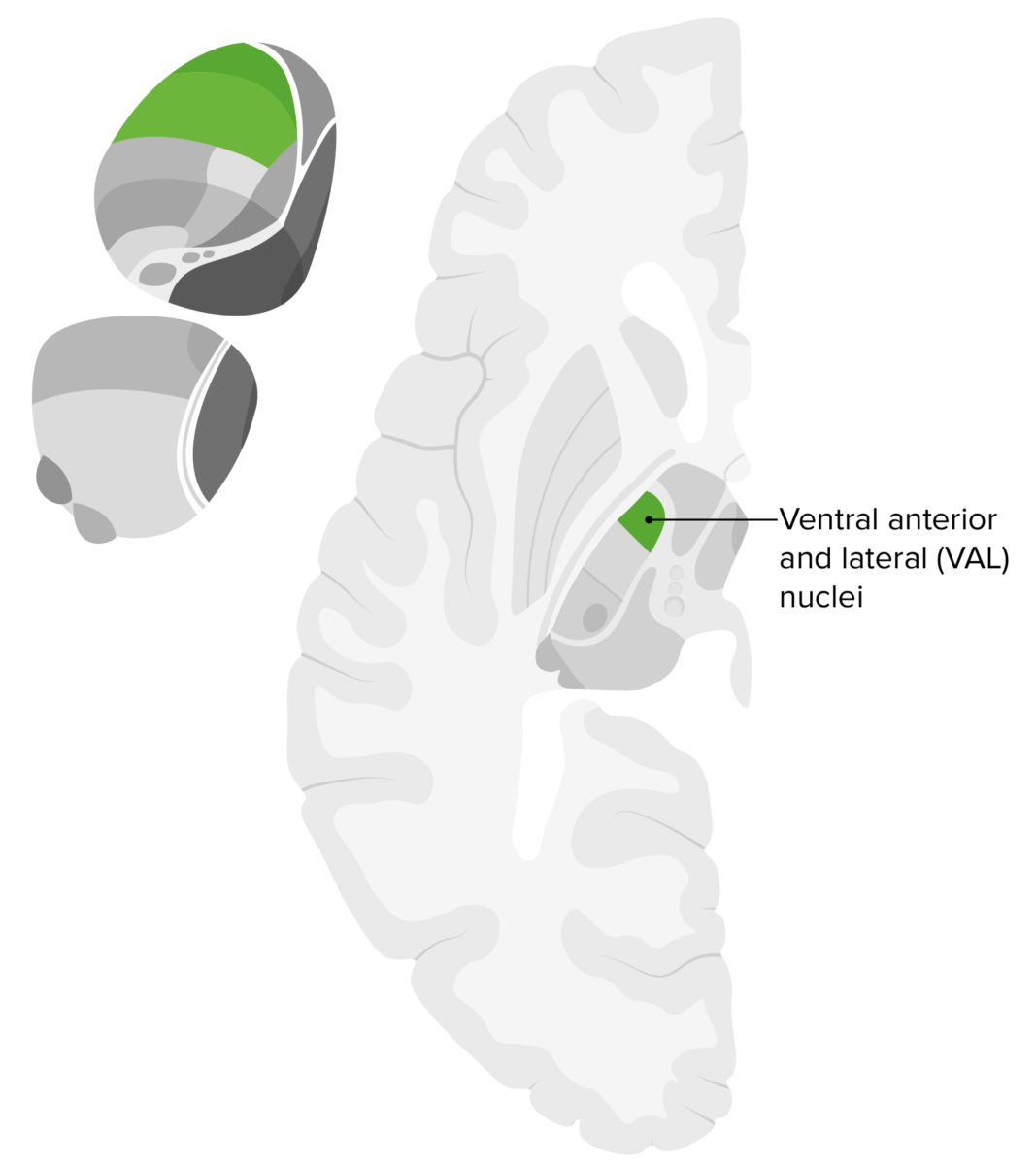
Ventral Anterior and Ventral Lateral Nuclei

The ventral anterior and ventral lateral nuclei are associated with inputs from the basal ganglia Basal Ganglia Basal ganglia are a group of subcortical nuclear agglomerations involved in movement, and are located deep to the cerebral hemispheres. Basal ganglia include the striatum (caudate nucleus and putamen), globus pallidus, substantia nigra, and subthalamic nucleus. Basal Ganglia: Anatomy and cerebellum Cerebellum The cerebellum, Latin for “little brain,” is located in the posterior cranial fossa, dorsal to the pons and midbrain, and its principal role is in the coordination of movements. The cerebellum consists of 3 lobes on either side of its 2 hemispheres and is connected in the middle by the vermis. Cerebellum: Anatomy and output to the frontal lobe Frontal lobe The part of the cerebral hemisphere anterior to the central sulcus, and anterior and superior to the lateral sulcus. Cerebral Cortex: Anatomy. Both nuclei are also associated with various motor pathways Motor pathways Spinal Cord: Anatomy.
  • Ventral anterior nucleus Nucleus Within a eukaryotic cell, a membrane-limited body which contains chromosomes and one or more nucleoli (cell nucleolus). The nuclear membrane consists of a double unit-type membrane which is perforated by a number of pores; the outermost membrane is continuous with the endoplasmic reticulum. A cell may contain more than one nucleus. The Cell: Organelles:
    • Input: basal ganglia Basal Ganglia Basal ganglia are a group of subcortical nuclear agglomerations involved in movement, and are located deep to the cerebral hemispheres. Basal ganglia include the striatum (caudate nucleus and putamen), globus pallidus, substantia nigra, and subthalamic nucleus. Basal Ganglia: Anatomy
    • Outputs: frontal lobe Frontal lobe The part of the cerebral hemisphere anterior to the central sulcus, and anterior and superior to the lateral sulcus. Cerebral Cortex: Anatomy to the motor Motor Neurons which send impulses peripherally to activate muscles or secretory cells. Nervous System: Histology, premotor, and supplementary motor Motor Neurons which send impulses peripherally to activate muscles or secretory cells. Nervous System: Histology cortices
    • Function: relays basal ganglia Basal Ganglia Basal ganglia are a group of subcortical nuclear agglomerations involved in movement, and are located deep to the cerebral hemispheres. Basal ganglia include the striatum (caudate nucleus and putamen), globus pallidus, substantia nigra, and subthalamic nucleus. Basal Ganglia: Anatomy inputs to motor Motor Neurons which send impulses peripherally to activate muscles or secretory cells. Nervous System: Histology cortices for motor Motor Neurons which send impulses peripherally to activate muscles or secretory cells. Nervous System: Histology planning
  • Ventral lateral nucleus Nucleus Within a eukaryotic cell, a membrane-limited body which contains chromosomes and one or more nucleoli (cell nucleolus). The nuclear membrane consists of a double unit-type membrane which is perforated by a number of pores; the outermost membrane is continuous with the endoplasmic reticulum. A cell may contain more than one nucleus. The Cell: Organelles:
    • Input: cerebellum Cerebellum The cerebellum, Latin for “little brain,” is located in the posterior cranial fossa, dorsal to the pons and midbrain, and its principal role is in the coordination of movements. The cerebellum consists of 3 lobes on either side of its 2 hemispheres and is connected in the middle by the vermis. Cerebellum: Anatomy
    • Outputs: frontal lobe Frontal lobe The part of the cerebral hemisphere anterior to the central sulcus, and anterior and superior to the lateral sulcus. Cerebral Cortex: Anatomy to the motor Motor Neurons which send impulses peripherally to activate muscles or secretory cells. Nervous System: Histology, premotor, and supplementary motor Motor Neurons which send impulses peripherally to activate muscles or secretory cells. Nervous System: Histology cortices
    • Function: relays cerebellar inputs to motor Motor Neurons which send impulses peripherally to activate muscles or secretory cells. Nervous System: Histology cortices for motor Motor Neurons which send impulses peripherally to activate muscles or secretory cells. Nervous System: Histology planning and control
Val nuclei of thalamus

Location of the ventral anterior and ventral lateral nuclei

Image by Lecturio.

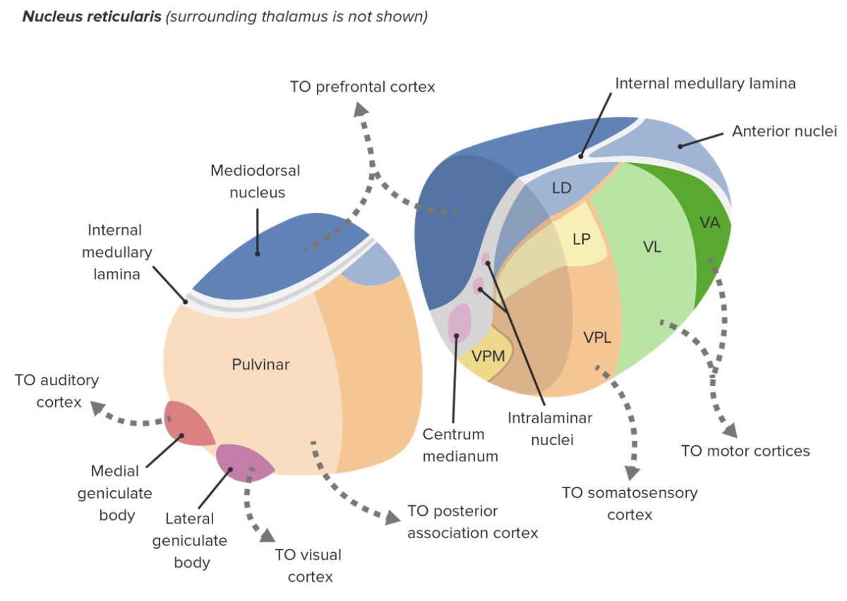
Dorsomedial Nuclei

The dorsomedial nuclei are associated with inputs from the amygdala Amygdala Almond-shaped group of basal nuclei anterior to the inferior horn of the lateral ventricle of the temporal lobe. The amygdala is part of the limbic system. Limbic System: Anatomy, olfactory centers, and basal ganglia Basal Ganglia Basal ganglia are a group of subcortical nuclear agglomerations involved in movement, and are located deep to the cerebral hemispheres. Basal ganglia include the striatum (caudate nucleus and putamen), globus pallidus, substantia nigra, and subthalamic nucleus. Basal Ganglia: Anatomy and output to the frontal Frontal The bone that forms the frontal aspect of the skull. Its flat part forms the forehead, articulating inferiorly with the nasal bone and the cheek bone on each side of the face. Skull: Anatomy cortex. The dorsomedial nuclei are associated with various limbic pathways.
  • Inputs:
    • Amygdala Amygdala Almond-shaped group of basal nuclei anterior to the inferior horn of the lateral ventricle of the temporal lobe. The amygdala is part of the limbic system. Limbic System: Anatomy
    • Olfactory
    • Limbic basal ganglia Basal Ganglia Basal ganglia are a group of subcortical nuclear agglomerations involved in movement, and are located deep to the cerebral hemispheres. Basal ganglia include the striatum (caudate nucleus and putamen), globus pallidus, substantia nigra, and subthalamic nucleus. Basal Ganglia: Anatomy
  • Outputs:
    • Frontal Frontal The bone that forms the frontal aspect of the skull. Its flat part forms the forehead, articulating inferiorly with the nasal bone and the cheek bone on each side of the face. Skull: Anatomy cortex
    • Prefrontal association areas
  • Functions:
    • Emotions
    • Cognition
    • Learning
Nuclei that make up the thalamus and their respective projections

Nuclei that make up the thalamus and their respective projections: Note the dorsomedial (mediodorsal) nucleus located medially on the dorsal aspect of the thalamus with projections to the prefrontal cortex.
VA: ventral anterior
VL: ventral lateral
VPL: ventral posterolateral
VPM: ventral posteromedial
LD: lateral dorsal
LP: lateral posterior

Image by Lecturio.

Pulvinar

The pulvinar is associated with inputs from the superior colliculus Superior Colliculus The anterior pair of the quadrigeminal bodies which coordinate the general behavioral orienting responses to visual stimuli, such as whole-body turning, and reaching. Cranial Nerve Palsies, visual areas, auditory complex, and other sensory pathways Sensory pathways Spinal Cord: Anatomy, and outputs to the parietotemporal association areas. The pulvinar is associated with various visual and sensory pathways Sensory pathways Spinal Cord: Anatomy.
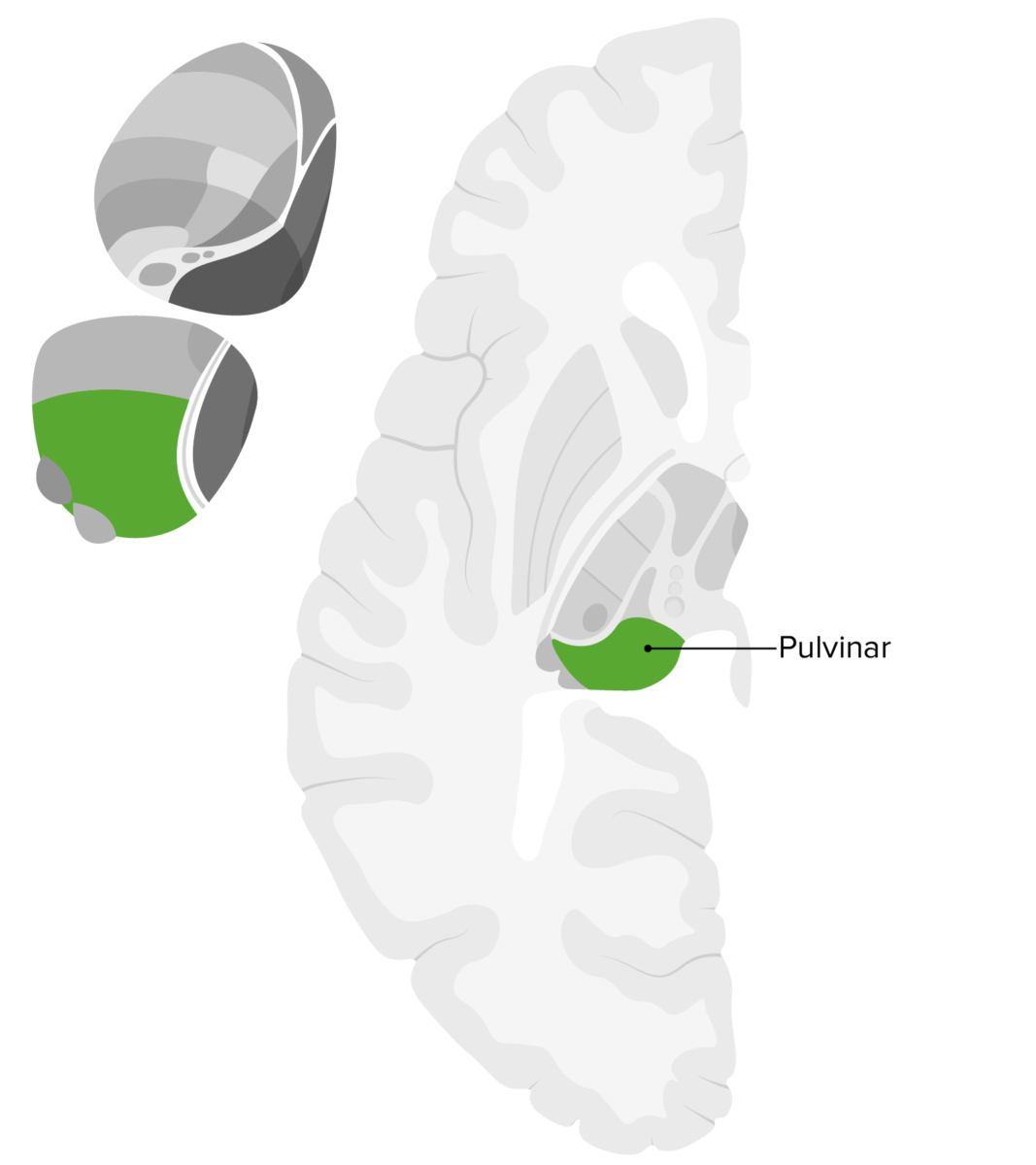
  • Consists of 3 primary subdivisions:
    • Medial
    • Lateral
    • Inferior
  • Inputs:
  • Output: parietotemporal-occipital association area
  • Functions:
Pulvinar nucleus of thalamus

Location of the pulvinar, which is the posterior expansion of the thalamus overhanging the superior colliculus

Image by Lecturio.

Medial and Lateral Geniculate Bodies

The medial and lateral geniculate bodies are associated with inputs from the inferior colliculus Inferior colliculus The posterior pair of the quadrigeminal bodies which contain centers for auditory function. Brain Stem: Anatomy and retina Retina The ten-layered nervous tissue membrane of the eye. It is continuous with the optic nerve and receives images of external objects and transmits visual impulses to the brain. Its outer surface is in contact with the choroid and the inner surface with the vitreous body. The outermost layer is pigmented, whereas the inner nine layers are transparent. Eye: Anatomy and outputs to the temporal lobe Temporal lobe Lower lateral part of the cerebral hemisphere responsible for auditory, olfactory, and semantic processing. It is located inferior to the lateral fissure and anterior to the occipital lobe. Cerebral Cortex: Anatomy and visual cortex Visual cortex Area of the occipital lobe concerned with the processing of visual information relayed via visual pathways. Cerebral Cortex: Anatomy, and are involved in various auditory and visual pathways.
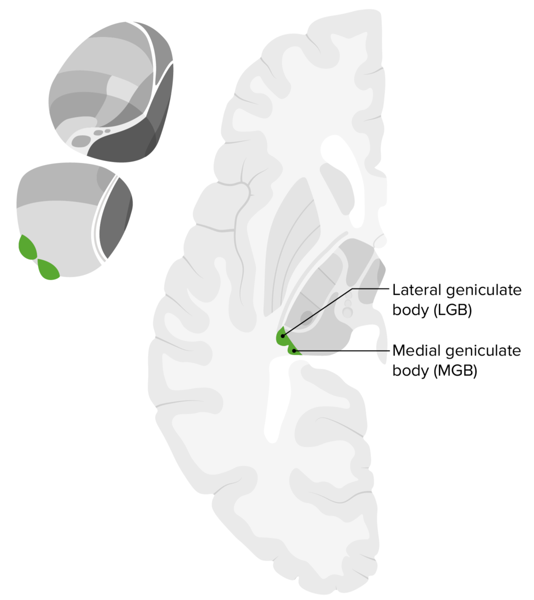
  • Medial geniculate body:
    • Part of the auditory pathway
    • Input: inferior colliculus Inferior colliculus The posterior pair of the quadrigeminal bodies which contain centers for auditory function. Brain Stem: Anatomy
    • Output: auditory cortex Auditory cortex The region of the cerebral cortex that receives the auditory radiation from the medial geniculate body. Auditory and Vestibular Pathways: Anatomy of the temporal lobe Temporal lobe Lower lateral part of the cerebral hemisphere responsible for auditory, olfactory, and semantic processing. It is located inferior to the lateral fissure and anterior to the occipital lobe. Cerebral Cortex: Anatomy
    • Function: relays auditory input to the cortex
  • Lateral geniculate body Lateral Geniculate Body The Visual Pathway and Related Disorders:
    • Main relay station of the visual pathway Visual pathway The primary visual pathway consists of a relay system, beginning at the retina, whose ganglion cell axons form the optic nerve. The optic nerve fibers from each eye hemidecussate in the optic chiasm (OC), with nasal fibers joining the temporal fibers of the contralateral nerve. Signals are then transmitted to the primary visual cortex of the occipital lobe. The Visual Pathway and Related Disorders
    • Organized in 6 layers:
      • Layers 1 and 2 consist of large neurons Neurons The basic cellular units of nervous tissue. Each neuron consists of a body, an axon, and dendrites. Their purpose is to receive, conduct, and transmit impulses in the nervous system. Nervous System: Histology.
      • Layers 3–6 consist of smaller neurons Neurons The basic cellular units of nervous tissue. Each neuron consists of a body, an axon, and dendrites. Their purpose is to receive, conduct, and transmit impulses in the nervous system. Nervous System: Histology.
    • Inputs:
      • Retina Retina The ten-layered nervous tissue membrane of the eye. It is continuous with the optic nerve and receives images of external objects and transmits visual impulses to the brain. Its outer surface is in contact with the choroid and the inner surface with the vitreous body. The outermost layer is pigmented, whereas the inner nine layers are transparent. Eye: Anatomy/ optic nerve Optic nerve The 2nd cranial nerve which conveys visual information from the retina to the brain. The nerve carries the axons of the retinal ganglion cells which sort at the optic chiasm and continue via the optic tracts to the brain. The largest projection is to the lateral geniculate nuclei; other targets include the superior colliculi and the suprachiasmatic nuclei. Though known as the second cranial nerve, it is considered part of the central nervous system. The 12 Cranial Nerves: Overview and Functions
      • Contralateral nasal hemiretina projects to layers 1, 4, and 6.
      • Ipsilateral temporal hemiretina projects to layers 2, 3, and 5.
    • Outputs:
    • Function: relays visual inputs to the primary visual cortex Primary Visual Cortex The Visual Pathway and Related Disorders
Mgb and lgb of thalamus

Location of the LGB and MGB of the thalamus

Image by Lecturio.

Intralaminar/Midline Nuclei

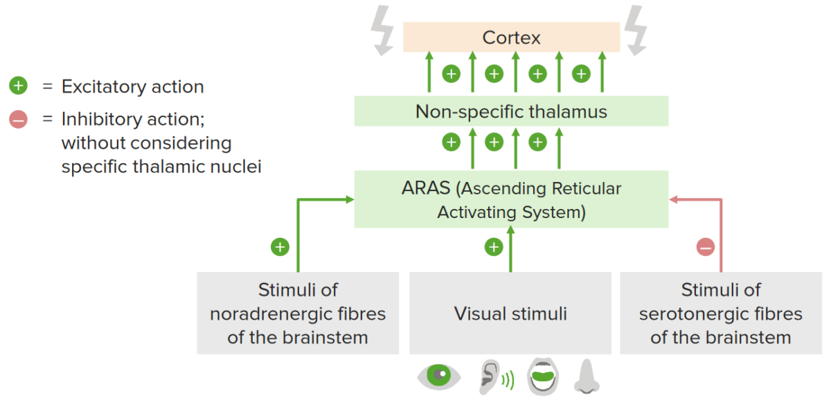
Intralaminar and midline nuclei are associated with inputs from the spinal cord Spinal cord The spinal cord is the major conduction pathway connecting the brain to the body; it is part of the CNS. In cross section, the spinal cord is divided into an H-shaped area of gray matter (consisting of synapsing neuronal cell bodies) and a surrounding area of white matter (consisting of ascending and descending tracts of myelinated axons). Spinal Cord: Anatomy, hypothalamus Hypothalamus The hypothalamus is a collection of various nuclei within the diencephalon in the center of the brain. The hypothalamus plays a vital role in endocrine regulation as the primary regulator of the pituitary gland, and it is the major point of integration between the central nervous and endocrine systems. Hypothalamus, and descending reticular formation Reticular Formation A region extending from the pons and medulla oblongata through the mesencephalon, characterized by a diversity of neurons of various sizes and shapes, arranged in different aggregations and enmeshed in a complicated fiber network. Brain Stem: Anatomy, and outputs to the basal ganglia Basal Ganglia Basal ganglia are a group of subcortical nuclear agglomerations involved in movement, and are located deep to the cerebral hemispheres. Basal ganglia include the striatum (caudate nucleus and putamen), globus pallidus, substantia nigra, and subthalamic nucleus. Basal Ganglia: Anatomy and limbic system Limbic system The limbic system is a neuronal network that mediates emotion and motivation, while also playing a role in learning and memory. The extended neural network is vital to numerous basic psychological functions and plays an invaluable role in processing and responding to environmental stimuli. Limbic System: Anatomy. Both nuclei are associated with various emotional and sensory pathways Sensory pathways Spinal Cord: Anatomy.
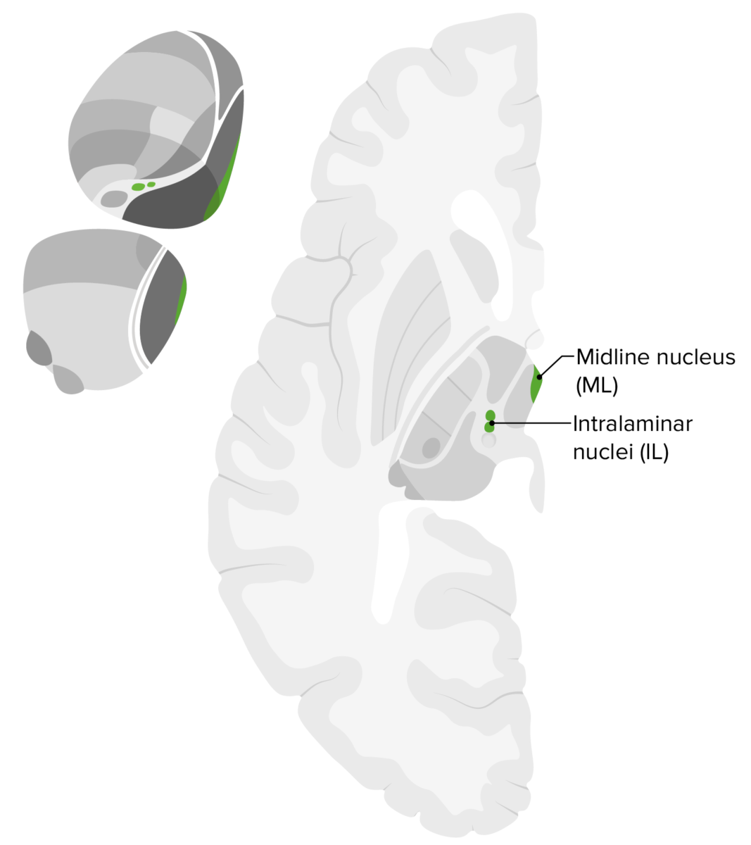
  • Involved in arousal and part of the reticular activating system
  • Inputs:
    • Ascending reticular formation Reticular Formation A region extending from the pons and medulla oblongata through the mesencephalon, characterized by a diversity of neurons of various sizes and shapes, arranged in different aggregations and enmeshed in a complicated fiber network. Brain Stem: Anatomy
    • Spinal cord Spinal cord The spinal cord is the major conduction pathway connecting the brain to the body; it is part of the CNS. In cross section, the spinal cord is divided into an H-shaped area of gray matter (consisting of synapsing neuronal cell bodies) and a surrounding area of white matter (consisting of ascending and descending tracts of myelinated axons). Spinal Cord: Anatomy
    • Hypothalamus Hypothalamus The hypothalamus is a collection of various nuclei within the diencephalon in the center of the brain. The hypothalamus plays a vital role in endocrine regulation as the primary regulator of the pituitary gland, and it is the major point of integration between the central nervous and endocrine systems. Hypothalamus
  • Outputs:
    • Limbic cortex
    • Basal ganglia Basal Ganglia Basal ganglia are a group of subcortical nuclear agglomerations involved in movement, and are located deep to the cerebral hemispheres. Basal ganglia include the striatum (caudate nucleus and putamen), globus pallidus, substantia nigra, and subthalamic nucleus. Basal Ganglia: Anatomy
  • Functions:
    • Arousal
    • Motivation
    • Affect
    • Pain Pain An unpleasant sensation induced by noxious stimuli which are detected by nerve endings of nociceptive neurons. Pain: Types and Pathways

Clinical Relevance

  • Auditory pathways: The auditory system consists of a series of interconnected parts from the external ear itself, to the middle and inner ear Inner ear The essential part of the hearing organ consists of two labyrinthine compartments: the bony labyrinthine and the membranous labyrinth. Ear: Anatomy, and to the cortical regions that process auditory stimuli.
  • Brainstem: consists of the midbrain Midbrain The middle of the three primitive cerebral vesicles of the embryonic brain. Without further subdivision, midbrain develops into a short, constricted portion connecting the pons and the diencephalon. Midbrain contains two major parts, the dorsal tectum mesencephali and the ventral tegmentum mesencephali, housing components of auditory, visual, and other sensorimotor systems. Brain Stem: Anatomy, pons Pons The front part of the hindbrain (rhombencephalon) that lies between the medulla and the midbrain (mesencephalon) ventral to the cerebellum. It is composed of two parts, the dorsal and the ventral. The pons serves as a relay station for neural pathways between the cerebellum to the cerebrum. Brain Stem: Anatomy, and medulla oblongata Medulla Oblongata The lower portion of the brain stem. It is inferior to the pons and anterior to the cerebellum. Medulla oblongata serves as a relay station between the brain and the spinal cord, and contains centers for regulating respiratory, vasomotor, cardiac, and reflex activities. Brain Stem: Anatomy. The brainstem is a stalk-like structure that connects the cerebrum with the spinal cord Spinal cord The spinal cord is the major conduction pathway connecting the brain to the body; it is part of the CNS. In cross section, the spinal cord is divided into an H-shaped area of gray matter (consisting of synapsing neuronal cell bodies) and a surrounding area of white matter (consisting of ascending and descending tracts of myelinated axons). Spinal Cord: Anatomy and is a major relay station for sensory Sensory Neurons which conduct nerve impulses to the central nervous system. Nervous System: Histology, motor Motor Neurons which send impulses peripherally to activate muscles or secretory cells. Nervous System: Histology, and autonomic information.
  • Cerebral cortex Cerebral cortex The cerebral cortex is the largest and most developed part of the human brain and CNS. Occupying the upper part of the cranial cavity, the cerebral cortex has 4 lobes and is divided into 2 hemispheres that are joined centrally by the corpus callosum. Cerebral Cortex: Anatomy: a layer of gray matter Gray matter Region of central nervous system that appears darker in color than the other type, white matter. It is composed of neuronal cell bodies; neuropil; glial cells and capillaries but few myelinated nerve fibers. Cerebral Cortex: Anatomy covering the surface of each cerebral hemisphere, folded into gyri that are separated by sulci. The cerebral cortex Cerebral cortex The cerebral cortex is the largest and most developed part of the human brain and CNS. Occupying the upper part of the cranial cavity, the cerebral cortex has 4 lobes and is divided into 2 hemispheres that are joined centrally by the corpus callosum. Cerebral Cortex: Anatomy functions in higher-order mental functions.
  • Visual system: consists of a series of interdependent parts from the organization of the eye and retina Retina The ten-layered nervous tissue membrane of the eye. It is continuous with the optic nerve and receives images of external objects and transmits visual impulses to the brain. Its outer surface is in contact with the choroid and the inner surface with the vitreous body. The outermost layer is pigmented, whereas the inner nine layers are transparent. Eye: Anatomy to the cortical regions. The visual system is primarily located in the occipital lobe Occipital lobe Posterior portion of the cerebral hemispheres responsible for processing visual sensory information. It is located posterior to the parieto-occipital sulcus and extends to the preoccipital notch. Cerebral Cortex: Anatomy and processes visual stimuli.

References

  1. Sadler, T. (2014). Langman’s Medical Embryology, 13th edition. Lippincott Williams & Wilkins.
  2. Shkoukani, M.A., Chen, M., Vong, A. (2013). Cleft Lip – A Comprehensive Review. Front Pediatr. 1, 53. https://doi.org/10.3389/fped.2013.00053
  3. Berkowitz, A.L. (Ed.). (2016). Clinical Neurology and Neuroanatomy: A Localization-Based Approach. McGraw-Hill. 
  4. Kibble, J.D. (Ed.). (2020). Neurophysiology in The Big Picture Physiology: Medical Course & Step 1 Review, 2e. McGraw-Hill.
  5. Martin, J. (2021). The visual system in Neuroanatomy: Text and Atlas, 5e. McGraw-Hill.
  6. Purves, D. (2001). Visual Field Defects in Neuroscience (2nd ed.). Sunderland (MA): Sinauer Associates. https://www.ncbi.nlm.nih.gov/books/NBK10912/
  7. Shakkottai, V.G., Lomen-Hoerth, C. (2019). Nervous System Disorders. In Hammer G.D., McPhee S.J. (Eds.), Pathophysiology of Disease: An Introduction to Clinical Medicine, 8e. McGraw-Hill.
  8. Waxman, S. (2020). The Visual System. In Clinical Neuroanatomy, 29e. McGraw-Hill.

Create your free account or log in to continue reading!

Sign up now and get free access to Lecturio with concept pages, medical videos, and questions for your medical education.

User Reviews

Details