Testicles: Anatomy

The testicles, also known as the testes or the male gonads Gonads The gamete-producing glands, ovary or testis. Hormones: Overview and Types, are a pair of egg-shaped glands suspended within the scrotum. The testicles have multiple layers: an outer tunica vaginalis, an intermediate tunica albuginea Tunica albuginea Penis: Anatomy, and an innermost tunica vasculosa. The testicles are composed of testicular lobules Lobules Breasts: Anatomy (contain interstitial tissue) and seminiferous tubules (produce spermatozoa). Blood supply to the testicles is primarily provided by the testicular artery. Venous drainage is through testicular veins Veins Veins are tubular collections of cells, which transport deoxygenated blood and waste from the capillary beds back to the heart. Veins are classified into 3 types: small veins/venules, medium veins, and large veins. Each type contains 3 primary layers: tunica intima, tunica media, and tunica adventitia. Veins: Histology.

Last updated: Nov 19, 2024

Editorial responsibility: Stanley Oiseth, Lindsay Jones, Evelin Maza

Embryology

Male development

  • Male development is driven by the Y chromosome Y chromosome The male sex chromosome, being the differential sex chromosome carried by half the male gametes and none of the female gametes in humans and in some other male-heterogametic species in which the homologue of the X chromosome has been retained. Basic Terms of Genetics:
    • The Y chromosome Y chromosome The male sex chromosome, being the differential sex chromosome carried by half the male gametes and none of the female gametes in humans and in some other male-heterogametic species in which the homologue of the X chromosome has been retained. Basic Terms of Genetics contains the testis-determining gene Gene A category of nucleic acid sequences that function as units of heredity and which code for the basic instructions for the development, reproduction, and maintenance of organisms. Basic Terms of Genetics SRY (sex-determining region on the Y chromosome Y chromosome The male sex chromosome, being the differential sex chromosome carried by half the male gametes and none of the female gametes in humans and in some other male-heterogametic species in which the homologue of the X chromosome has been retained. Basic Terms of Genetics).
    • Development of male genitalia occurs in the presence of the SRY gene Gene A category of nucleic acid sequences that function as units of heredity and which code for the basic instructions for the development, reproduction, and maintenance of organisms. Basic Terms of Genetics.
  • Gonads Gonads The gamete-producing glands, ovary or testis. Hormones: Overview and Types appear as a pair of longitudinal ridges (gonadal ridges).
  • Gonadal ridge Gonadal ridge Development of the Urogenital System:
    • Formed by a proliferation of the epithelium Epithelium The epithelium is a complex of specialized cellular organizations arranged into sheets and lining cavities and covering the surfaces of the body. The cells exhibit polarity, having an apical and a basal pole. Structures important for the epithelial integrity and function involve the basement membrane, the semipermeable sheet on which the cells rest, and interdigitations, as well as cellular junctions. Surface Epithelium: Histology and a condensation of mesenchyme
    • Epithelial cells penetrate the mesenchyme and form primitive sex Sex The totality of characteristics of reproductive structure, functions, phenotype, and genotype, differentiating the male from the female organism. Gender Dysphoria cords (at the stage of indifferent gonads Gonads The gamete-producing glands, ovary or testis. Hormones: Overview and Types).
    • Germ cells Germ Cells The reproductive cells in multicellular organisms at various stages during gametogenesis. Gametogenesis appear after the 6th week of gestation.
  • Primordial germ cells Primordial germ cells Gametogenesis originate in the epiblast Epiblast Embryoblast and Trophoblast Development and migrate:
    • By the 3rd week of gestation, the primordial germ cells Primordial germ cells Gametogenesis reside in the yolk sac Yolk Sac The first of four extra-embryonic membranes to form during embryogenesis. In reptiles and birds, it arises from endoderm and mesoderm to incorporate the egg yolk into the digestive tract for nourishing the embryo. In placental mammals, its nutritional function is vestigial; however, it is the source of intestinal mucosa; blood cells; and germ cells. It is sometimes called the vitelline sac, which should not be confused with the vitelline membrane of the egg. Embryoblast and Trophoblast Development close to the allantois Allantois An extra-embryonic membranous sac derived from the yolk sac of reptiles; birds; and mammals. It lies between two other extra-embryonic membranes, the amnion and the chorion. The allantois serves to store urinary wastes and mediate exchange of gas and nutrients for the developing embryo. Development of the Abdominal Organs.
    • By the 4th week of gestation, the primordial germ cells Primordial germ cells Gametogenesis reside along the dorsal mesentery Mesentery A layer of the peritoneum which attaches the abdominal viscera to the abdominal wall and conveys their blood vessels and nerves. Peritoneum: Anatomy of the hindgut Hindgut Development of the Abdominal Organs.
    • By the 7th week of gestation, the primordial germ cells Primordial germ cells Gametogenesis invade the gonadal ridges.

Testicular development

  • Under the influence of the SRY gene Gene A category of nucleic acid sequences that function as units of heredity and which code for the basic instructions for the development, reproduction, and maintenance of organisms. Basic Terms of Genetics
  • Mesonephric (wolffian) duct:
    • Present in males → development of testes Testes Gonadal Hormones 
    • Degenerates without the presence of the SRY gene Gene A category of nucleic acid sequences that function as units of heredity and which code for the basic instructions for the development, reproduction, and maintenance of organisms. Basic Terms of Genetics
    • Sertoli cells:
      • Secrete müllerian inhibitory factor
      • Blocks formation of female internal genitalia
    • Leydig cells: 
      • Secrete testosterone Testosterone A potent androgenic steroid and major product secreted by the leydig cells of the testis. Its production is stimulated by luteinizing hormone from the pituitary gland. In turn, testosterone exerts feedback control of the pituitary LH and FSH secretion. Depending on the tissues, testosterone can be further converted to dihydrotestosterone or estradiol. Androgens and Antiandrogens
      • Lead development of the wolffian duct Wolffian duct A pair of excretory ducts of the middle kidneys of an embryo, also called mesonephric ducts. In higher vertebrates, wolffian ducts persist in the male forming vas deferens, but atrophy into vestigial structures in the female. Kidneys: Anatomy to male internal genitalia
    • Testosterone Testosterone A potent androgenic steroid and major product secreted by the leydig cells of the testis. Its production is stimulated by luteinizing hormone from the pituitary gland. In turn, testosterone exerts feedback control of the pituitary LH and FSH secretion. Depending on the tissues, testosterone can be further converted to dihydrotestosterone or estradiol. Androgens and Antiandrogens:
      • Converted to dihydrotestosterone Dihydrotestosterone A potent androgenic metabolite of testosterone. It is produced by the action of the enzyme 3-oxo-5-alpha-steroid 4-dehydrogenase. Gonadal Hormones ( DHT DHT A potent androgenic metabolite of testosterone. It is produced by the action of the enzyme 3-oxo-5-alpha-steroid 4-dehydrogenase. Gonadal Hormones) by 5-alpha reductase
      • Important for prostate Prostate The prostate is a gland in the male reproductive system. The gland surrounds the bladder neck and a portion of the urethra. The prostate is an exocrine gland that produces a weakly acidic secretion, which accounts for roughly 20% of the seminal fluid. development 
    • At puberty Puberty Puberty is a complex series of physical, psychosocial, and cognitive transitions usually experienced by adolescents (11-19 years of age). Puberty is marked by a growth in stature and the development of secondary sexual characteristics, achievement of fertility, and changes in most body systems. Puberty:
      • Testis cords acquire a lumen, become seminiferous tubules, and join the rete testis tubules.
      • The wolffian duct Wolffian duct A pair of excretory ducts of the middle kidneys of an embryo, also called mesonephric ducts. In higher vertebrates, wolffian ducts persist in the male forming vas deferens, but atrophy into vestigial structures in the female. Kidneys: Anatomy becomes the vas deferens.

Gross Anatomy

Testes Testes Gonadal Hormones (the male gonads Gonads The gamete-producing glands, ovary or testis. Hormones: Overview and Types) are responsible for the production of sperm and testosterone Testosterone A potent androgenic steroid and major product secreted by the leydig cells of the testis. Its production is stimulated by luteinizing hormone from the pituitary gland. In turn, testosterone exerts feedback control of the pituitary LH and FSH secretion. Depending on the tissues, testosterone can be further converted to dihydrotestosterone or estradiol. Androgens and Antiandrogens:

  • The testes Testes Gonadal Hormones are enclosed by 3 layers:
    • Tunica vaginalis:
      • Outermost layer 
      • Continuation of the peritoneal processus vaginalis
    • Tunica albuginea Tunica albuginea Penis: Anatomy:
      • Intermediate layer
      • Where vessels, nerves, and ducts enter the testicular capsule Capsule An envelope of loose gel surrounding a bacterial cell which is associated with the virulence of pathogenic bacteria. Some capsules have a well-defined border, whereas others form a slime layer that trails off into the medium. Most capsules consist of relatively simple polysaccharides but there are some bacteria whose capsules are made of polypeptides. Bacteroides to begin formation of the testicular lobules Lobules Breasts: Anatomy
    • Tunica vasculosa:
  • Spermatic cord:
    • Testes Testes Gonadal Hormones are suspended by the spermatic cord within the scrotum. 
    • Allows for maintenance of a lower temperature
  • Scrotum:
    • A pouch composed of highly glandular, hair-bearing skin Skin The skin, also referred to as the integumentary system, is the largest organ of the body. The skin is primarily composed of the epidermis (outer layer) and dermis (deep layer). The epidermis is primarily composed of keratinocytes that undergo rapid turnover, while the dermis contains dense layers of connective tissue. Skin: Structure and Functions and multiple layers of fascia Fascia Layers of connective tissue of variable thickness. The superficial fascia is found immediately below the skin; the deep fascia invests muscles, nerves, and other organs. Cellulitis/muscle:
    • Contents:
      • Testes Testes Gonadal Hormones
      • Epididymis
      • Vas deferens (ductus deferens)
      • Cremaster muscle: lifts the scrotum to maintain an ideal temperature
      • Dartos muscle: corrugates skin Skin The skin, also referred to as the integumentary system, is the largest organ of the body. The skin is primarily composed of the epidermis (outer layer) and dermis (deep layer). The epidermis is primarily composed of keratinocytes that undergo rapid turnover, while the dermis contains dense layers of connective tissue. Skin: Structure and Functions to moderate temperature
      • Spermatic, cremasteric, and infundibuliform fascia Fascia Layers of connective tissue of variable thickness. The superficial fascia is found immediately below the skin; the deep fascia invests muscles, nerves, and other organs. Cellulitis (attached to the parietal Parietal One of a pair of irregularly shaped quadrilateral bones situated between the frontal bone and occipital bone, which together form the sides of the cranium. Skull: Anatomy layer of the tunica vaginalis) 
    • Raphe (scrotal midline):
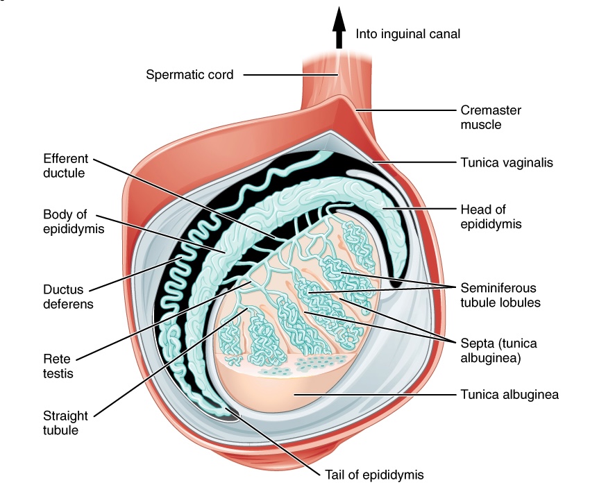
Anatomy of the testis

Various layers and structures comprising the testes:
Note the suspension from the spermatic cord, which permits temperature regulation.
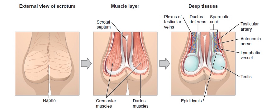
Image: “Anatomy of the testis” by Phil Schatz. License: CC BY 4.0
The scrotum and testes

External scrotal view:
The central raphe on the far left represents the “seam” where the genital tubercles fused in development. In the central image, note the cremaster and dartos muscles, which are important for temperature regulation. On the right, note a deep layer demonstrating the external testes, epididymis, and neurovasculature.

Image: “The Scrotum and Testes” by Phil Schatz. License: CC BY 4.0

Microscopic Anatomy

Blood vessels, lymphatics, and the genital ducts enter the mediastinum Mediastinum The mediastinum is the thoracic area between the 2 pleural cavities. The mediastinum contains vital structures of the circulatory, respiratory, digestive, and nervous systems including the heart and esophagus, and major thoracic vessels. Mediastinum and Great Vessels: Anatomy of the testis and give rise to numerous lobules Lobules Breasts: Anatomy composed of seminiferous tubules and interstitial tissue:

Seminiferous tubules

  • Site of spermatogenesis Spermatogenesis The process of germ cell development in the male from the primordial germ cells, through spermatogonia; spermatocytes; spermatids; to the mature haploid spermatozoa. Gametogenesis 
  • Contains spermatozoa of various stages of development:
    • Spermatogonia = germ cell for all spermatozoa
    • Primary spermatocytes → meiosis I Meiosis I Following DNA replication, meiosis I creates 2 daughter cells containing half the genetic information of the mother cell (1n) but the same number of chromosomes (2c) by segregating sister chromatids into the same daughter cell Meiosis → 2 secondary spermatocytes → meiosis II Meiosis II Meiosis II is a cellular division event wherein the number of chromosomes in the daughter cells is halved from that of the mother cell. Meiosis II: similar to meiosis I but not preceded by interphase (DNA replication) Meiosis → 4 haploid Haploid The chromosomal constitution of cells, in which each type of chromosome is represented once. Symbol: n. Basic Terms of Genetics spermatids
  • Accounts for the majority of testicular volume
  • Lumen lined by Sertoli cells:
    • Vital supporting cells in spermatogenesis Spermatogenesis The process of germ cell development in the male from the primordial germ cells, through spermatogonia; spermatocytes; spermatids; to the mature haploid spermatozoa. Gametogenesis
    • Secrete fluid to facilitate sperm movement
    • Regulate hormones Hormones Hormones are messenger molecules that are synthesized in one part of the body and move through the bloodstream to exert specific regulatory effects on another part of the body. Hormones play critical roles in coordinating cellular activities throughout the body in response to the constant changes in both the internal and external environments. Hormones: Overview and Types in the testicles via secretion Secretion Coagulation Studies of inhibin B and androgen-binding protein in response to gonadotropins

Interstitial tissue

  • Leydig cells: release testosterone Testosterone A potent androgenic steroid and major product secreted by the leydig cells of the testis. Its production is stimulated by luteinizing hormone from the pituitary gland. In turn, testosterone exerts feedback control of the pituitary LH and FSH secretion. Depending on the tissues, testosterone can be further converted to dihydrotestosterone or estradiol. Androgens and Antiandrogens in response to gonadotropins secreted by the pituitary Pituitary A small, unpaired gland situated in the sella turcica. It is connected to the hypothalamus by a short stalk which is called the infundibulum. Hormones: Overview and Types gland
  • Immune cells ( mast cells Mast cells Granulated cells that are found in almost all tissues, most abundantly in the skin and the gastrointestinal tract. Like the basophils, mast cells contain large amounts of histamine and heparin. Unlike basophils, mast cells normally remain in the tissues and do not circulate in the blood. Mast cells, derived from the bone marrow stem cells, are regulated by the stem cell factor. Innate Immunity: Phagocytes and Antigen Presentation and macrophages Macrophages The relatively long-lived phagocytic cell of mammalian tissues that are derived from blood monocytes. Main types are peritoneal macrophages; alveolar macrophages; histiocytes; kupffer cells of the liver; and osteoclasts. They may further differentiate within chronic inflammatory lesions to epithelioid cells or may fuse to form foreign body giant cells or langhans giant cells. Innate Immunity: Phagocytes and Antigen Presentation)
  • Nerves and blood vessels

Epididymis

The epididymis is posterolateral to the testis and consists of 3 sections:
  • Head:
    • Testicular tubules enter the epididymis at the location
    • Contains a high concentration of principal cells Principal cells Tubular System with stereocilia for reabsorption of secretory fluid
  • Body:
    • Contains smooth muscle aiding in the transmission of spermatozoa to the vas deferens during ejaculation
    • Plays a role in reabsorption of secretory fluid
  • Tail:
    • Leads to the vas deferens (ductus deferens)
    • Contains prominent smooth muscle

Vas deferens (ductus deferens)

  • A tubular structure originating at the tail of the epididymis 
  • Transports sperm to the urethra Urethra A tube that transports urine from the urinary bladder to the outside of the body in both the sexes. It also has a reproductive function in the male by providing a passage for sperm. Urinary Tract: Anatomy

Spermatic cord

  • Functions to suspend the testis
  • Terminates in the mediastinum Mediastinum The mediastinum is the thoracic area between the 2 pleural cavities. The mediastinum contains vital structures of the circulatory, respiratory, digestive, and nervous systems including the heart and esophagus, and major thoracic vessels. Mediastinum and Great Vessels: Anatomy of the testis
  • Arises at the deep inguinal ring Deep inguinal ring Inguinal Canal: Anatomy and Hernias, traverses the inguinal canal Inguinal canal The tunnel in the lower anterior abdominal wall through which the spermatic cord, in the male; round ligament, in the female; nerves; and vessels pass. Its internal end is at the deep inguinal ring and its external end is at the superficial inguinal ring. Inguinal Canal: Anatomy and Hernias, and exits at the external inguinal ring

Neurovasculature

Arterial supply

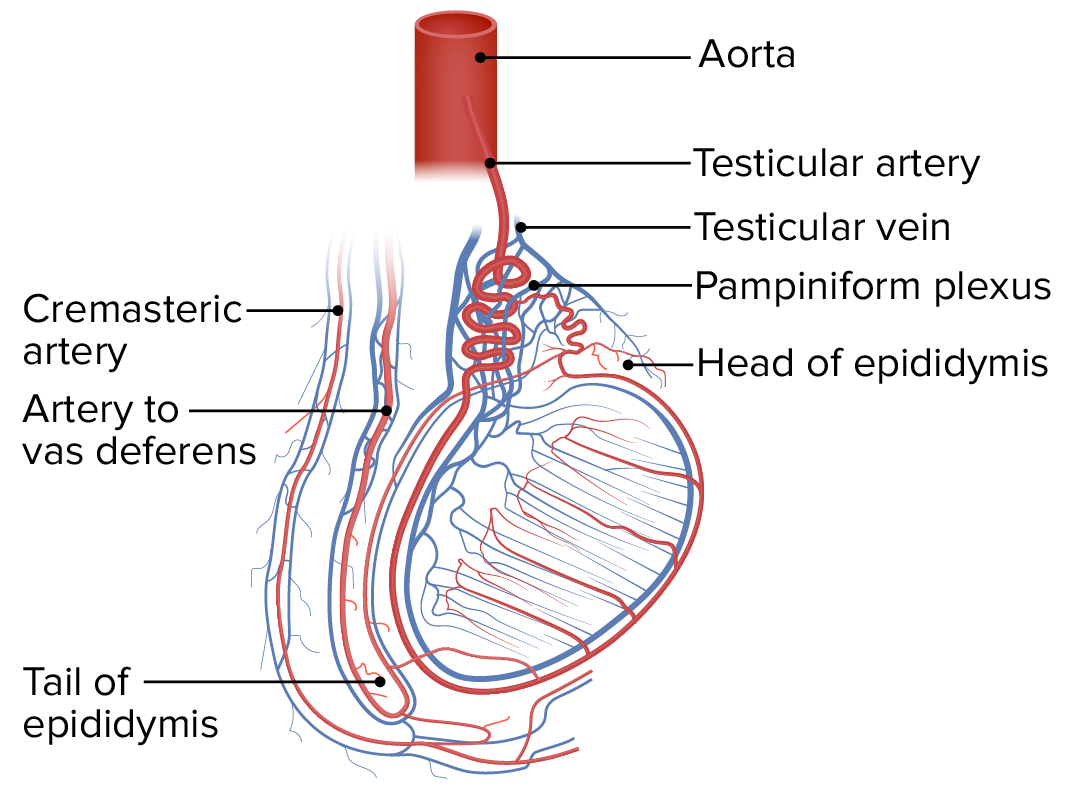
Vasculature of the testis

The robust vasculature of the testis and surrounding structures:
Note the largest arterial supply is the testicular artery, arising from the aorta. Also, note the large testicular vein and the pampiniform plexus.
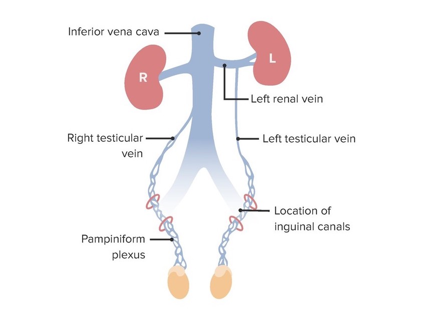
Image by Lecturio.

Venous drainage

Venous drainage of the testis

Venous drainage of the testes demonstrating the different routes of venous emptying between the left testicular vein and the right testicular vein:
Note the left testicular vein drains into the left renal vein, but the right testicular vein drains directly into the inferior vena cava.
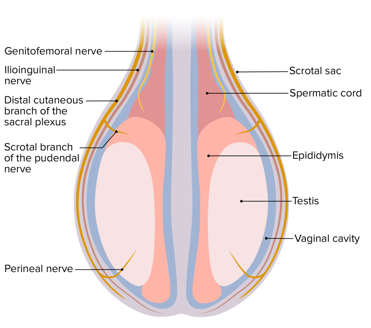
Image by Lecturio.

Lymphatic drainage

  • Testes Testes Gonadal Hormones:
    • The right testis drains to the interaortocaval nodes and paracaval nodes.
    • The left testis drains to the left para aortic and interaortocaval nodes.
  • Scrotum:
    • The right side drains to right superficial inguinal nodes.
    • The left side drains to left superficial inguinal nodes.

Innervation

Nerve supply to the testis

Nerve supply to the testes

Image by Lecturio.

Clinical Relevance

  • Hypogonadism Hypogonadism Hypogonadism is a condition characterized by reduced or no sex hormone production by the testes or ovaries. Hypogonadism can result from primary (hypergonadotropic) or secondary (hypogonadotropic) failure. Symptoms include infertility, increased risk of osteoporosis, erectile dysfunction, decreased libido, and regression (or absence) of secondary sexual characteristics. Hypogonadism: characterized by decreased sex Sex The totality of characteristics of reproductive structure, functions, phenotype, and genotype, differentiating the male from the female organism. Gender Dysphoria steroid production in the gonads Gonads The gamete-producing glands, ovary or testis. Hormones: Overview and Types. In men, hypogonadism Hypogonadism Hypogonadism is a condition characterized by reduced or no sex hormone production by the testes or ovaries. Hypogonadism can result from primary (hypergonadotropic) or secondary (hypogonadotropic) failure. Symptoms include infertility, increased risk of osteoporosis, erectile dysfunction, decreased libido, and regression (or absence) of secondary sexual characteristics. Hypogonadism can be the result of primary testicular failure or secondary testicular failure (secondary to pituitary Pituitary A small, unpaired gland situated in the sella turcica. It is connected to the hypothalamus by a short stalk which is called the infundibulum. Hormones: Overview and Types or hypothalamic disorders). Symptoms of hypogonadism Hypogonadism Hypogonadism is a condition characterized by reduced or no sex hormone production by the testes or ovaries. Hypogonadism can result from primary (hypergonadotropic) or secondary (hypogonadotropic) failure. Symptoms include infertility, increased risk of osteoporosis, erectile dysfunction, decreased libido, and regression (or absence) of secondary sexual characteristics. Hypogonadism include erectile dysfunction Erectile Dysfunction Erectile dysfunction (ED) is defined as the inability to achieve or maintain a penile erection, resulting in difficulty to perform penetrative sexual intercourse. Local penile factors and systemic diseases, including diabetes, cardiac disease, and neurological disorders, can cause ED. Erectile Dysfunction, decreased libido, and regression Regression Corneal Abrasions, Erosion, and Ulcers or absence of secondary sexual characteristics Secondary Sexual Characteristics Precocious Puberty.
  • Hydrocele Hydrocele Accumulation of serous fluid between the layers of membrane (tunica vaginalis) covering the testis in the scrotum. Varicocele, Hydrocele, and Spermatocele: scrotal masses can result from various causes, hydroceles are 1 of the most common. A hydrocele Hydrocele Accumulation of serous fluid between the layers of membrane (tunica vaginalis) covering the testis in the scrotum. Varicocele, Hydrocele, and Spermatocele is the pathological collection of fluid in the tunica vaginalis around the testes Testes Gonadal Hormones. Varicoceles occur when there is dilatation of the pampiniform venous plexus, which is connected to the internal spermatic vein. Varicoceles are associated with impaired testicular hormonal function and infertility Infertility Infertility is the inability to conceive in the context of regular intercourse. The most common causes of infertility in women are related to ovulatory dysfunction or tubal obstruction, whereas, in men, abnormal sperm is a common cause. Infertility. Spermatoceles are paratesticular epididymal cysts Cysts Any fluid-filled closed cavity or sac that is lined by an epithelium. Cysts can be of normal, abnormal, non-neoplastic, or neoplastic tissues. Fibrocystic Change with sperm-containing fluid.
  • Testicular torsion Testicular torsion Testicular torsion is the sudden rotation of the testicle, specifically the spermatic cord, around its axis in the inguinal canal or below. The acute rotation results in compromised blood flow to and from the testicle, which puts the testicle at risk for necrosis. Testicular Torsion: the sudden rotation Rotation Motion of an object in which either one or more points on a line are fixed. It is also the motion of a particle about a fixed point. X-rays of 1 of the testes Testes Gonadal Hormones around the axis. Testicular torsion Testicular torsion Testicular torsion is the sudden rotation of the testicle, specifically the spermatic cord, around its axis in the inguinal canal or below. The acute rotation results in compromised blood flow to and from the testicle, which puts the testicle at risk for necrosis. Testicular Torsion refers to the twisting of the spermatic cord structures either in or below the inguinal canal Inguinal canal The tunnel in the lower anterior abdominal wall through which the spermatic cord, in the male; round ligament, in the female; nerves; and vessels pass. Its internal end is at the deep inguinal ring and its external end is at the superficial inguinal ring. Inguinal Canal: Anatomy and Hernias. Compromised blood flow Blood flow Blood flow refers to the movement of a certain volume of blood through the vasculature over a given unit of time (e.g., mL per minute). Vascular Resistance, Flow, and Mean Arterial Pressure to and from the testis results from the acute rotation Rotation Motion of an object in which either one or more points on a line are fixed. It is also the motion of a particle about a fixed point. X-rays. The impeded venous return and reduced arterial perfusion of the testis can lead to hemorrhagic infarction of the testicular parenchyma. Testicular torsion Testicular torsion Testicular torsion is the sudden rotation of the testicle, specifically the spermatic cord, around its axis in the inguinal canal or below. The acute rotation results in compromised blood flow to and from the testicle, which puts the testicle at risk for necrosis. Testicular Torsion is a medical emergency and surgical intervention is usually necessary.

References

  1. Isidori A. M., Lenzi A. (2017). Scrotal and Testicular Anatomy. In: Ultrasound of the Testis for the Andrologist. Trends in Andrology and Sexual Medicine. Springer, Cham. https://doi.org/10.1007/978-3-319-51826-8_1
  2. Patel A. P. (2017). Anatomy and physiology of chronic scrotal pain. Translational andrology and urology, 6(Suppl 1), S51–S56. https://doi.org/10.21037/tau.2017.05.32
  3. Raghu M. R. (2020). Testicle and Epididymis Anesthesia: Overview, Indications, Contraindications. Emedicine. Com. https://emedicine.medscape.com/article/82983

Create your free account or log in to continue reading!

Sign up now and get free access to Lecturio with concept pages, medical videos, and questions for your medical education.

User Reviews

Details