Advertisement

Advertisement

Advertisement

Advertisement

Surface Epithelium: Histology

The epithelium is a complex of specialized cellular organizations arranged into sheets and lining cavities and covering the surfaces of the body. The cells exhibit polarity, having an apical and a basal pole. Structures important for the epithelial integrity and function involve the basement membrane Basement membrane A darkly stained mat-like extracellular matrix (ecm) that separates cell layers, such as epithelium from endothelium or a layer of connective tissue. The ecm layer that supports an overlying epithelium or endothelium is called basal lamina. Basement membrane (bm) can be formed by the fusion of either two adjacent basal laminae or a basal lamina with an adjacent reticular lamina of connective tissue. Bm, composed mainly of type IV collagen; glycoprotein laminin; and proteoglycan, provides barriers as well as channels between interacting cell layers. Thin Basement Membrane Nephropathy (TBMN), the semipermeable sheet on which the cells rest, and interdigitations, as well as cellular junctions. The epithelium is classified according to the cells (squamous, cuboidal, columnar), the number of layers, and other unique characteristics either due to function (transitional epithelium allowing distention) or appearance (pseudostratified epithelium giving a false impression of multiple layers). The surface epithelium has multiple functions, which include protection, secretion Secretion Coagulation Studies, filtration, and sensory Sensory Neurons which conduct nerve impulses to the central nervous system. Nervous System: Histology reception.

Last updated: Apr 18, 2023

Editorial responsibility: Stanley Oiseth, Lindsay Jones, Evelin Maza

Advertisement

Advertisement

Advertisement

Advertisement

Advertisement

Advertisement

Epithelial Tissue

Surface epithelium

A complex of specialized cellular organizations arranged into sheets and lining cavities and covering the surfaces of the body:

  • Derived from all 3 germ layers:
    • Ectoderm Ectoderm The outer of the three germ layers of an embryo. Gastrulation and Neurulation (e.g., epidermis Epidermis The external, nonvascular layer of the skin. It is made up, from within outward, of five layers of epithelium: (1) basal layer (stratum basale epidermidis); (2) spinous layer (stratum spinosum epidermidis); (3) granular layer (stratum granulosum epidermidis); (4) clear layer (stratum lucidum epidermidis); and (5) horny layer (stratum corneum epidermidis). Skin: Structure and Functions)
    • Mesoderm Mesoderm The middle germ layer of an embryo derived from three paired mesenchymal aggregates along the neural tube. Gastrulation and Neurulation (e.g., GI tract lining)
    • Endoderm Endoderm The inner of the three germ layers of an embryo. Gastrulation and Neurulation (e.g., linings of body cavities)
  • One of 4 major basic tissue types (the other 3 include nervous, muscular, and connective tissues)

These cells exhibit polarity:

  • Apical/luminal pole: derived from the apex, faces the surface/lumen
  • Basal pole: derived from the base, attached to the connective tissue Connective tissue Connective tissues originate from embryonic mesenchyme and are present throughout the body except inside the brain and spinal cord. The main function of connective tissues is to provide structural support to organs. Connective tissues consist of cells and an extracellular matrix. Connective Tissue: Histology located below the epithelium

The surface epithelium does not possess blood vessels; therefore, nutrients and oxygen are received from adjacent connective tissue Connective tissue Connective tissues originate from embryonic mesenchyme and are present throughout the body except inside the brain and spinal cord. The main function of connective tissues is to provide structural support to organs. Connective tissues consist of cells and an extracellular matrix. Connective Tissue: Histology.
Functions

  • Protection (provides covering surface or lining)
  • Secretion Secretion Coagulation Studies (release of hormones Hormones Hormones are messenger molecules that are synthesized in one part of the body and move through the bloodstream to exert specific regulatory effects on another part of the body. Hormones play critical roles in coordinating cellular activities throughout the body in response to the constant changes in both the internal and external environments. Hormones: Overview and Types, sweat, mucus, and enzymes Enzymes Enzymes are complex protein biocatalysts that accelerate chemical reactions without being consumed by them. Due to the body’s constant metabolic needs, the absence of enzymes would make life unsustainable, as reactions would occur too slowly without these molecules. Basics of Enzymes) as found in glands
  • Absorption Absorption Absorption involves the uptake of nutrient molecules and their transfer from the lumen of the GI tract across the enterocytes and into the interstitial space, where they can be taken up in the venous or lymphatic circulation. Digestion and Absorption (substance intake as seen in the intestinal lining)
  • Excretion and filtration of substances
  • Sensory Sensory Neurons which conduct nerve impulses to the central nervous system. Nervous System: Histology reception (detection of sensation) as found in olfactory epithelium Olfactory epithelium Olfaction: Anatomy

Related structures

  • Intercellular junctions:
    • Adhesion Adhesion The process whereby platelets adhere to something other than platelets, e.g., collagen; basement membrane; microfibrils; or other ‘foreign’ surfaces. Coagulation Studies of cells is facilitated by cadherins (calcium-dependent adhesion Adhesion The process whereby platelets adhere to something other than platelets, e.g., collagen; basement membrane; microfibrils; or other ‘foreign’ surfaces. Coagulation Studies molecules).
    • At the apical end, structures encircle the area (like a band A band Skeletal Muscle Contraction around the cell):
      • Tight or occluding junctions (zonulae occludens): have tight ridges, preventing passive flow Flow Blood flows through the heart, arteries, capillaries, and veins in a closed, continuous circuit. Flow is the movement of volume per unit of time. Flow is affected by the pressure gradient and the resistance fluid encounters between 2 points. Vascular resistance is the opposition to flow, which is caused primarily by blood friction against vessel walls. Vascular Resistance, Flow, and Mean Arterial Pressure of substances between cells
      • Adherent junctions (zonulae adherens Adherens Anchoring points where the cytoskeleton of neighboring cells are connected to each other. They are composed of specialized areas of the plasma membrane where bundles of the actin cytoskeleton attach to the membrane through the transmembrane linkers, cadherins, which in turn attach through their extracellular domains to cadherins in the neighboring cell membranes. In sheets of cells, they form into adhesion belts (zonula adherens) that go all the way around a cell. The Cell: Cell Junctions): help stabilize both junctions and hold cells together.
    • Desmosomes Desmosomes A type of junction that attaches one cell to its neighbor. One of a number of differentiated regions which occur, for example, where the cytoplasmic membranes of adjacent epithelial cells are closely apposed. It consists of a circular region of each membrane together with associated intracellular microfilaments and an intercellular material which may include, for example, mucopolysaccharides. Bullous Pemphigoid and Pemphigus Vulgaris ( macula Macula An oval area in the retina, 3 to 5 mm in diameter, usually located temporal to the posterior pole of the eye and slightly below the level of the optic disk. It is characterized by the presence of a yellow pigment diffusely permeating the inner layers, contains the fovea centralis in its center, and provides the best phototropic visual acuity. It is devoid of retinal blood vessels, except in its periphery, and receives nourishment from the choriocapillaris of the choroid. Eye: Anatomy adherens Adherens Anchoring points where the cytoskeleton of neighboring cells are connected to each other. They are composed of specialized areas of the plasma membrane where bundles of the actin cytoskeleton attach to the membrane through the transmembrane linkers, cadherins, which in turn attach through their extracellular domains to cadherins in the neighboring cell membranes. In sheets of cells, they form into adhesion belts (zonula adherens) that go all the way around a cell. The Cell: Cell Junctions) and gap junctions Gap Junctions Connections between cells which allow passage of small molecules and electric current. Gap junctions were first described anatomically as regions of close apposition between cells with a narrow (1-2 nm) gap between cell membranes. The variety in the properties of gap junctions is reflected in the number of connexins, the family of proteins which form the junctions. The Cell: Cell Junctions (communicating junctions):
      •  Structures bound to intracellular intermediate filaments Intermediate filaments Cytoplasmic filaments intermediate in diameter (about 10 nanometers) between the microfilaments and the microtubules. They may be composed of any of a number of different proteins and form a ring around the cell nucleus. The Cell: Cytosol and Cytoskeleton, aiding the zonulae adherens Adherens Anchoring points where the cytoskeleton of neighboring cells are connected to each other. They are composed of specialized areas of the plasma membrane where bundles of the actin cytoskeleton attach to the membrane through the transmembrane linkers, cadherins, which in turn attach through their extracellular domains to cadherins in the neighboring cell membranes. In sheets of cells, they form into adhesion belts (zonula adherens) that go all the way around a cell. The Cell: Cell Junctions
      • Additionally, gap junctions Gap Junctions Connections between cells which allow passage of small molecules and electric current. Gap junctions were first described anatomically as regions of close apposition between cells with a narrow (1-2 nm) gap between cell membranes. The variety in the properties of gap junctions is reflected in the number of connexins, the family of proteins which form the junctions. The Cell: Cell Junctions permit flow Flow Blood flows through the heart, arteries, capillaries, and veins in a closed, continuous circuit. Flow is the movement of volume per unit of time. Flow is affected by the pressure gradient and the resistance fluid encounters between 2 points. Vascular resistance is the opposition to flow, which is caused primarily by blood friction against vessel walls. Vascular Resistance, Flow, and Mean Arterial Pressure of molecules.
    • Hemidesmosomes: anchor epithelial cells to the basal lamina Basal Lamina Capillaries: Histology (of the basement membrane Basement membrane A darkly stained mat-like extracellular matrix (ecm) that separates cell layers, such as epithelium from endothelium or a layer of connective tissue. The ecm layer that supports an overlying epithelium or endothelium is called basal lamina. Basement membrane (bm) can be formed by the fusion of either two adjacent basal laminae or a basal lamina with an adjacent reticular lamina of connective tissue. Bm, composed mainly of type IV collagen; glycoprotein laminin; and proteoglycan, provides barriers as well as channels between interacting cell layers. Thin Basement Membrane Nephropathy (TBMN))
  • Interdigitations: cell membrane Cell Membrane A cell membrane (also known as the plasma membrane or plasmalemma) is a biological membrane that separates the cell contents from the outside environment. A cell membrane is composed of a phospholipid bilayer and proteins that function to protect cellular DNA and mediate the exchange of ions and molecules. The Cell: Cell Membrane foldings that increase the surface area (e.g., for ion or water transport)
  • Basement membrane Basement membrane A darkly stained mat-like extracellular matrix (ecm) that separates cell layers, such as epithelium from endothelium or a layer of connective tissue. The ecm layer that supports an overlying epithelium or endothelium is called basal lamina. Basement membrane (bm) can be formed by the fusion of either two adjacent basal laminae or a basal lamina with an adjacent reticular lamina of connective tissue. Bm, composed mainly of type IV collagen; glycoprotein laminin; and proteoglycan, provides barriers as well as channels between interacting cell layers. Thin Basement Membrane Nephropathy (TBMN): 
    • Thin semipermeable sheet on which the basal surface of epithelia rests
    • Attachment of cells achieved with laminin Laminin Large, noncollagenous glycoprotein with antigenic properties. It is localized in the basement membrane lamina lucida and functions to bind epithelial cells to the basement membrane. Evidence suggests that the protein plays a role in tumor invasion. Connective Tissue: Histology and integrin
    • Parts:
    • Function:
      • Attachment to underlying tissue
      • Separation of tissues
      • Filtration
      • Scaffold and guidance (repair)
      • Cell signaling
Characteristics of the epithelium

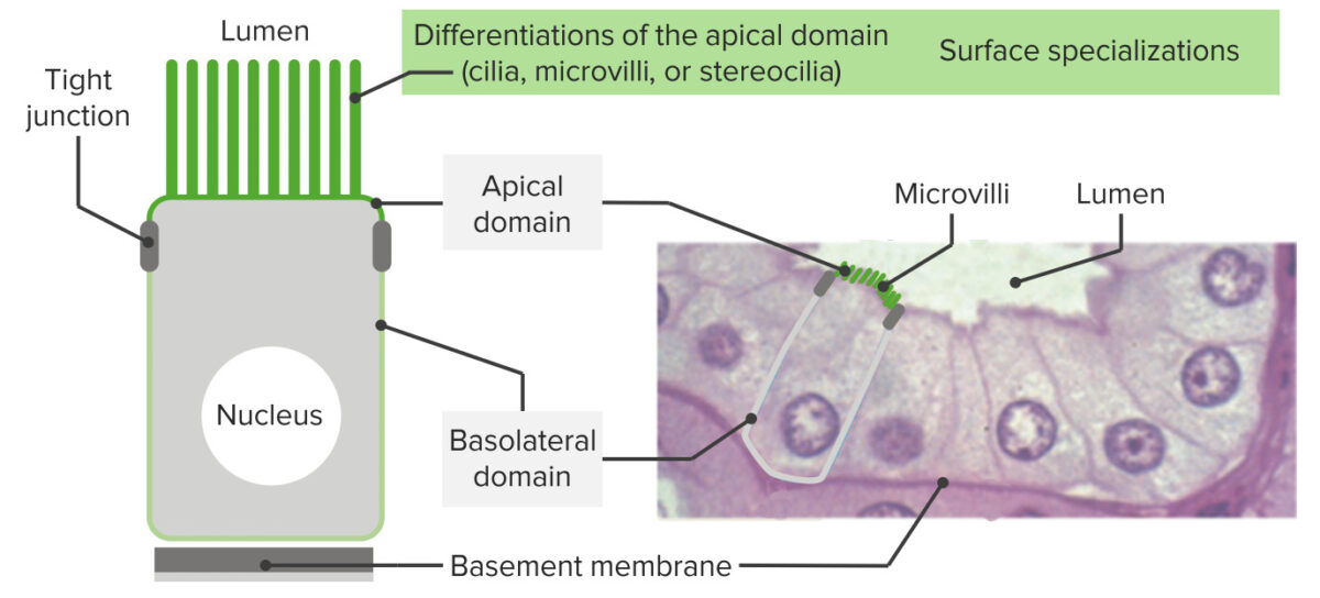
Epithelia have an apex, lateral borders, and a surface sitting on a basement membrane:
As seen in the image, these cells exhibit polarity (have apical domains and basal/basolateral domains). At the apical end, structures encircle the area (like a band around the cell) facilitating cell-to-cell adhesion (tight junctions and adherent junctions). Near the basement membrane, hemidesmosomes anchor the epithelia to the basal lamina. On the right is shown the histology of the intestinal epithelial lining with corresponding structures. On the apical domain, microvilli are seen.

Image by Lecturio.

Classification

By number of layers

  • Simple epithelium:
    • Consists of a single layer
    • Typically found where absorption Absorption Absorption involves the uptake of nutrient molecules and their transfer from the lumen of the GI tract across the enterocytes and into the interstitial space, where they can be taken up in the venous or lymphatic circulation. Digestion and Absorption, secretion Secretion Coagulation Studies, and filtration occur
  • Stratified epithelium:
    • Composed of ≥ 2 layers
    • Found in high-abrasion areas ( skin Skin The skin, also referred to as the integumentary system, is the largest organ of the body. The skin is primarily composed of the epidermis (outer layer) and dermis (deep layer). The epidermis is primarily composed of keratinocytes that undergo rapid turnover, while the dermis contains dense layers of connective tissue. Skin: Structure and Functions and lining of the mouth)
    • Name of cells in stratified epithelia based on shape of cells in apical layer
  • Pseudostratified epithelium:
    • All cells are attached to the basement membrane Basement membrane A darkly stained mat-like extracellular matrix (ecm) that separates cell layers, such as epithelium from endothelium or a layer of connective tissue. The ecm layer that supports an overlying epithelium or endothelium is called basal lamina. Basement membrane (bm) can be formed by the fusion of either two adjacent basal laminae or a basal lamina with an adjacent reticular lamina of connective tissue. Bm, composed mainly of type IV collagen; glycoprotein laminin; and proteoglycan, provides barriers as well as channels between interacting cell layers. Thin Basement Membrane Nephropathy (TBMN), but not all cells extend to the free surface.
    • The nuclei, being at different levels, give the epithelia a stratified appearance.

By shape of the cells

  • Squamous cells: thin or flattened cells
  • Cuboidal cells: cell width and thickness generally similar (cube-shaped)
  • Columnar cells: cells are taller than wide (column-shaped).
  • Transitional: shape of cells “goes through a transition”:
    • When the organ (e.g., bladder Bladder A musculomembranous sac along the urinary tract. Urine flows from the kidneys into the bladder via the ureters, and is held there until urination. Pyelonephritis and Perinephric Abscess) is relaxed, cells appear cuboidal.
    • When the organ is distended, cells flatten.

By function

Note that there are tissues that have both:
  • Covering epithelium: line the cavities or organs
  • Glandular epithelium Glandular epithelium Glandular epithelia, composed of epithelial tissue, are specialized structures that play a role in the production and release of enzymes, hormones, sweat, oil, and mucus in organisms. The secretion and release of these substances are prompted by either external or internal stimuli. Glandular Epithelium: Histology: secretory
Types of epithelium

The types of epithelia:
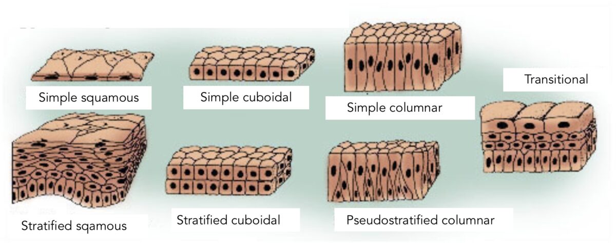
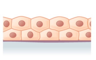
The differentiation and classification of the types are based on the shape of the cell and the layers. Simple epithelium indicates 1 layer of cells. Stratified epithelium indicates multiple layers. Pseudostratified epithelium gives a false impression of > 1 layer owing to different nuclei levels. Transitional epithelium is a type in which the shape of cells “go into transition” depending on the organ function (distention versus relaxation, as seen in the urinary bladder).
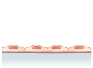
Image: “Types of epithelium” by U.S. National Cancer Institute’s Surveillance. License: Public Domain, edited by Lecturio.

Simple Epithelium

Simple squamous epithelium

  • Characteristics:
    • Single layer of flattened cells
    • Disc-shaped nuclei (most prominent structure)
    • Sparse cytoplasm
  • Typically found in lining of vessels, regulating passing substances to the tissue(s)
  • Located in:
    • Air sacs/ alveoli Alveoli Small polyhedral outpouchings along the walls of the alveolar sacs, alveolar ducts and terminal bronchioles through the walls of which gas exchange between alveolar air and pulmonary capillary blood takes place. Acute Respiratory Distress Syndrome (ARDS) of lungs Lungs Lungs are the main organs of the respiratory system. Lungs are paired viscera located in the thoracic cavity and are composed of spongy tissue. The primary function of the lungs is to oxygenate blood and eliminate CO2. Lungs: Anatomy
    • Lining of the heart
    • Blood vessels and lymphatic vessels Lymphatic Vessels Tubular vessels that are involved in the transport of lymph and lymphocytes. Lymphatic Drainage System: Anatomy
    • Renal loops of Henle
    • Cornea Cornea The transparent anterior portion of the fibrous coat of the eye consisting of five layers: stratified squamous corneal epithelium; bowman membrane; corneal stroma; descemet membrane; and mesenchymal corneal endothelium. It serves as the first refracting medium of the eye. Eye: Anatomy
  • Special naming:
    • Endothelium Endothelium A layer of epithelium that lines the heart, blood vessels (vascular endothelium), lymph vessels (lymphatic endothelium), and the serous cavities of the body. Arteries: Histology: simple squamous epithelium lining blood vessels and lymphatic vessels Lymphatic Vessels Tubular vessels that are involved in the transport of lymph and lymphocytes. Lymphatic Drainage System: Anatomy
    • Mesothelium: simple squamous epithelium found in large body cavities (secreting serous fluid)
Simple squamous epithelium

Structure noted in simple squamous epithelium (single layer of flattened cells)

Image: “Simple squamous epithelium” by Phil Schatz. License: CC BY 4.0
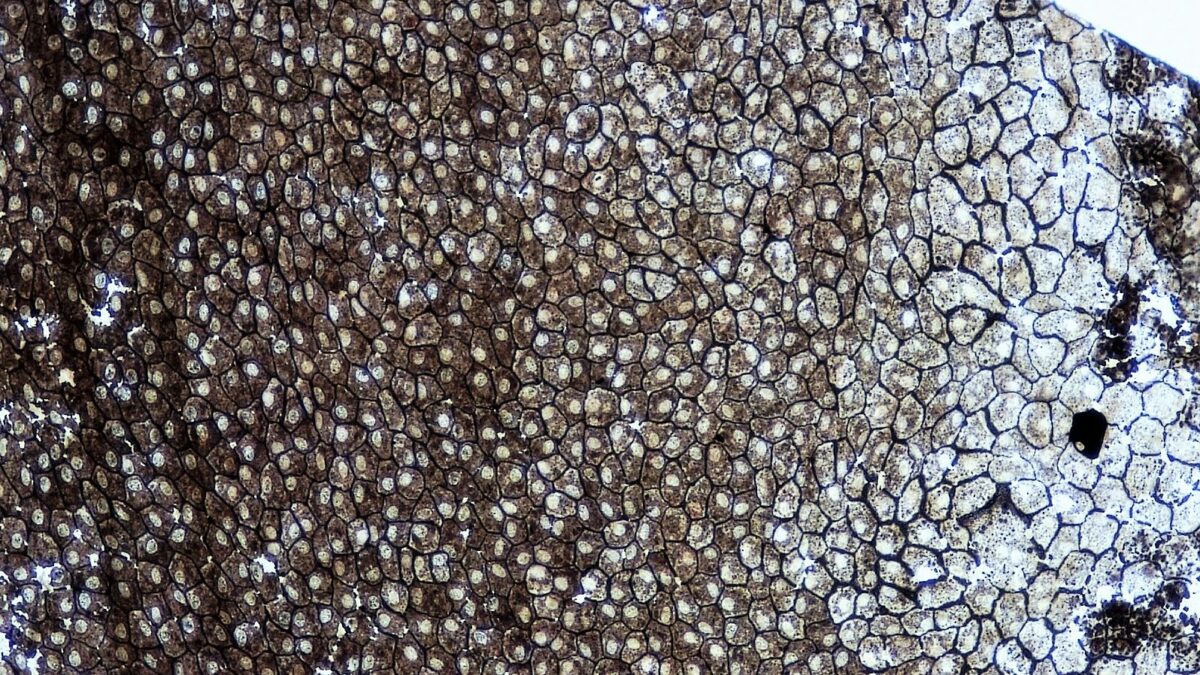
Epithelial tissues simple squamous epithelium

A whole mount of simple squamous epithelium

Image: “Epithelial Tissues Simple Squamous Epithelium” by Berkshire Community College Bioscience Image Library. License: CC0 1.0
Simple cuboidal epithelium

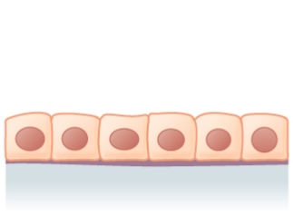
  • Single layer of cube-like cells
  • Cuboidal shape allows increased mitochondria Mitochondria Semiautonomous, self-reproducing organelles that occur in the cytoplasm of all cells of most, but not all, eukaryotes. Each mitochondrion is surrounded by a double limiting membrane. The inner membrane is highly invaginated, and its projections are called cristae. Mitochondria are the sites of the reactions of oxidative phosphorylation, which result in the formation of ATP. They contain distinctive ribosomes, transfer RNAs; amino Acyl tRNA synthetases; and elongation and termination factors. Mitochondria depend upon genes within the nucleus of the cells in which they reside for many essential messenger RNAs. Mitochondria are believed to have arisen from aerobic bacteria that established a symbiotic relationship with primitive protoeukaryotes. The Cell: Organelles and other organelles Organelles A cell is a complex unit that performs several complex functions. An organelle is a specialized subunit within a cell that fulfills a specific role or function. Organelles are enclosed within their own lipid bilayers or are unbound by membranes. The Cell: Organelles needed for functions such as active transport Active transport The movement of materials across cell membranes and epithelial layers against an electrochemical gradient, requiring the expenditure of metabolic energy. The Cell: Cell Membrane, secretion Secretion Coagulation Studies, and absorption Absorption Absorption involves the uptake of nutrient molecules and their transfer from the lumen of the GI tract across the enterocytes and into the interstitial space, where they can be taken up in the venous or lymphatic circulation. Digestion and Absorption
  • Found in:
    • Ducts and secretory portions of glands
    • Kidney tubules
Simple cuboidal epithelium

Simple cuboidal epithelium: 1 layer of cuboidal cells

Image: “Simple cuboidal epithelium” by Phil Schatz. License: CC BY 4.0
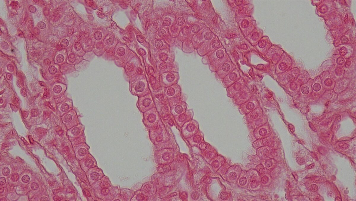
Simple cuboidal epithelium

Cross section of a kidney tubule showing a layer of cuboidal cells

Image: “Epithelial Tissues Simple Cuboidal Epithelium” by Berkshire Community College Bioscience Image Library. License: CC0 1.0
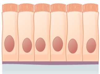
Simple columnar epithelium

  • Single-layer tall cells
  • Often with cilia or microvilli
  • Mostly involved in absorption Absorption Absorption involves the uptake of nutrient molecules and their transfer from the lumen of the GI tract across the enterocytes and into the interstitial space, where they can be taken up in the venous or lymphatic circulation. Digestion and Absorption and secretion Secretion Coagulation Studies
  • Found in:
    • Ciliated epithelium in the bronchi Bronchi The larger air passages of the lungs arising from the terminal bifurcation of the trachea. They include the largest two primary bronchi which branch out into secondary bronchi, and tertiary bronchi which extend into bronchioles and pulmonary alveoli. Bronchial Tree: Anatomy, fallopian tubes Fallopian tubes The uterus, cervix, and fallopian tubes are part of the internal female reproductive system. The fallopian tubes receive an ovum after ovulation and help move it and/or a fertilized embryo toward the uterus via ciliated cells lining the tubes and peristaltic movements of its smooth muscle. Uterus, Cervix, and Fallopian Tubes: Anatomy, and uterus Uterus The uterus, cervix, and fallopian tubes are part of the internal female reproductive system. The uterus has a thick wall made of smooth muscle (the myometrium) and an inner mucosal layer (the endometrium). The most inferior portion of the uterus is the cervix, which connects the uterine cavity to the vagina. Uterus, Cervix, and Fallopian Tubes: Anatomy
    • Smooth (nonciliated) epithelium in the digestive tract 
    • Lining of the gallbladder Gallbladder The gallbladder is a pear-shaped sac, located directly beneath the liver, that sits on top of the superior part of the duodenum. The primary functions of the gallbladder include concentrating and storing up to 50 mL of bile. Gallbladder and Biliary Tract: Anatomy
Simple columnar epithelium

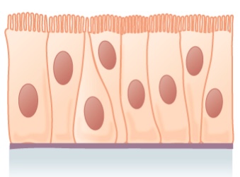
Simple columnar epithelium:
Shown is a layer of columnar cells with cilia
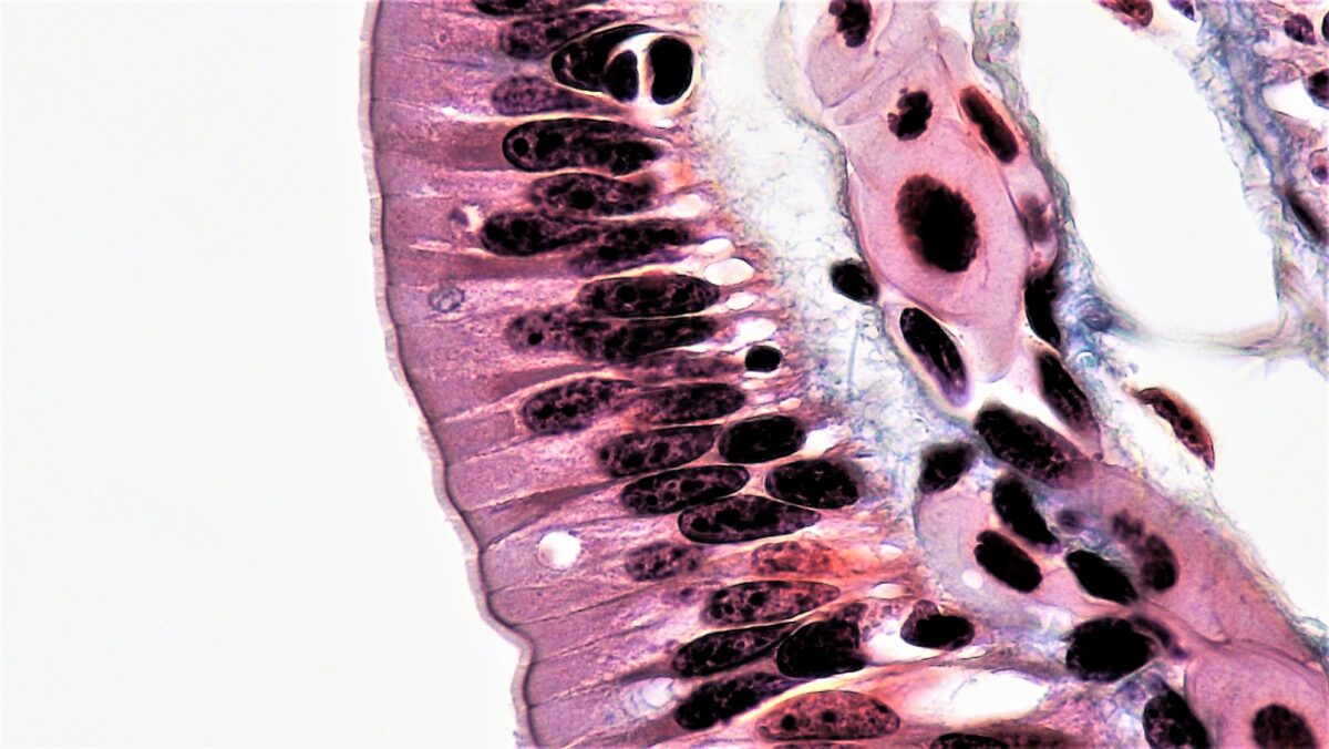
Image: “Simple columnar epithelium” by Phil Schatz. License: CC BY 4.0
Simple columnar epithelium

Simple columnar epithelium lining the intestinal tract

Image: “Epithelial Tissues Simple Columnar Epithelium” by Epithelial Tissues: Simple Columnar Epithelium. License: CC0 1.0
Pseudostratified, Stratified, and Transitional Epithelia

Pseudostratified columnar epithelium

  • Characteristics:
    • Cells vary in height, with all cells resting on the basement membrane Basement membrane A darkly stained mat-like extracellular matrix (ecm) that separates cell layers, such as epithelium from endothelium or a layer of connective tissue. The ecm layer that supports an overlying epithelium or endothelium is called basal lamina. Basement membrane (bm) can be formed by the fusion of either two adjacent basal laminae or a basal lamina with an adjacent reticular lamina of connective tissue. Bm, composed mainly of type IV collagen; glycoprotein laminin; and proteoglycan, provides barriers as well as channels between interacting cell layers. Thin Basement Membrane Nephropathy (TBMN) but only some reaching all the way to the apical surface.
    • Nuclei placed at different heights
    • Some have cilia.
  • Functions in secretion Secretion Coagulation Studies and absorption Absorption Absorption involves the uptake of nutrient molecules and their transfer from the lumen of the GI tract across the enterocytes and into the interstitial space, where they can be taken up in the venous or lymphatic circulation. Digestion and Absorption
  • Found in:
    • Ciliated epithelium of the trachea Trachea The trachea is a tubular structure that forms part of the lower respiratory tract. The trachea is continuous superiorly with the larynx and inferiorly becomes the bronchial tree within the lungs. The trachea consists of a support frame of semicircular, or C-shaped, rings made out of hyaline cartilage and reinforced by collagenous connective tissue. Trachea: Anatomy
    • Part of the upper digestive tract
Pseudostratified epithelium

Pseudostratified epithelium:
Shown is a single layer of columnar cells, with varying levels of nuclei
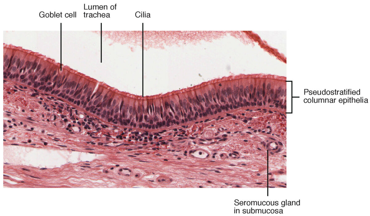
Image: “Pseudostratified epithelium” by Phil Schatz. License: CC BY 4.0
Cross-section of pseudostratified columnar epithelium

Pseudostratified columnar epithelium found in the trachea

Image: “Cross-section of pseudostratified columnar epithelium” by OpenStax College. License: CC BY 3.0
Stratified squamous epithelium

  • Most widespread of the stratified epithelia
  • Composed of several layers, providing protective function
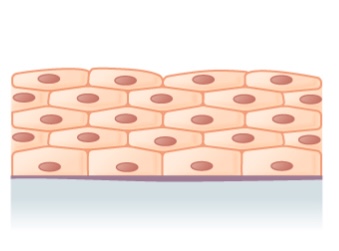
  • Free surface cells are squamous.
  • Deeper layer cells are cuboidal or columnar.
  • Found in areas subjected to wear and tear
  • Found in:
    • Skin Skin The skin, also referred to as the integumentary system, is the largest organ of the body. The skin is primarily composed of the epidermis (outer layer) and dermis (deep layer). The epidermis is primarily composed of keratinocytes that undergo rapid turnover, while the dermis contains dense layers of connective tissue. Skin: Structure and Functions, which is keratinized or filled with keratin Keratin A class of fibrous proteins or scleroproteins that represents the principal constituent of epidermis; hair; nails; horny tissues, and the organic matrix of tooth enamel. Two major conformational groups have been characterized, alpha-keratin, whose peptide backbone forms a coiled-coil alpha helical structure consisting of type I keratin and a type II keratin, and beta-keratin, whose backbone forms a zigzag or pleated sheet structure. Alpha-keratins have been classified into at least 20 subtypes. In addition multiple isoforms of subtypes have been found which may be due to gene duplication. Seborrheic Keratosis:
      • These cells will lose the organelles Organelles A cell is a complex unit that performs several complex functions. An organelle is a specialized subunit within a cell that fulfills a specific role or function. Organelles are enclosed within their own lipid bilayers or are unbound by membranes. The Cell: Organelles and nuclei as they flatten and accumulate keratin Keratin A class of fibrous proteins or scleroproteins that represents the principal constituent of epidermis; hair; nails; horny tissues, and the organic matrix of tooth enamel. Two major conformational groups have been characterized, alpha-keratin, whose peptide backbone forms a coiled-coil alpha helical structure consisting of type I keratin and a type II keratin, and beta-keratin, whose backbone forms a zigzag or pleated sheet structure. Alpha-keratins have been classified into at least 20 subtypes. In addition multiple isoforms of subtypes have been found which may be due to gene duplication. Seborrheic Keratosis.
      • Cells move toward the surface, becoming metabolically inactive and are sloughed off.
    • Esophagus Esophagus The esophagus is a muscular tube-shaped organ of around 25 centimeters in length that connects the pharynx to the stomach. The organ extends from approximately the 6th cervical vertebra to the 11th thoracic vertebra and can be divided grossly into 3 parts: the cervical part, the thoracic part, and the abdominal part. Esophagus: Anatomy, mouth, vagina Vagina The vagina is the female genital canal, extending from the vulva externally to the cervix uteri internally. The structures have sexual, reproductive, and urinary functions and a rich blood supply, mainly arising from the internal iliac artery. Vagina, Vulva, and Pelvic Floor: Anatomy, which have nonkeratinized cells (retain the nuclei)
Stratified squamous epithelium

Illustration of stratified squamous epithelium (multiple layers)

Image: “Stratified squamous epithelium” by Phil Schatz. License: CC BY 4.0
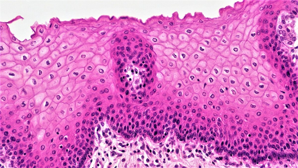
Epithelial tissues stratified squamous epithelium

Histologic picture of stratified squamous epithelium

Image: “Epithelial Tissues Stratified Squamous Epithelium” by Berkshire Community College Bioscience Image Library. License: CC0 1.0
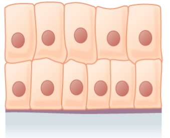
Stratified columnar epithelium and stratified cuboidal epithelium

  • Both are rare and have limited distribution.
  • Stratified columnar epithelium:
    • Secretory and protective function
    • Found in:
      • Male urethra Urethra A tube that transports urine from the urinary bladder to the outside of the body in both the sexes. It also has a reproductive function in the male by providing a passage for sperm. Urinary Tract: Anatomy
      • Some glandular ducts
  • Stratified cuboidal epithelium is found in ducts of large glands (e.g., sweat glands Sweat glands Sweat-producing structures that are embedded in the dermis. Each gland consists of a single tube, a coiled body, and a superficial duct. Soft Tissue Abscess, mammary glands).
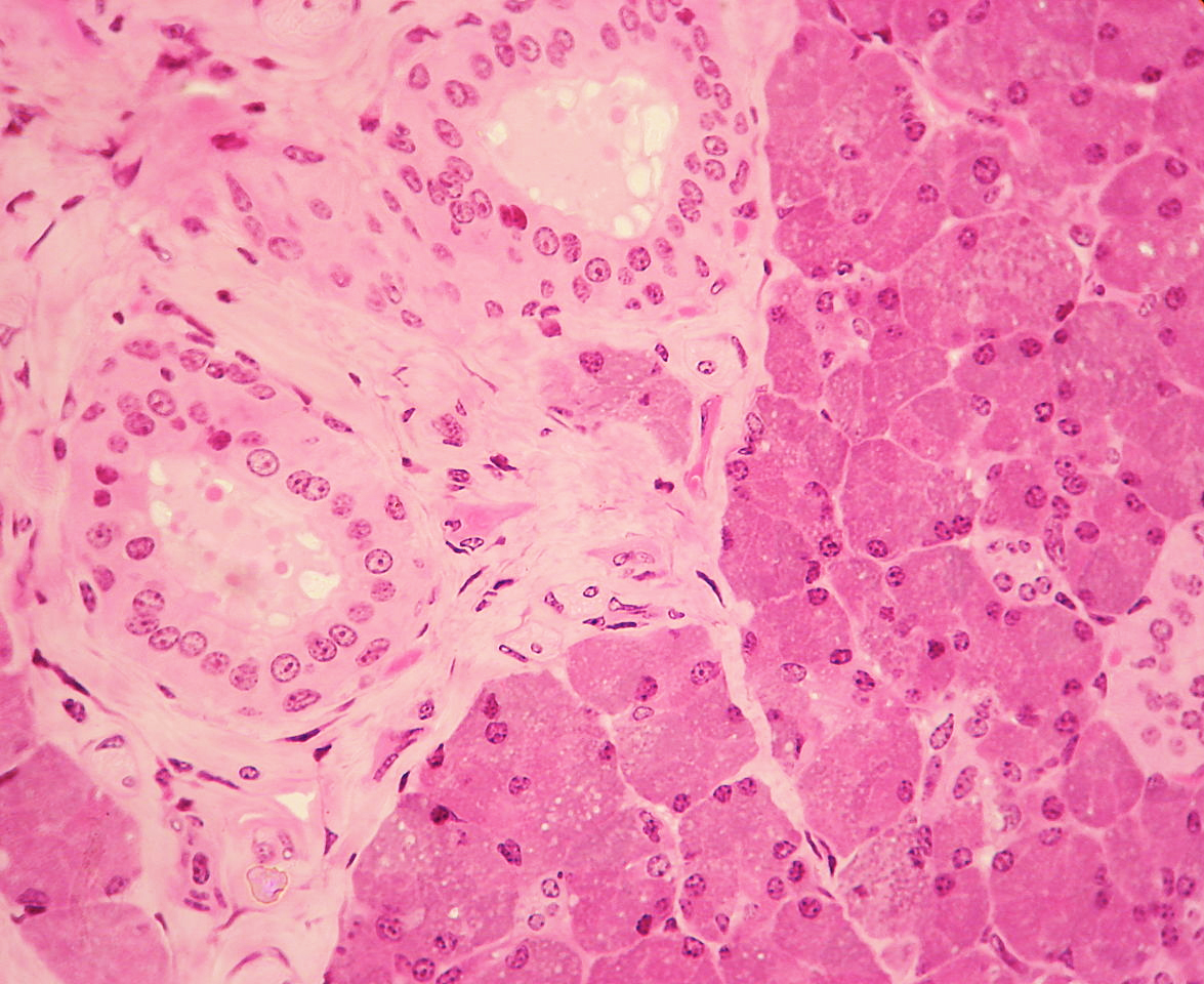
Parotid gland

Stratified cuboidal epithelium (seen on the left) is visible in a duct surrounded by connective tissue in the parotid gland.

Image: “WVSOM Parotid Gland1” by Wbensmith. License: CC BY 3.0

Transitional epithelium

  • Characteristics:
    • Surface layer of umbrella cells (dome-like cells)
    • Allows for stretching of the organs as they distend
    • Urothelium Urothelium The epithelial lining of the urinary tract. Urinary Tract: Anatomy: transitional epithelium in the urinary tract Urinary tract The urinary tract is located in the abdomen and pelvis and consists of the kidneys, ureters, urinary bladder, and urethra. The structures permit the excretion of urine from the body. Urine flows from the kidneys through the ureters to the urinary bladder and out through the urethra. Urinary Tract: Anatomy
  • Found in:
    • Urinary bladder Urinary Bladder A musculomembranous sac along the urinary tract. Urine flows from the kidneys into the bladder via the ureters (ureter), and is held there until urination. Urinary Tract: Anatomy
    • Urethra Urethra A tube that transports urine from the urinary bladder to the outside of the body in both the sexes. It also has a reproductive function in the male by providing a passage for sperm. Urinary Tract: Anatomy
    • Ureters Ureters One of a pair of thick-walled tubes that transports urine from the kidney pelvis to the urinary bladder. Urinary Tract: Anatomy
Transitional epithelium

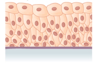
Transitional epithelium

Image: “Transitional epithelium” by Phil Schatz. License: CC BY 4.0
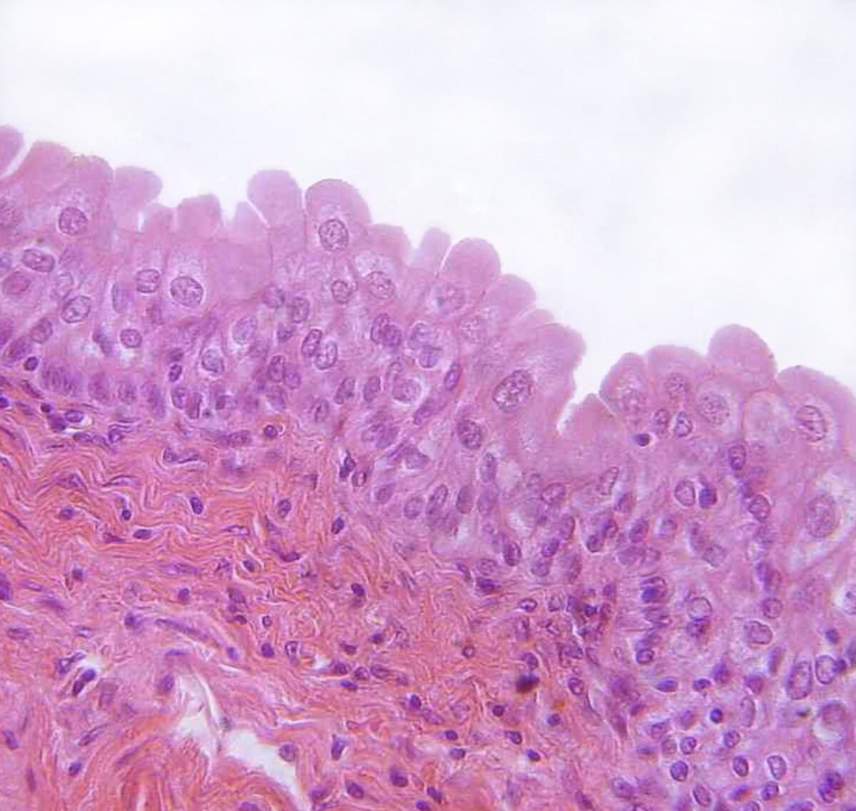
Transitional epithelium

Transitional epithelium found in the urinary bladder

Image: “urinary bladder, urothelium, haemalum-eosin stain” by Polarlys. License: CC BY 2.5

Clinical Relevance

  • Ichthyosis: dermatologic disease in which abnormal keratinization occurs. Ichthyosis is caused by an autosomal dominant Autosomal dominant Autosomal inheritance, both dominant and recessive, refers to the transmission of genes from the 22 autosomal chromosomes. Autosomal dominant diseases are expressed when only 1 copy of the dominant allele is inherited. Autosomal Recessive and Autosomal Dominant Inheritance mutation Mutation Genetic mutations are errors in DNA that can cause protein misfolding and dysfunction. There are various types of mutations, including chromosomal, point, frameshift, and expansion mutations. Types of Mutations in the filaggrin gene Gene A category of nucleic acid sequences that function as units of heredity and which code for the basic instructions for the development, reproduction, and maintenance of organisms. Basic Terms of Genetics, which results in skin Skin The skin, also referred to as the integumentary system, is the largest organ of the body. The skin is primarily composed of the epidermis (outer layer) and dermis (deep layer). The epidermis is primarily composed of keratinocytes that undergo rapid turnover, while the dermis contains dense layers of connective tissue. Skin: Structure and Functions barrier dysfunction. Clinical presentation features rough, dry, and scaly skin Skin The skin, also referred to as the integumentary system, is the largest organ of the body. The skin is primarily composed of the epidermis (outer layer) and dermis (deep layer). The epidermis is primarily composed of keratinocytes that undergo rapid turnover, while the dermis contains dense layers of connective tissue. Skin: Structure and Functions, with symptoms worsening during cold, dry months. The diagnosis is usually clinical but often aided with a skin biopsy Skin Biopsy Secondary Skin Lesions showing hyperkeratosis Hyperkeratosis Ichthyosis Vulgaris and a diminished stratum granulosum Stratum granulosum Skin: Structure and Functions
  • Transitional cell carcinoma: malignancy Malignancy Hemothorax affecting the urinary bladder Urinary Bladder A musculomembranous sac along the urinary tract. Urine flows from the kidneys into the bladder via the ureters (ureter), and is held there until urination. Urinary Tract: Anatomy. Transitional cell carcinoma is the most common cancer involving the urinary system. The predominant histologic type is urothelial (transitional cell) carcinoma in the United States and Europe. Risk factors include genetics Genetics Genetics is the study of genes and their functions and behaviors. Basic Terms of Genetics, smoking Smoking Willful or deliberate act of inhaling and exhaling smoke from burning substances or agents held by hand. Interstitial Lung Diseases, opium use, and occupational carcinogen exposure. Presentation is usually with painless hematuria Hematuria Presence of blood in the urine. Renal Cell Carcinoma, and the diagnostic approach includes cystoscopy, urine cytology, and biopsies. Transurethral resection of bladder Bladder A musculomembranous sac along the urinary tract. Urine flows from the kidneys into the bladder via the ureters, and is held there until urination. Pyelonephritis and Perinephric Abscess tumor Tumor Inflammation, radical cystectomy, and chemotherapy Chemotherapy Osteosarcoma are among the treatment options, which depend on the stage. 
  • Barrett’s esophagus Esophagus The esophagus is a muscular tube-shaped organ of around 25 centimeters in length that connects the pharynx to the stomach. The organ extends from approximately the 6th cervical vertebra to the 11th thoracic vertebra and can be divided grossly into 3 parts: the cervical part, the thoracic part, and the abdominal part. Esophagus: Anatomy: replacement of stratified squamous epithelium with gastric columnar epithelium in the esophagus Esophagus The esophagus is a muscular tube-shaped organ of around 25 centimeters in length that connects the pharynx to the stomach. The organ extends from approximately the 6th cervical vertebra to the 11th thoracic vertebra and can be divided grossly into 3 parts: the cervical part, the thoracic part, and the abdominal part. Esophagus: Anatomy caused by chronic GERD GERD Gastroesophageal reflux disease (GERD) occurs when the stomach acid frequently flows back into the esophagus. This backwash (acid reflux) can irritate the lining of the esophagus, causing symptoms such as retrosternal burning pain (heartburn). Gastroesophageal Reflux Disease (GERD). This is associated with an increased risk of esophageal adenocarcinoma Esophageal Adenocarcinoma Esophageal Cancer. Esophagogastroduodenoscopy would show proximal displacement Displacement The process by which an emotional or behavioral response that is appropriate for one situation appears in another situation for which it is inappropriate. Defense Mechanisms of the squamocolumnar junction Squamocolumnar junction Esophagus: Anatomy ( Z-line Z-line Esophagus: Anatomy) from the gastroesophageal junction Gastroesophageal junction The area covering the terminal portion of esophagus and the beginning of stomach at the cardiac orifice. Esophagus: Anatomy (GEJ). Biopsies reveal columnar epithelium and goblet cells Goblet cells A glandular epithelial cell or a unicellular gland. Goblet cells secrete mucus. They are scattered in the epithelial linings of many organs, especially the small intestine and the respiratory tract. Glandular Epithelium: Histology in the distal esophagus Esophagus The esophagus is a muscular tube-shaped organ of around 25 centimeters in length that connects the pharynx to the stomach. The organ extends from approximately the 6th cervical vertebra to the 11th thoracic vertebra and can be divided grossly into 3 parts: the cervical part, the thoracic part, and the abdominal part. Esophagus: Anatomy. Treatment is with proton pump Pump ACES and RUSH: Resuscitation Ultrasound Protocols inhibitors and lifestyle modifications. 
  • Cervical cancer Cervical cancer Cervical cancer, or invasive cervical carcinoma (ICC), is the 3rd most common cancer in women in the world, with > 50% of the cases being fatal. In the United States, ICC is the 13th most common cancer and the cause of < 3% of all cancer deaths due to the slow progression of precursor lesions and, more importantly, effective cancer screening. Cervical Cancer: very common cancer affecting women. Most cases of cervical cancer Cervical cancer Cervical cancer, or invasive cervical carcinoma (ICC), is the 3rd most common cancer in women in the world, with > 50% of the cases being fatal. In the United States, ICC is the 13th most common cancer and the cause of < 3% of all cancer deaths due to the slow progression of precursor lesions and, more importantly, effective cancer screening. Cervical Cancer are due to HPV HPV Human papillomavirus (HPV) is a nonenveloped, circular, double-stranded DNA virus belonging to the Papillomaviridae family. Humans are the only reservoir, and transmission occurs through close skin-to-skin or sexual contact. Human papillomaviruses infect basal epithelial cells and can affect cell-regulatory proteins to result in cell proliferation. Papillomavirus (HPV). The region around the external os External os Uterus, Cervix, and Fallopian Tubes: Anatomy is lined by nonkeratinized stratified squamous epithelium continuous with that of the vagina Vagina The vagina is the female genital canal, extending from the vulva externally to the cervix uteri internally. The structures have sexual, reproductive, and urinary functions and a rich blood supply, mainly arising from the internal iliac artery. Vagina, Vulva, and Pelvic Floor: Anatomy. This area meets the mucus-secreting columnar epithelium of the endocervix Endocervix Uterus, Cervix, and Fallopian Tubes: Anatomy in the transformation zone Transformation zone Diagnostic Procedures in Gynecology. Exposure of the transformation zone Transformation zone Diagnostic Procedures in Gynecology to HPV HPV Human papillomavirus (HPV) is a nonenveloped, circular, double-stranded DNA virus belonging to the Papillomaviridae family. Humans are the only reservoir, and transmission occurs through close skin-to-skin or sexual contact. Human papillomaviruses infect basal epithelial cells and can affect cell-regulatory proteins to result in cell proliferation. Papillomavirus (HPV) can lead to squamous differentiation into squamous intraepithelial lesion (leading to squamous cell carcinoma Squamous cell carcinoma Cutaneous squamous cell carcinoma (cSCC) is caused by malignant proliferation of atypical keratinocytes. This condition is the 2nd most common skin malignancy and usually affects sun-exposed areas of fair-skinned patients. The cancer presents as a firm, erythematous, keratotic plaque or papule. Squamous Cell Carcinoma (SCC)). The endocervical columnar epithelium can develop into glandular intraepithelial lesions (which can progress to adenocarcinoma).

References

  1. Kierszenbaum, A., Tres, L. (2019). Histology and Cell Biology: An Introduction to Pathology, 5th ed. Elsevier.
  2. Mescher A.L.(Ed.). (2021). Epithelial tissue. Chapter 4 of Junqueira’s Basic Histology Text and Atlas, 16th ed. McGraw-Hill. https://accessmedicine.mhmedical.com/content.aspx?bookid=3047&sectionid=255120320
  3. Mescher A.L.(Ed.), (2021). The female reproductive system. Chapter 22 of Junqueira’s Basic Histology Text and Atlas, 16th ed. McGraw-Hill. https://accessmedicine.mhmedical.com/content.aspx?bookid=3047&sectionid=255122848

Create your free account or log in to continue reading!

Sign up now and get free access to Lecturio with concept pages, medical videos, and questions for your medical education.

User Reviews

Details